BMP = bone morphogenetic protein; bp = brachypodism; Cbfa1 = core-binding factor alpha 1; CDMP1 = cartilage-derived morphogenetic protein 1; CNCC = cranial neural crest cell; EO = endochondral ossification; FGF = fibroblast growth factor; FGFR = fibroblast growth factor receptor; HMG = high-mobility group; Ihh = Indian hedgehog; PTHrP = parathyroid hormone-related peptide; VEGF = vascular endothelial growth factor.
Introduction
Cartilage is a connective tissue of diverse embryonic origin, that serves multiple prenatal and postnatal func-tions. Cartilage provides structural support for the early embryo, forms a template for developing endochondral bones, provides for rapid postnatal growth of the skeleton, cushions the joints, allows for flexible facial structure, and repairs fractured bones. Chondrocytes exhibit a life cycle of proliferation, differentiation, maturation, and apoptosis; the rate of each of these processes is dependent on
tem-poral and spatial cues within the body. Identifying and characterizing these cues will reveal the molecular basis of cartilage form and function. Mutations or deregulation of these determinants of chondrogenesis and cartilage development can lead to skeletal malformation, limited skeletal function, or predisposition to injury. Congenital skeletal malformations are common and can be caused by a number of factors: inherited individual or multiple gene mutations; or acquired gene–environment interactions. Recent progress in linkage analysis and positional cloning
Review
The life cycle of chondrocytes in the developing skeleton
Lillian Shum and Glen Nuckolls
Cartilage Biology and Orthopaedics Branch, National Institute of Arthritis and Musculoskeletal and Skin Diseases, National Institutes of Health, Bethesda, Maryland, USA
Correspondence: Dr Glen H Nuckolls, Cartilage Biology and Orthopaedics Branch, National Institute of Arthritis and Musculoskeletal and Skin Diseases, National Institutes of Health, 6 Center Drive, Building 6, Room 324, MSC2745, Bethesda, MD 20892-2745, USA. Tel: +1 301 435 8427; fax: +1 301 480 3313; e-mail: [email protected]
Abstract
Cartilage serves multiple functions in the developing embryo and in postnatal life. Genetic mutations affecting cartilage development are relatively common and lead to skeletal malformations, dysfunction or increased susceptibility to disease or injury. Characterization of these mutations and investigation of the molecular pathways in which these genes function have contributed to an understanding of the mechanisms regulating skeletal patterning, chondrogenesis, endochondral ossification and joint formation. Extracellular growth and differentiation factors including bone morphogenetic proteins, fibroblast growth factors, parathyroid hormone-related peptide, extracellular matrix components, and members of the hedgehog and Wnt families provide important signals for the regulation of cell proliferation, differentiation and apoptosis. Transduction of these signals within the developing mesenchymal cells and chondrocytes results in changes in gene expression mediated by transcription factors including Smads, Msx2, Sox9, signal transducer and activator of transcription (STAT), and core-binding factor alpha 1. Further investigation of the interactions of these signaling pathways will contribute to an understanding of cartilage growth and development, and will allow for the development of strategies for the early detection, prevention and treatment of diseases and disorders affecting the skeleton.
Keywords:cartilage, chondrogenesis, endochondral ossification, limb bud, neural crest cells Received: 13 August 2001
Revisions requested: 5 September 2001 Revisions received: 14 September 2001 Accepted: 19 September 2001 Published: 8 November 2001
Arthritis Res2002, 4:94-106
This article may contain supplementary data which can only be found online at http://arthritis-research.com/content/4/2/094
© 2002 BioMed Central Ltd
has identified many genetic mutations associated with human skeletal syndromes or predispositions to certain skeletal diseases (Table 1). Sequencing the human genome also allows for the identification of the genetic loci of many bone- and cartilage-associated genes that serve as candidate links with additional skeletal disorders. Because of genetic background or gene–environment interactions, single gene mutations can cause different disorders. Furthermore, the same phenotype can be the result of mutations in different genes [1]. Complex dis-eases that are subject to multifactorial influences, such as osteoarthritis [2,3] provide the greatest challenges ahead. An understanding of the mechanisms that regulate chon-drogenesis and cartilage development will, therefore, con-tribute to early gene-based detection of diseases and disorders that affect cartilage, and will provide the neces-sary foundation for novel prevention and treatment strate-gies, such as gene therapy and tissue engineering.
Genetic and biomechanical determinants of
chondrogenesis
Although chondrocytes appear to be a uniform cell type comprising the majority of the cells in cartilage, the origins and elaborations of the cartilage lineage are diverse. Chondrocytes arise from cranial neural crest cells (CNCCs) of the neural ectoderm, cephalic meso-derm, sclerotome of the paraxial mesomeso-derm, or somato-pleure of the lateral plate mesoderm. Terminal differentiation of chondrocytes results in different types of cartilage: hyaline; elastic; and fibrous. Chondrocyte differ-entiation, therefore, provides unique opportunities for the study of ‘what, when and how’ a repertoire of morpho-genetic signals are integrated into the developmental program. A number of molecules have been shown to function in cartilage formation. These include classes of extracellular ligands and their cognate receptors and cytoplasmic transducers [4], nuclear receptors [5], tran-scription factors or DNA-binding proteins [6], matrix proteins [7], matrix modifiers including matrix metallo-proteinases [8], adhesion molecules [9] and the cytoskeleton [10]. The functions of these molecules have been reviewed in the literature cited and references therein. Although much is known about the gene prod-ucts that characterize the cartilage phenotype, very little is known about the combinations of gene products that reflect the genesis of the cartilage cell lineage. Further-more, the growth and development of the skeleton are particularly susceptible to the influence of biomechanical forces [11]. Mechanical loading regulates the shape, repair, regeneration, and senescence of the skeleton. Mechanical signals are transduced through the extracellu-lar matrices, modify cell–matrix and cell–cell interactions, and impact on transcriptional responses. The interplay, therefore, between genetic and biomechanical determi-nants controls the integrity of cartilage produced both in vivoand in vitro[12].
Mesenchymal cell condensation
Chondroprogenitor mesenchymal cells aggregate into chondrogenic nodules as a necessary step in chondrocyte differentiation. This condensation process is dependent on signals initiated by cell–matrix and cell–cell adhesion, and these signals are modified by the cell’s response to growth and differentiation factors in the extracellular envi-ronment. Condensation is hallmarked by changes in cell adhesion and cytoskeletal architecture [9,13]. The roles of N-cadherin, fibronectin, syndecans, tenascins, throm-bospondins, neural cell adhesion molecule, focal adhesion kinase and paxillin in chondrogenic condensation have been reported. These molecules are expressed in restricted temporal and spatial patterns that correlate with chondroprogenitor cell condensation. Perturbations of the functions of these molecules leads to disruption in cell aggregation and inhibition of normal cartilage formation. Cell–cell and cell–matrix interactions activate cytoplasmic kinases, phosphatases and GTPases that can, in turn, be modulated by signaling from growth and differentiation factors such as the bone morphogenetic proteins (BMPs) and Wnts [14–16]. Although chondrogenesis is regulated by combinatorial signaling of a large number of factors, cell condensation can be regarded as the major event of the cell’s commitment to the cartilage lineage, after which tissue-specific transcription factors and structural proteins begin to accumulate.
Bone morphogenetic proteins
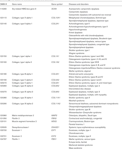
differentia-Table 1
Selected genes causal to skeletal diseases and disorders
OMIM # Gene name Gene symbol Diseases and disorders
114290 Sry-related HMG-box gene 9 SOX9 Acampomelic campomelic dysplasia Campomelic dysplasia
Campomelic dysplasia with autosomal sex reversal 120110 Collagen, type X alpha-1 COL10A1 Metaphyseal chondrodysplasia, Schmid type
Spondylometaphyseal dysplasia, Japanese type 120140 Collagen, type II alpha-1 COL2A1 Achondrogenesis, type II
Achondrogenesis-hypochondrogenesis, type II Hypochondrogenesis
Kniest dysplasia
Osteoarthritis with mild chondrodysplasia Spondyloepimetaphyseal dysplasia, Strudwick type Spondyloepiphyseal dysplasia, various types Spondylometaphyseal dysplasia, congenital type Spondyloperipheral dysplasia
Stickler syndrome, type I Wagner syndrome
120150 Collagen, type I alpha-1 COL1A1 Ehlers–Danlos syndrome, types I and VIIA Osteogenesis imperfecta, types I, II, III, and IV 120160 Collagen, type I alpha-2 COL1A2 Ehlers–Danlos syndrome, type VII-B
Osteogenesis imperfecta, types II, III, and IV
Osteogenesis imperfecta/Ehlers–Danlos crossover syndrome Marfan syndrome, atypical
120180 Collagen, type III alpha-1 COL3A1 Arterial and aortic aneurysms
Ehlers–Danlos syndrome, types III and IV 120190 Collagen, type V alpha-2 COL5A2 Ehlers–Danlos syndrome, types I and II
120215 Collagen, type V alpha-1 COL5A1 Ehlers–Danlos syndrome, types I, II, and mixed type 120260 Collagen, type IX alpha-2 COL9A2 Epiphyseal dysplasia, multiple, type 2
Intervertebral disc disease
120270 Collagen, type IX alpha-3 COL9A3 Epiphyseal dysplasia, multiple, type 3 Epiphyseal dysplasia, multiple, with myopathy 120280 Collagen, type XI alpha-1 COL11A1 Stickler syndrome, type II
Marshall syndrome
120290 Collagen, type XI alpha-2 COL11A2 Sensorineural deafness, autosomal dominant nonsyndromic Otospondylomegaepiphyseal dysplasia
Stickler syndrome, type III
Weissenbacher–Zweymuller syndrome 120360 Matrix metalloproteinase 2 MMP2 Osteolysis, idiopathic, Saudi type 121050 Fibrillin 2 FBN2 Contractural arachnodactyly, congenital 123101 Muscle segment homeobox 2 MSX2 Craniosynostosis, Boston-type
Parietal foramina 1
125505 Deoxyribonuclease I DNASE1 Systemic lupus erythematosus susceptibility
133700 Exostosin 1 EXT1 Exostoses, multiple, type 1
Chondrosarcoma
133701 Exostosin 2 EXT2 Exostoses, multiple, type II
134797 Fibrillin 1 FBN1 Marfan syndrome, various types
Ectopia lentis, familial Marfanoid skeletal syndrome Mass syndrome
Shprintzen–Goldberg syndrome
[image:3.612.58.555.113.718.2]Table 1
Continued
OMIM # Gene name Gene symbol Diseases and disorders
134934 Fibroblast growth factor receptor 3 FGFR3 Achondroplasia
Crouzon syndrome with acanthosis nigricans Hypochondroplasia
Muenke syndrome Multiple myeloma Saddan dysplasia
Thanatophoric dysplasia, types I and II 136350 Fibroblast growth factor receptor 1 FGFR1 Pfeiffer syndrome
139250 Growth hormone 1 GH1 Growth hormone deficiency
Isolated growth hormone deficiency, type I Kowarski syndrome
139320 Guanine nucleotide-binding protein, GNAS1 Mccune–Albright syndrome alpha-stimulating activity polypeptide 1 Albright hereditary osteodystrophy
Pituitary adenoma, ACTH-secreting 142461 Heparan sulfate proteoglycan HSPG2 Schwartz–Jampel syndrome, type 1
of basement membrane, perlecan Dyssegmental dysplasia, Silverman–Handmaker type 142958 Homeobox A11 HOXA11 Radioulnar synostosis with amegakaryocytic thrombocytopenia 147620 Interleukin 6 IL6 Interleukin 6 polymorphism associated with systemic onset
juvenile rheumatoid arthritis 154870 Matrix gamma-carboxyglutamic MGP Keutel syndrome
acid protein
156845 Microphthalmia-associated MITF Waardenburg syndrome, type IIA transcription factor Tietz albinism–deafness syndrome 157660 Mitochondrial RNA-processing RMRP Cartilage–hair hypoplasia
endoribonuclease (RNA component of)
168450 Parathyroid hormone PTH Hypoparathyroidism
168468 Parathyroid hormone receptor 1 PTHR1 Metaphyseal chondrodysplasia, Murk Jansen type Chondrodysplasia, Blomstrand type
176943 Fibroblast growth factor receptor 2 FGFR2 Apert syndrome
Beare–Stevenson cutis gyrata syndrome Craniosynostosis, nonsyndromic unicoronal Crouzon syndrome
Jackson–Weiss syndrome Pfeiffer syndrome
Saethre–Chotzen syndrome 190180 Transforming growth factor, beta-1 TGFB1 Camurati–Engelmann disease
193500 Paired box gene 3 PAX3 Waardenburg syndrome, types I, II, and III Waardenburg syndrome with meningomyelocele Rhabdomyosarcoma, alveolar
Craniofacial–deafness–hand syndrome 203500 Homogentisate 1,2-dioxygenase HGD Alkaptonuria
217000 Complement component 2 C2 Complement component 2 deficiency 222600 Solute carrier family 26, member 2 SLC26A2 Achondrogenesis, type IB
Atelosteogenesis, type II Diastrophic dysplasia 249100 Familial Mediterranean fever gene MEFV Familial Mediterranean fever 277900 ATPase, Cu(2+)-transporting, ATP7B Wilson Disease
beta polypeptide
300202 Sedlin SEDL Spondyloepiphyseal dysplasia, late
300300 Bruton agammaglobulinemia BTK Agammaglobulinemia, X-linked associated with
tyrosine kinase septic arthritis
Table 1
Continued
OMIM # Gene name Gene symbol Diseases and disorders
308000 Hypoxanthine guanine HPRT1 Gout, HPRT-related phosphoribosyltransferase 1 Lesch–Nyhan syndrome 311850 Phosphoribosylpyrophosphate PRPS1 Gout, PRPS-related
synthetase I
312865 Short stature homeobox SHOX Short stature, idiopathic Leri–Weill dyschondrosteosis Langer mesomelic dysplasia 600211 Runt-related transcription factor 2 RUNX2 Cleidocranial dysplasia 600310 Cartilage oligomeric matrix protein COMP Epiphyseal dysplasia
Pseudoachondroaplasia
600725 Sonic hedgehog SHH Holoprosencephaly 3
600726 Indian hedgehog IHH Brachydactyly type A1
600856 Cyclin-dependent kinase inhibitor 1C CDKN1C Beckwith–Wiedemann syndrome
600946 Growth hormone receptor GHR Laron syndrome
Short stature, autosomal dominantand idiopathic
601105 Cathepsin K CTSK Pycnodysostosis
601146 Growth/differentiation factor 5 GDF5 Acromesomelic dysplasia, Hunter–Thompson type Brachydactyly, type C
Chondrodysplasia, Grebe type 601199 Calcium-sensing receptor CASR Hypercalciuric hypercalcemia
Hypercalciuric hypocalcemia Hyperparathyroidism Hypocalcemia
Hypocalciuric hypercalcemia Hypoparathyroidism, various types
601309 Patched PTCH Basal cell nevus syndrome
Basal cell carcinoma, sporadic 601769 Vitamin D receptor VDR Vitamin D-resistant rickets, type II
602109 Matrilin 3 MATN3 Multiple epiphyseal dysplasia
602337 Receptor tyrosine kinase-like ROR2 Brachydactyly, type B1
Orphan receptor 2 Robinow syndrome, autosomal recessive
602365 Cathepsin C CTSC Papillon–Lefevre syndrome
Haim–Munk syndrome
602727 Chloride channel 7 CLCN7 Osteopetrosis, Autosomal Recessive, Infantile Malignant
602991 Noggin NOG Symphalangism, proximal
Multiple synostoses syndrome 1
603400 Wnt1-inducible signaling WISP3 Arthropathy, progressive pseudorheumatoid of childhood pathway protein 3
603499 Tumor necrosis factor receptor TNFRSF11A Expansile osteolysis, familial
superfamily, 11A Paget disease of bone 2
604142 Tyro protein tyrosine TYROBP Polycystic lipomembranous osteodysplasia with sclerosing
kinase-binding protein leukoencephalopathy
604283 Proteoglycan 4 PRG4 Camptodactyly–arthropathy–coxa vara–pericarditis syndrome 604592 T cell immune regulator 1 TCIRG1 Osteopetrosis, autosomal recessive
604831 Ellis–Van Creveld syndrome gene EVC Ellis–Van Creveld syndrome Weyers acrodental dysostosis
605145 Ank ANKH Craniometaphyseal dysplasia, autosomal dominant
605380 Fibroblast growth factor 23 FGF23 Hypophosphatemic rickets, autosomal dominant
605420 Aristaless-like 4, ALX4 Parietal foramina 2
605740 Sclerostin SOST Sclerosteosis
tion factors, resulting in combinatorial signaling and diver-gent outcomes dependent on the modifiers, which can be either genetic or environmental [23].
Sox9
The Sry-type, high-mobility group (HMG)-box containing transcription factor SOX9 comes closest to serving the function of a master regulator of the chondrocyte lineage of any molecule yet characterized. Sox9 expression is directly induced by BMP signaling [24–26]. In humans,
SOX9 haploinsufficiency (Online Mendelian Inheritance in Man [OMIM] number 114290) results in campomelic dys-plasia (a lethal skeletal malformation syndrome) with XY sex reversal [27]. During embryogenesis, Sox9 is expressed in all chondroprogenitors, coincident with the expression of type II collagen. Sox9 regulates chondroge-nesis through binding to essential DNA sequence motifs in chondrocyte-specific enhancer elements of the type II and type XI collagen genes and the cartilage-derived retinoic-acid-sensitive protein. Sox9 can even bind to, and activate, these DNA enhancer sequences in cartilage genes that have been transfected into nonchondrocytes [28–30]. Mouse embryonic stem cells with null mutations of Sox9 do not form cartilage in teratomas [31]. Animals that are heterozygous null for Sox9 exhibit defects in all cartilage primordia and present a phenotype similar to human campomelic dysplasia [32]. The phosphorylation of Sox9 by cAMP-dependent protein kinase A in response to parathyroid hormone-related peptide (PTHrP) signaling regulates the binding of Sox9 to responsive elements in the collagen promoters [33,34]. Furthermore, Sox9 is known to form complexes with L-Sox5 and Sox6, and may also inter-act with other chondrocyte-associated transcription finter-actors [35]. The regulation of this key player in chondrogenesis, therefore, is at the level of expression, protein modification, complex formation, and transcriptional activation.
Patterning and cell fate determination
Chondrogenesis can be divided into two interdependent processes: patterning; and cell fate determination. Pattern formation is the process during which number, size, and shape of the cartilaginous template is delineated and established. Cell fate determination is the process by which the combinatorial interactions of genetic and envi-ronmental factors serve to direct the developmental pro-gression of a cell lineage. Cell fate is progressively restricted, and tissue specificity is gradually committed. The actions of these determinants are dependent upon concentration, time, and position. Patterning and cell fate determination are governed by a series of tissue interac-tions, which include interactions between adjacent com-ponents of segmental structures, or between juxtaposed epithelium and mesenchyme. Chondrogenesis during craniofacial and limb development best illustrates the complexity and hierarchy of regulatory mechanisms under-lying the developmental program. During vertebrate
morphogenesis, CNCCs, as well as limb bud mesenchy-mal cells, respond to BMP4 [36]. Depending on the timing of exposure to BMP4, these mesenchymal cells may undergo apoptosis or chondrogenesis. The orchestration of the apoptotic and chondrogenic response results in the formation and delineation of cartilaginous structures in the developing face and limbs. Studies have shown that the regulation of BMP4-mediated divergent morphogenetic outcome is dependent on both positive and negative mod-ulators, at the level of ligands, cytoplasmic signals, and transcription factors.
Craniofacial development
such that these cells are allowed to migrate and arrive at their target site before overt differentiation occurs. A domi-nant negative form of Msx2 accelerates the rate and extent of chondrogenesis in CNCCs in cultures, demonstrating that, when the function of Msx2 is inhibited, cells are de-repressed, and allowed to differentiate [44]. Msx2 also functions as a repressor of chondrogenesis during the for-mation of Meckel’s cartilage in the mandibular division of the first branchial arch [26]. Overexpression of Msx2 in the developing mandible disrupts the formation of Meckel’s cartilage. Interestingly, Msx2 expression closely borders areas of cartilage differentiation and is tightly juxtaposed to the expression of Sox9. This suggests that Msx2 nor-mally functions to delineate and define the boundaries for cartilage formation. Implanting BMP4-soaked beads in the developing mandible induces the expression of both Sox9 and Msx2 [26]. The relative level of expression of these antagonistic molecules, however, is dependent on posi-tional cues within the mandible. These posiposi-tional cues may be genes that are locally expressed in a specific region of the developing mandible. These genes may modify cellular competence to respond to BMP4, and consequently the expression pattern and profile of Sox9 and Msx2 induced by BMP4. It is this relative expression of Sox9 and Msx2 that determines whether ectopic cartilage will form around the bead. The regional molecular differences in the mandible that account for differential expression of Sox9 and Msx2 remain to be explored.
Limb development
During skeletogenesis in the developing limb bud, chon-droprogenitor cells initiate their differentiation while neigh-boring cells undergo apoptosis, thus defining the boundaries of the developing skeletal elements. Mes-enchymal condensations followed by chondrocyte differ-entiation and maturation occur in digital zones, whereas mesenchymal cells undergo apoptotic elimination in inter-digital web zones to give rise to the delineation of the digits [45]. Failure of one of these processes results in limb malformations such as polydactyly or syndactyly of soft or hard tissues. BMPs regulate not only the chondro-genic and the apoptotic responses of the mesenchymal cells, but also specify digit identity, as well as participate in the generation, maintenance, and regression of the apical ectodermal ridge (a structure that governs the proxi-mal–distal patterning of the limb bud) [46–48]. BMPs, however, do not pattern each region of the limb bud indi-vidually. Rather, evidence supports the hypothesis that BMPs participate in communicating cell fate decisions interactively between adjacent regions of the limb bud. Interdigital mesenchyme destined to undergo apoptosis in vivoproduces cartilage when it is isolated away from the digits and developed in vitro [49,50]. Furthermore, digit identity is specified by the correspondingly more posterior interdigital tissue [48]. Interestingly, similar to the early patterning of the CNCC, Msx2 is also a mediator of
BMP-regulated apoptosis in the interdigital mesenchyme [51]. Recent data suggest that the specificity of BMP for multi-ple actions during limb morphogenesis reflects different activities of the receptor subtypes transducing the BMP signal [52]. BMP receptor type IB appears to be the nec-essary mediator of BMP-induced chondrogenesis [53–55], although overexpression of the receptor, or con-stitutive activation of the receptor can also cause exces-sive apoptosis [56,57]. A significant challenge of future research is to distinguish between the downstream signal-ing pathways from the BMP receptor subtypes. These dif-ferences may provide a molecular basis for the specific and often antithetic responses elicited by BMPs within developing limb buds and other tissues.
Chondrocyte maturation
Embryonic cartilage is destined for one of several fates; it can remain as permanent cartilage, such as on the articu-lar surfaces of bones, or it can provide a template for the formation of bones through the process of endochondral ossification (EO) [58,59]. Most of the bones of the axial and appendicular skeleton, and some of the bones of the craniofacial skeleton, develop through this process. The following two sections describe recent advances in under-standing the molecular regulation of chondrocyte matura-tion during EO and joint formamatura-tion.
Endochondral ossification
The anlagen of long bones develop as relatively homoge-neous elongated masses of cartilage tissue surrounded by a perichondrium. Signaling between the perichondrium and the chondrocytes (discussed below) causes cells in the center of the anlagen to initiate progression in their matura-tion program to prepare a site of ossificamatura-tion. These chon-drocytes undergo several rounds of more rapid proliferation, and then arrest in their cell cycle. The postmitotic cells change their morphology, alter their gene expression, and remodel their extracellular matrix to become hypertrophic chondrocytes (Fig. 1). Whereas proliferating and articular chondrocytes synthesize a cartilage matrix composed mostly of type II collagen, hypertrophic chondrocytes cease expressing type II and express type X collagen, which is rec-ognized as a marker of hypertrophic cells in the chondro-cyte lineage. The cartilage matrix also becomes mineralized, and the hypertrophic chondrocytes undergo apoptosis. Prior to their death, they deposit the angiogenic factor, vas-cular endothelial growth factor (VEGF), into their extracellu-lar matrix, which promotes the invasion of blood vessels into the cartilage tissue [60]. The blood vessels bring chondro-clasts, osteoblasts and osteoclasts into the new ossification center, which begin removing the mineralized cartilage matrix and forming bone tissue.
arrest, hypertrophy, and apoptosis arranged sequentially (Fig. 1). The perichondrium along the shaft differentiates into a collar of bone that expands toward the ends of the developing bone in pace with the advance of hypertrophic chondrocytes. During postnatal development, these carti-lage structures, called growth plates, are ‘sandwiched’ between the bony metaphysis and epiphysis, and serve as factories for the rapid production of new bone. Regulation and coordination of the rates of chondrocyte proliferation, hypertrophic maturation, apoptosis, and bone collar forma-tion are essential to normal bone morphogenesis. Human genetic disorders affecting EO, such as achondroplasia and chondrodysplasias, are relatively common. Positional cloning in affected pedigrees has contributed to the identi-fication of genes that regulate bone development [59,61–63]. Further analysis of the regulatory mechanisms in animal models has provided an understanding of the interactions of these genes. Recent advances in under-standing the regulation of EO have resulted from the
studies of fibroblast growth factor receptors (FGFRs), Indian hedgehog (Ihh), PTHrP, BMPs and core-binding factor (Cbfa1). Although retinoids, nitric oxide, hypoxia, vitamin D, estrogens, and other small molecules, as well as extracellular matrix molecules and biomechanical signals contribute to the regulation of chondrocyte differentiation and maturation, this review will focus primarily on peptide and glycoprotein growth factor signaling pathways.
The PTHrP/Ihh pathway
Chondrocyte proliferation and maturation in the growth plate is regulated by a negative feedback loop of intercellu-lar communication, mediated by the secreted signaling mol-ecules PTHrP and Ihh [64,65]. PTHrP, a peptide hormone with homology to parathyroid hormone, is synthesized and secreted by periarticular perichondrial cells, and by chon-drocytes later in development. It functions as a patterning molecule, inhibiting chondrocyte hypertrophy near the artic-ular ends of the developing bone, thus maintaining a pool of proliferating cells [66]. Mutations in the PTH/PTHrP recep-tor that result in constitutive activation cause Jansen’s chon-drodysplasia [67]. These patients have decreased skeletal growth, abnormal metaphases and other skeletal malforma-tions (OMIM 156400). Ihh, a member of the hedgehog family of cell-surface-associated ligands is expressed in the postmitotic, prehypertrophic cells, and provides the signal to maintain PTHrP expression at the ends of the developing bone [65]. By inhibiting chondrocyte maturation, PTHrP downregulates Ihh in the cells near the ends of the bone. Ihh promotes chondrocyte proliferation and specifies growth in the long axis through PTHrP-dependent and PTHrP-independent mechanisms [68]. Loss of Ihh function by gene targeting in mice results in decreased chondrocyte proliferation, loss of PTHrP expression, and abnormal posi-tioning of hypertrophic chondrocytes close to the articular surface [66]. Ihh is also necessary for the signaling between the postmitotic chondrocytes and the perichondrium to establish and advance the bone collar [66,69]. Point muta-tions in Ihh that may inhibit binding to its receptor causes shortening of the digits (brachydactyly type A-1; OMIM 112500), consistent with its role in chondrocyte prolifera-tion and bone growth [70].
The BMP pathways
BMP6 may serve a direct role in regulating chondrocyte maturation, while other BMPs may contribute to signaling between the chondrocytes and the perichondrium. BMP6 is expressed in prehypertrophic and hypertrophic chon-drocytes, and several other BMPs are expressed in the perichondrium [71–74]. Treatment of chondrogenic cul-tures with BMP6 promotes the expression of type X colla-gen and alkaline phosphatase [75]. Misexpression of constitutively active BMP receptor type IA in developing limbs, however, delays chondrocyte maturation, and like Ihh overexpression, upregulates PTHrP [56,65]. This sug-gests that BMP stimulation of its receptor in the diaphy-Figure 1
seal perichondrium mediates the signaling between Ihh from the prehypertrophic chondrocytes and PTHrP expression in the periarticular perichondrium.
The fibroblast growth factor pathways
Of the high affinity receptors for fibroblast growth factors (FGFs), three of the five family members FGFR1, FGFR2 and FGFR3 regulate skeletal development. The importance of FGFR3 in regulating chondrocyte proliferation and matu-ration has been revealed by analysis of patients with acti-vating mutations in this gene, which causes achondroplasia (OMIM 100800), hypochondroplasia (OMIM 146000), thanatophoric dysplasias (OMIM 187600), and other skeletal and soft tissue disorders, depending on the muta-tion (OMIM 134934)[63]. Su et al.[76] demonstrated that chondrocytes from a fetus with thanatophoric dysplasia type II exhibited increased activation of the transcription factor STAT1, and increased expression of the cyclin-dependent kinase inhibitor p21(Waf1/Cip1), a STAT-reg-ulated gene. This suggests that mutations in FGFR cause defects in EO by inhibiting chondrocyte proliferation. Sub-sequent studies in mice and tissue culture cells have sup-ported the hypothesis that increased FGFR activity disrupts the normal pattern of cartilage growth and matu-ration, at least in part by signaling through Stat molecules and increasing cyclin-dependent kinase inhibitor expres-sion [77–82]. Furthermore, increased FGFR signaling causes premature apoptosis of growth plate chondrocytes in a Stat1-dependent manner [82,83]. Conversely, the FGFR3 null mutant mice exhibit increased chondrocyte proliferation and increased bone growth [84]. Interest-ingly, Ihh and PTHrP expression is decreased in mice expressing activated mutant FGFR3 or in wild type metatarsals grown in culture in the presence of FGF2 [85,86]. This suggests interactions between the FGF sig-naling pathway and the PTHrP/Ihh pathways in regulating chondrocyte proliferation and maturation, although the precise mechanisms of these interactions is not clear.
The Cbfa1 pathway
Cbfa1 (also called Runx2, PEBP2A or Osf2; OMIM 600211) is a critical gene in the regulation of skele-tal development as it is necessary for endochondral and intramembranous bone formation, and it is sufficient to induce premature and ectopic chondrocyte hypertrophy [87–90]. Cbfa1encodes a transcription factor containing a conserved runt domain, that is expressed in mesenchy-mal condensations, chondrocytes, and cells of the osteoblast lineage [90–93]. Heterozygous loss of function mutations in Cbfa1 cause cleidocranial dysplasia (OMIM 119600), a syndrome that includes clavicle hypoplasia or aplasia, failure in closure of the anterior fontanel, and other skeletal and dental malformations [94]. In addition to the essential role that Cbfa1 plays in osteoblast differentiation, it also regulates chondrocyte maturation. Loss of Cbfa1 by gene targeting in mice
results in a complete lack of bone formation and a lack of chondrocyte hypertrophy in most of the skeleton [87,88,92,93]. Ectopic expression of Cbfa1 in nonhyper-trophic chondrocytes of transgenic mice promotes their hypertrophic differentiation and disrupts joint formation [90,95]. Cbfa1 is a direct regulator of osteocalcin and other genes in osteoblasts, and may also directly regulate hypertrophic-chondrocyte-specific genes [89]. VEGF, which is normally expressed in hypertrophic chondrocytes, is not expressed in the chondrocytes of Cbfa1null mutant mice. Furthermore, VEGF expression is upregulated by Cbfa1 in fibroblasts in tissue culture [96]. These data suggest that Cbfa1 is an important regulator of EO, con-trolling chondrocyte maturation, osteoblast differentiation, and angiogenesis in the developing bone.
The regulation of Cbfa1 expression and activity serves as a point of convergence of multiple signaling pathways. Cbfa1 expression is upregulated by BMP2, BMP4 or BMP7 treatment of multipotential, skeletal, or myogenic cell lines [91,97–99]. The regulation of Cbfa1 by BMP may be mediated by Msx2, since Cbfa1 expression is decreased in Msx2 null mutant mice [39]. Negative regula-tors of Cbfa1 expression in osteogenic cells include glu-cocorticoid, 1,25(OH)2D3, and TGF-β [91,100,101]. The function of Cbfa1 is repressed by its association with Smad3 in TGF-βstimulated cells [101]. Cbfa1 expression is upregulated in transgenic mice carrying an activated mutant FGFR1, and upregulated in a mesenchymal cell line treated with FGF2 or FGF8 [102]. Ihh may be both a regulator of Cbfa1 expression and a target of Cbfa1 tran-scriptional activity. Cbfa1 expression and bone collar for-mation is dependent on Ihh expression in prehypertrophic chondrocytes [66,69]. Expression of Cbfa1 in nonhyper-trophic chondrocytes induces Ihh expression in these cells and eventual hypertrophy [90,95]. Further study of the regulation of Cbfa1, its protein interactions, and the targets of its transcriptional activity will contribute to the detailed characterization of the molecular mechanisms regulating EO.
Joint formation
Another fate for embryonic skeletal cartilage is the forma-tion of joints. The cartilage template for the developing limb skeleton forms as a continuous, branched cartilage element from the humerus/femur to the digit rays, with only a few skeletal elements formed from independent conden-sations [103,104]. These cartilage structures are then segmented through the differentiation and apoptosis of chondrocytes to form joint cavities, through a process called cavitation. Concurrently, adjacent chondrocytes and perichondrial cells differentiate to form articular carti-lage and other joint-associated tissues [105,106].
superfamily, related to BMPs. Null mutation of Gdf5 causes shortening of the digits and defects in joint formation in the limbs as seen in the classical mouse mutant line brachy-podism (bp) [105,107]. Mutations in CDMP1 cause the human skeletal disorders Grebe type chondrodysplasia (OMIM 200700), Hunter-Thompson type acromesomelic dysplasia (OMIM 201250) and brachydactyly type C (OMIM 113100), which all include shortened or missing phalanges. Gdf5 is normally expressed in developing joints. It is one of the earliest markers of joint formation and is strongly expressed throughout cavitation [108–110]. Gli3, a transcription factor that functions in the hedgehog signal-ing pathway, is also expressed in developsignal-ing joints, and its expression pattern is expanded in bp mice [110]. Focal application of exogenous Gdf5 protein to the cartilage digit rays of bpmouse limb buds in culture inhibits the expanded expression of Gli3 [110]. This suggests that CDMP1/Gdf5 provides an important signal for the sites of joint formation, and this signal regulates the expression of genes that control chondrocyte differentiation.
Recent studies by Hartmann and Tabin [111] demon-strated the importance of Wnt-14 in initiating joint forma-tion and in the spacing of joints within the cartilage condensation. Wnt-14 is a member of the large Wnt family of secreted glycoproteins that bind to receptors of the friz-zled family. Wnt-14 is expressed in the early joint-forming regions of the developing chicken limb in a pattern similar to Gdf5. In fully developed joints, however, Wnt-14 is expressed in the joint capsule and synovial membrane, while Gdf5 is restricted to the joint fibroarticular cartilage. Ectopic expression of Wnt-14 in developing digits induces morphological and molecular changes indicative of ectopic joint formation, including inhibition of cartilage dif-ferentiation, and upregulation of Gli3, Gdf5, autotaxin, chordin, Wnt-4 and CD44rel expression in relative pat-terns similar to those seen in normal joint development [111]. Furthermore, ectopic Wnt-14 expression represses adjacent endogenous joint development in the same carti-lage condensation. This suggests that joint initiation by Wnt-14 activates a signal that regulates the positioning of the next joint in the patterning of the digits.
Study of the Wnt/frizzled pathway will contribute to an understanding of skeletal patterning and joint formation and may also provide molecular characterization of cellular changes in rheumatoid arthritis. Wnt-5a and frizzled 5 are both overexpressed in the synovial tissues of rheumatoid arthritis patients when compared to normal joint tissue or tissue from osteoarthritic joints [112]. This may suggest a change in the state of differentiation of synoviocytes that contributes to progression of the disease.
Future directions
Skeletal morphogenesis depends greatly on the patterning and formation of cartilage, and the subsequent remodeling
of cartilage into bones or joints. Molecules such as BMPs regulate important steps at different times in the life cycle of cartilage. This may suggest that BMPs instruct chon-drocytes when to change their patterns of gene expres-sion and behavior rather than providing specific instructions of which genes to express. The set of genes ready for expression may be determined by the history of signals to which the cell has been exposed, and the col-lection of receptors, signaling molecules and transcription factors accumulated in response to those signals. During chondrogenesis and endochondral ossification, chondro-cyte proliferation is regulated by FGFs, BMPs, PTHrP, Ihh, cell–cell and cell–matrix adhesion, and biomechanical signals. These multiple concurrent signals converge on the regulation of cell cycle progression and cell differentia-tion. Further study of the interaction of cartilage-associ-ated transcription factors such as Msx2 and Cbfa1 with cell-cycle regulators will contribute to an understanding of the important connection between proliferation and differ-entiation. Tissue engineering for the treatment of skeletal diseases and disorders will depend on effective tools for expanding populations of chondrocytes while maintaining or restoring their state of differentiation.
Acknowledgements
We are grateful to Harold C Slavkin for bringing our research together and for providing continued encouragement and guidance. We thank Alan Horner for critical reading of the manuscript and Sirinee Chiamvi-chitr for assisting in the preparation of the manuscript. Barbara Schmitt provided valuable assistance in compiling the information for Table I. Lillian Shum and Glen Nuckolls are supported by NIH Z01-AR41114.
References
1. Nuckolls GH, Shum L, Slavkin HC: Progress toward under-standing craniofacial malformations. Cleft Palate Craniofac J 1999, 36:12-26.
2. Felson DT, Lawrence RC, Dieppe PA, Hirsch R, Helmick CG, Jordan JM, Kington RS, Lane NE, Nevitt MC, Zhang Y, Sowers M, McAlindon T, Spector TD, Poole AR, Yanovski SZ, Ateshian G, Sharma L, Buckwalter JA, Brandt KD, Fries JF: Osteoarthritis: new insights. Part 1: the disease and its risk factors. Ann Intern Med2000, 133:635-646.
3. Loughlin J: Genetic epidemiology of primary osteoarthritis. Curr Opin Rheumatol2001, 13:111-116.
4. Hill DJ, Logan A: Peptide growth factors and their interactions during chondrogenesis. Prog Growth Factor Res1992, 4 :45-68.
5. Underhill TM, Sampaio AV, Weston AD: Retinoid signalling and skeletal development. Novartis Found Symp2001, 232 :171-185.
6. Mundlos S, Olsen BR: Heritable diseases of the skeleton. Part I: Molecular insights into skeletal development-transcription factors and signaling pathways. Faseb J1997, 11:125-132. 7. Mundlos S, Olsen BR: Heritable diseases of the skeleton. Part
II: Molecular insights into skeletal development-matrix com-ponents and their homeostasis. Faseb J1997, 11:227-233. 8. Wu W, Mwale F, Tchetina E, Kojima T, Yasuda T, Poole AR:
Car-tilage matrix resorption in skeletogenesis. Novartis Found Symp2001, 232:158-166.
9. DeLise AM, Fischer L, Tuan RS: Cellular interactions and sig-naling in cartilage development: Osteoarthritis Cartilage2000, 8:309-334.
10. Daniels K, Solursh M: Modulation of chondrogenesis by the cytoskeleton and extracellular matrix. J Cell Sci 1991, 100:249-254.
and biosynthetic response. Crit Rev Biomed Eng 1999, 27: 415-488.
12. Reddi AH: Morphogenesis and tissue engineering of bone and cartilage: inductive signals, stem cells, and biomimetic biomaterials. Tissue Eng2000, 6:351-359.
13. Hall BK, Miyake T: All for one and one for all: condensations and the initiation of skeletal development. Bioessays 2000, 22:138-147.
14. Haas AR, Tuan RS: Chondrogenic differentiation of murine C3H10T1/2 multipotential mesenchymal cells: II. Stimulation by bone morphogenetic protein-2 requires modulation of N-cadherin expression and function. Differentiation1999, 64 :77-89.
15. Stott NS, Jiang TX, Chuong CM: Successive formative stages of precartilaginous mesenchymal condensations in vitro: modulation of cell adhesion by Wnt-7A and BMP-2. J Cell Physiol1999, 180:314-324.
16. Oh CD, Chang SH, Yoon YM, Lee SJ, Lee YS, Kang SS, Chun JS: Opposing role of mitogen-activated protein kinase sub-types, erk-1/2 and p38, in the regulation of chondrogenesis of mesenchymes. J Biol Chem2000, 275:5613-5619. 17. Urist MR: Bone: formation by autoinduction. Science 1965,
150:893-899.
18. Ducy P, Karsenty G: The family of bone morphogenetic pro-teins. Kidney Int2000, 57:2207-2214.
19. Wozney JM: The bone morphogenetic protein family: multi-functional cellular regulators in the embryo and adult. Eur J Oral Sci1998, 106:160-166.
20. Miyazono K, Kusanagi K, Inoue H: Divergence and convergence of TGF-beta/BMP signaling. J Cell Physiol2001, 187:265-276. 21. Wrana JL: Regulation of Smad activity. Cell2000, 100
:189-192.
22. Reddi AH: Cartilage morphogenesis: role of bone and carti-lage morphogenetic proteins, homeobox genes and extracel-lular matrix. Matrix Biol1995, 14:599-606.
23. Reddi AH: Interplay between bone morphogenetic proteins and cognate binding proteins in bone and cartilage develop-ment: noggin, chordin and DAN. Arthritis Res2001, 3:1-5. 24. Healy C, Uwanogho D, Sharpe PT: Regulation and role of Sox9
in cartilage formation. Dev Dyn1999, 215:69-78.
25. Zehentner BK, Dony C, Burtscher H: The transcription factor Sox9 is involved in BMP-2 signaling. J Bone Miner Res1999, 14:1734-1741.
26. Semba I, Nonaka K, Takahashi I, Takahashi K, Dashner R, Shum L, Nuckolls GH, Slavkin HC: Positionally-dependent chondrogen-esis induced by BMP4 is co-regulated by Sox9 and Msx2. Dev Dyn2000, 217:401-414.
27. Wheatley S, Wright E, Jeske Y, McCormack A, Bowles J, Koopman P: Aetiology of the skeletal dysmorphology syn-drome campomelic dysplasia: expression of the Sox9 gene during chondrogenesis in mouse embryos. Ann N Y Acad Sci 1996, 785:350-352.
28. Lefebvre V, de Crombrugghe B: Toward understanding SOX9 function in chondrocyte differentiation. Matrix Biol1998, 16: 529-540.
29. Xie WF, Zhang X, Sakano S, Lefebvre V, Sandell LJ: Trans-acti-vation of the mouse cartilage-derived retinoic acid-sensitive protein gene by Sox9. J Bone Miner Res1999, 14:757-763. 30. de Crombrugghe B, Lefebvre V, Behringer RR, Bi W, Murakami S,
Huang W: Transcriptional mechanisms of chondrocyte differ-entiation. Matrix Biol2000, 19:389-394.
31. Bi W, Deng JM, Zhang Z, Behringer RR, de Crombrugghe B: Sox9 is required for cartilage formation. Nat Genet1999, 22: 85-89.
32. Bi W, Huang W, Whitworth DJ, Deng JM, Zhang Z, Behringer RR, de Crombrugghe B: Haploinsufficiency of Sox9 results in defective cartilage primordia and premature skeletal mineral-ization. Proc Natl Acad Sci USA2001, 98:6698-6703. 33. Huang W, Chung UI, Kronenberg HM, de Crombrugghe B: The
chondrogenic transcription factor Sox9 is a target of signaling by the parathyroid hormone-related peptide in the growth plate of endochondral bones. Proc Natl Acad Sci USA2001, 98:160-165.
34. Huang W, Zhou X, Lefebvre V, de Crombrugghe B: Phosphory-lation of SOX9 by cyclic AMP-dependent protein kinase A enhances SOX9’s ability to transactivate a Col2a1 chondro-cyte-specific enhancer. Mol Cell Biol2000, 20:4149-4158.
35. Lefebvre V, Li P, de Crombrugghe B: A new long form of Sox5 (L-Sox5), Sox6 and Sox9 are coexpressed in chondrogenesis and cooperatively activate the type II collagen gene. EMBO J 1998, 17:5718-5733.
36. Nuckolls GH, Slavkin HC, Shum L: Bone morphogenetic protein signaling in limb and craniofacial development. In Pro-ceedings of the Biological Mechanisms of Tooth Eruption, Resorption and Replacement by Implants. Edited by Davidowitch Z. Birmingham, AL: Harvard Society for the Advancement of Orthodontics, EBSCO media; 1998:39-47.
37. Shum L, Takahashi K, Takahashi I, Nagata M, Tan DP, Semba I, Tanaka O, Bringas P, Nuckolls GH, Slavkin HC: Embryogenesis and the classification of craniofacial dysmorphogenesis. In Oral and Maxillofacial Surgery. Edited by Fonseca RJ. Philadel-phia: WB Saunders; 2000:149-194.
38. Bendall AJ, Abate-Shen C: Roles for Msx and Dlx homeopro-teins in vertebrate development. Gene2000, 247:17-31. 39. Satokata I, Ma L, Ohshima H, Bei M, Woo I, Nishizawa K, Maeda
T, Takano Y, Uchiyama M, Heaney S, Peters H, Tang Z, Maxson R, Maas R: Msx2 deficiency in mice causes pleiotropic defects in bone growth and ectodermal organ formation. Nat Genet 2000, 24:391-395.
40. Wilkie AO, Oldridge M, Tang Z, Maxson RE Jr: Craniosynostosis and related limb anomalies. Novartis Found Symp2001, 232: 122-133.
41. Graham A, Francis-West P, Brickell P, Lumsden A: The sig-nalling molecule BMP4 mediates apoptosis in the rhomben-cephalic neural crest. Nature1994, 372:684-686.
42. Takahashi K, Nuckolls GH, Tanaka O, Semba I, Takahashi I, Dashner R, Shum L, Slavkin HC: Adenovirus-mediated ectopic expression of Msx2 in even-numbered rhombomeres induces apoptotic elimination of cranial neural crest cells in ovo. Development1998, 125:1627-1635.
43. Ellies DL, Church V, Francis-West P, Lumsden A: The WNT antagonist cSFRP2 modulates programmed cell death in the developing hindbrain. Development2000, 127:5285-5295. 44. Takahashi K, Nuckolls GH, Takahashi I, Nonaka K, Nagata M,
Ikura T, Slavkin HC, Shum L: Msx2 is a repressor of chondro-genic differentiation in migratory cranial neural crest cells. Dev Dynamics2001, 222:252-262.
45. Pizette S, Niswander L: Early steps in limb patterning and chondrogenesis. Novartis Found Symp2001, 232:23-36. 46. Dahn RD, Fallon JF: Limiting outgrowth: BMPs as negative
reg-ulators in limb development. Bioessays1999, 21:721-725. 47. Merino R, Ganan Y, Macias D, Rodriguez-Leon J, Hurle JM: Bone
morphogenetic proteins regulate interdigital cell death in the avian embryo. Ann N Y Acad Sci1999, 887:120-132.
48. Dahn RD, Fallon JF: Interdigital regulation of digit identity and homeotic transformation by modulated BMP signaling. Science2000, 289:438-441.
49. Ros MA, Piedra ME, Fallon JF, Hurle JM: Morphogenetic poten-tial of the chick leg interdigital mesoderm when diverted from the cell death program. Dev Dyn1997, 208:406-419.
50. Tang MK, Leung AK, Kwong WH, Chow PH, Chan JY, Ngo-Muller V, Li M, Lee KK: Bmp-4 requires the presence of the digits to initiate programmed cell death in limb interdigital tissues. Dev Biol2000, 218:89-98.
51. Ferrari D, Lichtler AC, Pan ZZ, Dealy CN, Upholt WB, Kosher RA: Ectopic expression of Msx-2 in posterior limb bud mesoderm impairs limb morphogenesis while inducing BMP-4 expres-sion, inhibiting cell proliferation, and promoting apoptosis. Dev Biol1998, 197:12-24.
52. Cheifetz S: BMP receptors in limb and tooth formation. Crit Rev Oral Biol Med1999, 10:182-198.
53. Nonaka K, Shum L, Takahashi I, Takahashi K, Ikura T, Dashner R, Nuckolls GH, Slavkin HC: Convergence of the BMP and EGF signaling pathways on Smad1 in the regulation of chondroge-nesis. Int J Dev Biol1999, 43:795-807.
54. Baur ST, Mai JJ, Dymecki SM: Combinatorial signaling through BMP receptor IB and GDF5: shaping of the distal mouse limb and the genetics of distal limb diversity. Development2000, 127:605-619.
55. Yi SE, Daluiski A, Pederson R, Rosen V, Lyons KM: The type I BMP receptor BMPRIB is required for chondrogenesis in the mouse limb. Development2000, 127:621-630.
and differentiation of cartilage. Genes Dev 1997, 11 :2191-2203.
57. Zhang Z, Yu X, Zhang Y, Geronimo B, Lovlie A, Fromm SH, Chen Y: Targeted misexpression of constitutively active BMP receptor-IB causes bifurcation, duplication, and posterior transformation of digit in mouse limb. Dev Biol 2000, 220: 154-167.
58. Erlebacher A, Filvaroff EH, Gitelman SE, Derynck R: Toward a molecular understanding of skeletal development. Cell1995, 80:371-378.
59. Olsen BR, Reginato AM, Wang W: Bone Development. Annu Rev Cell Dev Biol2000, 16:191-220.
60. Gerber HP, Vu TH, Ryan AM, Kowalski J, Werb Z, Ferrara N: VEGF couples hypertrophic cartilage remodeling, ossification and angiogenesis during endochondral bone formation. Nat Med1999, 5:623-628.
61. Francomano CA, McIntosh I, Wilkin DJ: Bone dysplasias in man: molecular insights. Curr Opin Genet Dev1996, 6:301-308. 62. Mundlos S, Olsen BR: Heritable diseases of the skeleton. Part
II: Molecular insights into skeletal development-matrix com-ponents and their homeostasis. FASEB J1997, 11:227-233. 63. Vajo Z, Francomano CA, Wilkin DJ: The molecular and genetic
basis of fibroblast growth factor receptor 3 disorders: the achondroplasia family of skeletal dysplasias, Muenke cran-iosynostosis, and Crouzon syndrome with acanthosis nigri-cans. Endocr Rev2000, 21:23-39.
64. Lanske B, Karaplis AC, Lee K, Luz A, Vortkamp A, Pirro A, Karpe-rien M, Defize LH, Ho C, Mulligan RC, Abou-Samra AB, Juppner H, Segre GV, Kronenberg HM: PTH/PTHrP receptor in early development and Indian hedgehog-regulated bone growth. Science1996, 273:663-666.
65. Vortkamp A, Lee K, Lanske B, Segre GV, Kronenberg HM, Tabin CJ: Regulation of rate of cartilage differentiation by Indian hedgehog and PTH-related protein. Science1996, 273 :613-622.
66. St-Jacques B, Hammerschmidt M, McMahon AP: Indian hedge-hog signaling regulates proliferation and differentiation of chondrocytes and is essential for bone formation. Genes Dev 1999, 13:2072-2086.
67. Schipani E, Kruse K, Juppner H: A constitutively active mutant PTH-PTHrP receptor in Jansen-type metaphyseal chon-drodysplasia. Science1995, 268:98-100.
68. Karp SJ, Schipani E, St-Jacques B, Hunzelman J, Kronenberg H, McMahon AP: Indian hedgehog coordinates endochondral bone growth and morphogenesis via parathyroid hormone related-protein-dependent and -independent pathways. Development2000, 127:543-548.
69. Chung UI, Schipani E, McMahon AP, Kronenberg HM: Indian hedgehog couples chondrogenesis to osteogenesis in endo-chondral bone development. J Clin Invest2001, 107:295-304. 70. Gao B, Guo J, She C, Shu A, Yang M, Tan Z, Yang X, Guo S,
Feng G, He L: Mutations in IHH, encoding Indian hedgehog, cause brachydactyly type A-1. Nat Genet2001, 28:386-388. 71. Lyons KM, Pelton RW, Hogan BL: Organogenesis and pattern
formation in the mouse: RNA distribution patterns suggest a role for bone morphogenetic protein-2A (BMP-2A). Develop-ment1990, 109:833-844.
72. Jones CM, Lyons KM, Hogan BL: Involvement of Bone Morpho-genetic Protein-4 (BMP-4) and Vgr-1 in morphogenesis and neurogenesis in the mouse. Development1991, 111:531-542. 73. Kingsley DM: What do BMPs do in mammals? Clues from the
mouse short-ear mutation. Trends Genet1994, 10:16-21. 74. Pathi S, Rutenberg JB, Johnson RL, Vortkamp A: Interaction of
Ihh and BMP/Noggin signaling during cartilage differen-tiation. Dev Biol1999, 209:239-253.
75. Grimsrud CD, Romano PR, D’Souza M, Puzas JE, Reynolds PR, Rosier RN, O’Keefe RJ: BMP-6 is an autocrine stimulator of chondrocyte differentiation. J Bone Miner Res1999, 14 :475-482.
76. Su WC, Kitagawa M, Xue N, Xie B, Garofalo S, Cho J, Deng C, Horton WA, Fu XY: Activation of Stat1 by mutant fibroblast growth-factor receptor in thanatophoric dysplasia type II dwarfism. Nature1997, 386:288-292.
77. Chen L, Adar R, Yang X, Monsonego EO, Li C, Hauschka PV, Yayon A, Deng CX: Gly369Cys mutation in mouse FGFR3 causes achondroplasia by affecting both chondrogenesis and osteogenesis. J Clin Invest1999, 104:1517-1525.
78. Li C, Chen L, Iwata T, Kitagawa M, Fu XY, Deng CX: A Lys644Glu substitution in fibroblast growth factor receptor 3 (FGFR3) causes dwarfism in mice by activation of STATs and ink4 cell cycle inhibitors. Hum Mol Genet1999, 8:35-44. 79. Sahni M, Ambrosetti DC, Mansukhani A, Gertner R, Levy D,
Basil-ico C: FGF signaling inhibits chondrocyte proliferation and regulates bone development through the STAT-1 pathway. Genes Dev1999, 13:1361-1366.
80. Hart KC, Robertson SC, Kanemitsu MY, Meyer AN, Tynan JA, Donoghue DJ: Transformation and Stat activation by deriva-tives of FGFR1, FGFR3, and FGFR4. Oncogene2000, 19 :3309-3320.
81. Aikawa T, Segre GV, Lee K: Fibroblast growth factor inhibits chondrocytic growth through induction of p21 and subse-quent inactivation of Cyclin E-Cdk2. J Biol Chem2001, 276: 29347-29352.
82. Sahni M, Raz R, Coffin JD, Levy D, Basilico C: STAT1 mediates the increased apoptosis and reduced chondrocyte prolifera-tion in mice overexpressing FGF2. Development 2001, 28: 2119-2129.
83. Legeai-Mallet L, Benoist-Lasselin C, Delezoide AL, Munnich A, Bonaventure J: Fibroblast growth factor receptor 3 mutations promote apoptosis but do not alter chondrocyte proliferation in thanatophoric dysplasia. J Biol Chem 1998, 273 :13007-13014.
84. Deng C, Wynshaw-Boris A, Zhou F, Kuo A, Leder P: Fibroblast growth factor receptor 3 is a negative regulator of bone growth. Cell1996, 84:911-921.
85. Naski MC, Colvin JS, Coffin JD, Ornitz DM: Repression of hedgehog signaling and BMP4 expression in growth plate cartilage by fibroblast growth factor receptor 3. Development 1998, 125:4977-4988.
86. Chen L, Li C, Qiao W, Xu X, Deng C: A Ser(365)—>Cys muta-tion of fibroblast growth factor receptor 3 in mouse downreg-ulates Ihh/PTHrP signals and causes severe achondroplasia. Hum Mol Genet2001, 10:457-465.
87. Komori T, Yagi H, Nomura S, Yamaguchi A, Sasaki K, Deguchi K, Shimizu Y, Bronson RT, Gao YH, Inada M, Sato M, Okamoto R, Kitamura Y, Yoshiki S, Kishimoto T: Targeted disruption of Cbfa1 results in a complete lack of bone formation owing to maturational arrest of osteoblasts. Cell1997, 89:755-764. 88. Otto F, Thornell AP, Crompton T, Denzel A, Gilmour KC, Rosewell
IR, Stamp GW, Beddington RS, Mundlos S, Olsen BR, Selby PB, Owen MJ: Cbfa1, a candidate gene for cleidocranial dysplasia syndrome, is essential for osteoblast differentiation and bone development. Cell1997, 89:765-771.
89. Ducy P: Cbfa1: a molecular switch in osteoblast biology. Dev Dyn2000, 219:461-471.
90. Takeda S, Bonnamy JP, Owen MJ, Ducy P, Karsenty G: Continu-ous expression of Cbfa1 in nonhypertrophic chondrocytes uncovers its ability to induce hypertrophic chondrocyte differ-entiation and partially rescues Cbfa1-deficient mice. Genes Dev2001, 15:467-481.
91. Ducy P, Zhang R, Geoffroy V, Ridall AL, Karsenty G: Osf2/Cbfa1: a transcriptional activator of osteoblast differentiation. Cell 1997, 89:747-754.
92. Inada M, Yasui T, Nomura S, Miyake S, Deguchi K, Himeno M, Sato M, Yamagiwa H, Kimura T, Yasui N, Ochi T, Endo N, Kita-mura Y, Kishimoto T, Komori T: Maturational disturbance of chondrocytes in Cbfa1-deficient mice. Dev Dyn 1999, 214: 279-290.
93. Kim IS, Otto F, Zabel B, Mundlos S: Regulation of chondrocyte differentiation by Cbfa1. Mech Dev1999, 80:159-170. 94. Mundlos S, Otto F, Mundlos C, Mulliken JB, Aylsworth AS,
Albright S, Lindhout D, Cole WG, Henn W, Knoll JH, Owen MJ, Mertelsmann R, Zabel BU, Olsen BR: Mutations involving the transcription factor CBFA1 cause cleidocranial dysplasia. Cell 1997, 89:773-779.
95. Ueta C, Iwamoto M, Kanatani N, Yoshida C, Liu Y, Enomoto-Iwamoto M, Ohmori T, Enomoto H, Nakata K, Takada K, Kurisu K, Komori T: Skeletal malformations caused by overexpression of Cbfa1 or its dominant negative form in chondrocytes. J Cell Biol2001, 153:87-100.
97. Tsuji K, Ito Y, Noda M: Expression of the PEBP2alphaA/AML3/ CBFA1 gene is regulated by BMP4/7 heterodimer and its overexpression suppresses type I collagen and osteocalcin gene expression in osteoblastic and nonosteoblastic mes-enchymal cells. Bone1998, 22:87-92.
98. Gori F, Thomas T, Hicok KC, Spelsberg TC, Riggs BL: Differenti-ation of human marrow stromal precursor cells: bone mor-phogenetic protein-2 increases OSF2/CBFA1, enhances osteoblast commitment, and inhibits late adipocyte matura-tion. J Bone Miner Res1999, 14:1522-1535.
99. Lee MH, Javed A, Kim HJ, Shin HI, Gutierrez S, Choi JY, Rosen V, Stein JL, van Wijnen AJ, Stein GS, Lian JB, Ryoo HM: Transient upregulation of CBFA1 in response to bone morphogenetic protein-2 and transforming growth factor beta 1 in C2C12 myogenic cells coincides with suppression of the myogenic phenotype but is not sufficient for osteoblast differentiation. J Cell Biochem1999, 73:114-125.
100. Chang DJ, Ji C, Kim KK, Casinghino S, McCarthy TL, Centrella M: Reduction in transforming growth factor beta receptor I expression and transcription factor CBFa1 on bone cells by glucocorticoid. J Biol Chem1998, 273:4892-4896.
101. Alliston T, Choy L, Ducy P, Karsenty G, Derynck R: TGF-beta-induced repression of CBFA1 by Smad3 decreases cbfa1 and osteocalcin expression and inhibits osteoblast differentiation. EMBO J2001, 20:2254-2272.
102. Zhou YX, Xu X, Chen L, Li C, Brodie SG, Deng CX: A Pro250Arg substitution in mouse Fgfr1 causes increased expression of Cbfa1 and premature fusion of calvarial sutures. Hum Mol Genet2000, 9:2001-2008.
103. Shubin NH, Alberch P: A morphogenetic approach to the origin and basic organization of the tetrapod limb.Evol Biol 1986, 20:319-387.
104. Oster GF, Shubin N, Murray JD, Alberch P: Evolution and mor-phogenetic rules: the shape of the vertebrate limb in ontogeny and phylogney. Evolution1988, 42:862-884. 105. Kingsley DM: Genetic control of bone and joint formation.
Novartis Found Symp2001, 232:213-22.
106. Pacifici M, Koyama E, Iwamoto M, Gentili C: Development of articular cartilage: what do we know about it and how may it occur?Connect Tissue Res2000, 41:175-184.
107. Storm EE, Huynh TV, Copeland NG, Jenkins NA, Kingsley DM, Lee SJ: Limb alterations in brachypodism mice due to muta-tions in a new member of the TGF beta-superfamily. Nature 1994, 368:639-643.
108. Storm EE, Kingsley DM: Joint patterning defects caused by single and double mutations in members of the bone mor-phogenetic protein (BMP) family. Development 1996, 122: 3969-3979.
109. Wolfman NM, Hattersley G, Cox K, Celeste AJ, Nelson R, Yamaji N, Dube JL, DiBlasio-Smith E, Nove J, Song JJ, Wozney JM, Rosen V: Ectopic induction of tendon and ligament in rats by growth and differentiation factors 5, 6, and 7, members of the TGF-beta gene family. J Clin Invest1997, 100:321-330. 110. Storm EE, Kingsley DM: GDF5 coordinates bone and joint
for-mation during digit development. Dev Biol1999, 209:11-27. 111. Hartmann C, Tabin CJ: Wnt-14 plays a pivotal role in inducing
synovial joint formation in the developing appendicular skele-ton. Cell2001, 104:341-351.
112. Sen M, Lauterbach K, El-Gabalawy H, Firestein GS, Corr M, Carson DA: Expression and function of wingless and frizzled homologs in rheumatoid arthritis. Proc Natl Acad Sci USA 2000, 97:2791-2796.


![4 Bromo 1 [2,6 dichloro 4 (trifluoromethyl)phenyl] 5 (4 methoxybenzylideneamino) 1H pyrazole 3 carbonitrile](data:image/gif;base64,R0lGODlhAQABAIAAAP///wAAACH5BAEAAAAALAAAAAABAAEAAAICRAEAOw==)