N A N O E X P R E S S
Probing Specific Interaction Forces Between Human IgG and Rat
Anti-Human IgG by Self-Assembled Monolayer and Atomic Force
Microscopy
Zhengjian Lv•Jianhua Wang• Guoping Chen• Linhong Deng
Received: 21 January 2010 / Accepted: 29 March 2010 / Published online: 14 April 2010
ÓThe Author(s) 2010. This article is published with open access at Springerlink.com
Abstract Interaction forces between biological mole-cules such as antigen and antibody play important roles in many biological processes, but probing these forces remains technically challenging. Here, we investigated the specific interaction and unbinding forces between human IgG and rat anti-human IgG using self assembled mono-layer (SAM) method for sample preparation and atomic force microscopy (AFM) for interaction force measure-ment. The specific interaction force between human IgG and rat anti-human IgG was found to be 0.6–1.0 nN, and the force required for unbinding a single pair of human IgG and rat anti-human IgG was calculated to be 144±11 pN. The results are consistent with those reported in the liter-atures. Therefore, SAM for sample preparation combined with AFM for interaction measurement is a relatively simple, sensitive and reliable technique to probe specific interactions between biological molecules such as antigen and antibody.
Keywords Interaction Human IgG
Rat anti-human IgGSelf assembled monolayer (SAM)
Atomic force microscopy (AFM)
Introduction
Structure, dynamics and function of biological molecules are largely determined by physical forces acting on and between the molecules. For example, the intermolecular adhesion force between an antigen and an antibody essen-tially determines whether the two molecules recognize and bind to each other to initiate a response in the immune system. Therefore, measurement of interaction forces between biological molecules is important to elucidating single molecule recognition processes such as antigen– antibody binding and unbinding, ligand-receptor attach-ment and activation [1–3]. Several techniques have been developed for measuring interaction forces between bio-logical molecules including surface forces apparatus (SFA), optical or magnetic tweezers and atomic force microscopy (AFM) [4–7]. SFA is a classical technique with high sen-sitivity to small interaction forces, but also with significant limitations such as being technically demanding, only applicable to surfaces of large area [4, 5]. More recent techniques are optical or magnetic tweezers and AFM [7]. The latter has emerged as widely used because (1) most reported single-molecule interaction forces have fallen well within the measuring range of AFM [2,8,9], (2) AFM can measure interaction forces under near physiological con-ditions with high resolution of both force and space [10,11], (3) with functionalized measuring tip, AFM is capable of sensing and mapping interaction forces across a large area such as the entire surface of a living cell [6,12–15].
One of the major challenges, however, for using AFM to measure interaction force between biological molecules is the sample preparation, in particular, the coating of mole-cules onto the surface of the substrate, and the AFM tip (functionalizing AFM tip). Currently, an extensively used method is to chemically link biological molecules onto the
Z. LvJ. WangG. ChenL. Deng
Key Laboratory of Biorheological Science and Technology, Ministry of Education, Chongqing University,
400044 Chongqing, China
Z. LvJ. Wang (&)G. ChenL. Deng (&) Institute of Biochemistry and Biophysics, College of Bioengineering, Chongqing University, 400044 Chongqing, China
e-mail: [email protected] L. Deng
surface either by silanization or by thiol-based self-assembled monolayers (SAM) [16, 17]. SAM has been developed over two decades ago and proven to be an effective and facile way to form well-defined and con-trolled films. Here, we demonstrate that by employing SAM method, rat anti-human immunoglobulin G (IgG) and human IgG could be linked onto the surface of gold sub-strate, and AFM tip, respectively, and used as a model system to probe antibody–antigen interaction forces by AFM. The single-molecule-specific adhesion force between human IgG and rat anti-human IgG was further calculated by Poisson statistical method. The results sug-gest that this method may provide a relative simple and reliable way to probe specific interactions between bio-logical molecules.
Methods and Materials
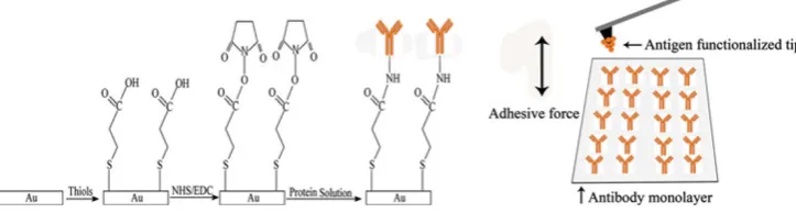
The simple mechanism for immobilizing proteins such as antibodies onto thiol-based SAM has been described by Ferretti et al. [17]. Briefly, sulfur-containing molecules (thiols, sulfides and disulfides) have a strong affinity for gold and will interact with it in near covalent manner. Therefore, when gold is immersed into a solution of thiols such as 16-Mercaptohexadecanoic acid (MHA), the thiol molecules will spontaneously react with gold and form a SAM of thiols on the gold surface with tightly packed and well-ordered chains. The terminal end of the thiol-based SAM consists of carboxyl tail groups that can be activated by the 1-ethyl-3-(dimethylaminopropyl) carbodiimide hydrochloride (EDC) and N-Hydroxysulfosuccinimide (NHS). The activated SAM can then be soaked into protein solution to form protein layer.
Gold-Coated Substrate
Gold-coated substrate was prepared by vapor deposition of gold onto mica substrate that had been freshly cleaved and preheated to 325°C for 2 h by a radiator heater. The high vacuum evaporator in which the gold was vapor deposited onto mica substrate was operated at the pressure of
*10-7Torr, and evaporation rate of 0.1–0.3 nm/s, resulting in a final thickness of the coated gold film at
*200 nm. A chromium layer was also vapor deposited and sandwiched between the gold and mica to strengthen the adhesion between the surfaces. The gold-coated substrate was then annealed in H2flame for 1 min before use.
SAM of Thiols on Gold Surface
The bare gold-coated substrate prepared as above was thoroughly cleaned in hot piranha solution (v/v
H2SO4:H2O2=3:1) for 30 min. The gold-coated substrate
was then immersed into the ethanol solution of 1 mM MHA for 24 h to produce the thiol-based SAM on the gold surface (Fig.1 left panel, columns 1–2), and unbound thiols were removed by ultrasonication in pure ethanol for 2 min. The prepared SAM was then rinsed sequentially with pure ethanol, ultra pure water, and finally dried in a N2
stream before use.
Protein Immobilization onto the SAM
Protein immobilization was carried out according to the method published by Wakayama et al. [18] with minor modification. In brief, the thiol-based SAM was treated in the solution of 2 mg/mL NHS and 2 mg/mL EDC in PBS for 1 h, which activated the carboxylic acid terminal groups of the SAM (Fig. 1left panel, column 3). After thoroughly rinsed with ultra pure water, and dried in N2stream, the
activated SAM was then immersed into the protein solution of 7lg/mL rat anti-human IgG in PBS and incubated at 4 °C for 8 h to immobilize the proteins onto the SAM (Fig.1left panel, column 4). The prepared sample of pro-tein layer was stored in PBS at 4°C before use.
Functionalization of AFM Tip
Functionalized AFM tip with human IgG coating was prepared similarly as described above. First, the AFM tip was cleaned in the hot piranha solution for 30 min and then rinsed with ultra pure water. Subsequently, the tip surface was coated with thiol-based SAM in the solution of MHA and then activated in the solution of EDC and NHS. Finally, the tip was functionalized with human IgG coating by incubating the activated tip in PBS solution of the protein at concentration of 7lg/mL, at 4 °C for 8 h. The functionalized tip was stored in PBS at 4 °C before use. Measurement of Antigen–Antibody Adhesion Force by AFM
Hooke’s law, i.e.,F=k9d, wheredis the deflection,kis the spring constant of the cantilever tip. Generally,kshould be small for AFM in order to minimize measurement noise [4]. In this study, commercially available gold-coated Si3N4cantilever tip (BudgetSensorsÒ, Innovative Solutions
Bulgaria Ltd. Bulgaria) was used of which the spring constant, calibrated by thermal fluctuation method [20], was 0.2–0.3 N/m. The tip has a pyramidal geometry, its tip radius is about 25 nm, and the thickness of the gold layer is 70 nm.
All force measurements were taken by using contact mode AFM with PBS as the medium between the tip and the protein monolayer, and the retraction velocity was estimated to be 0.04lm/s. From the ‘‘force–displacement’’ curve, the adhesion force between the rat anti-human IgG on the substrate and the human IgG on the tip was calcu-lated. Measurement was repeated many times at each of several randomly selected locations across the protein monolayer on the gold substrate.
Specificity of the Measured Adhesion Force
In order to consider specific adhesion force only, any nonspecific interaction force between the human IgG and the rat anti-human IgG should be measured and excluded. This was done by a blocking experiment performed as follows. First, the AFM tip coated with human IgG was incubated for 30 min in solution of rat anti-human IgG to block the binding sites of the antigen on the tip. Then, the nonspecific interaction force was obtained by the same force measurement as described above, but performed using the blocked tip.
Materials
16-Mercaptohexadecanoic acid (MHA), 1-ethyl-3-(dimeth-ylaminopropyl) carbodi-imide hydrochloride (EDC) and
N-Hydroxysulfosuccinimide (NHS) were purchased from Sigma–Aldrich Chemical Co. and used as received. Phos-phate-buffered saline (PBS, 140 mM NaCl, 3 mM KCl, pH 7.4) and ethanol (guaranteed grade) were purchased from Merck Co., and ultra pure water (resistivity of 18.2 MXcm) was obtained by Millpore purification system. Human IgG and rat anti-human IgG were purchased from Biosun Co. (China).
Results and Discussion
Although SAM method is relatively simple and easy to do, there are many aspects that need to be considered carefully in order to form a satisfactory protein monolayer on SAM-modified substrate [16,17,21,22]. These include, but not limited to, the following: (1) gold was used as substrate because it is chemically inert, and thiols bind to it with a high affinity; (2) MHA was used to form thiol-based SAM because of its flexible long carbon chain that served as a spacer to minimize interference between the protein mol-ecules and the gold substrate; (3) protein immobilization was carried out in PBS at 4°C and pH=7.4 because that pH and temperature may both affect protein activity; (4) the coated protein layer should not only provide optimally orientated protein molecules, but also give minimal steric hindrance to the protein molecules so that they can mimic their natural state; (5) in addition to that 1 mM thiol con-centration and 24 h immersion that were sufficient for forming well-ordered SAM of thiols [16], the protein concentration was also important for forming uniform protein monolayer. We found that 10lg/mL was the adequate protein concentration for forming uniform layer, and above this concentration the proteins might aggregate and form irregular layer. Considering that SAM method has been proven capable of ensuring the activity, mobility and stability of protein molecules [10, 16], and all
Fig. 1 A schematic illustration of the methodology used in this study. From left to right, the gold-coated surface of substrate was first modified by soaking it into the ethanol solution of 16-Mercaptohexa-decanoic acid (MHA) for 24 h, which formed an MHA film on the surface of gold substrate. Then, the MHA-modified surface of gold substrate was subject to NHS and EDC in PBS solution for 1 h to activate the MHA film. Afterward, the activated MHA film on the gold substrate was immersed into protein (rat anti-human IgG in this
[image:3.595.116.478.63.160.2]experimental aspects addressed properly as described above, the method presented here can be used to prepare reliable sample surface of biological molecules for AFM force measurement. Indeed, the topography of protein-modified surface prepared using this method had been examined by AFM imaging and confirmed satisfactory [23].
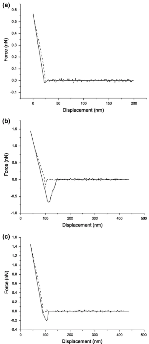
Figure2shows three representative force–displacement curves obtained by AFM measurement between rat anti-human IgG monolayer formed on thiol-based SAM sub-strate and (1) original bare tip, (2) blocked tip prepared as described in ‘‘Specificity of the Measured Adhesion Force’’, (3) tip coated with human IgG. These force–dis-placement curves characterize the binding and unbinding events between the AFM tip and the substrate when there were either no interactions, only nonspecific interactions, or specific interactions, respectively. The binding force and its probability distribution were calculated from repeated measurements and plotted in Fig.3. The results demon-strate that, considering the noise floor of the measurement, there were no interaction forces between the bare tip and rat anti-human IgG on the substrate. When the antigen-coated tip was blocked, there were no interactions for most of the time, but occasionally (approximately 20% proba-bility) there were small interaction forces occurring between the tip and rat anti-human IgG on the substrate. These occasional small binding could be attributed to nonspecific interaction between rat anti-human IgG mole-cules [24,25]. In contrast to these conditions, when the tip was coated with human IgG, there were marked binding forces measured between the human IgG and rat anti-human IgG. Although the magnitudes (refer to the maximal downward cantilever deflection during a retraction curve compared to the baseline) of measured interaction forces spread from 0.2 to 1.8 nN, the majority of them were between 0.6 and 1.0 nN. The variation of the measured interaction forces between the antigen and antibody could be attributed to the variation of contact areas between the tip and the protein monolayer when probed at different time and different locations, the density distribution of protein molecules on the substrate, and thermal fluctuation of AFM [26, 27]. The loading rate of force measurement might also contribute to the variation of measured binding force values [18,28]. In this study, the retraction velocity was estimated to be 0.04lm/s, all measurements were observed under this condition.
Since the contact area of AFM tip is very large relative to the size of protein molecule attached to it, there had to be multiple pairs of antigen–antibody involved during each single interaction event detected by AFM. Thus, the
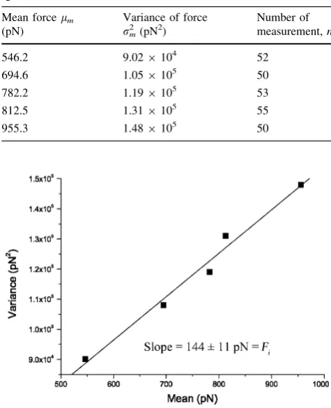
[image:4.595.305.545.55.606.2]interaction force measured by AFM was not that of a single antigen–antibody pair, but rather a collective result of interaction forces from multiple antigen/antibody pairs. However, the Poisson statistical method developed by Beebe et al. could be used to determine the unbinding force required to separate a single pair of antigen and antibody molecules [29–31]. The principal assumption of this method is that during each unbinding event as an AFM tip is pulled off the substrate, the number of antigen–antibody pairs that contribute to the total adhesive force is finite and, more importantly, follows a Poisson distribution when the unbinding event is observed repeatedly within the same fixed area of contact. The advantage of this method is that it provides an accurate calculation of single-molecule adhesive force in the presence of moderate-to-large varia-tion or noise of various types [32]. As defined by the Poisson distribution, the mean value equals the variance of the number (n) of interacting antigen–antibody pairs. Pro-vided that the measured total adhesion force is composed of a finite number of discrete interacting antigen–antibody pairs within a fixed contact area, the adhesion force between a single antigen–antibody pair (Fi) and possible
nonspecific interaction force (F0) can be derived from the
slope and interception of the linear regression curve of the variance (rm
2
) versus the mean (lm) of the measured total
adhesion force asr2
m¼lmFiFiF0 [29].
The total adhesion force between human IgG and rat anti-human IgG were measured repeated for 50–55 times at each of several randomly chosen locations of the rat anti-human IgG monolayer, and the mean (lm) and variance
(rm 2
) of these measurements are given in Table1, and
plotted with linear regression as shown in Fig. 4. From these results, the specific adhesion force between a single pair of human IgG and rat anti-human IgG, Fi and the
nonspecific force, F0, were calculated as 144±11 and
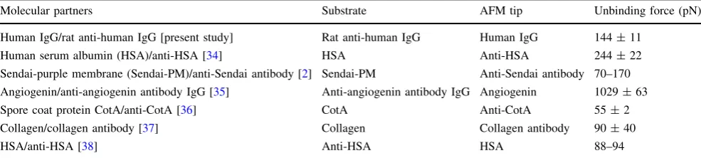
69 pN, respectively. This level of specific adhesion force was well within the range of 35–165 pN that has been reported as the estimated range of force required to rupture a single antigen–antibody complex [33]. Comparison among several antibody-based single molecular interaction forces is summarized in Table2. The specific interaction force value of human IgG/rat anti-human IgG was close to that of collagen/collagen antibody, but significantly lower than that of anti-angiogenin antibody IgG/angiogenin. The specific forces are largely influenced by the tip-sample system, the surface properties and the experimental setup. For example, the same molecular partners, namely human serum albumin (HSA) and anti-HSA, were measured by Hinterdorfer et al. [34] and Idiris et al. [38], they used different methods to immobilize the protein molecules, placed the antibody molecules on the contrary position (tip or substrate), the resulting force values were significantly different. Since in the Poisson distribution method, the
[image:5.595.52.287.55.236.2]Fig. 3 Distribution histogram of all measured adhesion forces. The adhesion forces measured when there were no interactions (between bare tip and rat anti-human IgG), only non-specific interactions (blocking experiment) and specific interactions (between human IgG and rat anti-human IgG) were represented byred,whiteand black bars, respectively. Thesolid linesare theoretical Gaussian distribu-tion curves
Table 1 Unbinding forces between human IgG and rat anti-human IgG measured at five different locations
Mean forcelm
(pN)
Variance of force
rm2 (pN2)
Number of measurement,n
546.2 9.029104 52 694.6 1.059105 50 782.2 1.199105 53
812.5 1.319105 55
955.3 1.489105 50
Fig. 4 The variance (rm2) was plotted versus the mean (lm) of the
[image:5.595.305.543.78.371.2]chemical bonds, hydrogen bonds and van der Waals force are considered as specific interactions yielding a total adhesion force, in this case, it is difficult to define or attribute the measured nonspecific force to certain forces.
Conclusions
Protein monolayers of rat anti-human IgG and human IgG were covalently bound on gold substrate and AFM tip using SAM method. Thus, the interactions between the antigen and antibody molecules on the gold substrate and the AFM tip were probed by atomic force microscopy. The specific interaction forces were determined to be largely within a range of 0.6–1.0 nN. Moreover, based on these measurements and the Poisson statistical method, it was calculated that the force required for unbinding a single antigen/antibody pair was 144±11 pN, and the nonspe-cific interaction force was 69 pN, respectively. These results are consistent with those measured by other, more complex methods, and suggest that when SAM method is properly used to prepare the sample surfaces, AFM can be a relatively simple, sensitive and reliable technique to probe specific interactions between biological molecules such as antigen/antibody pairs.
Acknowledgments This work was supported by the National Nat-ural Science Foundation of China (No. 30670496, 30770529) and the Scientific Research Foundation for the Returned Overseas Chinese Scholars, State Education Ministry (2006-331) and the Natural Sci-ence Foundation Project of CQ CSTC (2006BB5017).
Open Access This article is distributed under the terms of the Creative Commons Attribution Noncommercial License which per-mits any noncommercial use, distribution, and reproduction in any medium, provided the original author(s) and source are credited.
References
1. W. Hanley, O. McCarty, S. Jadhav, Y. Tseng, D. Wirtz, K. Konstantopoulos, J. Biol. Chem.278, 10556 (2003)
2. F. Kienberger, G. Kada, H. Mueller, P. Hinterdorfer, J. Mol. Biol.
347, 597 (2005)
3. J. Yu, Q. Wang, X. Shi, X. Ma, H. Yang, Y.G. Chen, X. Fang, J. Phys. Chem. B.111, 13619 (2007)
4. P. Hinterdorfer, Y.F. Dufrene, Nat. Methods3, 347 (2006) 5. S. Lin, J.L. Chen, L.S. Huang, H.W. Lin, Curr. Proteomics.2, 55
(2005)
6. C.K. Lee, Y.M. Wang, L.S. Huang, S. Lin, Micron38, 446 (2007) 7. K.C. Neuman, A. Nagy, Nat. Methods5, 491 (2008)
8. I. Lee, R.E. Marchant, Surf. Sci.491, 433 (2001)
9. T. Osada, A. Itoh, A. Ikai, Ultramicroscopy97, 353 (2003) 10. L. Li, S. Chen, S. Oh, S. Jiang, Anal. Chem.74, 6017 (2002) 11. N. Jalili, K. Laxminarayana, Mechatronics14, 907 (2004) 12. N.C. Santos, M.A.R.B. Castanho, Biophys. Chem. 107, 133
(2004)
13. B. Cappella, G. Dietler, Surf. Sci. Rep.34, 1 (1999)
14. M. Iijima, M. Yoshimura, T. Tsuchiya, M. Tsukada, H. Ichikawa, Y. Fukumori, H. Kamiya, Langmuir24, 3987 (2008)
15. T. Okada, M. Sano, Y. Yamamoto, H. Muramatsu, Langmuir24, 4050 (2008)
16. J.C. Love, L.A. Estroff, J.K. Kriebel, R.G. Nuzzo, G.M. White-sides, Chem. Rev.105, 1103 (2005)
17. S. Ferretti, S. Paynter, D.A. Russell, K.E. Sapsford, D.J. Rich-ardson, TRAC Trends Anal. Chem.19, 530 (2000)
18. J.I. Wakayama, H. Sekiguchi, S. Akanuma, T. Ohtani, S. Sug-iyama, Anal. Biochem.380, 51 (2008)
19. L. Li, S. Chen, S. Jiang, Langmuir19, 2974 (2003)
20. J.L. Hutter, J. Bechhoefer, Rev. Sci. Instrum.64, 1868 (1993) 21. S. Chen, L. Liu, J. Zhou, S. Jiang, Langmuir19, 2859 (2003) 22. G.B. Demirel, T. Caykara, Appl. Surf. Sci.255, 6571 (2009) 23. Z.J. Lv, J.H. Wang, L.H. Deng, G.P. Chen, Nanoscale Res. Lett.
4, 1403 (2009)
24. P.B. Chowdhury, P.F. Luckham, Colloids Surf. A143, 53 (1998) 25. Y. Miura, T. Yamauchi, H. Sato, T. Fukuda, Thin Solid Films
516, 2443 (2008)
26. X.X. He, R. Jin, L. Yang, K.M. Wang, W. Li, W.H. Tan, H.M. Li, Chin. Sci. Bull.53, 198 (2008)
27. F. Qin, Y.X. Jiang, X.Y. Ma, F. Chen, X.H. Fang, C.L. Bai, Y.Q. Li, Chin. Sci. Bull.49, 1376 (2004)
28. Y. Gan, Surf. Sci. Rep.64, 99 (2009)
29. Y.S. Lo, N.D. Huefner, W.S. Chan, F. Stevens, J.M. Harris, T.P. Beebe, Langmuir15, 1373 (1999)
30. Y.S. Lo, Y.J. Zhu, T.P. Beebe, Langmuir17, 3741 (2001) 31. W. Liu, V. Parpura, Ann. N. Y. Acad. Sci.1152, 113 (2009) 32. Y. Jiang, F. Qin, Y. Li, X. Fang, C. Bai, Nucleic Acids Res.32,
e101 (2004)
33. U. Dammer, M. Hegner, D. Anselmetti, P. Wagner, M. Dreier, W. Huber, H.J. Guntherodt, Biophys. J.70, 2437 (1996) 34. P. Hinterdorfer, W. Baumgartner, H.J. Gruber, K. Schilcher, H.
[image:6.595.51.544.73.184.2]Schindler, Proc. Natl. Acad. Sci.93, 3477 (1996)
Table 2 Comparison of several measurements of antibody-based protein–protein interactions by AFM
Molecular partners Substrate AFM tip Unbinding force (pN) Human IgG/rat anti-human IgG [present study] Rat anti-human IgG Human IgG 144±11
35. D.K. Kang, H. Park, I.C. Kang, S.I. Chang, Biochip. J.3, 339 (2009)
36. J. Tang, D. Krajcikova, R. Zhu, A. Ebner, S. Cutting, H.J. Gruber, I. Barak, P. Hinterdorfer, J. Mol. Recognit.20, 483 (2007) 37. R. Avci, M. Schweitzer, R.D. Boyd, J. Wittmeyer, A. Steele, J.
Toporski, W. Beech, F.T. Arce, B. Spangler, K.M. Cole, D.S. McKay, Langmuir20, 11053 (2004)