IJPSR (2019), Volume 10, Issue 10 (Research Article)
Received on 26 June 2019; received in revised form, 12 September 2019; accepted, 24 September 2019; published 01 October 2019
DEVELOPMENT AND INVESTIGATION OF THERMO-SENSITIVE ORGANOGEL OF
DICLOFENAC SODIUM FOR IN-SITU IMPLANTATION
A. Muralidhara and Marina Koland *
Department of Pharmaceutics, NGSM Institute of Pharmaceutical Sciences, Nitte (Deemed to be University), Deralakatte, Mangalore - 575018, Karnataka, India.
ABSTRACT: The aim of the present study was to develop and evaluate thermo-sensitive in-situ implant forming injectable organogel systems of diclofenac sodium for prolonged drug effects. The formulations were prepared by separately dissolving fatty acids such as arachidic acid, stearic acid or palmitic acid in injectable soybean oil. The organogels were evaluated for physicochemical parameters as well as for in-vitro, ex-vivo release and in-vivo fluorescence imaging. All the formulations showed gelation temperature and gelation time in the range of 36.6 to 37.1 ºC and 38 to 61 sec respectively. The pH of the formulation was found to be in the range of 6.67 to 7.14. All the formulations appeared opaque, off white and free from aggregates during the study and passed the test for syringeability and injectability. The drug content of the formulations was found in the range of 89.5 to 93.5 percent. The in-vitro drug release study showed that the release followed zero-order kinetics
and ex-vivo drug release was slower when compared to in-vitro drug
release. In-vivo fluorescence imaging and histological studies showed that the formulation F1 was able to form a depot at the injection site at body temperature and was able to retain the depot for about 4 days without any signs of inflammation or necrosis. The formulations can be considered promising in the sustained delivery of diclofenac sodium as an alternative to the oral formulations which are associated with several GI-related risks on long term use of the drug.
INTRODUCTION: Parenteral drug delivery is considered as the best suitable route for the administration of drugs with poor oral bioavailability. With parenteral administration, the onset of action can be achieved more rapidly than by any other route and provide better control over drug blood levels.
QUICK RESPONSE CODE
DOI:
10.13040/IJPSR.0975-8232.10(10).4738-47
This article can be accessed online on
www.ijpsr.com
DOI link: http://dx.doi.org/10.13040/IJPSR.0975-8232.10(10).4738-47
Oral sustained and controlled release drug delivery has made it possible to provide effective treatment of several chronic diseases owing to their ability to maintain systemic drug level and improving patient compliance 1. However, the majority of these dosage forms are unable to effectively reduce G.I. related side effects of certain classes of drugs, notably the NSAIDs.
Parenteral controlled release dosage forms such as sub-dermal implants can be expected to overcome these problems, but they are known to present problems such as the need for surgical intervention for implantation and removal, possible rejection by the body and tissue toxicity 2.
Keywords:
Diclofenac sodium,
In-situ organogel, Thermo-sensitive, Sustained release
Correspondence to Author: Dr. Marina Koland
Professor,
NGSM Institute of Pharmaceutical Sciences, Nitte (Deemed to be University), Deralakatte, Mangalore - 575018, Karnataka, India.
Recently a lot of research has been done to explore the use of in-situ gelling systems as possible cheaper alternatives to sub-dermal implants. These systems make use of biodegradable polymers whose solutions or dispersions are converted to the gel state in the presence of certain environmental stimuli at the site of injection such a‟ change in temperature, pH, ionic composition or chemical reaction retaining all the advantages of the solid implants without their disadvantages 3. A special group of in-situ depot forming gelling systems are the organogels. An organogel is defined as “a non-glassy thermorevesible semisolid system composed of an organic liquid phase entrapped in a three-dimensionally cross-linked network” 4.
In-situ solidifying organogels are the systems which are converted into solid depot after injecting into the body and form an implant at the site of injection which releases the drug for an extended period of time 4, 5. The system was formulated by employing different fatty acids such as arachidic acid, stearic acid or palmitic acid in soybean oil at different concentrations and the systems acquire thermo-sensitive gelling property because of these fatty acids. The important advantage of these organogels is the absence of any organic solvent which can cause tissue irritation. The fatty acids serve as the organogelators which form a gel when mixed with soybean oil 6, 7. The phase transition temperature and time required for transition depended on carbon chain length and concentration of the gelator. These compositions need to be preheated to 50 ºC before administration and can be injected safely by the subcutaneous route. At the injection site, thermo conversion occurs from sol to gel state at a body temperature of 37 ºC, resulting in the formation of a solid depot in the subcutaneous tissue which could be maintained for an extended period of time 8.
Diclofenac sodium, a commonly used Non-Steroidal Anti Inflammatory Drug, is known to produce its anti-inflammatory, analgesic and antipyretic effect by inhibiting cyclooxygenase and lipoxygenase pathways which in turn results in the decreased prostaglandin and leukotriene synthesis which is responsible for causing inflammation, pain and fever 9. It has a very short biological half-life and hence requires frequent administration in chronic inflammatory disorders.
The oral administration of this drug is frequently associated with G.I. related adverse effects such as gastric irritation, ulceration and bleeding on long term use. Hence in this investigation, an attempt was made to design thermo-sensitive in-situ gelling implantation system of the organogel type which will be useful for providing sustained release of drugs for an extended period of time and thereby avoiding disadvantages such as frequent dosing, surgical implantation, patient non-compliance, and drug toxicity. Thus, these formulations can be useful in the management of pain and inflammation associated with rheumatoid arthritis.
MATERIALS AND METHODS:
Materials: Diclofenac sodium was gifted by Micro Labs Bangalore. Arachidic acid, injectable soybean oil, was purchased from Sigma-Aldrich. Stearic acid, Palmitic acid, Formalin, and Sodium fluorescein was obtained from Loba Chemie Ltd., Mumbai. Methanol HPLC grade, Acetonitrile HPLC grade, Water HPLC grade were procured from Merck laboratories. Fluid thioglycollate and Soybean casein digest medium were obtained from Himedia Laboratories Ltd., Mumbai.
Methods:
Preparation of Injectable Thermo-sensitive Organogel System: The required quantities of Arachidic acid, stearic acid, and palmitic acid were weighed accurately along with measured quantities of injectable Soybean oil. The in-situ implant forming organogel system was prepared by separately dissolving each fatty acid at different concentrations in injectable soybean oil and the drug, diclofenac sodium was directly incorporated into the system with continuous stirring under magnetic stirrer at 60 ºC and subjected to sterilization by autoclaving at 121 ºC for 15 min 8. The compositions are shown in Table 1.
Evaluation of the Organogels of Diclofenac Sodium: Appearance: The appearance of the prepared organogel system was determined by visual observation of the gel against white and black background under a strong light 10.
equilibrium. Then readings were taken in triplicate 11
.
Drug Content Determination by HPLC: Each formulation equivalent to 100 mg of the drug was taken and shaken vigorously with 100 ml of methanol for about 30 min. Then this solution is subjected to sonication for 15 min for complete extraction of drug and the resultant solution is filtered. The filtrate was suitably diluted with the mobile phase of methanol, acetonitrile and deionized water (60:20:20) to get the concentrations of the drug within the range of 10-100 μg/ml. Then 20 μl of the above solution was injected into the C18 column of HPLC system and area under the peak was determined 12.
Gelation Temperature and Gelation Time: The gelation temperature was measured by taking 0.5 ml of the preheated gel in a test tube which is then placed in the constant temperature water bath. The initial temperature of the bath was maintained at 50 ºC and thereafter the temperature was reduced at increments of 1 ºC. With every degree drop in temperature, the transition from solution to gel was detected by inverting the test tube vertically and the temperature at which the gelation occurred was
recorded as the gelation temperature. The gelation time was recorded by taking the preheated gels in the test tube which is then placed in the 37 ºC water bath and the time required by the gel to transfer from solution to gel phase was recorded as a gelation time 8.
Syringeability and Injectability: Syringeability and Injectability are evaluated on a qualitative basis. The Syringeability is evaluated on the basis of the ease with which it passes through the needle and injectability is evaluated as the ease with which it can be injected into the isolated extensor digitorium muscle 13, 14. For the purpose of this study, the extensor digitorum muscle was excised fresh from Gallus gallusdomesticus, from a reputable hatchery. A volume of 2 ml of the formulation was drawn into a syringe with a 20 gauge needle and injected into the muscle. In order to assess the Syringeability and Injectability the following scores were given for both these parameters:
+++ Easily passed/injected ++ Moderate
[image:3.612.48.567.435.547.2]+ Difficult
TABLE 1: COMPOSITION OF INJECTABLE IN-SITU ORGANOGEL SYSTEM Formulation
code
Lipids Arachidic acid
(% w/v)
Stearic acid (% w/v)
Palmitic acid (% w/v)
Injectable soybean oil (% w/v)
F1 F2 F3 F4 F5 F6
4.0 5.0 - - - -
- - 7.5 10.0
- -
- - - - 10.0 12.5
96.0 95.0 92.5 90.0 90.0 87.5
Viscosity: The viscosity of the formulation was determined by using Brook field viscometer (Brookfield DV-11+ Pro) with the T-bar spindle. The instrument was equipped with a temperature controlling unit and the sample to be measured is allowed to attain equilibrium for about 10 min. The viscosity of the formulation was measured at 50, 37 and 25 ºC.
In-vitro diffusion study: The in-vitro diffusion study was carried out by using the Franz diffusion cell. The cellophane membrane was used as a diffusion membrane which was soaked in the phosphate buffer overnight for saturation and
Ex-vivo Drug Release Study of the in-situ Forming Gel: The ex-vivo drug release study was carried out by using Extensor digitorium muscle (chick muscle) from Gallus gallusdomesticus. The study was carried out in two sets, one set serves for the visual observation of the formed gel in the muscle and another set was used to study the ex-vivo drug release from the muscle.
For each set, the muscle weighing about 4.5 g was cut and the formulation was injected into the muscle through a 20 gauge needle. The muscle was then suspended in the vessel of an organ bath containing 25 ml of phosphate buffer of pH 7.4 and maintained at 37 ± 5 ºC with constant aeration of 10-12 bubbles per second. In the first set, the formulation was stained with crystal violet dye before injection to increase the visibility of the gel. The formation of a gel was confirmed by taking a section of the muscle through the injection site after about 5 min and observing the violet-colored gel produced. In the second set, the ex-vivo drug release profile was determined by taking aliquots of 3 ml from the organ bath at regular time intervals and the absorbance was measured using UV- visible spectrophotometer 16, 17.
Study of the Effect of Sterilization on the Physico-Chemical Properties of the Optimized Formulation: Dosage forms for parenteral administration must be sterile and therefore sterilization is an important step in the manufacture of these formulations which are meant to be administered by the subcutaneous route. In the present study autoclaving was selected for the sterilization of formulation, hence it is very important to know the effect of thermal and pressure stresses associated with this method on the physicochemical properties of the formulation. The formulations were sealed in glass ampoules and sterilized by autoclaving at 121 ºC for 15 min. After sterilization, the formulations were evaluated for parameters like appearance, gelation time, gelation temperature, viscosity, and percentage drug content and the results analyzed.
In-vivo Fluorescence Imaging of the Depot: This study was approved by the Institutional Animal Ethics Committee of the NGSM Institute of Pharmaceutical Sciences. (Approval number: NGSMIPS/IAEC/MAY-2016/6)
The in-situ gelling of the selected formulation at the injection site was confirmed by using fluorescence imaging of albino mice who were administered the formulation. Healthy mice of either sex were divided into two groups with three animals in each group. A volume of 0.1 ml of the optimized formulation containing sodium fluorescein was subcutaneously injected into one group of pre-anesthetized six-week-old mice and second group received aqueous solution of sodium fluorescein, which served as control. Fluorescence images were taken at 535 nm for 7 days, to visualize the fate of implants. At the end of the study, the same animals were subjected to histological evaluation for determining the biocompatibility as described under 3, 18.
Histological Evaluation for Assessment of Local Inflammation or Necrosis at Injection Site: A biocompatibility evaluation of the organogel was carried out on albino mice via subcutaneous injection of optimized formulation. After six days, the animals were sacrificed by giving Ketamine overdose via the intraperitoneal route and the tissue adjacent to the site was excised and fixed with 10% neutral buffered formalin. Tissue sections were embedded in paraffin and stained by hematoxylin-eosin for assessment of local inflammatory response 3.
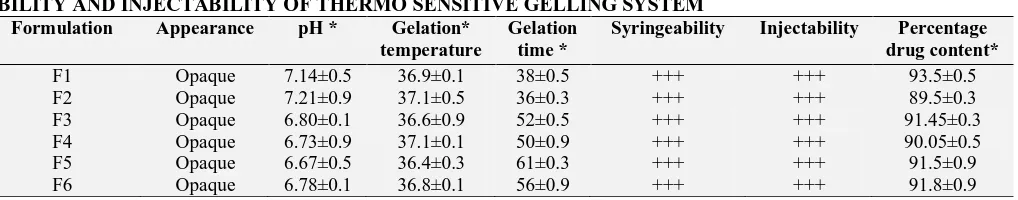
Appearance: The visual appearance of the formulation is a very important factor to be considered in the preparation of formulation meant for parenteral administration. The presence of particulate matter in the formulation of injectables may cause tissue irritation, may even be harmful to the patient, may cause difficulty for injection and also ruin patient compliance. All the formulations were visually observed and results are tabulated in Table 2. All the formulations were found to be uniformly opaque, off white in color and free from foreign particles or aggregates.
pH Determination: The pH of the formulations to be injected has the potential to produce pain and tissue irritation and preferably should be formulated to be close to the physiological pH 7.4. Table 2 shows that the pH of all the formulation was found in the range of 5.9 to 7.21. This means that all the formulations would be well tolerated after administration and possibly free from irritation. Moreover, the small volumes of 1.5 -2 ml normally administered subcutaneously means that they are readily diluted by tissue fluids to bring their pH to physiological values.
Drug Content Determination by HPLC: The results for drug content estimation are tabulated in Table 2 which showed that all the formulations had drug content in the range of 89.5 to 93.5 percent. This indicates that sufficient drug is solubilized in the formulation.
Gelation Temperature and Gelation Time: The measurement of gelation temperature and gelation time are very important in the development of
in-situ implant forming injectable systems because it is necessary to confirm the ability of the formulation to form a depot at the site of injection at body temperature. Otherwise, the formulation will remain as a solution and sustained delivery of drugs will not be achieved. The gelation temperature and time depends upon the organo-gelator which is the fatty acid, and its concentration and the stimulus responsible for gelation is the temperature. The results shown in Table 2 revealed that, as the concentration of the organogelator increased, gelation temperature was increased and gelation time was shortened.
Syringeability and Injectability: The word syringe ability refers to the ability or ease of the formulation to pass through the needle of syringe and injectability refers to the performance of formulation during injection like force or pressure required for injection 13, 19. The syringeability and injectability are important criteria that influence injecting the formulation without causing any pain to the patient. Both these attributes depend upon the viscosity of the formulations prior to thermo-conversion. Viscosity creates significant challenges in injectability since high viscosity requires high injection force that leads to increased pressure upon injection inevitably causing pain. High viscous products can also deter the completeness of the injection (i.e. the percentage of the dose delivered). All the formulations are evaluated for these parameters and results of the test are given in Table 2 which showed all formulation possess good syringe ability and injectability characteristics.
TABLE 2: RESULTS OF pH, APPEARANCE, GELATION TIME, GELATION TEMPERATURE, SYRINGE ABILITY AND INJECTABILITY OF THERMO SENSITIVE GELLING SYSTEM
Formulation Appearance pH * Gelation*
temperature
Gelation time *
Syringeability Injectability Percentage
drug content* F1 F2 F3 F4 F5 F6 Opaque Opaque Opaque Opaque Opaque Opaque 7.14±0.5 7.21±0.9 6.80±0.1 6.73±0.9 6.67±0.5 6.78±0.1 36.9±0.1 37.1±0.5 36.6±0.9 37.1±0.1 36.4±0.3 36.8±0.1 38±0.5 36±0.3 52±0.5 50±0.9 61±0.3 56±0.9 +++ +++ +++ +++ +++ +++ +++ +++ +++ +++ +++ +++ 93.5±0.5 89.5±0.3 91.45±0.3 90.05±0.5 91.5±0.9 91.8±0.9 * Mean and S.D. of „n‟ determinations, n=3
Viscosity Measurement: Viscosity is one of the major factors to be considered during formulation of parenteral products because this is responsible for the passage of formulation through syringe needle. If the solution is too viscous, it creates
[image:5.612.57.566.565.665.2]gels at body temperature (37 ºC). Gelation at body temperature is imperative to ensure the formation of a depot at the site of injection from which sustained release of the drug into the surrounding tissues will be possible.
All the six optimum formulations were subjected to viscosity measurement at 50 ºC, 37 ºC, and 25 ºC and the change in viscosity with temperature is compared as shown in Fig. 1. After thermo-conversion from sol to gel state, it is observed that there is a sharp increase in viscosity at body temperature and further increased on cooling to 25ºC.
FIG. 1: CHANGE IN VISCOSITY OF THE FORMULATIONS AT DIFFERENT TEMPERATURES
In-vitro Drug Release Study: All the optimized formulations F1, F2, F3, F4, F5, and F6 were subjected to in-vitro drug release studies in phosphate buffer of pH 7.4 by using Franz diffusion cell as described under methodology. The in-vitro drug release profiles in Fig. 2 shows that the drug release behavior from the formulation was affected by the type and concentration of fatty acids used in the formulation. The release pattern showed that as the concentration of the fatty acids increases, the drug release was sustained for a longer period of time. This is because, as the concentration of fatty acid increased, there was the formation of a stronger or firmer gel. Thus, there would be greater resistance to the diffusion of the drug through the gelled matrix at the site of injection before reaching the release medium. In the first hour of the study, there appears to be a burst release of the drug from all the formulations, probably due to the immediate drug release from the sol form of the preparation prior to conversion to gel in that short period of time.
FIG. 2: IN-VITRO DRUG RELEASE PROFILE OF DICLOFENAC SODIUM FROM FORMULATION F1, F2, F3, F4, F5, AND F6
Kinetic Analysis of in-vitro Release Data:
Drug Release Kinetics Study: In order to determine the drug release kinetics and mechanism of release from the formulations the in-vitro drug release data were fitted to different models such as the Zero order, First order, Higuchi model and Korsmeyer-Peppas model 20.
The best fit models were selected based on Regression coefficient values which were obtained from the analysis of the data.
Zero Order Release: The release data was fitted into the following equation 20:
Q = Q0 + K0t
Where, Q = Amount of drug released or dissolved (assuming that release occurs rapidly after the drug dissolves), Q0 = is the initial amount of drug in solution (it is usually zero), and K0 = is the zero-order release constant.
Zero-order kinetic model is a plot of % drug release versus time.
First Order Release: In this model, the release rate data were fitted into following equation 20:
logC = logCo – kt / 2.303
Where, C = Amount of drug released at time t, Co = Initial amount of drug in the solution and K = First order release constant.
[image:6.612.314.564.55.203.2] [image:6.612.48.301.254.405.2]Higuchi Model: This model was applied since the gelled systems serve as diffusion matrices for the drug release. The following equation was used 15:
Q = Kt1/2
Where K is the constant reflecting the design variables of the system
Higuchi model is a plot of % Cumulative drug remaining versus square root of time
Korsmeyer-Peppas Model: In order to better characterize the drug release behavior and to understand the mechanism, the Korsmeyer-Peppas model was applied by using the following equation 20, 21
.
Mt / M∞ = Ktn
Where Mt/ M∞ are fraction of drug released at time t, k is the rate constant, and n is the release exponent.
The results of kinetic analysis of in-vitro drug release data are given in Table 3. From the results it was observed that drug release from all the formulations followed zero-order kinetics. The mechanism of drug release from the formulation can be determined from the „n‟ value of Kos-meyer-Peppas equation. It was observed that for all formulations, the release exponent, „n‟ was found to be between 0.45 < n <0.89, thus indicating non-Fickian transport 20.
[image:7.612.49.569.337.653.2]Moreover the formulations after gelling can be considered as typical matrix systems, and therefore the Higuchi‟s model can be applied for determining the mechanism of release. The regression coefficient (R2) values for Higuchi model for all the formulation are in the range of 0.8838 to 0.9474. Therefore, we can conclude that the mechanism of drug release also followed the matrix diffusion process.
TABLE 3: DATA FOR DRUG RELEASE KINETICS FROM THE FORMULATIONS
Release Models F1 F2 F3 F4 F5 F6
Zero order R2 0.9853 0.9949 0.9953 0.9976 0.9842 0.9947 First order R2 0.9521 0.9721 0.9601 0.9807 0.9483 0.9826 Higuchi matrix R2 0.9138 0.9474 0.9828 0.9409 0.9312 0.9441
Korsmeyer- Peppas
R2 0.9097 0.9032 0.9282 0.9101 0.9013 0.8456
n 0.491 0.472 0.757 0.621 0.485 0.462
Best Fitting Model Higuchi matrix
Higuchi matrix
Higuchi matrix
Higuchi matrix
Higuchi matrix
Higuchi matrix
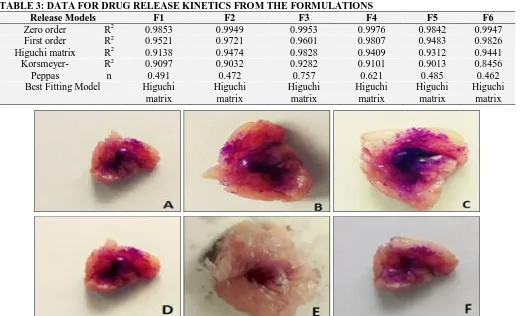
FIG. 3: FORMATION OF DEPOT INSIDE THE EXTENSOR DIGITORUM MUSCLE: (A) FORMULATION F1, (B) FORMULATION F2, (C) FORMULATION F3, (D) FORMULATION F4, (E) FORMULATION F5, (F) FORMULATION F6
Ex-vivo Drug Release Study of in-situ Forming Gel: All the optimized formulations were subjected to ex-vivo studies for the confirmation of depot formation after injecting into the muscle and to
violet-colored semi-solid depot as shown in Fig. 3 due to the crystal violet dye added to the formulation. In case of in-situ implant forming systems, it is necessary to retain the formation as depot in the subcutaneous region for a longer period so that drug can be released slowly from the depot which acts as a controlled or sustained release drug delivery system for the incorporated drug.
The ex-vivo studies were carried out for 8 h when compared to 30 h in case of in-vitro drug release studies. This is because it is difficult to maintain the viability of the tissue for 30 h. The ex-vivo drug release profile curves as demonstrated in Fig. 4 indicate that the drug release from the muscle was slower when compared to in-vitro drug release because, in case of the former, the drug must diffuse through the muscle before reaching the release medium in the organ bath. As could be expected, formulations of greater viscosity of in-situ gels, namely F5 and F6 produced slower release profiles when compared to others, since these compositions had higher fatty acid concentration. Hence, there would be a greater resistance for drug diffusion through the muscle into the release medium. As in the case of the in-vitro release study, there appears to be burst release of the drug from all formulations in the first 30-60 min due to the immediate drug release from the sol
form of the formulations before conversion to the gel form.
FIG. 4: EX-VIVO DRUG RELEASE PROFILE OF DICLOFENAC SODIUM FROM DIFFERENT FORMULATIONS
[image:8.612.315.567.89.233.2]Study of the Effect of Sterilization on the Physico-Chemical Properties of the Optimized Formulation: The viscosity, appearance, gelation temperature, gelation time and drug content for the formulations after sterilization by autoclaving were compared with the results obtained before this process. These results are shown in Table 4 and 5 which revealed that no significant change was observed for the evaluated parameters. Hence, autoclaving could be recommended as the method for sterilizing these injectable in-situ implant forming formulations.
TABLE 4: EFFECT OF AUTOCLAVING ON THE PHYSICAL PROPERTIES OF OPTIMIZED FORMULATIONS BEFORE AND AFTER AUTOCLAVING
Formulation Gelation temperature Gelation time Appearance Before*
autoclaving
After* autoclaving
Before* autoclaving
After* autoclaving
Before* autoclaving
Before* autoclaving
F1 36.9±0.06 36.6±0.03 38±0.03 40±0.02 Opaque Opaque F2 37.1±0.03 36.9±0.06 36±0.06 37±0.08 Opaque Opaque F3 36.6±0.03 36.5±0.03 52±0.03 54±0.02 Opaque Opaque F4 37.1±0.06 37.0±0.06 50±0.06 53±0.03 Opaque Opaque F5 36.4±0.03 36.4±0.08 61±0.08 60±0.02 Opaque Opaque F6 36.8±0.03 36.7±0.02 56±0.03 55±0.08 Opaque Opaque *Mean and S.D. of „n‟ determinations, n=3
TABLE 4: DATA FOR PERCENTAGE DRUG CONTENT AND VISCOSITY OF THE OPTIMIZED FORMULATIONS BEFORE AND AFTER AUTOCLAVING
Formulation % Drug content * Viscosity at 60 rpm(cps) Before*
autoclaving
After* autoclaving
Before* autoclaving After* autoclaving
At 50oC At 37oC At 25oC At 50oC At 37oC At 25oC
F1 36 400.1 909.1 37 403.2 909.5
[image:8.612.49.566.482.590.2] [image:8.612.53.565.615.741.2]In-vivo Fluorescence Imaging Studies: The optimized formulation F1 was selected for in-vivo study based on results from physicochemical parameters such as gelation temperature, gelation time, pH and drug release studies. The selected formulation was subjected to in-vivo fluorescence imaging studies to visualize the fate of the implant in albino mice and the images are shown in Fig. 5. It was observed that the depot was formed immediately at the injection site after injecting the
formulation by the subcutaneous route. The images taken daily for 7 days showed that the depot was clearly visible for 4 days, but the areas of fluorescence were gradually decreasing in diameter indicating the slow erosion of the gelled implants or depots. After the 6th day, there were no signs of fluorescence at the sites of injection in any of the animals which meant that the organogel matrix or depot was completely absorbed.
FIG. 5: IN-VIVO FLUORESCENCE IMAGING OF THE MICE AFTER S.C. INJECTION (A) DAY OF ADMINISTRATION (B) FIRST DAY (C) SECOND DAY (D) THIRD DAY (E) 6TH DAY AFTER ADMINISTRATION
[image:9.612.60.556.202.353.2]Histological Evaluations for Assessment of Local Inflammation or Necrosis at Injection Site: The histological evaluations were carried out as described under methodology to check the presence or absence of inflammation or necrosis at the injection site after 7 days. The image of the tissue section as shown in Fig. 6 did not indicate any sign of inflammation or necrosis around the injection site. In addition, there was no infiltration of inflammatory cells in the fibrous granulation tissue. Hence the formulation can be considered as safe for injection.
FIG. 6: HISTOLOGICAL STUDY OF THE INJECTION SITE OF MICE 7 DAYS AFTER INJECTION
CONCLUSION: This investigation demonstrates that thermo-sensitive injectable in-situ implant forming organogel systems could be successfully formulated by employing different fatty acids such as arachidic acid, stearic acid or palmitic acid in different concentrations with injectable soybean oil.
[image:9.612.48.296.566.729.2]The in-vivo studies of the selected formulation F1 showed that the formulation was successful in forming a depot at the injection site which was retained for at least 3 days before starting to erode. The histological study proved that the formulation did not produce any signs of inflammation or necrosis at the injection site.
Thus, this formulation can be used as an alternative to the conventional parenteral formulations of Diclofenac sodium or the sustained-release tablets for the long term management of pain and inflammation.
ACKNOWLEDGEMENT: The authors thank the Nitte (Deemed to be University) for the use of the facilities for carrying out this research work
CONFLICT OF INTEREST: The authors report no conflicts of interest. The authors alone are responsible for the content and writing of this article.
REFERENCES:
1. Lee T and Robinson JR: Remington: The Science and Practice of Pharmacy, Lippincott Williams & Wilkins, Baltimore, MD, Edition 20, 2000: 903-04.
2. Yie WC: Novel Drug Delivery System,Taylor & Francis, New York, Edition 2, 1992: 381-28.
3. Boylan JC and Nail SL: Parenteral Products. In: Banker GL, Siepmann J, Rhodes CT, editors. Modern Pharmaceutics, Marcel Dekker Inc., New York, Edition 4, 2002: 576-85.
4. Bastiat G, Plourde F, Motulsky A, Furtos A, Dumont Y and Lerou CJ: Tyrosine based rivastigmine- loaded organogels in the treatment of Alzheimer‟s disease. Biomaterials 2010; 31(1): 6031-38.
5. Hitesh BA: Prolonged-release parenteral drug delivery system– an overview. International Journal of Pharmaceutical Review and Research 2010; 3(1): 1-10. 6. Nikiforidis C, Gilbert E and Scholten E: Organogel
formation via supramolecular assembly of oleic acid and sodium oleate. RSC Advances 2015; 5(59): 47466-75. 7. Rocha J, Lopes J, Mascarenhas M, Arellano D, Guerreiro
L and da Cunha R: Thermal and rheological properties of
organogels formed by sugarcane or candelilla wax in soybean oil. Food Research International 2013; 50(1): 318-23.
8. Wang D, Zhao J, Liu F, Zhou Y, Teng L and Li Y: Parenteral thermo-sensitive organogel for Schizophrenia therapy, in-vitro and in-vivo evaluation. European Journal ofPharmaceutical Sciences 2014; 60(1): 40-48.
9. Aher SP, Patil SR, Sonje HA and Surwase KR: Formulation and evaluation of Diclofenac sodium sustained release tablet using factorial design. Journal of Current Pharmaceutical Research 2015; 5(3): 1528-38. 10. Indian Pharmacopoeia. Buffer solutions. 6th ed. Ghaziabad:
Indian Pharmacopoeia commission. 2014; Vol (1): 757- 61.
11. Ricci EJ, Bentley MVLB, Farah M, Bretas RES and Marchetti JM: Rheological characterization of Poloxamer 407 lidocaine hydrochloride gels. European Journal of Pharmaceutical Sciences 2002; 17: 161-67.
12. Riyadh AA: New method for determination of Diclofenac sodium by HPLC method. Tikrit Journal of Pharmaceutical Sciences 2012; 8(1): 60-67.
13. Patel RM: Parenteral Suspension: An overview. International Journal ofCurrent Pharmaceutical Research 2010; 2(3): 4-13.
14. Liberman HA, Rieger MM and Banker GS: Pharmaceutical Dosage Forms: Disperse systems, Marcel Dekker, New York, 1989: 549.
15. Aggarwal D and Kaur IP: Improved pharmacodynamics of Timolol maleate from a mucoadhesive niosomal ophthalmic drug delivery system. International Journal of Pharmaceutics 2005; 290: 155-59.
16. Dawre S and Devarajan PV: Novel ex-vivo method for intramuscular in-situ depot formulation. International symposium on Dissolution Asia 2014; 43.
17. Avachat AM and Kapure SS: Asenapine maleate in situ forming biodegradable implant: An approach to enhance bioavailability. International Journal of Pharmaceutics 2014; 477: 64-72.
18. Groves MJ: Parenteral Technology Manual. Inter pharm Press 1988; 9: 99-100.
19. Cabana A, Ait-Kadi A and Juhasz J: Study of the gelation process of polyethylene polypropylene oxide-polyethylene oxide copolymer (poloxamer 407) aqueous solutions. Journal of Colloid Interface Science 1997; 190: 307-12.
20. Peppas N and Narasimhan B: Mathematical models in drug delivery: How modeling has shaped the way we design new drug delivery systems. Journal of Controlled Release 2014; 190: 75-81.
21. Peppas NA: Analysis of Fickian and non Fickian drug release from polymers. Pharmaceutica Acta Helvetiae 1985; 60(4): 110-1.
All © 2013 are reserved by International Journal of Pharmaceutical Sciences and Research. This Journal licensed under a Creative Commons Attribution-NonCommercial-ShareAlike 3.0 Unported License. This article can be downloaded to Android OS based mobile. Scan QR Code using Code/Bar Scanner from your mobile. (Scanners are available on Google Play store)
How to cite this article: