MicroRNA-181b regulates NF-
kk
B–mediated
vascular inflammation
Xinghui Sun, … , Rebecca M. Baron, Mark W. Feinberg
J Clin Invest. 2012;
122(6)
:1973-1990.
https://doi.org/10.1172/JCI61495
.
EC activation and dysfunction have been linked to a variety of vascular inflammatory
disease states. The function of microRNAs (miRNAs) in vascular EC activation and
inflammation remains poorly understood. Herein, we report that microRNA-181b (miR-181b)
serves as a potent regulator of downstream NF-
k
B signaling in the vascular endothelium by
targeting importin-
a
3, a protein that is required for nuclear translocation of NF-
k
B.
Overexpression of miR-181b inhibited importin-
a
3 expression and an enriched set of
NF-k
B–responsive genes such as adhesion molecules VCAM-1 and E-selectin in ECs in vitro
and in vivo. In addition, treatment of mice with proinflammatory stimuli reduced miR-181b
expression. Rescue of miR-181b levels by systemic administration of miR-181b “mimics”
reduced downstream NF-
k
B signaling and leukocyte influx in the vascular endothelium and
decreased lung injury and mortality in endotoxemic mice. In contrast, miR-181b inhibition
exacerbated endotoxin-induced NF-
k
B activity, leukocyte influx, and lung injury. Finally, we
observed that critically ill patients with sepsis had reduced levels of miR-181b compared
with control intensive care unit (ICU) subjects. Collectively, these findings demonstrate that
miR-181b regulates NF-
k
B–mediated EC activation and vascular inflammation in response
to proinflammatory stimuli and that rescue of miR-181b expression could provide a new
target for antiinflammatory therapy and critical illness.
Research Article
Vascular biology
Find the latest version:
MicroRNA-181b regulates NF-
κ
B–mediated
vascular inflammation
Xinghui Sun,1 Basak Icli,1 Akm Khyrul Wara,1 Nathan Belkin,1 Shaolin He,1 Lester Kobzik,2 Gary M. Hunninghake,3 Miguel Pinilla Vera,3 MICU Registry,3 Timothy S. Blackwell,4
Rebecca M. Baron,3 and Mark W. Feinberg1
1Department of Medicine, Cardiovascular Division, 2Department of Pathology, and 3Department of Medicine, Pulmonary and Critical Care Division, Brigham and Women’s Hospital, Harvard Medical School, Boston, Massachusetts, USA. 4Department of Medicine, Division of Allergy,
Pulmonary and Critical Care Division, Vanderbilt University School of Medicine, Nashville, Tennessee, USA.
EC activation and dysfunction have been linked to a variety of vascular inflammatory disease states. The
func-tion of microRNAs (miRNAs) in vascular EC activafunc-tion and inflammafunc-tion remains poorly understood. Herein,
we report that microRNA-181b (miR-181b) serves as a potent regulator of downstream NF-
κ
B signaling in the
vascular endothelium by targeting importin-
α
3, a protein that is required for nuclear translocation of NF-
κ
B.
Overexpression of miR-181b inhibited importin-
α
3 expression and an enriched set of NF-
κ
B–responsive genes
such as adhesion molecules VCAM-1 and E-selectin in ECs in vitro and in vivo. In addition, treatment of mice
with proinflammatory stimuli reduced miR-181b expression. Rescue of miR-181b levels by systemic
admin-istration of miR-181b “mimics” reduced downstream NF-
κ
B signaling and leukocyte influx in the vascular
endothelium and decreased lung injury and mortality in endotoxemic mice. In contrast, miR-181b inhibition
exacerbated endotoxin-induced NF-
κ
B activity, leukocyte influx, and lung injury. Finally, we observed that
critically ill patients with sepsis had reduced levels of miR-181b compared with control intensive care unit
(ICU) subjects. Collectively, these findings demonstrate that miR-181b regulates NF-
κ
B–mediated EC
activa-tion and vascular inflammaactiva-tion in response to proinflammatory stimuli and that rescue of miR-181b
expres-sion could provide a new target for antiinflammatory therapy and critical illness.
Introduction
The vascular endothelium represents a critical interface between blood and all tissues. Endothelial dysfunction contributes to the development of both acute inflammatory disease states, such as endotoxemia and sepsis, and chronic inflammatory disease states, such as atherosclerosis, diabetes, rheumatoid arthritis, and inflammatory bowel disease (1–8). In response to inflammatory stimuli, the vascular endothelium expresses a number of adhesion molecules that play key roles in the recruitment of leukocytes to sites of inflammation (9, 10). In particular, vascular cell adhesion molecule 1 (VCAM-1), E-selectin, and intercellular adhesion mol-ecule 1 (ICAM-1) mediate early leukocyte attachment and rolling events. An inflammatory response within tissues is subsequently generated after events, such as firm adhesion and transmigration, occur (9). Clinical studies have found that the soluble forms of these adhesion molecules are increased in patients experiencing vascular inflammatory disease (11, 12).
Induction of VCAM-1, E-selectin, and ICAM-1 in ECs is primarily mediated by the activation of the NF-κB pathway (13–17). Activa-tion of NF-κB transcription factors has been implicated in many physiological and pathological processes (3, 18, 19). The transcrip-tional activity of NF-κB can be induced by a variety of stimuli, including proinflammatory cytokines TNF-α and IL-1β, exposure to bacterial products such as LPS, engagement of the T cell receptor, or stimulation of the CD40 and lymphotoxin-β receptors (19). In the canonical NF-κB signaling pathway, stimulus-mediated activa-tion of IκB kinase (IKK) complex leads to IKK rapidly
phosphorylat-ing IκBα at 2 N-terminal serines, which in turn results in its ubiq-uitin-induced degradation by the 26S proteasome (20). This event then leads to the release of NF-κB heterodimers, which then trans-locate to the nucleus via importin proteins and drive a wide range of gene expression by binding to various κB elements. In the vascular endothelium, activation of NF-κB leads to the expression of pro-inflammatory genes, including those encoding cytokines, adhesion molecules, and chemoattractant proteins that together play criti-cal roles in all aspects of the inflammatory and immune responses (21–26). Thus, targeting NF-κB–mediated EC activation holds promise for the development of novel antiinflammatory therapies.
miRNAs are emerging as new regulators of inflammatory and immune responses (27, 28). miRNAs are a class of single-stranded, small, noncoding RNAs that typically bind to the 3′ UTR of tar-get mRNA sequences, an effect leading to the reduction of protein expression predominantly by destabilizing target mRNAs and/or by translation inhibition (29–32). While over 1,000 human mature miRNA sequences are listed in the miRNA registry (33), only a handful have been characterized as functional regulators of leu-kocyte or EC inflammatory responses (28, 34, 35). In this study, we present data identifying microRNA-181b (miR-181b) as an essen-tial regulator of downstream NF-κB signaling, EC activation, and vascular inflammation in vivo by direct targeting of importin-α3, a protein critical for NF-κB nuclear translocation. These results may provide insights into the pathophysiology of a number of acute and chronic inflammatory disease states associated with dysregu-lated NF-κB signaling.
Results
miR-181b expression in ECs is regulated by TNF-α. In an attempt to identify how proinflammatory stimuli regulate endothelial
research article
tion, microarray miRNA profiling studies were undertaken using RNA from HUVECs exposed to vehicle alone or TNF-α for 4 hours, and reduced expression of miR-181b was noted (Supplemental Figure 1A; supplemental material available online with this article; doi:10.1172/JCI61495DS1). Using real-time PCR analysis, we veri-fied that miR-181b was rapidly reduced in response to TNF-α at 1 and 4 hours by 31% and 24%, respectively (Figure 1A), whereas at 24 hours, miR-181b expression increased before returning to base-line (Supplemental Figure 1B). miR-181b belongs to the miR-181 family, which comprises 4 mature miRNAs: miR-181a, miR-181b, miR-181c, and miR-181d. These mature sequences are encoded by 6 primary miRNA sequences located on 3 different chromosomes. The expression of miR-181b was determined to be about 12-fold higher than that of miR-181a and 274-fold higher than that of miR-181c by real-time PCR (Figure 1B). Since the level of primary miR-181d was very low (data not shown), we did not examine the level of this mature miR-181d. Collectively, these data suggest that miR-181b is the dominantly expressed miR-181 family member in HUVECs and that its expression is rapidly reduced in response to stimulation by the inflammatory cytokine TNF-α.
miR-181b inhibits TNF-α–induced expression of adhesion molecules and inhibits leukocyte adhesion to activated EC monolayers. To assess the potential role of miR-181b in endothelial activation, we exam-ined the effect of miR-181b on TNF-α–induced gene expression in HUVECs by using gain- and loss-of-function experiments. Overexpression of miR-181b inhibited TNF-α–induced VCAM-1, E-selectin, and ICAM-1 protein expression by 78%, 54%, and 44%, respectively, while miR-181b inhibitors (complementary antago-nist) increased their expression by 122%, 81%, and 48%, respectively (Figure 1C and Supplemental Figure 1G). In agreement with these results, the mRNA levels of VCAM-1, E-selectin, and ICAM-1 were lower in cells overexpressing miR-181b than in cells overexpressing the miRNA negative control; moreover, these mRNA levels were higher in the presence of the miR-181b inhibitor (Figure 1D). After 1 hour of TNF-α treatment, cells overexpressing miR-181b exhibit-ed rexhibit-educexhibit-ed mRNA levels of VCAM-1, E-selectin, and ICAM-1 (by 69%, 65%, and 57%, respectively); after 4 hours of TNF-α treatment, the levels were 74%, 41%, and 17%, respectively. In contrast, in cells transfected with miR-181b inhibitors, TNF-α–induced VCAM-1
mRNA was increased by 62% after 1 hour of TNF-α treatment, and after 4 hours of TNF-α treatment, VCAM-1, E-selectin, and ICAM-1 mRNA levels were increased by 35%, 52%, and 34%, respectively (Figure 1D). The effects of miR-181b on levels of soluble VCAM-1, E-selectin, and ICAM-1 in the culture medium, as measured by ELISA, were also consistent with its effects on the mRNA and pro-tein expression of these adhesion molecules (Figure 1E). Likewise, miR-181b also reduced VCAM-1 expression at both the protein and mRNA levels in HUVECs in response to LPS treatment (Sup-plemental Figure 1, C–E). Similar effects on VCAM-1 expression were observed for miR-181a (data not shown). Considering our finding that VCAM-1 expression was most sensitive to miR-181b, we determined whether the VCAM-1 gene might be a direct tar-get of the miRNA. However, overexpression of miR-181b did not alter the luciferase activity of a VCAM-1 3′ UTR construct, suggest-ing that the VCAM-1 3′ UTR is not directly targeted by miR-181b (Supplemental Figure 1F). Since VCAM-1, E-selectin, and ICAM-1 are typical proinflammatory molecules induced by TNF-α (14, 15, 36–38), these data suggested that miR-181b may be involved in the regulation of EC activation. In response to EC activation, adhesion molecules, such as VCAM-1, E-selectin, and ICAM-1, act to initiate,
promote, and sustain leukocyte attachment to the vascular endo-thelium. To determine the functional consequence of miR-181b effects on adhesion molecule expression, we employed an in vitro cell adhesion assay to assess leukocyte-EC interactions. As expect-ed, TNF-α treatment markedly increased the adhesion capabilities of THP-1 cells to HUVECs transfected with miRNA negative con-trol. However, adhesion was markedly reduced (by 44%) with miR-181b overexpression, whereas inhibition of miR-miR-181b increased the adherence by 50% (Figure 1F). Taken together, these findings suggest that miR-181b is able to negatively affect the expression of key adhesion molecules induced by proinflammatory stimuli and that miR-181b dynamically regulates leukocyte adhesion to stimulated EC monolayers.
miR-181b suppresses TNF-α–induced expression of adhesion molecules in vivo. We next investigated whether systemic administration of miR-181b could inhibit TNF-α–induced gene expression in vivo. To assess the effects of miR-181b mimics on TNF-α–induced expression of VCAM-1 in vivo, miR-181b or a miRNA negative control (1 nmol/mouse) was admixed with atelocollagen and tail vein injected 24 hours prior to TNF-α treatment. VCAM-1 pro-tein expression was first examined in lung tissues. At 4 hours after TNF-α i.p. injection, VCAM-1 was found to be induced by approxi-mately 3.4-fold in lung tissues in the presence of NS control mim-ics. In contrast, administration of miR-181b potently reduced the induction of VCAM-1 protein expression (Figure 2A) and
Figure 1
research article
Figure 2
miR-181b inhibits the NF-κB signaling pathway in activated ECs. In response to proinflammatory stimuli, both the NF-κB and MAPK pathways are involved in the inflammatory responses in ECs (21, 43). To determine whether miR-181b affects NF-κB activation, we first tested to determine whether miR-181b has any effect on the NF-κB concatemer and VCAM-1 promoter-reporter. As shown in Figure 3A, treatment of HUVECs with TNF-α induced the activ-ity of both the NF-κB concatemer and the VCAM-1 promoter-reporter, and cotransfection of miR-181b significantly attenuated this induction. In contrast, inhibition of miR-181b potentiated TNF-α–induced activity (Figure 3A). We next explored the effect of miR-181b on NF-κB nuclear accumulation by immunostaining for p65. We observed a nearly 40% reduction in p65 nuclear stain-ing in HUVECs transfected with miR-181b as compared with cells transfected with a miRNA-negative control (Figure 3B). After its release from the IκB complex, translocation of NF-κB from the cytoplasm to the nucleus is an essential step for the activation of NF-κB target genes (44, 45), an effect that can be revealed by detec-tion of the p50 and p65 protein levels in cytoplasmic and nuclear fractions. As shown in Figure 3, C and D, HUVECs overexpressing miR-181b exhibited reduced p65 and p50 expression by 39% and 28%, respectively, in the nuclear fraction, whereas the cytoplasmic fraction had increased p65 and p50 expression by 35% and 37%, respectively. Importantly, we did not observe any significant dif-ferences between miR-181b and the miRNA negative control on the expression of upstream components of the NF-κB pathway, including phosphorylated IκBα (in the presence or absence of pro-teosome inhibitor MG-132), total IκBα, or phosphorylated IKKβ
(Figure 3E and Supplemental Figure 3A) in response to TNF-α, suggesting that it is unlikely that miR-181b affects cell-surface receptors or activation of the IKK complex. Since several MAPKs have been implicated in TNF-α–induced expression of adhesion molecules (46–48), we next asked whether miR-181b had any effect on the activation of 3 MAPKs (ERK, p38, and JNK) in response to TNF-α. As shown in Supplemental Figure 3B, the phosphoryla-tion of ERK, p38, and JNK was robustly induced and peaked at 15 minutes after TNF-α treatment. However, miR-181b overexpres-sion had no inhibitory effect on their phosphorylation at 5, 15, and 30 minutes after TNF-α treatment. These data suggest that the inhibitory role of miR-181b on TNF-α–induced gene expres-sion is primarily due to its effects on the NF-κB signaling pathway by repression of NF-κB nuclear translocation.
miR-181b directly targets expression of importin-α3, a protein critical for NF-κB nuclear translocation. Previous studies have shown that NF-κBs are transported into the nucleus via a subset of importin-α
molecules (49, 50). Because miR-181b overexpression had no effect on upstream phosphorylated IκBα or IKKβ expression, we hypoth-esized that miR-181b may inhibit downstream NF-κB signaling by targeting importin-α family members. There are 6 importin-α par-alogs in humans (importin-α1, -α3, -α4, -α5, -α6, and -α7) that are characterized by distinct affinities to their substrates (51). Among these molecules, only importin-α3 and importin-α5 are predicted to be miR-181b targets, according to the algorithms of TargetScan (52), PITA (53), and miRanda (54). In miR-181b–overexpressing cells, importin-α3 expression, but not that of importin-α1 or importin-α5, was reduced by 40% and 25%, respectively, in the presence or absence of TNF-α (Figure 4A). Overexpression of miR-181b inhibited the activity of a luciferase reporter construct containing importin-α3 3′ UTR in a dose-dependent manner (Fig-ure 4B). In contrast, the activity of luciferase constructs
contain-ing the 3′ UTR of importin-α1, -α4, or -α5 was not inhibited by overexpressed miR-181b (Figure 4C).
The rna22 miRNA target detection and predication algorithm allows for seed mismatches between mature miRNA and targeting mRNA sequence (55) and has been successfully used to identify direct targets that were not predicted by other algorithms (56). To identify additional miR-181b–binding sites that may exist in the 3′ UTR of importin-α3, we applied the rna22 prediction algo-rithm and found 8 potential miR-181b–binding sites in the region of interest (Supplemental Figure 4A). Overexpression of miR-181b decreased luciferase activity by 31% and 20%, respectively, for lucif-erase-reporter constructs containing binding site 1 and site 2, but not for any of the other potential binding sites (Figure 4D). Site-directed mutations of binding site 1 and site 2 rescued the miR-181b–mediated inhibitory effects on both of these constructs (Fig-ure 4D). While site 1 has imperfect complementarity to miR-181b, it is evolutionarily conserved across species (Supplemental Figure 4B). Interestingly, the mRNA level of importin-α3 was not altered by miR-181b overexpression (Supplemental Figure 4C), an effect indicating that the reduction of importin-α3 at the protein level is likely due to translation inhibition and not mRNA decay. To further verify that miR-181b directly targets importin-α3, we per-formed Argonaute2 (AGO2) micro-ribonucleoprotein IP (miRNP-IP) studies to assess whether importin-α3 mRNA is enriched in the RNA-induced silencing complex following miR-181b overexpres-sion. An approximately 4-fold enrichment of importin-α3 mRNA was observed after AGO2 miRNP-IP in the presence of miR-181b, as compared with that with the miRNA-negative control (Figure 4E). In contrast, AGO2 miRNP-IP did not enrich the mRNA for
Smad1, a gene that was not predicted to be an miR-181b target (Figure 4E). Moreover, expression of importin-α3 lacking its 3′ UTR was able to rescue the inhibitory effect of miR-181b on NF-κB activation (Figure 4F). Systemic delivery of miR-181b also reduced importin-α3 expression by 40% in aortic endothe-lium (Figure 4G) and nearly 50% in lung ECs (Figure 4H). Con-versely, inhibition of miR-181b increased importin-α3 expres-sion by approximately 40% in HUVECs (Supplemental Figure 4E) and approximately 50% in the lungs in vivo (Figure 4I). To explore whether “knockdown” of importin-α3 expression could “phenocopy” the inhibitory effects of miR-181b on NF-κB, we systemically delivered importin-α3 siRNA by tail-vein injections. As shown in Supplemental Figure 5, A and B, importin-α3 siRNA reduced importin-α3 mRNA and protein expression in freshly iso-lated lung ECs. Importantly, importin-α3 siRNA reduced NF-κB targets VCAM-1 and E-selectin (Supplemental Figure 5, C and D) and NF-κB activity in the lungs in vivo (Supplemental Figure 5E) as well as VCAM-1 and E-selectin expression in HUVECs in vitro (Supplemental Figure 5, F and G). Moreover, systemic delivery of importin-α3 lacking its 3′ UTR was capable of rescuing the miR-181b–mediated inhibitory effect on VCAM-1 and E-selectin expression and NF-κB activity in the lungs of mice treated with LPS (Supplemental Figure 5, H and I). In HUVECs, overexpres-sion of importin-α3 lacking its 3′ UTR also rescued the miR-181b– mediated reduced expression for VCAM-1 and E-selectin com-pared with control (Supplemental Figure 5, J and K). Collectively, these data indicate that miR-181b inhibits the NF-κB signaling pathway by directly targeting importin-α3 expression.
research article
profiles of HUVECs transfected with miRNA negative control or miR-181b by using Agilent whole human genome microarrays. Transfected HUVECs were treated with TNF-α for 4 hours, and total RNA was isolated and processed for gene chip analysis. Out of the approximately 44,000 transcripts screened, we deter-mined that 841 genes were downregulated and 928 genes were
[image:7.585.51.534.84.521.2]upregulated by at least 1.5-fold in miR-181b–overexpressing cells as compared with control cells. Over 200 of those genes are known to be NF-κB regulated. Moreover, of 24 selected genes known to be associated with inflammatory disease states, all were inhibited by overexpression of miR-181b. These reduced gene expression changes were verified by qPCR analysis (Figure 5,
Figure 3
miR-181b inhibits the activation of the NF-κB signaling pathway. (A) Luciferase activity of reporters containing either the NF-κB concatemer or VCAM-1 promoter in HUVECs transfected with miRNA negative control or miR-181b mimics and miRNA inhibitor negative control or miR-181b inhibitor after 12 hours treatment with 10 ng/ml of TNF-α. #P < 0.05; *P < 0.01. Values represent mean ± SD; n = 3. (B) p65 staining in HUVECs
Figure 4
research article
A and B). Because the dominant-negative IκBα is a specific inhib-itor of the canonical NF-κB signaling, we examined the gene expression profile of the 24 selected genes by real-time qPCR in HUVECs transduced in the presence of an adenovirus express-ing the dominant-negative IκBα. We found that the expression of each of the 24 genes was reduced by expression of the dom-inant-negative IκBα in response to TNF-α treatment (Figure 5C), revealing a similar profile to cells overexpressing miR-181b mimics (Figure 5B). PAI-1, COX-2, CX3CL-1, and VCAM-1 were chosen to verify concordant directional changes at the protein level in miR-181b–overexpressing ECs (Figure 5D). To identify highly regulated biological processes in miR-181b–overexpress-ing cells, we performed gene set enrichment analysis (GSEA), a
[image:9.585.79.510.79.485.2]computational method that determines whether a defined set of genes shows significant differences between 2 biological states (57). We found 6 enriched biological processes that were signifi-cantly represented by the reduced genes in miR-181b–overex-pressing cells: response to cytokine stimulus; positive regulation of cell migration; regulation of inflammatory response; inflam-matory response; chemotaxis; and IKK/NF-κB cascade (Table 1). An abundance of targets that interconnected with the NF-κB signaling pathway was also observed using the Ingenuity web-based pathway analysis program (Supplemental Figure 6). Taken together, these data suggest that miR-181b selectively suppresses an enriched set of NF-κB–regulated genes and components of inflammatory signaling pathways in response to TNF-α in ECs.
Figure 5
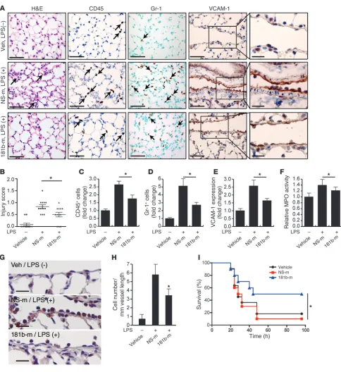
miR-181b reduces LPS-induced EC activation, leukocyte accumulation, lung inflammation/injury, and mortality. Sepsis is a medical condition with high morbidity and mortality and increasing incidence over the past few decades (58). The endotoxin LPS is a component of the outer membrane of Gram-negative bacteria that plays a significant role in the pathogenesis of about 25% to 30% of sepsis cases (59). LPS activates the TLR-mediated signaling pathway, induces the release of critical proinflammatory cytokines including TNF-α, and elicits a systemic inflammatory response syndrome (SIRS) (60, 61). During sepsis, activation of the vascular endothelium plays a critical role in the recruitment of neutrophils and monocytes/mac-rophages as well as subsequent exacerbation of the inflammatory response (1, 62). To determine whether miR-181b contributes to this process, we assessed whether the level of endothelial miR-181b could be affected in response to LPS and whether in vivo overex-pression of miR-181b could reduce leukocyte recruitment and EC activation in a systemic LPS mouse model of vascular inflamma-tion. As shown in Supplemental Figure 7A, the expression of miR-181b was reduced in freshly isolated aortic intima by 50% and 47% after 4 hours of systemic treatment with LPS or TNF-α, respec-tively. In contrast, VCAM-1 mRNA was induced about 5.0-fold and 7.2-fold by TNF-α and LPS, respectively, in freshly isolated aortic intima (Supplemental Figure 7B). In vivo i.v. administration of miR-181b mimics reduced the induction of VCAM-1 expression in response to LPS by 60% (Figure 6, A and E). Analysis of lung sections taken from mice injected with miR-181b revealed a sig-nificant reduction of CD45-positive (common leukocyte antigen) and Gr-1–positive (predominantly neutrophil) leukocytes (by 54% and 58%, respectively) in response to LPS treatment (Figure 6, A, C, and D). Importantly, the number of Gr-1–positive leukocytes adherent to the pulmonary vascular endothelium was reduced 47% by miR-181b overexpression (Figure 6, G and H). We also observed reduced interstitial edema and lung injury scores in the lungs of mice in which miR-181b was overexpressed (Figure 6, A and B). Myeloperoxidase activity, reflecting the presence of per-oxidase enzyme expressed most abundantly in neutrophils, was also reduced by miR-181b overexpression (Figure 6F). Finally, sys-temic delivery of miR-181b mimics in mice (2 injections before and 1 injection after the initiation of LPS-induced endotoxic shock) reduced mortality from endotoxemia by 75% over 4 days (Figure 6I). Taken together, these results demonstrate a critical role for miR-181b in endotoxin-induced endothelial activation and leuko-cyte accumulation and suggest that overexpression of miR-181b could represent a novel class of therapeutic avenues for limiting endotoxin-induced vascular inflammation and may ultimately have a beneficial effect in critical illness.
miR-181b inhibits the activation of the NF-κB pathway in vascu-lar endothelium in vivo. In response to inflammatory stimuli, activation of the NF-κB pathway, which involves transloca-tion of NF-κB heterodimers from cytoplasm to nucleus, is critical for vascular inflammation in vivo (24, 26). To deter-mine whether miR-181b regulates NF-κB translocation in vivo, miR-181b was systemically administered by tail-vein injection, and mice were challenged with or without LPS. In mice not exposed to LPS, the majority of NF-κB p65 remained cytoplasmic in pulmonary ECs (Figure 7A). In contrast, NF-κB p65 colocalized more with DAPI-stained endothelial nuclei by immunofluorescence in mice chal-lenged with LPS by i.p. injection (Figure 7A). Remarkably, the accumulation of NF-κB p65 in endothelial nuclei in response to LPS was reduced by 45% in the presence of systemically delivered miR-181b compared with mice injected with NS control miRNA (Figure 7A). Using a complementary approach, we also ana-lyzed the effect of miR-181b on NF-κB activation in vivo in NF-κB transgenic mice, which express a luciferase reporter driven by an NF-κB–responsive promoter (63, 64). Indeed, systemic delivery of miR-181b reduced LPS-induced luciferase activity in lungs by 57% (Figure 7B), NF-κB target genes VCAM-1 and E-selectin in lungs by 43% and 59%, respectively (Figure 7, C and D), and VCAM-1 expression in isolated mouse lung ECs by 52% (Figure 7E). These effects were associated with approximately 4.5-fold overexpression of miR-181b in the lungs by real-time qPCR compared with mice injected with NS control miRNA (Figure 7F). Importantly, miR-181b reduced LPS-induced luciferase to a degree similar to that of adenoviral vectors expressing a dominant-negative IκBα, and no additional inhibitory effect of miR-181b was observed when deliv-ered in combination with adenoviral vectors expressing a domi-nant-negative IκBα (Figure 7G). Finally, miR-181b overexpression had no effect on the expression of a panel of miRNAs in mouse lungs, which have been shown to regulate inflammatory genes or NF-κB activation, including miR-10a, miR-146a, miR-155, miR-31, and miR-17-3p (Supplemental Figure 8). These data suggest that systemic delivery of miR-181b inhibits endothelial NF-κB p65 nuclear translocation and, subsequently, the activation of NF-κB signaling pathway in vascular ECs in vivo.
[image:10.585.54.301.127.206.2]Inhibition of miR-181b potentiated LPS-induced NF-κB–regulated gene expression and NF-κB activity in vivo. To determine whether the endogenous expression of miR-181b has any effect on NF-κB sig-naling and EC activation in vivo, mice were injected i.v. with miR-181b inhibitors in the presence of LPS. As shown in Figure 8, A and B, systemic delivery of miR-181b inhibitors reduced endogenous miR-181b expression by approximately 62% and potentiated LPS-induced VCAM-1 protein expression by approximately 1.9-fold in lung tissues. The expression of VCAM-1 and E-selectin mRNA also increased by approximately 1.9- and 1.5-fold, respectively (Figure 8, C and D). Furthermore, miR-181b inhibition enhanced NF-κB activ-ity in lung lysates from NF-κB–luciferase transgenic mice treated with LPS (Figure 8E) and exacerbated lung injury, as shown by scor-ing (Figure 8G). Finally, in response to LPS, leukocyte accumulation in the lungs of mice treated with miR-181b inhibitors was markedly increased by immunohistochemistry studies compared with that in mice treated with NS control inhibitors (Figure 8, F, H, and I). These findings indicate that inhibition of endogenous miR-181b potentiates NF-κB signaling, EC activation, and lung inflammation in the presence of inflammatory stimuli, suggesting that reduced miR-181b expression may play a role in the pathogenesis of sepsis.
Table 1
Enriched GO biological processes downregulated in miR-181b overexpression cells, identified by GSEA at 25% FDR
GO biological processes Description of gene set FDR
GO:0034097 Response to cytokine stimulus 0.013 GO:0030335 Positive regulation of cell migration 0.171 GO:0050727 Regulation of inflammatory response 0.178 GO:0006954 Inflammatory response 0.194
GO:0006935 Chemotaxis 0.195
research article
Figure 6
Figure 7
research article
Circulating levels of miR-181b are reduced in patients with sepsis. Accu-mulating studies have reported that miRNA can be detected in the plasma and other body fluids (65–67). To examine the regula-tion of circulating miR-181b in an acute inflammatory disease, we determined the levels of miR-181b in plasma from patients with sepsis or with sepsis and acute respiratory distress syndrome (ARDS). As shown in Figure 9, A and B, the levels of circulating miR-181b were reduced by approximately 40% in patients with sepsis or sepsis plus sepsis/ARDS compared with control subjects admitted to the intensive care unit (ICU) without sepsis. In a mul-tivariate logistic regression model adjusting for the Acute Physiol-ogy and Chronic Health Evaluation II (APACHE II) score, a classi-fication system to reflect the severity of disease upon admission to an ICU in the hospital (68), the miR-181b level was independently associated with sepsis (odds ratio [OR] 0.49, 95% CI 0.25–0.95,
P = 0.03) and sepsis plus sepsis/ARDS (OR 0.52, 95% CI 0.29–0.92,
P = 0.03). Thus, after adjusting for APACHE II score, each one unit increase in miR-181b level was associated with an approximately 50% decrease in the odds of having sepsis. The characteristics of control subjects and septic patients are shown in Table 2. Collec-tively, these data indicate that reduced circulating miR-181b levels in the plasma are associated with patients with sepsis.
Discussion
Sustained EC activation may adversely contribute to the pathogen-esis of both acute and chronic inflammatory disease states. Herein, we provide evidence that miR-181b is dynamically regulated in response to proinflammatory stimuli and functions to suppress the expression of an enriched set of NF-κB target genes associated with inflammatory disease states, such as adhesion molecules (e.g., VCAM-1, E-selectin), chemokines and chemokine receptors (e.g., CCL1, CCL7, CX3CL1, CXCL1, CCR2), and other key inflamma-tory mediators (e.g., COX-2, PAI-1). In support, using complemen-tary gain- and loss-of-function approaches, we demonstrated that miR-181b inhibits downstream NF-κB signaling by directly target-ing importin-α3 in vitro and in vivo, an effect that inhibits nuclear accumulation of p50 and p65, and reduced expression of adhesion molecules, leukocyte accumulation, and vascular inflammation in vivo. Importantly, systemic delivery of miR-181b inhibited activa-tion of the NF-κB pathway and reduced mortality of endotoxemic mice by reducing NF-κB nuclear translocation. Moreover, systemic overexpression of a dominant-negative IκBα completely blocked the inhibitory effect of miR-181b on LPS-induced NF-κB activa-tion in the lungs. In light of these findings, these studies identify miR-181b as a novel regulator of EC activation and downstream NF-κB signaling in vitro and in vivo.
ECs perform multiple functions that are critical to vascular homeostasis, including controlling leukocyte trafficking, regulat-ing vessel wall permeability, and maintainregulat-ing blood fluidity. The recruitment of leukocytes and extravasation into the blood vessel wall are essential early events in normal inflammatory responses and related disease states (9, 69–71). Our findings that miR-181b can potently inhibit adhesion molecules, chemokines, and other NF-κB–responsive mediators suggest that it may serve to dampen the early stages of vascular inflammation. Furthermore, miR-181b–mediated inhibition of NF-κB targets was observed for sever-al major physiologic proinflammatory mediators, such as TNF-α
and LPS (Figures 1, 2, 5, and 6). Remarkably, miR-181b expres-sion was also rapidly reduced in response to these stimuli in vitro and in vivo, an effect that may allow for enhanced NF-κB
signal-ing. Consistent with this hypothesis, use of miR-181b inhibitors increased (a) leukocyte adhesion to stimulated EC monolayers; (b) leukocyte accumulation and lung injury scores; (c) NF-κB activity in lung lysates; and (d) expression of NF-κB target genes (VCAM-1
and E-selectin) in vivo (Figure 1F and Figure 8). The effect of miR-181b was determined to be specific for the NF-κB signaling path-way, as the majority of the TNF-α–inducible genes examined were inhibited by miR-181b and were “phenocopied” by overexpression of a dominant-negative IκBα (Figure 5). Moreover, interrogation of the entire set of over 800 miR-181b–reduced genes identified by microarray analysis revealed 6 biological signaling pathways associated with NF-κB activation. In addition, miR-181b had no inhibitory effect on phosphorylation of the MAPK downstream mediators, ERK, p38, and JNK (Supplemental Figure 3B). Interest-ingly, in the presence of LPS, systemic delivery of miR-181b mim-ics failed to inhibit NF-κB–regulated targets COX-2 and IL-1β and NF-κB activity in PBMCs (Supplemental Figure 9). These findings indicate that the maintenance of miR-181b expression in the vas-cular endothelium is a key feature in controlling NF-κB signaling events and vascular inflammation.
miR-181b belongs to the miR-181 family, which consists of 4 members, miR-181a, miR-181b, miR-181c, and miR-181d. The biological functions of this miRNA family were first identified when miR-181a was recognized as a contributor to hematopoietic lineage commitment and differentiation (72, 73). Later studies revealed that increased miR-181a activity in primary embryonic lymphatic ECs resulted in substantially reduced levels of Prox1
and is capable of potently suppressing EC activation by targeting importin-α3 in vitro and in vivo.
The role of miRNAs in ECs related to inflammatory and immune responses has only recently begun to emerge. miR-126, an EC-enriched miRNA, was found to regulate endothelial expres-sion of VCAM-1 by directly targeting its 3′ UTR (91). A more recent microarray-based study showed that miR-31 and miR-17-3p are inducible by TNF-α and target the 3′ UTRs of E-selectin and ICAM-1, respectively, to reduce their expression (35). The above 2
[image:14.585.43.528.75.523.2]studies suggest miRNAs can reduce proinflammatory gene expres-sion in ECs by targeting the 3′ UTR of these genes. Recently, an antiinflammatory role was demonstrated for miR-10a in ECs (34). The expression of endothelial miR-10a was lower in the athero-susceptible regions of the aortic arch than in the nearby regions of the descending thoracic aorta (34). miR-10a was reported to inhib-it NF-κB–mediated EC activation in vitro by reducing the expres-sion of mitogen-activated kinase kinase kinase 7 and β-transducin repeat-containing gene, 2 key regulators of TNF-α degradation;
Figure 8
research article
furthermore, an inverse association was observed between the expression of miR-10a and mitogen-activated kinase kinase kinase 7, β-transducin repeat-containing gene, and nuclear p65 (34). Thus, in response to inflammatory stimuli, the expression of several miRNAs would be expected to be differentially regulated in a spatial and temporal manner to fine-tune the output of the NF-κB signaling pathway via multiple upstream, downstream, and feedback regulatory mechanisms. While some miRNAs have been reported to directly alter the expression of other miRNAs in patho-physiological states, we found no effect of systemically delivered miR-181b in vivo on the expression of any of the miRNAs impli-cated in affecting NF-κB activation, including the following: miR-10a (targets mitogen-activated kinase kinase kinase 7 [MAP3K7; TAK1] and β-transducin repeat-containing gene [β-TRC]), miR-146a (targets TNF receptor-associated factor 6 [TRAF6] and IL-1 receptor-associated kinase 1 [IRAK1]), miR-155 (targets IKKβ and IKKε), miR-31 (targets E-selectin), and miR-17-3p (targets ICAM-1) (Supplemental Figure 8). Considering these findings, we have identified an unexpected role for miR-181b in its unique ability to inhibit downstream NF-κB nuclear translocation via targeting importin-α3, an effect that is distinct from that of other miRs that have been implicated in regulating upstream NF-κB signals.
Importins are a family of proteins involved in nuclear transloca-tion. We demonstrate by a combination of experimental approaches, including bioinformatics, 3′ UTR reporter assays, miRNP-IP, and in vivo expression and functional analyses, that importin-α3 is necessary and sufficient to mediate miR-181b’s inhibitory effect on NF-κB and NF-κB–responsive gene expression in response to proinflammatory stimuli. Indeed, overexpression of importin-α3 (lacking its 3′ UTR) effectively rescued miR-181b–
mediated inhibition of NF-κB–induced activity in vitro and in vivo (Figure 4F and Supplemental Figure 5I). Conversely, siRNA knockdown of importin-α3 “phenocopied” miR-181b’s inhibitory effects on NF-κB activity and targets (VCAM-1 and E-selectin) in vitro and in vivo (Supplemental Figure 5, A–G). Importin-α3 has been previously implicated in regu-lating nuclear translocation of p50 and p65 in other cell systems; for example, alteration of importin-α3 expression is the mechanism by which prohibitin reduces NF-κB activation in intestinal epithelial cells
(92). Because we did not observe any effect of miR-181b on IKK complex proteins or IκBα phosphorylation (Figure 3, C–E, and Supplemental Figure 3A), miR-181b–mediated inhibitory effects are not likely to be associated with these upstream events. Indeed, multiple prediction databases did not reveal miR-181b–binding sites in the 3′ UTRs of IκBα, IKKα, IKKβ, or IKKγ. In addition, while miR-181b inhibits the expression of upstream receptors implicated in NF-κB signaling, such as TRAF1, TNFRSF11B, and IL1R1 (Figure 5), we found that miR-181b could not inhibit the 3′ UTRs for these genes (Supplemental Figure 4D). Indeed, these genes are known to be NF-κB responsive (93–95).
Accumulating studies have implicated circulating miRNAs in acute inflammatory diseases such as sepsis, a life-threatening con-dition associated with poor outcome. We identified patients with sepsis or sepsis plus sepsis/ARDS as having reduced circulating plasma levels for miR-181b compared with control patients admit-ted to the ICU without sepsis (Figure 9). Furthermore, after adjust-ing for the APACHE II score (a measure of severity of disease at presentation to the ICU), miR-181b was independently associated with sepsis or sepsis plus sepsis/ARDS. These findings coupled with the improved survival observed in miR-181b “rescue” studies in septic mice (Figure 6I) suggest that therapies directed at restor-ing miR-181b expression may ameliorate this acute inflammatory process. To date, 3 other miRNAs have been identified in plasma as potential biomarkers for sepsis patients. miR-150, initially identi-fied from leukocyte profiling, was also reduced in plasma of sep-sis patients and correlated with increasing severity of Sequential Organ Failure Assessment (SOFA) score (96). Circulating levels of miR-146a and miR-223 were also found reduced in sepsis patients compared with patients with SIRS or healthy controls (97). Collec-tively, these findings indicate that miRNAs correlate with varying levels of severity of sepsis for patients admitted to the ICU.
In summary, we have identified miR-181b as a cytokine-respon-sive miRNA that regulates the expression of key NF-κB–regulated genes involved in the endothelial response to inflammation in vitro and in vivo by regulating the NF-κB signaling pathway. These findings also revealed an unexpected mechanistic role for this miRNA in targeting downstream NF-κB signaling by directly targeting importin-α3. These studies support the possibility that miR-181b may serve as an important “inflamiR” by controlling critical aspects of EC homeostasis under physiologic or patho-logic conditions. Strategies aimed at restoring miR-181b expres-sion may provide a novel therapeutic approach to limiting acute inflammatory disease states.
Methods
[image:15.585.70.259.82.186.2]Reagents. InSolution MG-132 (no. 474791) was from Calbiochem. Cy3 dye–labeled Pre-miR Negative Control #1 (AM17120), Pre-miR miRNA
Table 2
The characteristics of control subjects and septic patients
Control subjects Sepsis Sepsis plus (n = 17) (n = 24) sepsis/ARDS (n = 36)
Age (years) 65.2 ± 16.2 54.3 ± 15.8 53.2 ± 14.5 Male/female 7/10 16/8 21/15 APACHE II score 20.3 ± 6.6 24.9 ± 9.2 26.2 ± 9.2 28-day mortality (%) 17.7% 20.8% 27.8%
[image:15.585.258.532.657.731.2]Age and APACHE II score are shown in mean ± SD.
Figure 9
Precursor Molecules Negative Control #1 (AM17110), Pre-miR miRNA Precursor Molecules miR-181b (PM12442), Anti-miR miRNA Inhibitors Negative Control #1 (AM17010), and miR-181b Inhibitor (AM12442) were from Ambion. For in vivo studies, oligomers with the same sequences were synthesized on a larger scale. For more information, see Supplemental Methods.
Cell culture, transfection, and adenoviral transduction. THP-1 cells were from ATCC and cultured in ATCC-formulated RPMI 1640 medium (no. 30-2001) supplemented with 10% fetal bovine serum and 0.05 mM 2-mer-captoethanol. HUVECs were obtained from Lonza (cc-2159) and cultured in EC growth medium EGM-2 (cc-3162). Cells passaged less than 5 times were used for all experiments. Lipofectamine 2000 transfection reagent from Invitrogen was used for transfection, following the manufacturer’s instructions. miRNA negative control or miR-181b mimics were transfected at 10 nM concentration, and miRNA inhibitor negative control or miR-181b inhibitor were transfected at 50 nM except where indicated. Cells were allowed to grow for 36 hours before treatment of 10 ng/ml recom-binant human TNF-α from R&D Systems (210-TA/CF) for various times, according to the experiment: Western blot, 8 hours; real-time qPCR, 1, 4, 8, or 24 hours; and ELISA, 16 hours. HUVECs were transduced with recom-binant adenoviruses, Ad-GFP, or Ad-DN-IκBα (expressing dominant nega-tive IκBα) at 100 PFU. For reporter studies, HUVECs were plated (50,000/ well) in triplicate on a 12-well plate, grown to 70%–80% confluency, and transfected with 200 ng of the indicated reporter constructs and 100 ng
β-gal expression plasmids. miR-181b mimics or inhibitors were cotrans-fected at 10 or 50 nM final concentration where indicated; after 36 hours of incubation, cells were treated with 10 ng/ml TNF-α for 8 hours. In some experiments, pcDNA3.1–miR-181b or empty vector was cotransfected with 200 ng reporter constructs and 100 ng β-gal expression plasmids. For res-cue studies, NF-κB concatemer luciferase reporter was cotransfected with pcDNA3.1–miR-181b or empty vector in the presence or absence of open reading frame cDNA of importin-α3 into 293T cells. Transfected cells were collected in 200 μl Reporter Lysis Buffer (Promega). The activity of lucifer-ase and β-gal was measured. Each reading of luciferase activity was normal-ized to the β-gal activity read for the same lysate.
Cell adhesion assay. HUVECs grown in 12-well plates were transfected with miRNA mimics or inhibitors. Twenty-four hours later, transfected cells were replated onto 96-well fluorescence plates (353948; BD) for overnight growth. The following day, 10 ng/ml TNF-α was added for 5 hours. THP-1 cells (ATCC) were washed with serum-free RPMI 1640 medium and sus-pended at 5 × 10⁶ cells/ml in medium with 5 μM of Calcein AM (C3100MP;
Invitrogen). Cells were kept in an incubator containing 5% CO2 at 37°C for
30 minutes. The labeling reaction was stopped by the addition of the cell growth medium, and cells were washed with growth medium twice and resuspended in growth medium at 5 × 10⁵ cells/ml. After 5 hours of TNF-α
treatment, HUVECs were washed once with THP-1 cell growth medium, and 200 μl Calcein AM–loaded THP-1 cells were added to each well. After 1 hour of incubation, nonadherent cells were removed carefully. Adher-ent cells were gAdher-ently washed with prewarmed RPMI 1640 medium 4 times. Fluorescence was measured by using a fluorescence plate reader at 485 nm excitation. The number of THP-1 cells per view was also quantified from randomly acquired images.
Real-time qPCR. HUVECs were suspended in TRIzol reagent (Invitrogen),
and total RNA was isolated according to the manufacturer’s instructions. Reverse transcriptions were performed by using miScript Reverse Transcrip-tion Kit (218061; QIAGEN). Either QuantiTect SYBR Green RT-PCR Kit (204243; QIAGEN) or miScript SYBR Green PCR Kit (218073; QIAGEN) was used for real-time qPCR analysis with the Mx3000P Real-Time PCR System (Stratagene) following the manufacturer’s instructions. Gene- and species-specific primers were used to detect human or mouse VCAM-1,
E-selectin, PAI-1, and ICAM-1. To amplify mature miRNA sequences, hsa–miR-181a (PN4373117), hsa–miR-181b (PN4373116), hsa–miR-181c (PN4373115), hsa–miR-181d (PN4373180), RNU6B (PN4373381), TaqMan MicroRNA Reverse Transcription Kit (PN4366596), TaqMan Universal PCR Master Mix, No AmpErase UNG (PN4324018), or miScript primer assays for Hs_RN5S1_1 (MS00007574) and Hs_miR-181b_1 (MS00006699) from QIAGEN were used. Please see Supplemental Tables 1 and 2.
miRNA microarray, DNA microarray gene chip analysis, and bioinformatics. HUVECs were treated with 10 ng/ml TNF-α for 4 hours and collected into TRIzol; total RNAs were prepared for miRNA microarray analysis (LC Sci-ences). For DNA microarray gene chip analysis, HUVECs were transfected with 10 nM miRNA negative control or miR-181b mimics for 36 hours and treated with 10 ng/ml TNF-α for 4 hours. Cells were collected into TRIzol and sent for Two-Color, 4 × 44 K format, Human Whole Genome Microarray Service (Miltenyi Biotec Inc.). Microarray data have been deposited in the GEO database (accession no. GSE35030). Differentially expressed genes were identified by using the fold change lower threshold of 1.5. GSEA (57) was used to determine whether a known biological pathway/ process or molecular function was suppressed by overexpression of miR-181b. Gene sets used for enrichment analyses were downloaded directly from the Broad Institute web site (www.broadinstitute.org/gsea/msigdb/ index.jsp) or the Gene Ontology web site (http://www.geneontology.org/). Gene sets discovered with a false discovery rate (FDR) of less than 25% were considered as significantly enriched. The primers used for real-time qPCR validation are listed in Supplemental Table 2.
In vivo miR-181b overexpression or inhibition and animal experiments. Animals were housed in pathogen-free barrier facilities and regularly monitored by the veterinary staff. Male C57BL/6 mice from 8 to 10 weeks old were pur-chased from Charles River. An equal volume of atelocollagen and miRNAs (Ambion) was mixed to form complexes, according to the manufacturer’s guidelines. Each mouse (n = 3–6 per group) was administered 200 μl mix-tures containing 50 μg negative control or miR-181b mimics (∼1 nmol/ mouse) by tail-vein injection. Recombinant mouse TNF-α (2 μg/mouse) from R&D Systems (410-MT/CF) was i.p. injected on the following day. Four hours later, mice were sacrificed to harvest tissues and organs for analysis. Lungs were used for Western blot analysis, total RNA extraction, and immunohistochemistry. Aorta arches were snap-frozen for total RNA extraction, while the descending aorta was fixed with 4% paraformaldehyde and embedded in paraffin for immunohistochemistry. Hearts, spleens, and livers were used for total RNA isolation. Sections of 6 μm were prepared from paraffin-embedded lung and aorta; sections were then deparaffinized with xylene and rehydrated in water through a graded ethanol series, followed by antigen retrieval performed for 5 minutes in a pressure cooker using Tris-EDTA solution, pH 8.0. The sections were treated with TBST buffer and 3% H2O2, blocked with blocking reagent, incubated sequentially with
goat anti-mouse VCAM-1, or anti–importin-α3 (ab6039; Abcam), second-ary antibody, and substrates, and counterstained with hematoxylin. Images of staining in paraffin sections from vehicle (n = 3), miRNA negative con-trol (n = 5), or miR-181b mimics–treated mice (n = 5) were acquired with an Olympus FluoView FV1000 microscope at ×40 magnification. The per-centage of VCAM-1 or importin-α3 positivity along the endothelium (1 slide per mouse; 3 random fields) was measured using computer-assisted image analysis (Image-Pro Plus software; Media Cybernetics) by 2 blinded observ-ers. Lung injury was evaluated from H&E staining by a blinded pathologist using a lung injury score as follows: score 0, no lung injury; score 0.1 to 2.5, mild-to-moderate lung injury; score 2.6 to 4.0, severe lung injury.
research article
(saline). Lung tissues were processed for immunohistochemistry 4 hours after LPS treatment and stained for Gr-1 (BD Biosciences — Pharmingen), CD45 (BD Biosciences — Pharmingen), or VCAM-1. Myeloperoxidase (MPO) activity was measured in lung homogenates using the Amplex Red Peroxidase Assay Kit (Invitrogen) according to the manufacturer’s instruc-tions. In a separate experiment, 6-week-old male C57BL/6 mice were injected i.v. with vehicle, miRNA negative control, or miR-181b mimics (1 nmol/mouse) at 48 and 24 hours before and 1.5 hours after LPS administration (50 mg per kg i.p., n = 10 to 11 per group), and monitored 6 times daily for survival. To determine the effect of miR-181b on NF-κB activation in vivo, 8- to 10-week-old C57BL/6 male mice were injected (i.v.) 3 times with a mixture of Lipofectamine 2000 and miR-181b mimics or miRNA NS negative controls on days 1, 2, and 3, and challenged with 10 mg/kg LPS (026:B6) on day 4 for 2 hours. Lungs were harvested for histological analysis and stained with DAPI, anti-CD31, and anti-p65 fol-lowed by secondary antibodies. Images were acquired on an upright Carl Zeiss LSM 510 confocal microscope equipped with Plan-Neofluar ×40/1.3 oil-immersion objective using the 405 nm diode laser, the 543 nm line of a HeNe543 laser, and the 633 nm line of a HeNe633 laser. Fluorescence intensity of nuclear p65 was measured using ImageJ software (http:// rsbweb.nih.gov/ij/). NGL mice (NF-κB promoter with GFP/luciferase fusion reporter) fully backcrossed into C57BL/6 were reported previously (63, 64). Male 6- to 8-week-old mice were injected (i.v.) with a mixture of Lipofectamine 2000 and mimics (miRNA negative control, or miR-181b; 1 nmol/mouse) or inhibitors (miRNA inhibitor negative control, or miR-181b inhibitor; 2 nmol/mouse) 3 times on days 1, 2, and 3 and challenged with 10 mg/kg LPS (026:B6) on day 4 for 4 hours; tissues were harvested for analyses. NGL mice were injected with recombinant adenovirus, Ad-GFP, or Ad-DN-IκBα (expressing dominant-negative IκBα) at 3 × 10⁹
PFU/mouse, followed by 3 mimics injections as described above. To mea-sure luciferase activity, 400 μl 1× Reporter Lysis Buffer from Promega was added for a small piece of tissue (about 20 mg wet weight). The lysates were spun at 11,700 g for 5 minutes after homogenization, and 40 μl of lysate was loaded to measure luciferase activity.
Clinical sepsis plasma samples and real-time qPCR analysis. Human plas-ma samples were obtained from a prospectively enrolled cohort of patients admitted to the adult medical ICU Registry of Critical Illness in accordance with the Institutional Review Board–approved protocol at Brigham and Women’s Hospital. Enrolled subjects were characterized as sepsis, sepsis plus ARDS, or control subjects (ICU patients without sepsis or ARDS) by a group of blinded critical care physicians. A subset of randomly selected subjects from the MICU Registry with the above diagnoses was selected for analysis. Anonymized plasma samples were
generated from blood collected in EDTA-containing tubes obtained from patients within 24 to 48 hours of admission and stored at −80°C. Plasma was isolated from whole blood at 1500 g for 15 minutes at room tempera-ture. No significant differences were observed for miR-181b expression in microparticle-enriched or nonenriched plasma in 3 healthy human sub-jects (data not shown). Total RNA was isolated from plasma by using the Total RNA Purification Kit from Norgen Biotek Corporation and reverse transcription and real-time qPCR were performed as described above. Human plasma samples were provided by the following MICU Registry members: Tamas Dolinay (University of Tennessee, Chattanooga, Ten-nessee, USA); and Lee Gazourian, Laura Fredenburgh, Anthony Massaro, Augustine Choi (Brigham and Women’s Hospital).
Statistics. Differences between 2 groups were examined using Student’s
t test (2-tailed). Mann-Whitney U test was used to determine whether a difference exists between 2 groups for Figure 6B and Figure 8G. One-tailed log-rank test was used for Figure 6I. P < 0.05 was considered significant.
Study approval. Human study protocols were approved by the Institutional Review Board at Brigham and Women’s Hospital. Written informed con-sent was obtained from all participants or their appropriate surrogates. All protocols concerning animal use were approved by the Institutional Ani-mal Care and Use Committee at Harvard Medical School and conducted in accordance with the NIH Guide for the Care and Use of Laboratory Animals.
Acknowledgments
We acknowledge Yevgenia Tesmenitsky (Brigham and Women’s Hospital) for technical help with some of the intravenous injec-tions and Bonna Ith (Brigham and Women’s Hospital) for tech-nical help with Gr-1 staining. This work was supported by fund-ing from the NIH (HL091076 and HL115141 to M.W. Feinberg; F32HL088819 to A.K.M. Wara), the American Cancer Society (Research Scholar Grant RSG0719501-LIB to M.W. Feinberg), the Boston Area Diabetes Endocrinology Research Center Pilot Grant P30DK057521 to M.W. Feinberg), and a Watkins Cardiovascular Medicine Discovery Award (to M.W. Feinberg).
Received for publication October 14, 2011, and accepted in revised form March 14, 2012.
Address correspondence to: Mark W. Feinberg, Department of Medicine, Cardiovascular Division, Brigham and Women’s Hospi-tal, Harvard Medical School, 77 Avenue Louis Pasteur, NRB-742F, Boston, Massachusetts 02115, USA. Phone: 617.525.4381; Fax: 617.525.4380; E-mail: mfeinberg@rics.bwh.harvard.edu.
1. Aird WC. The role of the endothelium in severe sepsis and multiple organ dysfunction syndrome. Blood. 2003;101(10):3765–3777.
2. Aird WC. Endothelium as a therapeutic target in sepsis. Curr Drug Targets. 2007;8(4):501–507. 3. Baker RG, Hayden MS, Ghosh S. NF-kappaB,
inflammation, and metabolic disease. Cell Metab. 2011;13(1):11–22.
4. Gareus R, et al. Endothelial cell-specific NF-kap-paB inhibition protects mice from atherosclerosis. Cell Metab. 2008;8(5):372–383.
5. Guerci B, Bohme P, Kearney-Schwartz A, Zannad F, Drouin P. Endothelial dysfunction and type 2 diabetes. Part 2: altered endothelial function and the effects of treatments in type 2 diabetes mellitus. Diabetes Metab. 2001;27(4 pt 1):436–447. 6. Hansson GK, Libby P. The immune response in
atherosclerosis: a double-edged sword. Nat Rev Immunol. 2006;6(7):508–519.
7. Khan F, Galarraga B, Belch JJ. The role of endo-thelial function and its assessment in rheumatoid
arthritis. Nat Rev Rheumatol. 2010;6(5):253–261. 8. Roifman I, et al. Evidence of endothelial
dysfunc-tion in patients with inflammatory bowel disease. Clin Gastroenterol Hepatol. 2009;7(2):175–182. 9. Ley K, Laudanna C, Cybulsky MI, Nourshargh S.
Getting to the site of inflammation: the leuko-cyte adhesion cascade updated. Nat Rev Immunol. 2007;7(9):678–689.
10. Pober JS, Sessa WC. Evolving functions of endo-thelial cells in inflammation. Nat Rev Immunol. 2007;7(10):803–815.
11. Shapiro NI, et al. The association of endothelial cell signaling, severity of illness, and organ dysfunction in sepsis. Crit Care. 2010;14(5):R182.
12. Xu DY, Zhao SP, Peng WP. Elevated plasma levels of soluble P-selectin in patients with acute myocar-dial infarction and unstable angina. An inverse link to lipoprotein(a). Int J Cardiol. 1998;64(3):253–258. 13. Hou J, Baichwal V, Cao Z. Regulatory elements
and transcription factors controlling basal and cytokine-induced expression of the gene encoding
intercellular adhesion molecule 1. Proc Natl Acad Sci U S A. 1994;91(24):11641–11645.
14. Montgomery KF, et al. Activation of endothelial-leu-kocyte adhesion molecule 1 (ELAM-1) gene transcrip-tion. Proc Natl Acad Sci U S A. 1991;88(15):6523–6527. 15. Neish AS, Williams AJ, Palmer HJ, Whitley MZ,
Collins T. Functional analysis of the human vas-cular cell adhesion molecule 1 promoter. J Exp Med. 1992;176(6):1583–1593.
16. Pober JS, Bevilacqua MP, Mendrick DL, Lapierre LA, Fiers W, Gimbrone MA Jr. Two distinct mono-kines, interleukin 1 and tumor necrosis factor, each independently induce biosynthesis and transient expression of the same antigen on the surface of cultured human vascular endothelial cells. J Immu-nol. 1986;136(5):1680–1687.
18. Ghosh S, Hayden MS. New regulators of NF-kappaB in inflammation. Nat Rev Immunol. 2008; 8(11):837–848.
19. Perkins ND. Integrating cell-signalling pathways with NF-kappaB and IKK function. Nat Rev Mol Cell Biol. 2007;8(1):49–62.
20. Karin M, Ben-Neriah Y. Phosphorylation meets ubiquitination: the control of NF-[kappa]B activ-ity. Annu Rev Immunol. 2000;18:621–663. 21. Kempe S, Kestler H, Lasar A, Wirth T. NF-kappaB
controls the global pro-inflammatory response in endothelial cells: evidence for the regulation of a pro-atherogenic program. Nucleic Acids Res. 2005;33(16):5308–5319.
22. Molestina RE, Miller RD, Lentsch AB, Ramirez JA, Summersgill JT. Requirement for NF-kappaB in trans criptional activation of monocyte chemotactic protein 1 by Chlamydia pneumoniae in human endo-thelial cells. Infect Immun. 2000;68(7):4282–4288. 23. Zhou Z, Connell MC, MacEwan DJ.
TNFR1-induced NF-kappaB, but not ERK, p38MAPK or JNK activation, mediates TNF-induced ICAM-1 and VCAM-1 expression on endothelial cells. Cell Signal. 2007;19(6):1238–1248.
24. Hajra L, Evans AI, Chen M, Hyduk SJ, Collins T, Cybulsky MI. The NF-kappa B signal transduction pathway in aortic endothelial cells is primed for activation in regions predisposed to atheroscle-rotic lesion formation. Proc Natl Acad Sci U S A. 2000;97(16):9052–9057.
25. Blackwell TS, Christman JW. The role of nuclear factor-kappa B in cytokine gene regulation. Am J Respir Cell Mol Biol. 1997;17(1):3–9.
26. Cuhlmann S, et al. Disturbed blood flow induces RelA expression via c-Jun N-terminal kinase 1: a novel mode of NF-kappaB regulation that promotes arterial inflammation. Circ Res. 2011;108(8):950–959. 27. O’Connell RM, Rao DS, Chaudhuri AA, Baltimore D. Physiological and pathological roles for micro-RNAs in the immune system. Nat Rev Immunol. 2010;10(2):111–122.
28. Taganov KD, Boldin MP, Chang KJ, Baltimore D. NF-kappaB-dependent induction of microRNA miR-146, an inhibitor targeted to signaling pro-teins of innate immune responses. Proc Natl Acad Sci U S A. 2006;103(33):12481–12486.
29. Bartel DP. MicroRNAs: target recognition and regulatory functions. Cell. 2009;136(2):215–233. 30. Guo H, Ingolia NT, Weissman JS, Bartel DP.
Mamma-lian microRNAs predominantly act to decrease target mRNA levels. Nature. 2010;466(7308):835–840. 31. Valencia-Sanchez MA, Liu J, Hannon GJ, Parker
R. Control of translation and mRNA degrada-tion by miRNAs and siRNAs. Genes Dev. 2006; 20(5):515–524.
32. Baek D, Villen J, Shin C, Camargo FD, Gygi SP, Bartel DP. The impact of microRNAs on protein output. Nature. 2008;455(7209):64–71.
33. Griffiths-Jones S. The microRNA Registry. Nucleic Acids Res. 2004;32(Database issue):D109–D111. 34. Fang Y, Shi C, Manduchi E, Civelek M, Davies
PF. MicroRNA-10a regulation of proinflamma-tory phenotype in athero-susceptible endothe-lium in vivo and in vitro. Proc Natl Acad Sci U S A. 2010;107(30):13450–13455.
35. Suarez Y, Wang C, Manes TD, Pober JS. Cutting edge: induced microRNAs regulate TNF-induced expression of E-selectin and intercellular adhesion molecule-1 on human endothelial cells: feedback control of inflammation. J Immunol. 2010; 184(1):21–25.
36. Bevilacqua MP, Pober JS, Mendrick DL, Cotran RS, Gimbrone MA Jr. Identification of an inducible endothelial-leukocyte adhesion molecule. Proc Natl Acad Sci U S A. 1987;84(24):9238–9242.
37. Pober JS, et al. Activation of cultured human endothelial cells by recombinant lymphotoxin: com-parison with tumor necrosis factor and interleukin 1
species. J Immunol. 1987;138(10):3319–3324. 38. Dustin ML, Rothlein R, Bhan AK, Dinarello CA,
Springer TA. Induction by IL 1 and interferon-gam-ma: tissue distribution, biochemistry, and func-tion of a natural adherence molecule (ICAM-1). J Immunol. 1986;137(1):245–254.
39. Behm CZ, et al. Molecular imaging of endothelial vascular cell adhesion molecule-1 expression and inflammatory cell recruitment during vasculogen-esis and ischemia-mediated arteriogenvasculogen-esis. Circula-tion. 2008;117(22):2902–2911.
40. Kaufmann BA, et al. Molecular imaging of the initial inflammatory response in atherosclerosis: implications for early detection of disease. Arterio-scler Thromb Vasc Biol. 2010;30(1):54–59. 41. Kaufmann BA, et al. Molecular imaging of
inflam-mation in atherosclerosis with targeted ultrasound detection of vascular cell adhesion molecule-1. Cir-culation. 2007;116(3):276–284.
42. Lindner JR. Molecular imaging of cardiovascular disease with contrast-enhanced ultrasonography. Nat Rev Cardiol. 2009;6(7):475–481.
43. Hoefen RJ, Berk BC. The role of MAP kinases in endothelial activation. Vascul Pharmacol. 2002; 38(5):271–273.
44. Oeckinghaus A, Ghosh S. The NF-kappaB fam-ily of transcription factors and its regulation. Cold Spring Harb Perspect Biol. 2009;1(4):a000034. 45. Vallabhapurapu S, Karin M. Regulation and function
of NF-kappaB transcription factors in the immune system. Annu Rev Immunol. 2009;27:693–733. 46. Berk BC, Abe JI, Min W, Surapisitchat J, Yan C.
Endothelial atheroprotective and anti–inflammato-ry mechanisms. Ann N Y Acad Sci. 2001;947:93–109. 47. Read MA, et al. Tumor necrosis factor alpha-induced E-selectin expression is activated by the nuclear factor-kappaB and c-JUN N-terminal kinase/p38 mitogen-activated protein kinase pathways. J Biol Chem. 1997;272(5):2753–2761.
48. Surapisitchat J, Hoefen RJ, Pi X, Yoshizumi M, Yan C, Berk BC. Fluid shear stress inhibits TNF-alpha activation of JNK but not ERK1/2 or p38 in human umbilical vein endothelial cells: Inhibitory cross-talk among MAPK family members. Proc Natl Acad Sci U S A. 2001;98(11):6476–6481.
49. Fagerlund R, Kinnunen L, Kohler M, Julkunen I, Melen K. NF-{kappa}B is transported into the nucleus by importin {alpha}3 and importin {alpha}4. J Biol Chem. 2005;280(16):15942–15951. 50. Fagerlund R, Melen K, Cao X, Julkunen I.
NF-kap-paB p52, RelB and c-Rel are transported into the nucleus via a subset of importin alpha molecules. Cell Signal. 2008;20(8):1442–1451.
51. Kohler M, et al. Evidence for distinct substrate speci-ficities of importin alpha family members in nuclear protein import. Mol Cell Biol. 1999;19(11):7782–7791. 52. Lewis BP, Burge CB, Bartel DP. Conserved seed
pairing, often flanked by adenosines, indicates that thousands of human genes are microRNA targets. Cell. 2005;120(1):15–20.
53. Kertesz M, Iovino N, Unnerstall U, Gaul U, Segal E. The role of site accessibility in microRNA target recognition. Nat Genet. 2007;39(10):1278–1284. 54. John B, Enright AJ, Aravin A, Tuschl T, Sander C,
Marks DS. Human microRNA targets. PLoS Biol. 2004;2(11):e363.
55. Miranda KC, et al. A pattern-based method for the identification of microRNA binding sites and their corresponding heteroduplexes. Cell. 2006; 126(6):1203–1217.
56. Lal A, et al. miR-24 Inhibits cell proliferation by targeting E2F2, MYC, and other cell-cycle genes via binding to “seedless” 3′UTR microRNA recogni-tion elements. Mol Cell. 2009;35(5):610–625. 57. Subramanian A, et al. Gene set enrichment
analy-sis: a knowledge-based approach for interpreting genome-wide expression profiles. Proc Natl Acad Sci U S A. 2005;102(43):15545–15550.
58. Lever A, Mackenzie I. Sepsis: definition, epidemiol-ogy, and diagnosis. BMJ. 2007;335(7625):879–883. 59. Annane D, Bellissant E, Cavaillon JM. Septic shock.
Lancet. 2005;365(9453):63–78.
60. Lu YC, Yeh WC, Ohashi PS. LPS/TLR4 signal trans-duction pathway. Cytokine. 2008;42(2):145–151. 61. Nduka OO, Parrillo JE. The pathophysiology of
sep-tic shock. Crit Care Clin. 2009;25(4):677–702, vii. 62. Woodman RC, Teoh D, Payne D, Kubes P.
Throm-bin and leukocyte recruitment in endotoxemia. Am J Physiol Heart Circ Physiol. 2000;279(3):H1338–H1345. 63. Everhart MB, et al. Duration and intensity of NF-kappaB activity determine the severity of endo-toxin-induced acute lung injury. J Immunol. 2006; 176(8):4995–5005.
64. Lawson WE, et al. Endoplasmic reticulum stress enhances fibrotic remodeling in the lungs. Proc Natl Acad Sci U S A. 2011;108(26):10562–10567. 65. Ji X, Takahashi R, Hiura Y, Hirokawa G, Fukushima
Y, Iwai N. Plasma miR-208 as a biomarker of myo-cardial injury. Clin Chem. 2009;55(11):1944–1949. 66. Mitchell PS, et al. Circulating microRNAs as stable
blood-based markers for cancer detection. Proc Natl Acad Sci U S A. 2008;105(30):10513–10518. 67. Recchiuti A, Krishnamoorthy S, Fredman G,
Chi-ang N, Serhan CN. MicroRNAs in resolution of acute inflammation: identification of novel resolvin D1-miRNA circuits. FASEB J. 2011;25(2):544–560. 68. Knaus WA, Draper EA, Wagner DP, Zimmerman
JE. APACHE II: a severity of disease classification system. Crit Care Med. 1985;13(10):818–829. 69. Berlin C, et al. alpha 4 integrins mediate
lympho-cyte attachment and rolling under physiologic flow. Cell. 1995;80(3):413–422.
70. Bevilacqua MP, Stengelin S, Gimbrone MA Jr, Seed B. Endothelial leukocyte adhesion molecule 1: an inducible receptor for neutrophils related to com-plement regulatory proteins and lectins. Science. 1989;243(4895):1160–1165.
71. Kansas GS. Selectins and their ligands: cur-rent concepts and controversies. Blood. 1996; 88(9):3259–3287.
72. Chen CZ, Li L, Lodish HF, Bartel DP. MicroRNAs modulate hematopoietic lineage differentiation. Science. 2004;303(5654):83–86.
73. Li QJ, et al. miR-181a is an intrinsic modula-tor of T cell sensitivity and selection. Cell. 2007; 129(1):147–161.
74. Kazenwadel J, Michael MZ, Harvey NL. Prox1 expression is negatively regulated by miR-181 in endothelial cells. Blood. 2010;116(13):2395–2401. 75. de Yebenes VG, et al. miR-181b negatively regulates
activation-induced cytidine deaminase in B cells. J Exp Med. 2008;205(10):2199–2206.
76. Ma X, Becker Buscaglia LE, Barker JR, Li Y. MicroRNAs in NF-kappaB signaling. J Mol Cell Biol. 2011;3(3):159–166.
77. Marcucci G, et al. MicroRNA expression in cytoge-netically normal acute myeloid leukemia. N Engl J Med. 2008;358(18):1919–1928.
78. Schaefer A, et al. Diagnostic and prognostic impli-cations of microRNA profiling in prostate carci-noma. Int J Cancer. 2010;126(5):1166–1176. 79. Schwind S, et al. Prognostic significance of
expres-sion of a single microRNA, miR-181a, in cytoge-netically normal acute myeloid leukemia: a Cancer and Leukemia Group B study. J Clin Oncol. 2010; 28(36):5257–5264.
80. Slaby O, et al. MicroRNA-181 family predicts response to concomitant chemoradiotherapy with temozolomide in glioblastoma patients. Neoplasma. 2010;57(3):264–269.
81. Visone R, et al. miR-181b is a biomarker of disease progression in chronic lymphocytic leukemia. Blood. 2011;118(11):3072–3079.