The pseudokinase MLKL mediates programmed
hepatocellular necrosis independently of RIPK3
during hepatitis
Claudia Günther, … , Christoph Becker, Stefan Wirtz
J Clin Invest.
2016;
126(11)
:4346-4360.
https://doi.org/10.1172/JCI87545
.
Although necrosis and necroinflammation are central features of many liver diseases, the
role of programmed necrosis in the context of inflammation-dependent hepatocellular death
remains to be fully determined. Here, we have demonstrated that the pseudokinase mixed
lineage kinase domain–like protein (MLKL), which plays a key role in the execution of
receptor-interacting protein (RIP) kinase–dependent necroptosis, is upregulated and
activated in human autoimmune hepatitis and in a murine model of inflammation-dependent
hepatitis. Using genetic and pharmacologic approaches, we determined that hepatocellular
necrosis in experimental hepatitis is driven by an MLKL-dependent pathway that occurs
independently of RIPK3. Moreover, we have provided evidence that the cytotoxic activity of
the proinflammatory cytokine IFN-
g
in hepatic inflammation is strongly connected to
induction of MLKL expression via activation of the transcription factor STAT1. In summary,
our results reveal a pathway for MLKL-dependent programmed necrosis that is executed in
the absence of RIPK3 and potentially drives the pathogenesis of severe liver diseases.
Research Article
Autoimmunity
Find the latest version:
The Journal of Clinical Investigation
R E S E A R C H A R T I C L EIntroduction
Although the liver is an organ of remarkable regenerative capaci-ty, cell death–related compensatory tissue injury responses com-monly culminate in fibrosis and eventually cirrhosis, a major cause of morbidity worldwide. With regard to this vital contribu-tion of hepatocellular death to virtually all hepatic diseases, pre-cise mechanistic knowledge of cell death regulation is essential to understand the pathophysiology of liver diseases. While for a long time apoptosis and necrosis were the most widely recog-nized forms of cell death, the concept of regulated cell death was recently challenged by the discovery of necroptosis (1, 2). Necro-ptosis has been described as a form of cell death mediated by the receptor-interacting protein kinase RIPK3 and mixed lineage kinase domain–like protein (MLKL) that is sensitized under cer-tain conditions, such as caspase-8 inhibition (3–8). In the absence of functional caspase-8, receptor-interacting protein kinases (RIP kinases) drive the assembly of a macromolecular complex, the so-called necrosome (9). It is currently believed that necro-some formation is a critical step during necroptosis, as it leads
to recruitment and activation of the RIPK3 substrate MLKL (7). Activated MLKL subsequently forms oligomers and translocates to the plasma membrane and other membranous cellular struc-tures to cause membrane disintegration, a critical step required for cell death execution (10, 11). While apoptosis is considered to be rather immunosuppressive, necroptosis has been suggested to be proinflammatory and to initiate inflammation. Accordingly, stud-ies implicated necroptosis in the pathogenesis of several inflam-matory diseases, such as inflaminflam-matory bowel disease and kidney diseases (8, 12–14). Conversely, the role of programmed necrotic cell death in human inflammatory liver diseases still remains to be fully elucidated (15). In patients suffering from drug-induced liver injury (DILI), cell death was demonstrated to be associated with activation of MLKL (16). However, the role of programmed hepatocellular death in acetaminophen-induced (APAP-induced) murine liver damage remains controversial. Although inhibition of necroptosis by deficiency of RIPK3 (17) or pharmacological block-age of RIPK1 kinase activity (18) reduced cell death at early time points during APAP-induced hepatic injury, RIPK3 and MLKL deficiency was unable to prevent liver injury in this model (19). Other studies further demonstrated that ethanol-induced hepatic injury is independent of RIPK1 kinase activity but dependent on RIPK3, suggesting that necroptosis does not always require RIPK1 function (20). A differential damage-dependent requirement of RIPK1 and RIPK3 for induction of liver tissue damage was also supported by another study (21), indicating that, in addition to
Although necrosis and necroinflammation are central features of many liver diseases, the role of programmed necrosis in the context of inflammation-dependent hepatocellular death remains to be fully determined. Here, we have demonstrated that the pseudokinase mixed lineage kinase domain–like protein (MLKL), which plays a key role in the execution of receptor-interacting protein (RIP) kinase–dependent necroptosis, is upregulated and activated in human autoimmune hepatitis and in a murine model of inflammation-dependent hepatitis. Using genetic and pharmacologic approaches, we determined that hepatocellular necrosis in experimental hepatitis is driven by an MLKL-dependent pathway that occurs independently of RIPK3. Moreover, we have provided evidence that the cytotoxic activity of the proinflammatory cytokine IFN-γ in hepatic inflammation is strongly connected to induction of MLKL expression via activation of the transcription factor STAT1. In summary, our results reveal a pathway for MLKL-dependent programmed necrosis that is executed in the absence of RIPK3 and potentially drives the pathogenesis of severe liver diseases.
The pseudokinase MLKL mediates programmed
hepatocellular necrosis independently of RIPK3
during hepatitis
Claudia Günther,1 Gui-Wei He,1 Andreas E. Kremer,1 James M. Murphy,2,3 Emma J. Petrie,2,3 Kerstin Amann,4
Peter Vandenabeele,5,6 Andreas Linkermann,7 Christopher Poremba,8 Ulrike Schleicher,9 Christin Dewitz,7
Stefan Krautwald,7 Markus F. Neurath,1 Christoph Becker,1 and Stefan Wirtz1
1Department of Medicine, Friedrich-Alexander-University, Erlangen, Germany. 2Walter and Eliza Hall Institute of Medical Research, Parkville, Victoria, Australia. 3Department of Medical Biology,
University of Melbourne, Parkville, Victoria, Australia. 4Department of Nephropathology, Friedrich-Alexander-University, Erlangen, Germany. 5Department of Biomedical Molecular Biology, Ghent University,
Ghent, Belgium. 6Inflammation Research Center, VIB, Ghent, Belgium. 7Department of Nephrology and Hypertension, University Hospital Schleswig-Holstein, Kiel, Germany. 8Department of Pathology,
Pathology Munich-North, Munich, Germany. 9Mikrobiologisches Institut, Universitätsklinikum Erlangen, Erlangen, Germany.
Related Commentary: doi:10.1172/JCI90830
Authorship note: C. Becker and S. Wirtz are co-senior authors.
Conflict of interest: The authors have declared that no conflict of interest exists.
Submitted: March 14, 2016; Accepted: September 1, 2016.
detailed understanding of the complex roles of IFN-γ in the devel-opment of organ-specific autoimmunity is still lacking.
Here, we demonstrate that the pseudokinase MLKL, a key factor in the cellular necrosome, is upregulated and activated in human AIH and in a model of inflammation-dependent hepatitis.
Using MLKL-deficient (Mlkl–/–) mice, we have identified MLKL
as a key trigger of experimental hepatitis in vivo. Surprisingly, we found that hepatocellular necrosis in experimental hepatitis is driven by a previously unrecognized RIPK3-independent function of MLKL. Moreover, we provide compelling evidence that the well-known cytotoxic activity of the proinflammatory cytokine IFN-γ in hepatic inflammation is strongly connected to induction of MLKL expression via activation of the transcription factor STAT1.
Results
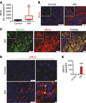
MLKL is strongly upregulated in hepatocytes of AIH patients and in experimental hepatitis. Although necrosis and necroinflammation
are central features of many liver diseases, the role of programmed necrosis in the context of inflammation-dependent hepatocellular death remains to be fully determined. Given that MLKL has been identified as a key mediator and potential biomarker of regulat-ed necrosis (7, 16), we initially determinregulat-ed hepatic expression of MLKL in patient cohorts with diverse liver diseases of viral, tox-ic, or autoimmune origin. While the abundance of MLKL tran-scripts was very low in healthy controls, patients with steatosis, and patients with primary biliary cirrhosis, we observed highly canonical necroptosis, other related pathways of programmed
necrosis may contribute to hepatocellular death. In particular, the significance of MLKL, the so-far most-terminal known end-stage effector of the necroptosis pathway, in the context of liver injury remains unclear at present.
[image:3.585.46.332.53.397.2]Autoimmune hepatitis (AIH) is a severe disease associated with chronic inflammation and fibrotic reorganization of liver tis-sue. The pathology of AIH is characterized by progressive destruc-tion of the hepatic parenchyma due to incompletely understood immune mechanisms that include activation of components of both the innate and the adaptive immune system (22). It has been shown that the severity of AIH correlated with the hepatic pres-ence of immune cells that stain positive for the proinflammatory cytokines IFN-γ and TNF-α (23). Moreover, several compelling findings in mouse models clearly demonstrated that the presence and upregulation of these cytokines directly drives hepatocellular cytotoxicity (24). Liver-resident or recruited T lymphocytes, NKT cells, and NK cells have been identified as primary producers of IFN-γ during hepatic inflammation, and IFN-γ signaling provokes death of hepatocytes through mechanisms that are poorly under-stood and likely involve several signaling pathways (25). IFN-γ rapidly stimulates STAT1 phosphorylation in vivo and in primary hepatocytes (26), and the IFN-γ/STAT1 axis has been suggested to block hepatocyte proliferation and induce hepatocyte death via apoptosis (27). However, to what extent IFN-γ–induced apoptosis contributes to death of hepatocytes in vivo remains unclear, and a
The Journal of Clinical Investigation
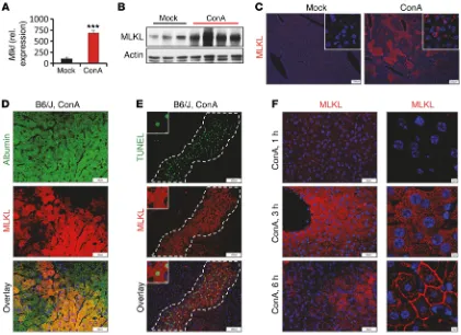
R E S E A R C H A R T I C L Ein steady-state murine liver tissue was weak, treatment of mice with ConA resulted in a rapid and strong increase in both Mlkl transcripts and protein, as evidenced by qPCR (Figure 2A and Sup-plemental Figure 2A), Western blot analysis (Figure 2B and Supple-mental Figure 2B), and immunohistochemistry (Figure 2C and
Sup-plemental Figure 2F). The absence of specific signals in Mlkl–/– mice
confirmed the specificity and suitability of the antibody used for detection (Supplemental Figure 2C). Immunofluorescent micros-copy further localized inflammation-dependent MLKL accumula-tion to hepatocytes, as demonstrated by double staining for MLKL and the hepatocyte marker albumin (Figure 2D and Supplemen-tal Figure 2D). Moreover, expression of Mlkl was pronounced in necrotic areas stained positive for the cell death marker TUNEL (Figure 2E). Accordingly, we observed that hepatic Mlkl mRNA expression correlated well with concentrations of circulating trans-aminases (Supplemental Figure 2E). Notably, at 3 hours after ConA injection, the MLKL protein was equally distributed throughout the cytoplasm of hepatocytes (Figure 2F). By contrast and similar to the staining pattern we observed in human AIH, at later stages, when hepatocytes stained positive for TUNEL, MLKL immunostaining was strongly localized to membranous compartments, including increased hepatic MLKL expression in patients with AIH (Figure
1A and Supplemental Figure 1A; supplemental material available online with this article; doi:10.1172/JCI87545DS1). Moreover, we also observed strong MLKL immunostaining in biopsies obtained from AIH patients. Remarkably, MLKL immunostaining in these samples was particularly restricted to hepatocytes in areas of severe inflammation and hepatocellular death (Figure 1, B and C). Phosphorylation of MLKL (pMLKL) and translocation of pMLKL to the plasma membrane has been demonstrated to be essential for necroptotic cell death (10, 28). Accordingly, strong pMLKL signals were present in affected areas in AIH tissue, whereas no pMLKL immunostaining was identified in healthy control tis-sues (Figure 1, D and E). Moreover, confocal microscopy clearly revealed subcellular MLKL and pMLKL localization to plasma membranes (Figure 1D and Supplemental Figure 1B), suggesting that MLKL activation is linked to disease activity in AIH.
[image:4.585.79.500.56.362.2]In order to study a potential role for MLKL in liver inflam-mation, we next took advantage of a mouse model in which hepatitis is induced by intravenous injection of the lectin con-canavalin A (ConA), which resembles some aspects of immune- mediated hepatitis in humans (29, 30). Whereas MLKL expression
during inflammation-dependent cell death. Consistently, IFN-γ serum concentrations and production by immune cells, which has been shown to be a central trigger for hepatocellular death
in the ConA model, were not significantly altered in Mlkl–/– mice
(Figure 3D and Supplemental Figure 3A). We have previously shown that STAT1 activation triggers IFN-γ–dependent hepato-cellular death (26). In order to investigate whether MLKL defi-ciency affects hepatic STAT1 signaling, we compared STAT1
activation in ConA-challenged wild-type and Mlkl–/– mice. While
hepatic pSTAT1Tyr701 was not detectable in unchallenged animals,
ConA administration induced a marked induction of pSTAT1Tyr701
in livers of control and Mlkl–/– mice (Supplemental Figure 3B). In
addition, primary mouse hepatocytes (PMHs) stimulated ex situ
with sera of ConA-challenged wild-type or Mlkl–/– mice released
similar amounts of the cytotoxicity marker LDH (Supplemental Figure 3C), suggesting that proinflammatory cytokine responses
in Mlkl–/– mice are not reduced to an extent that they
substantive-the plasma membrane, substantive-the nuclear membrane, and membranous compartments with morphological features of mitochondria (Fig-ure 2F and Supplemental Fig(Fig-ure 2, F and G). As it was previously shown that phosphorylation of MLKL coincides with MLKL oligo-merization and membrane translocation, these data strongly impli-cate MLKL activation in hepatocellular necrosis in the context of human and mouse inflammation–dependent liver disease.
Hepatocellular MLKL expression drives ConA-induced hep-atitis. In order to provide direct functional evidence for a role
of the MLKL protein in experimental hepatitis, we subjected
Mlkl–/– mice to ConA treatment. Interestingly, Mlkl–/– animals had
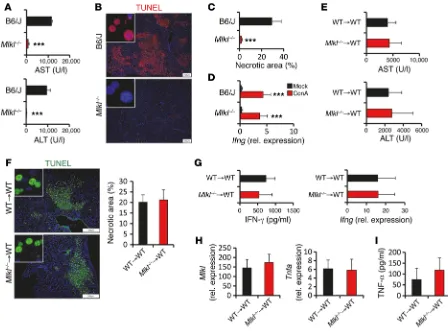
largely diminished plasma aspartate aminotransferase (AST) and alanine aminotransferase (ALT) concentrations (Figure 3A), compared with those in wild-type controls. Moreover, TUNEL staining revealed significantly reduced liver tissue necrosis and
death of hepatocytes in Mlkl–/– mice (Figure 3, B and C). Our
[image:5.585.66.514.55.386.2]data so far clearly point to a hepatocyte-intrinsic role of MLKL
Figure 3. MLKL expression in hepatocytes drives ConA-induced necrotic cell death. (A–D) C57BL/6 and Mlkl–/– mice were i.v. injected with ConA and
analyzed 7 hours later. Experiments were repeated 3 times with similar results. (A) Plasma AST/ALT concentrations (n = 4 per group). ***P < 0.001 by paired Student’s t test.(B) Representative images of liver sections stained by TUNEL assay. (C) Quantification of necrotic areas in TUNEL assay–stained livers of ConA-challenged mice (n = 4 per group). ***P < 0.001 by paired Student’s t test.(D) Quantification of Ifng transcripts in liver lysates of mock- or ConA-challenged control and Mlkl–/– mice by qPCR (n > 3 per group). ***P < 0.001 by paired Student’s t test.(E–I) 1 × 10⁷ bone marrow cells isolated from
control or Mlkl–/– mice were i.v. injected into lethally irradiated C57BL/6 recipient mice. Eight weeks later, mice were injected with ConA (n = 5 per group). (E)
The Journal of Clinical Investigation
R E S E A R C H A R T I C L Eer, in contrast to that in the ConA model, hepatic Mlkl transcripts were not increased and were comparable to saline-treated animals (Supplemental Figure 3D), suggesting that MLKL activation is not a pivotal cell death trigger in response to APAP. In line with this
and previous reports (19), Mlkl–/– mice developed strong APAP-
dependent liver injury, as demonstrated by plasma AST and ALT determination (Supplemental Figure 3E), H&E and TUNEL stain-ing, and quantification of the necrotic areas (Supplemental Figure 3F). Similarly, no changes in the cell death response were evident
in PMHs that were isolated from wild-type and Mlkl–/– mice and
treated ex situ with APAP (Supplemental Figure 3G). Collectively, our data demonstrate that MLKL is essential for inflammation- induced necrotic hepatocellular death.
RIPK1 but not RIPK3 activity is required for induction of experi-mental hepatitis. RIPK1 is known to regulate the RIPK3-dependent
phosphorylation and activation of MLKL, which is a hallmark of the canonical necroptosis pathway (7, 31–37). However, the role of these RIP kinases in models of experimental induced liver injury remains controversial and incompletely understood. Therefore, we next aimed to characterize the role of these RIPK proteins for the execution of MLKL-dependent hepatocellular death in the ly contribute to the protected phenotype of these mice in ConA
hepatitis. Although it was previously shown that Mlkl–/– mice did
not display a hematological disturbance under steady-state con-ditions (31), we further addressed the role of MLKL in hemato-poietic cells in chimeric mice generated by adoptive transfer of wild-type or Mlkl-deficient bone marrow into lethally irradiated wild-type recipient mice. Subsequently, administration of ConA induced a similar degree of liver injury in both chimeric strains, as AST and ALT plasma concentrations and TUNEL positivity of hepatocytes were comparable in both groups (Figure 3, E and F). Accordingly, in both chimeric strains, hepatic Mlkl, Ifng, and
Tnfa transcripts; serum concentrations of IFN-γ; and production of TNF-α by splenocytes were similarly increased (Figure 3, G–I). These data suggest that hepatocyte-intrinsic MLKL expression rather than functions in hematopoietic cells are required to medi-ate ConA-induced liver injury.
Recently, increased pMLKL staining was described around central veins in areas with significant damage in patients suffer-ing from DILI (16). Therefore, we elucidated the potential contri-bution of MLKL to murine DILI by measurement of hepatic Mlkl expression in livers of mice subjected to APAP overdose.
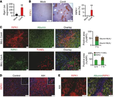
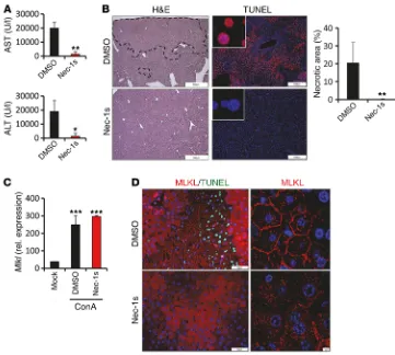
[image:6.585.95.485.56.389.2]ConA model. Similar to MLKL, RIPK1 was profoundly and rap-idly upregulated at both the mRNA and the protein level in livers after ConA treatment (Figure 4, A and B, and Supplemental Figure 4, A and B). Immunohistochemistry further revealed that RIPK1 staining clustered around hepatocytes in large foci at areas of pro-nounced tissue necrosis (Figure 4C and Supplemental Figure 4, B and C). In analogy to these findings in mice, we also observed a clustered RIPK1 staining pattern in hepatocytes in human AIH, whereas RIPK1 was only weakly expressed in the healthy liver (Figure 4, D and E). To ascertain whether ConA-induced liver injury depends on the kinase activity of RIPK1, we treated wild-type mice with the RIP1 kinase inhibitor necrostatin-1s (nec-1s) prior to ConA injection. Indeed, we observed low serum transam-inase concentrations (Figure 5A) and reduced necrosis (Figure 5B) in mice that received additionally nec-1s. Importantly, inhibition of RIPK1 kinase activity by nec-1s blocked the translocation of MLKL to the plasma membrane, while the induction of Mlkl gene expression in response to ConA was not affected (Figure 5, C and D). Notably, pretreatment with nec-1s had no impact on hepatic expression of Ifng, suggesting that the kinase activity of RIPK1 is involved in ConA-induced hepatocellular death downstream of T cell activation (Supplemental Figure 4D). In summary, these data indicate that RIPK1 is involved in MLKL-mediated necrotic dam-age during hepatic inflammation.
Although we observed only weak expression of RIPK3 in healthy murine liver tissue as compared with other organs (Supple-mental Figure 5, A and C), RIPK3 has been reported to be involved in the development of liver damage induced by APAP or alcohol
overdose (17, 20). However, in contrast to Ripk1 and Mlkl, Ripk3 hepatic transcripts were not increased in response to ConA or in human AIH (Supplemental Figure 5B). In line with these findings, we were unable to detect RIPK3 in hepatocytes by immunohisto-chemistry, and even late necrotic hepatocytes, in which the break-down of plasma membranes is traceable by β-catenin staining, did not stain positive for RIPK3 (Figure 6A). By contrast, we observed RIPK3 protein positivity in stromal cells that primarily represent
liver-resident F4/80+ Kupffer cells (Figure 6A and
Supplemen-tal Figure 5E). The absence of specific signals in Ripk3–/– mice
confirmed the specificity and suitability of the antibody used for detection (Supplemental Figure 5, C and D). Consistently, Ripk3 transcription was low in cultured PMHs (Supplemental Figure 5A). To study the functional contribution of RIPK3 to ConA-induced
hepatocellular death, we next subjected Ripk3–/– mice to ConA
treatment. In sharp contrast to the protection of mice in the
con-text of RIPK1 inhibition and MLKL deficiency, Ripk3–/– mice had
high plasma aminotransferase concentrations and developed a similar degree of histological liver damage as wild-type control mice (Figure 6, B and C, and Supplemental Figure 5, F and G). In addition, TUNEL staining and quantification of TUNEL-positive cells identified no changes in the degree of hepatocellular necrosis
in livers of Ripk3–/– mice (Figure 6, B and C). To further evaluate
these unexpected findings, we analyzed the subcellular
localiza-tion of MLKL in hepatocytes of ConA-injected Ripk3–/– mice by
confocal microscopy and Western blotting. Interestingly, similar to observations in wild-type mice, the MLKL protein clearly
[image:7.585.40.401.54.378.2]accu-mulated in affected areas of the plasma membrane in Ripk3–/–
Figure 5. ConA-induced necrotic cell death depends on the kinase activity of RIPK1. C57BL/6 mice were treat-ed with vehicle (DMSO) or nec-1s 30 minutes prior to ConA administration. Experiments were repeated 3 times with similar results. (A) Plasma concentrations of ALT and AST (n = 4 per group). *P < 0.05, **P < 0.01 by paired Student’s t test.(B) Represen-tative images of H&E- (dashed lines represent necrotic areas) and TUNEL assay–stained liver tissue sections and quantification of necrotic area (n = 4 per group). **P < 0.01 by paired Student’s
The Journal of Clinical Investigation
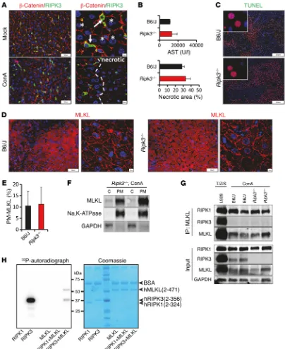
R E S E A R C H A R T I C L EFigure 6. RIPK3 is dispensable for ConA-induced hepatic injury. C57BL/6 and Ripk3–/– mice were i.v. injected with ConA and analyzed 7 hours later.
Exper-iments were repeated 3 times with similar results. (A) Representative images of liver tissue sections double stained for RIPK3 and β-catenin (confocal images are on the right; the dashed line separates necrotic areas from nonnecrotic areas; arrows demonstrate RIPK3-positive immune cells; asterisks mark representative hepatocytes). (B) Plasma AST concentrations of ConA-challenged control and Ripk3–/– mice (n > 3 per group) and evaluation of necrotic
areas in TUNEL assay–stained liver sections of control and Ripk3–/– mice (from C, n > 3 per group).(C) Representative images of immunohistochemical
(TUNEL assay) staining analysis of hepatic tissue sections. (D) Representative images of MLKL-stained liver tissue sections. (E) Quantification of plasma membrane localized MLKL (from D, n > 4 per group).(F) Western Blot analysis demonstrating that endogenous MLKL locates at the plasma membrane (PM) in Ripk3–/– mice following ConA treatment. C; cytoplasm. (G) Endogenous MLKL was immunoprecipitated with anti-MLKL antibody in lysates of L929
cells (treated for 3 hours with TNF-α/zVAD/SMAC mimetic) or in liver lysates of ConA-challenged wild-type or Ripk3–/– mice. (H)Recombinant hRIPK3,
but not hRIPK1, kinase domain undergoes autophosphorylation and can mediate hMLKL phosphorylation in vitro. Phosphoryl transfer of 32P by 25 ng/μl
Thus, in the setting of inflammation-dependent liver disease, a noncanonical type of regulated necrosis that does not require MLKL phosphorylation by RIPK3 is a critical trigger of hepatocellular death. Notably, RIPK1 itself was not able to directly phosphorylate MLKL in in vitro kinase assays, suggesting that additional mecha-nisms beyond RIPK3-mediated phosphorylation exist, where as-yet unidentified kinases could mediate MLKL activation (Figure 6H).
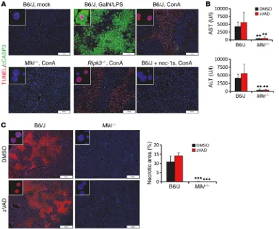
MLKL is not required for LPS/GalN-dependent hepatocellular apoptosis. While tissue necrosis has been clearly implicated in
ConA-induced liver inflammation, the role of hepatocellular apop-tosis in this model remains controversial. In comparison to mice challenged with LPS/D-galactosamine (LPS/GalN) (Figure 7A and Supplemental Figure 6A), a model known to be primarily mediat-ed by apoptotic hepatocellular death, we observmediat-ed only weak acti-vation of caspase-3 in liver areas with strong hepatocellular death in C57BL/6 wild-type mice subjected to ConA treatment (Figure 7A and Supplemental Figure 6A). Similarly, no cleaved caspase-3
signals were present in ConA-treated Mlkl–/– or Ripk3–/– mice and
mice pretreated with nec-1s (Figure 7A and Supplemental Figure 6A). These data suggest that apoptotic cell death does not substan-tially contribute to cell death during ConA-induced liver injury. In mice, as demonstrated by immunohistochemical analysis and
Western blotting (Figure 6, D–F, and Supplemental Figure 5H), suggesting that hepatocellular MLKL activation in ConA hepati-tis does not require RIPK3. Moreover, similar to ConA-challenged wild-type animals, hepatocellular necrosis was accompanied by profound clustering of RIPK1 into large foci in hepatocytes of
Ripk3–/– mice (Supplemental Figure 5I). During canonical TNF-α– mediated necroptosis, MLKL interacts with RIPK3 through its kinase-like domain within the necrosome complex (7). In order to test whether MLKL interacts with RIP kinases in the context of inflammation-dependent programmed hepatocellular necrosis, we next immunoprecipitated endogenous MLKL in liver lysates
of ConA-challenged wild-type and Ripk3–/– animals. Treatment of
L929 cells with TNF-α plus zVAD and SMAC mimetic served as a positive control for transient interaction of MLKL with RIPK3. Interestingly, we observed that MLKL is part of a complex that contains RIPK1 but not RIPK3 in the setting of ConA hepatitis, suggesting that MLKL- and RIPK1-containing protein complexes may assemble in the absence of RIPK3 (Figure 6G).
[image:9.585.95.500.56.389.2]Collectively, these data clearly indicate that RIPK3 but not RIPK1 is dispensable for ConA-induced hepatocellular necrosis.
Figure 7. Caspases are not required for ConA-induced hepatocellular death. (A) Representative images showing double immunofluorescence staining of cleaved caspase-3 and TUNEL assay in liver paraffin sections of unchallenged (mock), LPS/-GalN, or ConA-treated mice. (B and C) C57BL/6 or Mlkl–/– mice
The Journal of Clinical Investigation
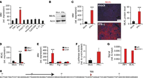
R E S E A R C H A R T I C L EIFN-γ–dependent STAT1 activation regulates MLKL expression in experimental hepatitis. Having shown that MLKL is strongly
upregulated in experimental hepatitis and AIH, our next objec-tive was to identify potential extracellular activators of Mlkl gene transcription. We therefore exposed PMHs ex situ to well-known triggers for necroptosis or cytokines involved in the pathogenesis of inflammatory liver disease. Interestingly, IFN-γ, a key driver of liver pathology, profoundly increased Mlkl transcription (Figure 8A) and protein abundance (Figure 8B). Furthermore, in vivo over-expression of IFN-γ by hydrodynamic delivery of DNA minicircles was sufficient to induce strong hepatic Mlkl gene expression that line with this notion, injection of a broad-spectrum caspase
inhibi-tor (zVAD) into control or Mlkl–/– mice before treatment with ConA
had no effect on tissue necrosis and the levels of serum transami-nases (Figure 7, B and C, and Supplemental Figure 6B).
In order to study a potential role of MLKL for hepatocellular
apoptosis, we subjected Mlkl–/– mice to LPS/GalN. However,
dif-ferent from the ConA model, Mlkl–/– mice developed strong LPS/
[image:10.585.40.536.56.328.2]GalN-dependent liver injury, as demonstrated by plasma AST and ALT determination and H&E and cleaved caspase-3 staining (Sup-plemental Figure 6, C–E), indicating that MLKL deficiency does not attenuate hepatocyte apoptosis in this model.
Figure 8. IFN-γ–dependent STAT1 activation regulates MLKL expression in experimental hepatitis. (A) Mlkl transcripts and (B) MLKL protein were quantified in PMHs stimulated ex situ with indicated factors (n = 3 per group). (C) IFN-γ expression constructs or empty control vectors (mock) were injected into C57BL/6 mice. Four days later, hepatic Mlkl mRNA or MLKL protein (n = 3 per group) was quantified by qPCR or Western blot. The presence of MLKL protein in livers was determined by specific immunofluorescent staining. (D) Relative Mlkl mRNA abundance in primary hepatocytes of wild-type or Stat1–/– mice stimulated with IFN-γ for 6 hours (n = 3 per group). (E) Quantification of hepatic Mlkl mRNA in vehicle- (mock) or ConA-challenged control
C57BL/6 mice, Ifng–/– mice, and Stat1–/– mice (n > 3 per group). (F) PMHs were transfected with a Mlkl luciferase promoter construct. Twenty-four hours
later, cells were stimulated with IFN-γ for 16 hours and firefly luciferase activity was determined (relative to mock group, n = 3 per group). (G) PMHs were stimulated with IFN-γ for 45 minutes. Subsequently ChIP was performed using a control antibody (IgG) or anti STAT1pTyr701 mAbs, as detailed in the
in vivo (Supplemental Figure 7J), and IFN-β stimulation of murine BNL liver cells (Supplemental Figure 7K) or MEFs (data not shown) resulted in increased Mlkl mRNA abundance, indicating that induction of MLKL expression may be a more general feature of IFN signaling. As IFN treatment is clinically important in sev-eral human diseases, this observation may be highly relevant for improving such therapeutic strategies. Notably, Ripk1 gene tran-scription was not regulated by IFN-dependent STAT1 activation (Supplemental Figure 8).
Contribution of TNF-α signaling to MLKL-mediated hepatocellu-lar death. While IFN-γ in vivo overexpression profoundly increased ConA-induced liver injury (Supplemental Figure 9, A and B), IFN-γ alone was not sufficient to rapidly induce pronounced hepatocel-lular death (data not shown). Consistent with this, treatment of primary hepatocytes with IFN-γ did not affect LDH release in vitro (Supplemental Figure 9C), suggesting that, in addition to IFN-γ– dependent MLKL upregulation, further signals are required for induction of inflammation-dependent hepatocellular death. In addition to IFN-γ, release of the proinflammatory cytokine TNF-α has been identified as an important contributor to liver injury (39). Given the well-known role of TNF-α in regulated necrosis, we therefore next addressed the role of this factor in the context of hepatic MLKL and RIPK1 activation. Although Tnfa transcripts
were strongly induced in the course of ConA challenge and Tnfr1–/–
mice displayed protection, confirming previously published data (40) (Supplemental Figure 10, A and B, and data not shown), hepatic Mlkl and Ripk1 expression was comparable between ConA-
challenged control and Tnfr1–/– mice (Supplemental Figure 8E and
Supplemental Figure 10, C and D), indicating that TNF-α signal-ing is not required for induction of Mlkl or Ripk1 gene transcrip-tion. By contrast, RIPK1 foci formation and MLKL translocation to membranous cellular compartments were profoundly reduced in
ConA-treated Tnfr1–/– mice as compared with controls
(Supplemen-tal Figure 10, E and F). In summary, these data implicate TNF-α in MLKL activation during experimentally induced hepatitis. Thus, IFN-γ and TNF-α synergistically drive inflammation-dependent hepatocellular death by regulation of MLKL transcription and its subsequent activation (Supplemental Figure 11).
Discussion
Irrespective of the cause of liver injury, hepatocellular death is a pathophysiologically important feature of many acute and chronic liver diseases (15). In some liver disorders with a strong inflamma-tory component, such as AIH, destruction of the liver parenchyma by cellular or soluble immune factors is a central trigger of dis-ease manifestation and progression. However, multiple signaling pathways are activated during hepatic inflammation, and so far precise therapeutically relevant mechanistic knowledge of the cell death machinery in hepatic diseases is limited. Here, we provide compelling evidence that the pseudokinase MLKL is an essential mediator of inflammation-driven hepatocellular necrosis in mice. By contrast, the activity of RIPK3 was not required for MLKL- directed death of hepatocytes in this setting, indicating the presence of a previously undescribed form of MLKL activation that may be physiologically relevant in liver diseases. On a molecular level, we further unraveled that the highly increased Mlkl gene expression in hepatocytes during inflammation was linked to STAT1 activation was restricted to hepatocytes, as demonstrated by MLKL-specific
immunostaining (Figure 8C). Given that IFN-γ has been shown to primarily mediate its biological function via the transcription factor STAT1, we stimulated primary hepatocytes of control and
Stat1–/– mice with IFN-γ and measured Mlkl expression. In support of an IFN-γ/STAT1–dependent regulation of Mlkl gene
expres-sion, IFN-γ did not induce Mlkl transcription in Stat1–/– mice or
STAT1-deficient hepatocytes (Figure 8D and Supplemental Fig-ure 7A). IFN-γ/STAT1 signaling is essential for ConA-mediated liver injury, and T cells and NKT cells were identified as primary producers of IFN-γ in this model (26, 29). Thus, we next analyzed
the expression of Mlkl in ConA-treated Stat1–/–, Ifng–/–, and
lymph-openic Rag1–/– mice. Remarkably, in all three strains hepatic Mlkl
expression was not induced in vivo, and this correlated to reduced hepatocyte death and tissue damage (Figure 8E and Supplemen-tal Figure 7, B–E). Collectively, these data demonstrate that in vivo
Mlkl expression is induced by an IFN-γ/STAT1–dependent path-way requiring the presence of lymphocytes.
Interestingly, in silico analysis identified several poten-tial STAT1-binding sites in the genomic region upstream of the
Mlkl-coding sequence (Figure 8H), indicating that Mlkl gene
tran-scription may be directly induced by IFN-γ–dependent STAT1 activation. To address this question, we cloned upstream DNA fragments of the Mlkl genomic region and generated luciferase reporter constructs for reporter gene assays. Interestingly, basal luciferase activity after transient transfection of an approximate-ly 1-kb fragment, including the putative Mlkl transcriptional start site, into primary hepatocytes strongly increased upon IFN-γ stimulation (Figure 8F). Similar results were obtained in IFN-γ– treated human embryonic kidney cells, BNL cells, and mouse embryonic fibroblasts (MEFs) (Supplemental Figure 7F and data not shown). Moreover, ChIP assays and quantitative ChIP using a pSTAT1-specific antibody for precipitation identified STAT1 bind-ing to predicted γ-activated sequences in the Mlkl promoter region in IFN-γ–stimulated BNL liver cells (Figure 8G). Binding of STAT1 to a previously described site in the Irf1 promoter served as a pos-itive control (38) (Figure 8G). In summary, these data suggest that the proinflammatory cytokine IFN-γ directly regulates Mlkl gene transcription via activation of STAT1.
Notably, while previous studies implicated the IFN-γ/STAT1 pathway in provoking hepatocellular death by induction of apop-tosis (25), our data support the notion that this pathway promotes MLKL-mediated programmed necrosis rather than apoptosis.
The Journal of Clinical Investigation
R E S E A R C H A R T I C L Eoligomerization, membrane translocation, and permeabilization. Moreover, additional phosphorylation sites in the MLKL protein might control its activation and subsequent programmed necrosis independent of RIPK3 (28). As a result, the responsible kinases and mechanisms underlying MLKL regulation by phosphorylation at these additional sites remain matters of ongoing interest.
In summary, these results provide a rationale for investigation of MLKL as a possible diagnostic biomarker for hepatocellular necrosis in AIH and other inflammation-dependent liver diseases. In addition, our findings support the concept that specific mod-ulation of the MLKL pathway may be therapeutically relevant to decrease inflammation-dependent hepatocellular death and its fatal consequences in chronic human liver disorders.
While we provided compelling evidence for an essential role of MLKL during lymphocyte-mediated hepatitis, MLKL deficien-cy did not ameliorate APAP-induced liver injury in mice. Although previous studies have shown that RIPK3 contributes to necrotic cell death after APAP overdose (17, 21), oxidative stress–related mitochondrial damage may be closely related to death of hepato-cytes in this model (47). This indicates that in hepatohepato-cytes sev-eral parallel mechanisms exist to execute necrotic cell death in vivo as previously shown in vitro for a variety of other cell types (15). Whereas APAP overdose and other forms of DILI are char-acterized by sterile inflammation, hepatocellular death in the ConA model and potentially AIH depends to a large extent on the release of cytokines by T lymphocytes, NKT cells, and NK cells. Accordingly, we identified IFN-γ, a cytokine previously described as essential for ConA-induced hepatitis, as a critical mediator of MLKL-dependent hepatocellular death. For the first time to our knowledge, we describe that IFN-γ signal transduction in hepato-cytes, via activation of STAT1 and transactivation of the Mlkl moter, rapidly increased the amount of MLKL transcripts and pro-tein, which are low under steady-state conditions.
PMHs display spontaneous cell death during long-term cell culture but show rather low sensitivity to necroptosis induction by TNF-α/zVAD/SMAC mimetic treatment (43, 48). Howev-er, pretreatment of mice or cells with IFN-γ upregulated Mlkl in hepatocytes and increased their susceptibility toward cell death (Supplemental Figure 12). Similarly, it was recently reported that MEFs may require IFN pretreatment for effective necroptosis induction (49), indicating that sensitization of cells with IFNs may represent a general mechanism supporting programmed necrosis. IFN-induced necroptosis was described in macrophages in the
presence of caspase inhibitors. Interestingly, in this study Ifnar1–/–
macrophages were resistant to TNF-α/zVAD–induced necropto-sis, indicating that feedback signaling through IFNs is essential for sustained activation of RIP kinases in response to TNF-α, LPS, or poly(I:C) (50). Our data now provide compelling evidence that, in addition to such other cell death–promoting properties of IFNs, the transcriptional regulation of MLKL by these factors is par-ticularly important in cells that do not express substantial basal levels of MLKL, such as hepatocytes. Thus, while previous stud-ies implicated the IFN-γ/STAT1 pathway in promoting hepato-cellular death by induction of apoptosis (25), our data support the notion that this pathway provokes MLKL-mediated programmed necrosis of hepatocytes rather than apoptosis. These data add to the molecular understanding of IFN-mediated tissue damage by the proinflammatory and cytotoxic cytokine IFN-γ, suggesting
that IFN signaling–dependent regulation of MLKL is an important mechanism of sensitization of cells to regulated necrosis.
We demonstrate that elevated levels of MLKL mRNA and pro-tein in hepatocytes relate to hepatic injury both during experimen-tally induced hepatitis and AIH in humans. Importantly, its upreg-ulation in hepatitis clearly correlated to strong MLKL translocation to the plasma membrane and other membranous compartments, indicating that MLKL-dependent disruption of cellular integrity contributes to inflammation-dependent death of hepatocytes. In current models of necroptosis, MLKL plasma membrane transloca-tion is driven by RIPK3-dependent phosphorylatransloca-tion (10, 11, 16, 41, 42). Importantly, while we observed a strong attenuation of acute
liver damage in Mlkl–/– mice, our data provided several lines of
evi-dence that, during experimental hepatitis, MLKL activation and translocation to membranous compartments occurs
independent-ly of RIPK3. Consistentindependent-ly, Ripk3–/– mice developed ConA-induced
hepatitis as severe as control mice. Accordingly, we observed only low basal levels of RIPK3 in hepatocytes, and similar to other reports (19, 43), RIPK3 positivity was rather limited to nonparen-chymal cells, particularly Kupffer cells. Although we have previ-ously shown that RIPK3 is upregulated in intestinal epithelial cells undergoing TNF-α–triggered necroptosis (8), we were unable to detect induction of RIPK3 mRNA or protein in hepatocytes during the course of experimentally induced hepatitis or in human AIH. In line with this, a recent study demonstrated that RIPK3 expression in hepatocytes is dispensable for inflammatory liver injury (43).
In some experiments, PMHs, BNL cells, or MEFs were stimulated with 100 ng/ml of recombinant mouse IFN-β (R&D Systems), 100 ng/ ml recombinant mouse IFN-γ (Preprotech), 100 ng/ml recombinant mouse IL-28 (IFN-λ, R&D Systems), 8 mM APAP, or murine sera in a final concentration of 3%. HEK293T cell were stimulated with 100 ng/ml recombinant human IFN-γ (Roche). For cytokine release, iso-lated splenocytes were cultured in RPMI 1640 supplemented with PMA (50 ng/ml, Calbiochem) in combination with ionomycin (500 ng/ml, Cayman) for 24 hours. Cell viability was measured using the Cytotoxitiy Detection Kit (LDH, Roche) or the Cell Death Detection ELISAPLUS Kit (Roche) accordingly to the manufacturer’s instruction.
Cell viability was calculated as follows: cell viability level = 1/mean cell viability of the control group.
Cytokine measurements. For determination of mouse IFN-γ or TNF-α, specific DuoSet ELISA Kits from R&D Systems were used. ELISA was performed according to manufacturer’s instructions.
Transient transfections and luciferase assays. An approximately 1-kb fragment, including the putative mouse Mlkl gene promoter sequence, was identified using the Genomatix software suite (http://www. genomatix.de) and amplified using the PfuUltra II Fusion HS DNA Polymerase (Aligent Technologies) out of tail genomic DNA using the primers 5′-ccctacttgggtaacttcac-3′ and 5′-gaatgccattcctcgatctc-3′. The PCR product was finally cloned upstream of the firefly luciferase open reading frame in the pGL-4.1 vector (Promega) using the NheI and HindIII sites. For the transfection of HEK293T or PMH cells, the cells were seeded at 300,000 (HEK293T) or 500,000 (PMH) cells per 6-well plate and transfected 1 day after with 5 μg DNA using Lipo-fectamine 2000 transfection reagent according to the manufacturer’s instructions. The Luciferase Reporter Assay System from Promega was used for the analysis of luciferase activity in cell lysates according to the manufacturer’s instructions.
ChIP assay. Chip assay was performed using the SimpleChIP Enzy-matic Chromatin IP Kit (Magnetic Beads) from Cell Signaling accord-ingly to the manufacturer’s instruction. In brief, BNL cells were stimu-lated with IFN-γ for 45 minutes, fixed by addition of 1% formaldehyde, lysed, and treated with 1,200 U Micrococcus nuclease. Protein-DNA complexes were immunoprecipitated overnight using anti-pSTAT1Tyr701
(Cell Signaling, catalog 9167) or control antibodies (Cell Signaling, cat-alog 2729) and protein A–coupled magnetic beads. Real-time PCR on silica column–purified DNA was performed using the following prim-ers: STAT1-Mlkl 5′-gaactgagcgatagac-3′ and 5′-cgaaaccaaccctaag-3′ or STAT1-Irf1 5′-agcacagctgacttcc-3′ and 5′-cttagactgtacgtcc-3′ Quantifi-cation of DNA by qPCR was performed as follows: percentage input = 2% × 2 (C[T] 2% input sample – C[T] IP sample), where C[T] denotes the threshold cycle of the PCR reaction.
IP and immunoblotting. Proteins were isolated from liver biopsies using Cell lysis buffer (Cell Signaling) supplemented with 1 mM PMSF (Cell Signaling). Isolation of plasma membrane proteins was performed using the Plasma Membrane Protein Extraction Kit (catalog ab65400, abcam) accordingly to the manufacturer’s instructions. Lysates were then centrifuged at 18,000 g for 20 minutes. For IP experiments super-natants were precleared and subsequently immunoprecipitated with anti-MLKL antibody (1:50, Merck Millipore, clone 3H1) at 4°C over-night. Subsequently, lysates were further incubated with protein G– coupled magnetic beads for 20 minutes at room temperature. After IP, the beads were washed 4 times with lysis buffer, and the immunopre-cipitated proteins were subsequently eluted with SDS sample buffer.
and establish a link between proinflammatory cytokine produc-tion during inflammaproduc-tion and regulated necrosis beyond TNF-α– induced signal transduction. Of note, IFNs are not only implicated in many autoimmune diseases but broadly used in the therapy of, for example, viral hepatitis and multiple sclerosis. Therapy is com-monly associated with adverse side effects, such as hepatoxicity, autoantibody production, and AIH, and this leads to dose reduc-tions or termination of therapy (51, 52). IFN-mediated induction of MLKL and subsequent activation of hepatocellular necrosis may significantly contribute to these side effects, and this obser-vation may thus be relevant for the future of IFN-related therapies. Such strategies may involve cotreatment of patients with IFNs and chemical compounds that inhibit the activity of MLKL.
Collectively, our data uncover a form of programmed necrot-ic cell death that is dependent on the RIPK1/MLKL axis but does not require RIPK3. Depending on the physiological context, MLKL has the capacity to promote RIPK3-mediated necroptosis or alter-natively triggers IFN-mediated RIPK3-independent programmed necrosis. In addition, our results suggest a physiological relevance for IFN-triggered MLKL-dependent death in the pathogenesis of inflammation-dependent liver injury such as AIH.
Methods
Animals and housing of mice. C57BL/6, Rag1–/–, Stat1–/–, and Tnfr1–/–
mice were obtained from The Jackson Laboratory. Ripk3−/− mice were
described previously (53). Ifng–/– and Ifnar–/– mice were provided by
U. Schleicher. Mlkl–/– mice were provided by J. Murphy. Throughout,
we used either C57BL/6J or littermate controls to exclude strain- dependent differences in disease susceptibility. Mice were routinely screened for pathogens according to FELASA guidelines.
Animal model of liver injury. LPS and GalN (catalog G1639) were obtained from Sigma-Aldrich. ConA was from Merck Millipore (cata-log 234567). APAP was from Braun. Z-Val-Ala-DL-Asp-fluoromethyl-ketone (zVAD) was from Bachem (catalog N-1510). Nec-1s was from BioVision (catalog 2263). ConA was administered intravenously (25 mg/kg body weight) alone or in combination with nec-1s (400 μg, i.p., 30 minutes) or zVAD (250 μg/mouse, i.p., 15 minutes) before ConA administration. Stock solutions of nec-1s were prepared in DMSO (final concentration 0.5%). D-GalN (1 mg/kg) was injected i.p. 1 hour prior to administration of LPS (100 μg/kg, i.p.). APAP (400 mg/kg) was injected i.p. after overnight fasting. Plasma concentrations of AST and ALT were measured in the clinical chemistry unit of the University Medical Center Erlangen.
For generation of bone marrow chimeras, 1 × 10⁷ cells from femurs
and tibias of donor mice were intravenously injected into lethally irra-diated (10 Gy) recipient C57BL/6 mice. Mice were left to recover for 8 weeks before ConA was administrated.
The Journal of Clinical Investigation
R E S E A R C H A R T I C L Edex-200 gel filtration columns (GE Healthcare) in 200 mM NaCl, 20 mM HEPES, pH 7.5, 5% glycerol; concentrated by centrifugal ultrafil-tration to 6.5–7 mg/ml; aliquoted; and snap frozen in liquid N2, before storage at –80°C until required.
In vitro kinase assays on purified proteins. Kinase assays for purified recombinant kinase domains of hRIPK1 and hRIPK3 in the presence or absence of recombinant human MLKL were performed essential-ly as described previousessential-ly (31, 58). Briefessential-ly, 3 μg recombinant hMLKL was incubated in the absence or presence of 100 ng of recombinant hRIPK1 or hRIPK3 for 5 minutes at 25°C after the reaction was initi-ated by addition of 5 μCi 32P-γ-ATP and 37.5 μM cold ATP in a 40-μl
reaction. In parallel, 1 μg of either hRIPK1 or hRIPK3 kinase domain was reacted analogously in the absence of substrate. Kinase reaction buffer comprised 100 mM NaCl, 20 mM Tris, pH 8, 4 mM MgCl2, 0.1 mM DTT, and 1 mg/ml BSA. Reactions were terminated by boiling in 1× reducing SDS-PAGE loading buffer for 5 to 10 minutes before one-quarter of each reaction was resolved by 4%–12% Bis-Tris reduc-ing SDS-PAGE. The gel stained with SimplyBlue SafeStain (Thermo Fisher) was dried under a vacuum and exposed to a phosphor screen for 16 hours before scanning using a Typhoon imager (GE Healthcare).
Statistics. Statistical analysis was performed using the 2-tailed Student’s t test. P values of less than 0.05 were considered significant.
Study approval. Animal protocols were approved by the Institu-tional Animal Care and Use Committee of the Regierung von Unter-franken. All studies with human material were approved by the ethics committee of the University Hospital of Erlangen. The diagnosis of AIH was defined according to the guidelines of the European Associ-ation for the Study of Liver Disease (59) and was based on compatible symptoms and signs of AIH, elevated ALT and AST, increased serum IgG, serological findings (elevated antinuclear antibodies, smooth muscle antibody, soluble liver antigen, or liver kidney/microsome antibodies), and histological findings, such as interface hepatitis and plasma cell infiltration. The acute forms of DILI can be divided into hepatocellular, cholestatic, and mixed forms. In this study cohort of DILI patients, all cases presented mixed forms indicated by the pres-ence of hepatocellular and cholangiocellular damage. The extent of hepatocellular and cholangiocellular toxicity was variable but never purely hepatocellular or cholestatic. Other conditions that may cause hepatitis, including viral, drug-induced, cholestatic, metabolic, and hereditary disorders, have been excluded in these patients.
Author contributions
CG, SW, and CB designed the research. CG, GWH, JMM, and EJP performed the experiments. AEK, KA, PV, AL, CP, CD, SK, US, and MFN supplied materials that made this study possible. CG, CB, and SW analyzed the data and wrote the paper. PV, JMM, and SK gave feedback on the draft paper.
Acknowledgments
The authors thank C. Lindner, G. Förtsch, A. Taut, S. Wallmüller, H. Dorner, and V. Thonn for excellent technical assistance. We
thank W. Alexander for providing the Mlkl–/– mice. We thank N.
Takahashi and Y. Dondelinger for providing materials and S. Zopf for sampling of liver biopsies. We thank Sam Young for assistance with preparing recombinant protein expression constructs and bacmids. This research has received funding from DFG proj-ects within SFB796, SPP1656, SFB1181-A08, project BE3686/2,
Proteins were separated using a MiniProtean-TGX gel (4%–15% poly-acrylamide; Bio-Rad) and transferred from the gel to a nitrocellulose membrane (Whatman). Membranes were probed with the following primary antibodies: murine MLKL (Biorbyt, catalog orb32399); RIPK1 (catalog 3493), Na, K-ATPase (catalog 3010), pSTAT1 (catalog 9167), and STAT1 (catalog 14994) (all from Cell Signalling); and murine RIPK3 (catalog ab62344), GAPDH (catalog ab9482), and ACTIN (cat-alog ab49900) (Abcam). HRP-linked anti-rabbit (cat(cat-alog 7074) (Cell Signaling) was used as a secondary antibody.
Liver-specific vectors and hydrodynamic tail vein injection. The generation of vectors encoding secreted version of IFNs controlled by liver-specific regulatory elements was described previously (55). Sustained in vivo transduction of hepatocytes with IFNs was achieved by hydrodynamic delivery of 10 μg vector in ringer solution (10% of body weight). Overexpression of IFNs in hepatocytes was confirmed by ELISA. Mice injected with empty control vector served as controls.
Histology and immunohistochemistry. Histopathological analyses were performed on formalin-fixed paraffin-embedded tissue after Mayer’s H&E staining. Immunofluorescence of tissue sections was performed using the TSA Cy3/Fluorescein system, as recommended by the manufacturer (Perkin Elmer). The following antibodies were used: murine MLKL (Biorbyt, catalog orb32399); β-catenin (catalog 8480), human MLKL (catalog 14993), human pMLKL (catalog 14516), RIPK1 (catalog 3493), and cleaved caspase-3 (catalog 9661) (all from Cell Signaling); murine RIPK3 (catalog ab62344), albumin (catalog ab106582), and anti-chicken (catalog ab150169) (all from Abcam); F4/80 (AbD Serotec, catalog MCA497); and anti-rabbit (Dianova, catalog 111-065-144). Nuclei were counterstained with Hoechst 3342 (Invitrogen). Cell death (TUNEL) was analyzed using the In Situ Cell Death Detection Kit (Roche). Murine RIPK1 expression was analyzed using the Envision+ system (Dako). Images were obtained using a
con-focal fluorescence microscope (LEICA TCS SP5 II) or the microscope LEICA DMI 4000B, together with the LEICA DFC360 FX or LEICA DFC420 C camera and the imaging software “LAS AF” (Leica).
Gene expression. Total RNA was extracted from liver tissue using the peqGOLD Total RNA Kit and from cells using the peqGOLD Microspin Total RNA Kit (Peqlab). cDNA was synthesized using the SCRIPT cDNA Synthesis Kit from Jena Bioscience and analyzed by real-time PCR using specific QuantiTect Primer assays (Qiagen). Experimental values were normalized to levels of the housekeeping gene hypoxanthine guanine phosphoribosyl transferase (Hprt).
Super-ture Support Scheme. SK acknowledges support from Dr. Werner Jackstädt-Stiftung and Fresenius Medical Care Germany.
Address correspondence to: Stefan Wirtz, Medical Department 1, University of Erlangen-Nuremberg Hartmannstrasse 14, 91052 Erlangen, Germany. Phone: 49.0.9131.85.35882; E-mail: stefan. wirtz@uk-erlangen.de.
KR4391/1-1, and the clinical research unit KFO257. Further support was given by the Interdisciplinary Center for Clinical Research of the University Erlangen-Nuremberg. JMM acknowl-edges support from the National Health and Medical Research Council of Australia (project 1057905, fellowship 1105754, and Independent Research Institute Infrastructure Support Scheme, 9000220) and the Victorian Government Operational
1. Vandenabeele P, Galluzzi L, Vanden Berghe T, Kroemer G. Molecular mechanisms of necropto-sis: an ordered cellular explosion. Nat Rev Mol
Cell Biol. 2010;11(10):700–714.
2. Degterev A, et al. Chemical inhibitor of nonapoptotic cell death with therapeutic poten-tial for ischemic brain injury. Nat Chem Biol. 2005;1(2):112–119.
3. Vercammen D, et al. Dual signaling of the Fas receptor: initiation of both apoptotic and necrotic cell death pathways. J Exp Med. 1998;188(5):919–930.
4. Zhang H, Zhou X, McQuade T, Li J, Chan FK, Zhang J. Functional complementation between FADD and RIP1 in embryos and lymphocytes.
Nature. 2011;471(7338):373–376.
5. Kaiser WJ, et al. RIP3 mediates the embryonic lethality of caspase-8-deficient mice. Nature. 2011;471(7338):368–372.
6. Oberst A, et al. Catalytic activity of the caspase-8-FLIP(L) complex inhibits RIPK3-dependent necrosis. Nature. 2011;471(7338):363–367. 7. Sun L, et al. Mixed lineage kinase domain-like
protein mediates necrosis signaling downstream of RIP3 kinase. Cell. 2012;148(1–2):213–227. 8. Günther C, et al. Caspase-8 regulates TNF-α-
induced epithelial necroptosis and terminal ileitis. Nature. 2011;477(7364):335–339. 9. Feoktistova M, et al. cIAPs block Ripoptosome
formation, a RIP1/caspase-8 containing intracel-lular cell death complex differentially regulated by cFLIP isoforms. Mol Cell. 2011;43(3):449–463. 10. Cai Z, et al. Plasma membrane
transloca-tion of trimerized MLKL protein is required for TNF-induced necroptosis. Nat Cell Biol. 2014;16(1):55–65.
11. Hildebrand JM, et al. Activation of the pseudo-kinase MLKL unleashes the four-helix bundle domain to induce membrane localization and necroptotic cell death. Proc Natl Acad Sci U S A. 2014;111(42):15072–15077.
12. Bonnet MC, et al. The adaptor protein FADD pro-tects epidermal keratinocytes from necroptosis in vivo and prevents skin inflammation. Immunity. 2011;35(4):572–582.
13. Ma X, et al. The oncogenic microRNA miR-21 promotes regulated necrosis in mice. Nat
Com-mun. 2015;6:7151.
14. Linkermann A, et al. Two independent path-ways of regulated necrosis mediate isch-emia-reperfusion injury. Proc Natl Acad Sci U S A. 2013;110(29):12024–12029.
15. Luedde T, Kaplowitz N, Schwabe RF. Cell death and cell death responses in liver disease: mech-anisms and clinical relevance. Gastroenterology. 2014;147(4):765–783.e4.
16. Wang H, et al. Mixed lineage kinase domain-like protein MLKL causes necrotic membrane
dis-ruption upon phosphorylation by RIP3. Mol Cell. 2014;54(1):133–146.
17. Ramachandran A, McGill MR, Xie Y, Ni HM, Ding WX, Jaeschke H. Receptor interacting pro-tein kinase 3 is a critical early mediator of acet-aminophen-induced hepatocyte necrosis in mice.
Hepatology. 2013;58(6):2099–2108.
18. Takemoto K, et al. Necrostatin-1 protects against reactive oxygen species (ROS)-induced hepato-toxicity in acetaminophen-induced acute liver failure. FEBS Open Bio. 2014;4:777–787. 19. Dara L, et al. Receptor interacting protein kinase
1 mediates murine acetaminophen toxicity independent of the necrosome and not through necroptosis. Hepatology. 2015;62(6):1847–1857. 20. Roychowdhury S, McMullen MR, Pisano SG,
Liu X, Nagy LE. Absence of receptor interacting protein kinase 3 prevents ethanol-induced liver injury. Hepatology. 2013;57(5):1773–1783. 21. Deutsch M, et al. Divergent effects of RIP1 or
RIP3 blockade in murine models of acute liver injury. Cell Death Dis. 2015;6:e1759.
22. Manns MP, Lohse AW, Vergani D. Autoimmune hepatitis--Update 2015. J Hepatol. 2015;62(1 Sup-pl):S100–S111.
23. Hussain MJ, Mustafa A, Gallati H, Mowat AP, Mieli-Vergani G, Vergani D. Cellular expression of tumour necrosis factor-alpha and interferon-gam-ma in the liver biopsies of children with chronic liver disease. J Hepatol. 1994;21(5):816–821. 24. Küsters S, Gantner F, Künstle G, Tiegs G.
Inter-feron gamma plays a critical role in T cell-depen-dent liver injury in mice initiated by concanavalin A. Gastroenterology. 1996;111(2):462–471. 25. Horras CJ, Lamb CL, Mitchell KA. Regulation of
hepatocyte fate by interferon-γ. Cytokine Growth
Factor Rev. 2011;22(1):35–43.
26. Siebler J, et al. A key pathogenic role for the STAT1/T-bet signaling pathway in T-cell- mediated liver inflammation. Hepatology. 2003;38(6):1573–1580.
27. Sun R, et al. STAT1 contributes to dsRNA inhibi-tion of liver regenerainhibi-tion after partial hepatecto-my in mice. Hepatology. 2006;44(4):955–966. 28. Tanzer MC, et al. Necroptosis signalling is tuned
by phosphorylation of MLKL residues outside the pseudokinase domain activation loop. Biochem J. 2015;471(2):255–265.
29. Tiegs G, Hentschel J, Wendel A. A T cell-depen-dent experimental liver injury in mice inducible by concanavalin A. J Clin Invest. 1992;90(1):196–203. 30. Heymann F, Hamesch K, Weiskirchen R, Tacke
F. The concanavalin A model of acute hepatitis in mice. Lab Anim. 2015;49(1 Suppl):12–20. 31. Murphy JM, et al. The pseudokinase MLKL
medi-ates necroptosis via a molecular switch mecha-nism. Immunity. 2013;39(3):443–453. 32. Degterev A, et al. Identification of RIP1 kinase
as a specific cellular target of necrostatins. Nat
Chem Biol. 2008;4(5):313–321.
33. Dillon CP, et al. RIPK1 blocks early postnatal lethality mediated by caspase-8 and RIPK3. Cell. 2014;157(5):1189–1202.
34. Rickard JA, et al. RIPK1 regulates RIPK3-MLKL-driven systemic inflammation and emergency hematopoiesis. Cell. 2014;157(5):1175–1188. 35. Kaiser WJ, et al. RIP1 suppresses innate immune
necrotic as well as apoptotic cell death during mammalian parturition. Proc Natl Acad Sci U S A. 2014;111(21):7753–7758.
36. Duprez L, et al. RIP kinase-dependent necrosis drives lethal systemic inflammatory response syndrome. Immunity. 2011;35(6):908–918. 37. Rodriguez DA, et al. Characterization of
RIPK3-mediated phosphorylation of the activa-tion loop of MLKL during necroptosis. Cell Death
Differ. 2016;23(1):76–88.
38. Pine R, Canova A, Schindler C. Tyrosine phosphor-ylated p91 binds to a single element in the ISGF2/ IRF-1 promoter to mediate induction by IFN alpha and IFN gamma, and is likely to autoregulate the p91 gene. EMBO J. 1994;13(1):158–167.
39. Maeda S, Chang L, Li ZW, Luo JL, Leffert H, Karin M. IKKbeta is required for prevention of apoptosis mediated by cell-bound but not by circulating TNFalpha. Immunity. 2003;19(5):725–737. 40. Küsters S, et al. In vivo evidence for a functional
role of both tumor necrosis factor (TNF) recep-tors and transmembrane TNF in experimental hepatitis. Eur J Immunol. 1997;27(11):2870–2875. 41. Chen X, et al. Translocation of mixed lineage
kinase domain-like protein to plasma mem-brane leads to necrotic cell death. Cell Res. 2014;24(1):105–121.
42. Dondelinger Y, et al. MLKL compromises plasma membrane integrity by binding to phosphatidylinositol phosphates. Cell Rep. 2014;7(4):971–981.
43. Kang YJ, et al. Regulation of NKT cell-mediated immune responses to tumours and liver inflam-mation by mitochondrial PGAM5-Drp1 signal-ling. Nat Commun. 2015;6:8371.
44. Newton K, Manning G. Necroptosis and inflam-mation. Annu Rev Biochem. 2016;85:743–763. 45. Dondelinger Y, Hulpiau P, Saeys Y, Bertrand MJ,
Vandenabeele P. An evolutionary perspective on the necroptotic pathway [published online ahead of print June 28, 2016]. Trends Cell Biol. doi: 10.1016/j.tcb.2016.06.004.
46. Tanzer MC, et al. Evolutionary divergence of the necroptosis effector MLKL. Cell Death Differ. 2016;23(7):1185–1197.
The Journal of Clinical Investigation
R E S E A R C H A R T I C L Edamage and nuclear DNA fragmentation. J Clin
Invest. 2012;122(4):1574–1583.
48. Vinken M, et al. Primary hepatocytes and their cultures in liver apoptosis research. Arch Toxicol. 2014;88(2):199–212.
49. Rodriguez DA, et al. Characterization of RIPK3-mediated phosphorylation of the activa-tion loop of MLKL during necroptosis. Cell Death
Differ. 2016;23(1):76–88.
50. McComb S, et al. Type-I interferon signal-ing through ISGF3 complex is required for sustained Rip3 activation and necroptosis in macrophages. Proc Natl Acad Sci U S A. 2014;111(31):E3206–E3213.
51. Dusheiko G. Side effects of alpha interferon in chronic hepatitis C. Hepatology. 1997; 26(3 Suppl 1):112S–121S.
52. García-Buey L, et al. Latent autoimmune
hepati-tis triggered during interferon therapy in patients with chronic hepatitis C. Gastroenterology. 1995;108(6):1770–1777.
53. Newton K, Sun X, Dixit VM. Kinase RIP3 is dis-pensable for normal NF-kappa Bs, signaling by the B-cell and T-cell receptors, tumor necrosis factor receptor 1, and Toll-like receptors 2 and 4.
Mol Cell Biol. 2004;24(4):1464–1469.
54. Bajt ML, Knight TR, Lemasters JJ, Jaeschke H. Acetaminophen-induced oxidant stress and cell injury in cultured mouse hepatocytes: protection by N-acetyl cysteine. Toxicol Sci. 2004;80(2):343–349.
55. McHedlidze T, et al. Interleukin-33-dependent innate lymphoid cells mediate hepatic fibrosis.
Immunity. 2013;39(2):357–371.
56. Murphy JM, et al. A robust methodology to subclas-sify pseudokinases based on their
nucleotide-bind-ing properties. Biochem J. 2014;457(2):323–334. 57. Babon JJ, Murphy JM. In vitro JAK kinase
activ-ity and inhibition assays. Methods Mol Biol. 2013;967:39–55.
58. Cook WD, et al. RIPK1- and RIPK3-induced cell death mode is determined by target availability.
Cell Death Differ. 2014;21(10):1600–1612.
59. European Association for the Study of the Liver. EASL Clinical Practice Guidelines: Autoimmune hepatitis. J Hepatol. 2015;63(4):971–1004. 60. Cartharius K, et al. MatInspector and beyond:
pro-moter analysis based on transcription factor bind-ing sites. Bioinformatics. 2005;21(13):2933–2942. 61. Marinescu VD, Kohane IS, Riva A. MAPPER: a