© 2017, IRJET | Impact Factor value: 5.181 | ISO 9001:2008 Certified Journal | Page 1734
Detection of Macular Edema by Using Various Techniques of Feature
Extraction and Classification by SVM Classifier
Ajay Advani
1, Ashish Thawrani
2, Amit Thaware
3,Abhinay Gaonkar
4,Mrs.Vaishali Kulloli
51,2,3,4Student, Information Technology, Pimpri Chinchwad College of Engineering, Maharashtra, India. 5AssistantProfessor, Information Technology, Pimpri Chinchwad College of Engineering, Maharashtra, India.
---***---Abstract -
Diabetic retinopathy could be a visionthreatening complication as a results of DM that is that the main cause of impairment and visual defect in diabetic patients. In several cases the patient isn't responsive to the disease till it's too late for effective treatment. The prevalence of retinopathy varies with the age of polygenic disorder and the period of illness. Early diagnosing by regular screening and treatment is useful in preventing visual impairment and visual defect. This paper presents the review of automatic detection of diabetic retinopathy.
1. INTRODUCTION
[image:1.595.348.519.208.382.2]Diabetic Retinopathy could be a complication of polygenic disorder and is an eye illness which may cause loss of sight. It affects almost eightieth of the patients having polygenic disorder for over tenyears. Diabetic Retinopathy is caused by the harm to the blood vessels that cause unseaworthy of blood and different sorts of fluids on the tissue layer. These leakages type patterns like venous loops, laborious exudates, small Aneurysms (MA’s),cotton wool spots, etc. Diabetic macular puffiness (DMA) could be acomplication caused attributable to diabetic retinopathy and is that the true explanation for visual defect and visual loss. Diabetic macularedema can be diagnosed attributable to ECF run from the blood vessels at intervals the macula region. Leakage is caused attributable to the breakdown of epithelium tight junctionspresent within the small aneurysms or retinal vessels. Thus the lipid deposition accumulated within the tissue layer attributable to run is called exudates. Exudates once clinically seen seem as yellow white intra-retinal deposits on digital fundus image. Since the screening of patients affected by diabetes is incredibly slow, thereby abundant effort needs to be placeup for the event of reliable computer aideddiagnosis (CAD) systems strictly acting on colorfundus pictures.
Figure type of diseases [14]
Due to the presence of an outsized variety of patients, the workload of associate specialist is extremely in substantialand automated detection systems square measure a requirement to limit the severity of the illness. There's a requirement to develop associate algorithm to aid ophthalmologists for early diagnosingand remedy of the illness with abundant ease and potency.To build associate economical automatic system, there's a requirement to analyze region, optic disc, common diabetic pathologies like exudates, small aneurysms, hemorrhages, found in large number round the immediate areas round the macula.
2. LITERATURE SURVEY
M. Gandhi and R. Dhanasekaran et al. [1] projected method to classify bodily structure pictures victimisation SVM supported exudates and also the difficultness of the lesions. K. SaiDeepak and JayanthiSivaswamy et al. [2] projected newfeature
extraction technique to capture the
© 2017, IRJET | Impact Factor value: 5.181 | ISO 9001:2008 Certified Journal | Page 1735 K. S. and V.K. Govindan et al [4] bestowed the automatic
unsupervised methodology to classify severity of diabetic macular puffiness in color bodily structure pictures. A.Punnolil et al. [5] projected novel approach for diagnosing and severity grading of diabetic maculopathy. They detected point and region victimization superior and inferior arcades at intervals the tissue layer, victimization multi-class SVM for severity grading So far, most of the algorithms studied, consisted of processing of whole pictures that were complicated and time overwhelming, therefore the potency of the system was greatly influenced. during this paper, our aim is to enhance effectiveness of the pc power-assisted diagnosing by extracting the feel options of the metameric region(region of interest) round the macula. Typically, the camera present within the twenty first century gift sensible progressive resolution for texture feature extraction. The detection of abnormalities round the immediate region of the macula is of utmost importance, because the texture options between normal and abnormal vary greatly with the progress of the disease. Therefore texture feature analysis holds the key for correct detection of DR within the bodily structure pictures. The projected methodology detects the risky macular puffiness with the highest accuracy. Our formula targets immediate region around the macula, with a radius of 1DD, thereby covering all the exudates gift within the high risk macular puffiness. Our significant contributions square measure to methodically distinguish the high-risk macular puffiness cases from traditional eyes with high accuracy, and conjointly to cut back the process time within the processing of a picture while not compromising the classification accuracy.
Segmentation related work
In the retinal pictures acquired using the setup described above, the matter of inaccurate segmentation comes chiefly as a results of heterogenous illumination of the background. Moreover, the variable distance of various retinal areas from the camera causes additional degradation owing to the expected loss of focus in some areas. These effects cause grey level variations at intervals a similar image and gray level variations between totally different pictures. This ends up in vital complications in police work
The retinal images have several objects to be extracted and totally recognized owing to their importance in diagnosing and treatment of retinal disorders. These objects are: the vas tree, the optic disk, the macula, the
region between the macula and therefore the optic disc and the exudates if present. The following section describes the recommended methods for segmenting these objects and gives the results of applying these methods on our images.
The blood vessel tree appears as dark structure in brighter background in the normal image (i.e., the images with no injected dye). If the patient is injected with an Indo-Cyanine Green (ICG), the blood vessel tree appears as bright structure in a darker background. Several Studies have been conducted in the area of blood vessel extraction from retinal images as well as from other medical images as extracting the coronary artery in the cardiac images.
The Canny edge detector is a promising method in detecting the boundaries of the blood vessels. Canny edge detector first smoothes the image by a Gaussian filter to eliminate noise. It then finds the image gradient to highlight regions with high spatial derivatives. An edge point is defined to be a point whose strength is regionally most in the direction of the gradient. the sting points determined create to ridges within the gradient magnitude image. The formula then tracks on the highest of those ridges and set to zero all pixels that don't seem to be truly on the ridge prime therefore as to provides a skinny line within the output, a method called nonmaximal suppression. The ridge pixels square measure then thresholded mistreatment 2 thresholds T1 and T2, with T1<T2. Ridge pixels with values larger than T2 square measure said to be robust edge pixels. Ridge pixels with values between T1 and T2 square measure aforementioned to be week edge pixels. Finally the formula performs edge linking by incorporating the week pixels that square measure 8-connected to the robust pixels.
Image classification related work
W. L. Lye et al., have proposed [4] a system consisting of two parts: Iris Localizaton and Iris Pattern Recognition. They used digital camera for capturing image. Iris is extracted. Only the portion of hand-picked iris then reconstructed into rectangle format, from that Iris pattern is recognized.
© 2017, IRJET | Impact Factor value: 5.181 | ISO 9001:2008 Certified Journal | Page 1736 measure enforced. AN regular camera with a camera
lens captures video pictures of the iris. Several useful findings were reached from alittle info. The iris codes square measure found to contain the majority the discriminating information. Correlation approach not to mention nearest neighbor’s classification outperforms the traditional thresholding methodology for iris recognition with degraded images.
Well-known strategies like Integro-differential, Hough transform and active contour models are booming techniques in police work the boundaries.
In 1993, J. Daugman [7] have introduced an integral differential operator that acts as a circular edge detector, is employed for decisive the inner and outer boundaries of the iris likewise because the higher and lower eyelids. They have used a texture-based method to predetermine iris. Multi scale 2D Gabor rippling remodel has worn to form a 256-byte iris code. playacting distance is next used as a measure to establish the proximity of 2 iris codes.
Wildes [8] has used Laplacian of a Gaussian filter to require out features as of the iris image. A Hough transform-based method has used for segmentation. Also, the higher and lower of the eyelids square measure approximated by parabolic curves.
Masek and Kovesi [9] used weighted gradients employing a combination of Kovesi’s changed clever edge detector and the circular Hough remodel to section the iris.
J. Koh et al., have proposed[10] sturdy iris localization that uses a full of life contour model and a circular Hough remodel. The segmentation is meant to be accurately mine the iris region despite the presence of noises like varied pupil sizes, shadows, mirror like reflections and highlights. Taking under consideration these obstacles, variety of tries have been ready in sturdy iris localization and segmentation.
R. Abduljalil et al., have proposed[11] live-wire technique which has been applied to localize lid boundaries based mostly on the intersection points among the lid and outer iris boundary. The lid detection formula increased the iris segmentation accuracy. The saturation color options of the sclera region of the HSI color house of the iris image square measure exploited to decide on the 2 intersection points between each eyelid and therefore the outer iris boundary. The powerfully connected edges between
these 2 points square measure detected victimisation the live wire technique that's probable to be the lid boundary.
Feature Encoding
Huang et al. [16] coarsely section the iris by means that of edge detection filters and Hough rework before normalizing it. The noise attributable to eyelids is then localized by the sting information supported the section congruency.
N. Singh et al., have[17] projected a fusion mechanism that amalgamates each, a smart Edge Detection and a Circular Hough rework, to sight the iris boundaries within the eye’s digital image. They applied the Haar rippling so as to require out the settled patterns in an exceedingly person’s iris within the sort of a feature vector.
S. M. Rajbhoj et al., have proposed[18] a way for iris recognition supported Haar rippling approach of Iris texture extraction. The feature extraction formula extracts haar wavelet packet energies of the normalized iris image (local features) to get a singular code by quantizing these energies into one bit in line with an adapted threshold.
Hamming distance live is employed in like better to get similarity minvolved within the iris pictures.
S. Lokhande et al., have proposed[19], iris recognition system victimization Haar rippling packet. rippling Packet Transform (WPT ) that is extension of separate rippling transform has multi-resolution approach. during this iris information is encoded supported energy of rippling packets.
© 2017, IRJET | Impact Factor value: 5.181 | ISO 9001:2008 Certified Journal | Page 1737
[image:4.595.96.227.123.424.2]3.SYSTEM OVERVIEW
Figure 2 System Architecture [13]
Image Acquisition
The images used were real time images and were got from Natasha eye Research Centre.Images can also be acquired from the web through an ASCII text file information Messidor [6]. The database consisted of 1200 eye structure color numerical images that were noninheritable by 3 medical specialty department mistreatment coloured video 3CCD camera on a TOPCON TRC NW6 non-mydriaticretino graph with a forty five degree field of read. pictures were captured using the 8-bit color plane at 1440*960, 2240*1488 or 2304*1536 pixels. The images obtained were classified by a doctor into three stages i.e. stage 0, stage1, and stage two in keeping with ETRDS grading scale.
Image Pre-Processing
The input structure pictures were pre-processed before the next step of segmentation. The most aim here was to remove the noise present within the image and smoothing the image. Removal of noise was tired the inexperienced channel, by applying the accommodative median filter (i.e. targeting salt and
pepper noise). The filtered image was additional subjected to accommodative bar chart deed, which adjusts the native variations gift within the contrast by increasing the contrast of the low contrast space and lowering of the contrast gift within the high contrast [7].
Image Segmentation
In our work, the area of interest was the region around the macula because the severity of the macular puffiness will be calculable by the immediate space round the region (centre of the macula) and also the severity reduces as we move radially far from the centre of the macula. The pre-processed image forms the input for all resultant processing. The centre of the optic disc is detected using [8] and macula is then set by proscribing the search to a neighborhood region. Since the point has the same brightness characteristics to exhausting exudates, it's detected and masked. The results of the optic disc and macula detection are shown in figure two, with macula and optic disc shown as a circular patch.
Figure a Optic disk detected 3b Macula detected [13]
Feature Extraction and Reduction
[image:4.595.348.515.411.507.2]© 2017, IRJET | Impact Factor value: 5.181 | ISO 9001:2008 Certified Journal | Page 1738 Feature ReductionImage based methods adopt ways in
which Analyze the image as an array of pixels with a gray scale.
Feature based ways treat the image with regard to its pure mathematics and analyze consistent with the anthropomorphic options. The Combined ways are an amalgamation of the higher than 2 ways and are enforced by extracting the features then designing algorithms. The algorithms of the appearance or image based model are a lot of faster and additional efficient as compared to the other model.
[image:5.595.333.542.262.377.2]Out of many face recognition algorithms some of the popular algorithms are Principal component Analysis (PCA) or eigen faces, Kahunen – Loeve transformation, Fisher faces or Linear Discriminant Analysis (LDA) and independent element Analysis (ICA). PCA searches for directions within the dataset that have the biggest variance and outline a projection matrix to project the data onto it. This ends up in a lower dimensional presentation of the information, and so removes some of the reedy directions [2].
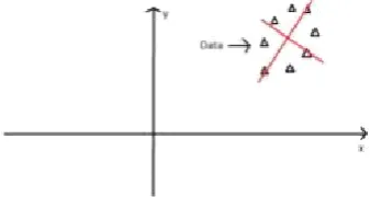
[image:5.595.79.248.404.494.2]Figure Data mapped on x and y axes
Figure : Change of frame w.r.t. Principal Component
In our projected methodology the feature reduction was implemented through Principal elements Analysis (PCA). PCA compresses the information from k-dimensions to n-k-dimensions. It tries to seek out a lower dimensional surface onto that to project the information, so the total of squares of the orthogonal projection error are reduced. In our proposed methodology, the higher than methodology is
implemented. The feature matrix may be a 25x6 matrix wherever 25 is that the variety of pictures (samples) used for coaching and 8 is that the variety of options. Then mean normalization is applied on the feature matrix, i.e. every feature features a zero mean. Then the co-variance matrix is computed and then the eigen| values and Eigen vectors of the variance matrix is computed. we elect the number of principal components such the ninety nine of the variance is retained. the total feature matrix is reduced from 25x6 to 25x2.
Classification
© 2017, IRJET | Impact Factor value: 5.181 | ISO 9001:2008 Certified Journal | Page 1739 BalintAntal et al.[3] projected AN for screening
[image:6.595.59.264.238.354.2]systemof diabetic retinopathy with ensemble feature extraction model during which options area unit extracted from many retinal pictures. The algorithms like image level, lesion specific, anatomical area unit used because the elements of image extraction. the selections area unit taken by the ensemble of machine learning classifier. The info MESSIDOR produces ninetieth sensitivity, ninety one specificity and ninety the troubles accuracy.
Figure difference of normal and diabetic macula [13]
In our methodology, a binary SVM classifier is utilized. It is the supervised learning model used for classification. Given a feature matrix for training, an SVM training algorithm classifies the information into 2 categories i.e. Normal or Abnormal. an SVM rule separates the information into two categories by selecting the most effective hyper plane that has the largest margin i.e. the largest distance between the nearest data points. The training data points that are closest to the hyper plane or setup are called Support Vectors.SVM is strong because it tries to separate the information with as large as a margin as doable. The classification in our methodis a binary classification exploitation SVM classifier. The SVM classifies the images into 2 category stage zero and stage 2. As the stage a pair of macular hydrops is found in the region round the macula with a radius of 1Disc Diameter from the region. All the fundus pictures are segmental in order that an area of one Disc Diameter is extracted out.
Result
To estimate the potency of the urged technique, the algorithm was run on the dataset and result obtained were tabulated. The dataset taken consisted of twentyfive pictures of which 15 training and 10 testing pictures were taken. The database was divided into training and testing pictures. The training dataset was given as input to coach the SVM classifier. The testing
info was used to test the classification accuracy of the SVM classifier. The results were computed in a circular area round the macula. The radius of the circle was 1DD or 1 Disc Diameter. In our classification we tend to took forty five pictures for training and fifteen pictures for testing. Out of forty five pictures utilized in training, twenty three pictures were of stage two and twenty two pictures were of Stage 0. we classified the information through the SVM classifier using linear in addition as non-linear kernels. numerous non-linear kernels were used like Polynomial, RBF, Multi-layer perceptron and Quadratic.
3. CONCLUSIONS
An economical methodology has been projected to classify diabetic macular puffiness into stage zero (Normal) and stage a pair of (Abnormal) supported texture feature extraction . Since there's significant changes in texture feature within the immediate area around 1disc diameter from the centre of the macula. These options during this metameric space facilitate in vital classification between stage zero and stage a pair of. The results show that an automatic diagnosing of diabetic macular puffiness is feasible to assist the doctor (ophthalmologist) in call making thereby leading to quicker and higher results with the combination of input from doctors and automatic system. Also taking the feel options of the metameric region reduces the process time drastically.
REFERENCES
[1] Rubini, S. S., &Kunthavai, A. (2015). Diabetic
Retinopathy Detection Based on Eigenvalues of the Hessian Matrix. Procedia-Procedia Computer Science, 47, 311-318.
[2] Dash, J. (2015). A Survey on Blood Vessel detection
Methodologies in Retinal Images.R. Nicole, “Title of paper with only first word capitalized,” J. Name Stand. Abbrev., in press.
[3] Antal, B., &Hajdu, A. (2014). Knowledge-Based
Systems An ensemble-based system for automatic screening of diabetic retinopathy, 60, 20-27.
[4] Syed, A. M., Akbar, M. U., Akram, M. U., & Fatima, J.
(2014). Automated Laser Mark Segmentation from Colored Retinal Images.
[5] Pola, M., &Donoso, R. (2015). A Web-Based
© 2017, IRJET | Impact Factor value: 5.181 | ISO 9001:2008 Certified Journal | Page 1740
[6] Ruba, T. (2015). Identification and segmentation of
exudates using SVM classifier.
[7] Mane, V. M., &Kawadiwale, R. B. (2015). Detection
of Red Lesions in Diabetic Retinopathy Affected Fundus Images Preprocessing B. Extraction of Retinal Blood Vessels, 56-60.
[8] Aloudat, M., &Faezipour, M. (2015). Histogram
Analysis for Automatic Blood Vessels Detection/: First Step of IOP, 146-151.
[9] Gupta, S., &Jadhav, R. (2015). Diabetic Retinopathy
using Morphological Operations and Machine Learning, 617-622.
[10] Kunwar, A., Magotra, S., &Sarathi, M. P. (2015).
Detection of High-Risk Macular Edema using Texture features and Classification using SVM Classifier, 2285-2289.
[11] Mahendran, G., &Dhanasekaran, R. (2015).
Investigation of the severity level of diabetic retinopathy using supervised classifier algorithms q. Computers and Electrical Engineering, 45, 312-323.
[12] Welikala, R. A., Fraz, M. M., Dehmeshki, J., Hoppe, A.,
Tah, V., Mann, S., Barman, S. A. (2015). Computerized Medical Imaging and Graphics Genetic algorithm based feature selection combined with dual classification for the automated detection of proliferative diabetic retinopathy. Computerized Medical Imaging and Graphics, 43, 64-77.
[13] Aditya Kunwar1,Shrey Magotra1,M Partha
Sarathi1,” Detection of High-Risk Macular Edema using Texture features and Classification using SVM Classifier” 2015 IEEE
BIOGRAPHIES
Mr. Ajay Advani is presently pursuing her B.E in Information Technology from Savitribai Phule Pune University. His area of interests is Image Processing and Data Mining.
Mr.Ashish Thawrani is presently pursuing her B.E in Information Technology from Savitribai Phule Pune University. His area of interests is Image Processing and Data Mining.
Mr. Amit Thaware is presently pursuing her B.E in Information Technology from Savitribai Phule Pune University. His area of interests is Image Processing and Data Mining.
Mr.Abhinay Goankar is presently pursuing her B.E in Information Technology from Savitribai Phule Pune University. His area of interests is Image Processing and Data Mining.
Mrs. V. C. Kulloli received her M.E (CSE) from Pune University. She is pursuing her Ph.D. (CI) from Pune University. She has Seventeen years long experience in the field of teaching. Her research areas are Image Processing, Data Mining. Her research work has been published in many national and international journals and Conferences.
2nd Author
Photo
![Figure type of diseases [14]](https://thumb-us.123doks.com/thumbv2/123dok_us/8156768.804360/1.595.348.519.208.382/figure-type-of-diseases.webp)
![Figure 2 System Architecture [13]](https://thumb-us.123doks.com/thumbv2/123dok_us/8156768.804360/4.595.348.515.411.507/figure-system-architecture.webp)
![Figure difference of normal and diabetic macula [13]](https://thumb-us.123doks.com/thumbv2/123dok_us/8156768.804360/6.595.59.264.238.354/figure-difference-normal-diabetic-macula.webp)
