Copyright © 1999, American Society for Microbiology. All Rights Reserved.
Fatal Case of
Trichoderma harzianum
Infection in a Renal
Transplant Recipient
JOSEP GUARRO,1* MARI´A ISABEL ANTOLI´N-AYALA,2JOSEPA GENE´,1
JESU´ S GUTIE´RREZ-CALZADA,2CARLOS NIEVES-DI´EZ,3 ANDMONTSERRAT ORTONEDA1
Unitat de Microbiologia, Facultat de Medicina i Cie`ncies de la Salut, Universitat Rovira i Virgili,
43201 Reus,1and Servicio de Microbiologı´a2and Servicio de Anatomı´a Patolo´gica,3
Hospital de Leo´n, 28002 Leo´n, Spain
Received 19 April 1999/Returned for modification 22 June 1999/Accepted 18 August 1999
We describe the second known case of human infection byTrichoderma harzianum. A disseminated fungal
infection was detected in the postmortem examination of a renal transplant recipient and confirmed in culture. The only other reported infection by this fungus caused peritonitis in a diabetic patient. The in vitro antifungal
susceptibilities of the clinical strain and three other strains ofTrichodermaspecies to six antifungal drugs are
provided. This case illustrates the widening spectrum of opportunisticTrichodermaspp. in
immunocompro-mised patients and emphasizes the problems in diagnosing invasive fungal diseases.
Opportunistic fungal infections have occurred with increas-ing frequency in recent years in immunosuppressed patients.
Trichodermaspp. are fungi distributed worldwide which rarely
infect humans but can cause from localized infections to fatal disseminated disease (5, 6, 8, 10, 12, 13). In this report, we describe a systemicT. harzianuminfection in a renal transplant patient which was detected in the necropsy study. The fungus was recovered from abscesses in brain and lung tissues.
Trichodermaspp. have been associated with 12 cases of
hu-man infections, half of which were peritonitis. Apart from the cases reported in the review by Munoz et al. (13), there have been two additional cases ofTrichodermaperitonitis, caused by
Trichoderma koningii(5) andTrichoderma harzianum(10), in
two patients who were undergoing peritoneal dialysis. Both patients died after being treated unsuccessfully with different antifungal drugs.Trichoderma longibrachiatumwas responsible for a case of invasive sinusitis in a recipient of a liver and small bowel transplant (6). The patient was successfully treated by fungical debridement and by administration of amphotericin B followed by oral itraconazole.
Case report. A 68-year-old man with chronic renal failure
had a transplant on 3 January 1996. He was then put on cyclosporine and prednisone. His past medical history was un-remarkable except for moderate systemic hypertension. Shortly after the renal transplant, there was an acute graft rejection, but this was quickly and successfully treated with high doses of steroids and cyclosporine; antibiotics were also used because Legionella pneumophila and Listeria sp. were identified on sputum cultures. At the end of January, the pa-tient suffered headaches and there were changes in his per-sonality. A computed tomography scan revealed a hypodense lesion at the subcortical left frontal area; an electroencepha-logram showed nonspecific signs of poor cerebral activity at both the frontal and parietal hemispheres. A cranial nuclear magnetic resonance scan showed signs of attenuation from the infra- and supratentorial regions of a nonspecific nature but
consistent with encephalitis. The patient’s blood biochemistry profile was normal at that time, except for creatinine (3 mg/dl) and urea (150 mg/dl) levels. Precipitin tests for a variety of antibodies were negative except for cytomegalovirus (CMV) (cell cultures from blood and urine were negative). A lumbar puncture resulted in cerebrospinal fluid (CSF) with a normal biochemical profile. India ink- and carbol fuchsin-stained prep-arations of CFS were negative for Cryptococcus neoformans
and mycobacteria, respectively. CFS cultures for mycobacteria were also negative. The patient was discharged and was to receive follow-up at the outpatient clinic. The patient was stable but complained of a moderate headache until 10 March, when he was found unconscious on the floor and then trans-ferred to the emergency room. He recovered spontaneously, and general and neurologic examinations were normal except for a low level of consciousness and a persistent headache. A computed tomography scan revealed hyperdense lesions at the cortical and parenchyma right frontal region and Silvio fissure, consistent with subarachnoid hemorrhaging. The patient was admitted to the hospital, and treatment with corticosteroids and ganciclovir was initiated; cyclosporine treatment was halted. Twelve hours later, neurologic deterioration associated with a progressive loss of consciousness began, and 48 h after admission, the patient collapsed and died.
Necropsy study. The brain weighed 1,500 g. It exhibited
edema and diffuse subarachnoid hemorrhaging, mostly in the basal distribution. On coronal cuts, there was also intraventric-ular hemorrhaging with tele-encephalic ventricintraventric-ular dilation; a silvanic mycotic aneurysm with massive thrombosis was iden-tified. Throughout the white substance at the oval centers in both frontal lobules, there were small microabscesses (0.3 to 0.5 cm in diameter) full of necrotic material. Similar lesions were also identified on the brain stem cuts. Histological exam-ination (Gomori methenamine silver and periodic acid-Schiff stains) of these lesions demonstrated neutrophil proliferation without a granulomatous reaction and ramified and septate hyphae (Fig. 1), which also invaded the vascular walls of the thrombotic aneurysm. These mycotic lesions were ultimately the cause of the brain hemorrhage. No other, coexistent brain infection was identified. The lungs had multiple microab-scesses spread across the subpleural and parenchyma spaces. These lesions, 0.8 to 1.2 cm in diameter, had a white purulent * Corresponding author. Mailing address: Unitat de Microbiologia,
Departament de Cie`ncies Me`diques Ba`siques, Facultat de Medicina i Cie`ncies de la Salut, Universitat Rovira i Virgili, Carrer Sant Llorenc¸ 21, 43201 Reus, Tarragona, Spain. Phone: 34 977759359. Fax: 34 977759322. E-mail: umb@fmcs.urv.es.
3751
on May 15, 2020 by guest
http://jcm.asm.org/
aspect, and some of them were cavitate. The findings of the microscopic examination were the same as for the brain (Fig. 2). There were also some patchy areas of consolidation and acute bronchiolitis surrounding the abscesses. The liver weighted 1,500 g and was full of small yellow nodules (0.1 to 0.6 cm in diameter) on both lobules. Histological examination
[image:2.612.116.489.72.336.2]showed that these lesions were due to parenchyma necrotic foci. Some inclusion bodies of CMV were also identified throughout the liver. No other lesions like those in the brain and lungs were found in the liver. The kidneys weighed less than normal and showed atrophic changes. There were also signs of interstitial nephritis and foci of CMV infection. Small FIG. 1. Brain abscess showing a radiating pattern of hyphae. Gomori methenamine silver stain; magnification,⫻128.
FIG. 2. Branching pattern of hyphae from a lung lesion. Periodic acid-Schiff stain; magnification,⫻1280.
3752 NOTES J. CLIN. MICROBIOL.
on May 15, 2020 by guest
http://jcm.asm.org/
[image:2.612.116.488.459.716.2]portions of necropsy specimens (brain and lungs) were re-peatedly cultured on Sabouraud dextrose agar. All yielded a filamentous fungus which was identified asTrichodermasp. This was sent to the Microbiology Unit of the Faculty of Medicine at the Rovira i Virgili University in Reus, Spain, for a conclusive mycological diagnosis.
Mycological study. The fungus was subcultured on potato
dextrose agar and oatmeal agar and incubated at room tem-perature in the dark. Fungal colonies grew very quickly on both media and after 4 days occupied the whole surface of the petri dish. On potato dextrose agar they were dense and cottony, with a yellowish green area at the center and white toward the periphery. The whole surface rapidly turned granular with dark green masses because of the abundant production of conidia in tufts. The colony reverse was colorless. A yellowish exudate was produced on this medium. Colonies on oatmeal agar were cottony and whitish green, becoming olive green in tufted conidial areas; the reverse was colorless. Exudate was absent on this medium. Microscopically, conidiophores were pyrami-dally branched, with short branches near the apex (Fig. 3). Phialides were usually in groups of two to five. They were ampulliform or lageniform and markedly constricted at the base, generally 4 to 7 by 3 to 3.5m; phialides near the apex of the conidiophore were usually longer and slender, up to 15
by 2.5 to 3m. Conidia were subglobose or short obovoid, 2.5 to 3 by 2 to 2.5m; they were subhyaline to pale green and smooth walled. Chlamydospores were observed in old cultures. They were usually intercalary, subglobose or ellipsoidal, hya-line, smooth walled, and up to 12m in diameter. This clinical strain was identified asT. harzianum, and one isolate was kept in the mycology laboratory of the Faculty of Medicine in Reus as isolate no. FMR 6424. A living culture of this isolate has been deposited in the Centraalbureau voor Schimmelcultures of The Netherlands under accession no. CBS 102174.
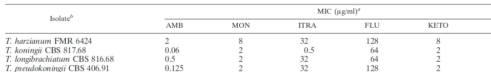
Antifungal susceptibility testing. This clinical isolate and
three additional isolates (T. koningii,T. longibrachiatum, andT.
pseudokoningii) from various sources were tested to determine
their susceptibilities to six antifungal drugs (amphotericin B, flucytosine, fluconazole, itraconazole, ketoconazole, and mi-conazole). The isolates were tested by a previously described microdilution method (14), mainly according to the guidelines of the National Committee for Clinical Laboratory Standards for molds, using RPMI 1640 medium buffered to pH 7 with 0.165 M morpholinepropanesulfonic acid (MOPS), an inocu-lum of 1.7⫻104to 3.1⫻104CFU/ml, an incubation
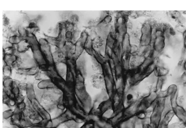
[image:3.612.55.547.70.437.2]temper-ature of 30°C, a second-day reading (48 h), and an additive drug dilution procedure. Table 1 shows the MICs of the six antifungals for the four isolates. The MIC for the case isolate FIG. 3.T. harzianum(FMR 6424). Pyramidal structure of a conidiophore with phialides and smooth conidia. (A) Nomarski optics; magnification,⫻1,600. (B) Scanning electron microscopy; magnification,⫻2,300.
on May 15, 2020 by guest
http://jcm.asm.org/
was clearly the highest of those for the four isolates; only ketoconazole displayed moderately high MICs. MICs for the remaining isolates were variable. In general, the MICs of am-photericin B and ketoconazole were low. The MIC of itracon-azole was also low for theT. koningiiisolate.
In our case, the patient did not receive any antifungal treat-ment because the fungal infection was discovered after post-mortem examination. However, the in vitro antifungal suscep-tibility of the strain showed that the MICs of the six antifungals tested were very high, and they would probably also have been ineffective in vivo. Munoz et al. (13) summarized the antifungal susceptibilities of the previously reported Trichoderma spp. clinical isolates. Most isolates were resistant to fluconazole and flucytosine, and approximately half were resistant to ampho-tericin B, although they were susceptible (or moderately so) to itraconazole, ketoconazole, and miconazole. In the last three reported cases, one isolate ofT. longibrachiatumwas sensitive in vitro to amphotericin B and itraconazole, and the patient was treated successfully with these two drugs (6); another isolate was sensitive to ketoconazole, miconazole, and flucy-tosine and resistant to amphotericin B. The patient died after treatment with amphotericin B (5). In the third case, ketoconazole and flucytosine were also administered unsuc-cessfully (10).
T. harzianum is one of the most common species of the
genus. It is well known as a biological control agent for various plant-pathogenic fungi (7). There is some controversy about the taxonomy of this species (1, 2, 15). It was recently reviewed by Gams and Meyer (7) and was defined as having regularly verticillate conidiophores, forming a pyramidal structure. The phialides are ampulliform to lageniform, usually in groups of three to four, and they generally measure 5.5 to 7.5 by 2.5m. The conidia were subglobose to obovoid, generally measuring 2.8 to 3.5 by 2.3 to 3 m, and are smooth and subhyaline to pale green.
Liu et al. (11) demonstrated that some histological features, such as the type of hyphae and the presence of characteristic reproductive structures like adventitious conidia, can be very useful for a preliminary identification of some unusual human-pathogenic fungi. This was shown by the diagnosis of hya-lohyphomycosis caused by Fusarium, Paecilomyces, and
Acremonium species. In our case, a detailed examination of
histological sections, especially those from the lung, showed an arborescent pattern of hyphal ramification (Fig. 2). The dichot-omous branching hyphae of Aspergillusspecies in tissues are similar to those that we observed. However,Trichoderma de-velops a more complex branching pattern, which is similar to that seen when it grows in culture. Further study is required to determine the usefulness of this finding for recognizing
Tricho-dermastrains in tissue sections.
The number of patients with infections caused by
Tricho-dermaspp. is likely to increase because certain therapies used
in current medical practice abrogate the immune response of
the host and because these fungi are common in the air my-cobiota (4). Our purpose here was to report the second known case ofT. harzianumhyalohyphomycosis with a fatal outcome and alert physicians and clinical microbiologists to the emer-gence of these opportunists with a high degree of fatality (ap-proximately half of the reported cases have resulted in death). Methods for identifying molds from histological specimens by development of fluorescent antibody conjugates and the use of molecular techniques would also provide definitive diagnoses. Equally important is the development of serologic tests for infections caused byTrichodermaspp., which can provide early presumptive diagnoses. Many physicians are currently aware of fungal diseases, and guidelines for preventing, diagnosing, and managing opportunistic fungal infections have been published. In spite of that, the incidence of mycotic infections diagnosed after postmortem examination is still remarkably high (3, 9, 16). If opportunistic infections were diagnosed early enough, morbidity and mortality would be significantly reduced in many cases.
We thank Arvind A. Padhye (Centers for Disease Control and Prevention, Atlanta, Ga.) for reviewing the manuscript and J. M. Marin-Trigo (Hospital Miguel Servet, Zaragoza, Spain) for kind col-laboration.
This work was supported by CICYT (Ministerio de Educacio´n y Ciencia of Spain) grant PM98-0059 and Fundacio´ Cie`ncia i Salut.
REFERENCES
1.Bisset, J.1991. A revision of the genusTrichoderma. III. Section Pachyba-sium. Can. J. Bot.69:2373–2417.
2.Bisset, J.1992.Trichoderma atroviride. Can. J. Bot.70:639–641.
3.Bodey, G. P., and S. Vartivarian.1989. Aspergillosis. Eur. J. Clin. Microbiol. Infect. Dis.8:413–437.
4.Calvo, M. A., J. Guarro, G. Suarez, and C. Ramı´rez.1980. Air-borne fungi in the air of Barcelona (Spain). IV. Various related genera. Mycopathologia
71:119–123.
5.Campos-Herrero, M. I., A. Bordes, A. Perera, M. C. Ruiz, and A. Ferna´ndez.
1996.Trichoderma koningiiperitonitis in a patient undergoing peritoneal dialysis. Clin. Microbiol. Newsl.18:150–152.
6.Furukawa, H., S. Kusne, D. A. Sutton, R. Manez, R. Carrau, L. Nichols, K. Abu-Elmaged, D. Skedros, S. Todo, and M. C. Rinaldi.1998. Acute invasive sinusitis due toTrichoderma longibrachiatum in a liver and small bowel transplant recipient. Clin. Infect. Dis.26:487–489.
7.Gams, W., and W. Meyer.1998. What exactly isTrichoderma harzianum? Mycologia90:904–915.
8.Gautheret, A., F. Dromer, J. Bourhis, and A. Andremont.1995.Trichoderma pseudokoningiias a cause of fatal infection in a bone marrow transplant recipient. Clin. Infect. Dis.20:1063–1064.
9.Goodrich, J. M., E. C. Reed, M. Mori, L. D. Fisher, S. Skerrett, P. S. Dandliker, B. Klis, G. W. Counts, and J. D. Meyers.1991. Clinical features and analysis of risk factors for invasive candidal infection after marrow transplantation. J. Infect. Dis.164:731–740.
10. Guiserix, J., R. Ramdane, P. Finielz, A. Michault, and P. Rajaonarivelo.
1996.Trichoderma harzianumperitonitis in peritoneal dialysis. Nephron74:
473–474.
11. Liu, K., D. N. Howell, J. R. Perfect, and W. A. Schell.1998. Morphological criteria for the preliminary identification ofFusarium,Paecilomyces, and Acremoniumspecies by histopathology. Am. J. Clin. Pathol.109:45–54. 12. Loeppky, C. B., R. F. Sprouse, J. V. Carlson, and E. D. Everett.1983.
[image:4.612.50.553.83.157.2]Trichoderma virideperitonitis. South. Med. J.76:798–799. TABLE 1. Antifungal susceptibilities ofTrichodermaspp. isolates
Isolateb MIC (g/ml)
a
AMB MON ITRA FLU KETO 5-FC
T. harzianumFMR 6424 2 8 32 128 8 256
T. koningiiCBS 817.68 0.06 2 0.5 64 2 256
T. longibrachiatumCBS 816.68 0.5 2 32 64 2 256
T. pseudokoningiiCBS 406.91 0.125 2 32 128 2 256
aAMB, amphotericin B; MON, miconazole; ITRA, itraconazole; FLU, fluconazole; KETO, ketoconazole; 5-FC, flucytosine.
bFMR, Faculty of Medicine, Universitat Rovira i Virgili, Reus, Spain; CBS, Centraalbureau voor Schimmelcultures, Baarn, The Netherlands.
3754 NOTES J. CLIN. MICROBIOL.
on May 15, 2020 by guest
http://jcm.asm.org/
13.Munoz, F. M., G. J. Demmler, W. R. Travis, A. K. Ogden, S. N. Rossmann, and M. G. Rinaldi.1997.Trichoderma longibrachiatuminfection in a pedi-atric patient with aplastic anemia. J. Clin. Microbiol.35:499–503. 14. Pujol, I., J. Guarro, C. Llop, L. Soler, and J. Ferna´ndez-Ballart.1996.
Comparison study of broth macrodilution and microdilution antifungal susceptibility tests for the filamentous fungi. Antimicrob. Agents.
Chemother.40:2106–2110.
15. Rifai, M. A.1969. A revision of the genusTrichoderma. Mycol. Pap.116:1– 56.
16. Tollemar, J., O. Ringden, L. Bostrom, B. Nilsson, and B. Sundberg.1989. Variables predicting deep fungal infections in bone marrow transplant re-cipients. Bone Marrow Transplant.4:635–641.