0095-1137/06/$08.00⫹0 doi:10.1128/JCM.44.1.206–213.2006
Copyright © 2006, American Society for Microbiology. All Rights Reserved.
Development and Validation of a Rotor-Gene Real-Time PCR Assay
for Detection, Identification, and Quantification of
Chlamydia
trachomatis
in a Single Reaction
Hamid Jalal,
1* Hannah Stephen,
1Martin D. Curran,
1Janet Burton,
1Michelle Bradley,
2and Christopher Carne
3Clinical Microbiology & Public Health Laboratory, Box 236, Addenbrooke’s Hospital, Hills Road, Cambridge CB2 2QW,1Centre
for Applied Medical Statistics, Institute of Public Health, University Forvie Site, Department of Public Health and Primary Care, Robison Way, Cambridge CB2 2SR,2and Department of Genitourinary Medicine, Clinic 1A,
Box 38 Addenbrooke’s Hospital, Hills Road, Cambridge,3United Kingdom
Received 29 July 2005/Returned for modification 20 September 2005/Accepted 19 October 2005
A multitarget real-time PCR (MRT-PCR) for detection of Chlamydia trachomatisDNA was developed and
validated. There were three targets for amplification in a single reaction: the cryptic plasmid (CP), the major outer membrane protein (MOMP) gene, and an internal control. The assay had the following characteristics:
(i) detection and confirmation of the presence ofC. trachomatisDNA in a single reaction, (ii) detection of all
genovars ofC. trachomatiswithout any cross-reactivity with pathogenic bacteria or commensal organisms of the
oropharynx and genital tract, (iii) a 95% probability of detection with three copies of MOMP and one copy of CP per reaction mixture, (iv) identification of the inhibition of amplification, (v) a quantitative dynamic range of 25 to 250,000 genome copies per reaction mixture, (vi) high intra- and interassay reproducibilities, and (vii) correct identification of all samples in the validation panel. There were 146 COBAS Amplicor PCR (Amplicor PCR)-positive samples and 122 Amplicor PCR-negative samples in the panel. MRT-PCR detected CP DNA alone in 6 (4%) Amplicor PCR-positive samples and both CP and MOMP DNAs in 140 (96%) of 146 Amplicor PCR-positive samples. The quantity of MOMP DNA in 95 (68%) of 140 samples was within the dynamic range
of the assay. The medianC. trachomatisload in these samples was 321 genome copies per reaction mixture
(range, 26 to 40,137 genome copies per reaction mixture). Due to the inclusion of two differentC. trachomatis
-specific targets, the assay confirmed 259 (97%) of 268 results in a single reaction. This assay could be used in
the qualitative format for the routine detection ofC. trachomatisand in the quantitative format for study of the
pathogenesis ofC. trachomatis-associated diseases. The assay demonstrated the potential to eliminate the need
for confirmatory testing in almost all samples, thus reducing the turnaround time and the workload.
Chlamydia trachomatisis a bacterial infection of global pub-lic health significance (10). It is an intracellular pathogen with 15 serotypes. It causes trachoma (serotypes A, B, B1, and C),
lymphogranuloma venereum (serotypes L1, L2, and L3), and
oculogenital infections (serotypes D to K). There are many highly conserved nucleotide sequences in different chromo-somal genes and in the cryptic plasmid (CP) ofC. trachomatis. These sequences are used as targets for nucleic acid amplifi-cation tests (NAATs). NAATs have surpassed cell culture and antigen detection for the diagnosis ofC. trachomatisinfections due to their enhanced sensitivities.
A large number of in-house NAATs for the detection ofC. trachomatishave been reported (16). The majority of these are gel based. A few non-gel-based NAATs have also been de-scribed, i.e., a Qreplicase-amplified assay (2) and PCR-en-zyme immunoassays (4, 11, 21). All previously reported in-house NAATs are manual and use open systems with separate steps for amplification and amplicon analysis. Due to the risk of contamination and a lack of automation, these assays are
inappropriate for diagnostic laboratories with high workloads. Real-time PCR is performed in a closed system, amplification and detection of target are done in a single step, and there is the potential for automation. Real-time PCR has been used for pharmacodynamic studies for C. trachomatis (19, 23). Two real-time PCR assays for the detection ofC. trachomatishave also been described (8, 13). Both these assays used the SYBR green technology in the LightCycler system (Roche Molecular Systems, Inc., Pleasanton, CA) and were used for the qualita-tive detection of C. trachomatis. Roche Molecular Systems, GEN-PROBE Inc. (San Diego, CA), BD Diagnostics (Sparks, MD), and artus GmbH (Hamburg, Germany) are the four major commercial companies which supply NAATs for the detection ofC. trachomatisin Europe. At present, artus GmbH is the only commercial company that provides an assay for the quantification ofC. trachomatisin clinical samples.
A single gene of C. trachomatisper amplification reaction was targeted in all of the in-house and commercial assays mentioned above. The targets of amplification were the CP, the major outer membrane protein (MOMP) gene, the 23S rRNA gene, and the cysteine-rich outer membrane protein gene (16). In the present study, we report on a multitarget real-time PCR (MTR-PCR) assay for the detection, identifi-cation, and quantification ofC. trachomatisin a single reaction. Three targets of amplification were used in this assay: the
* Corresponding author. Mailing address: Clinical Microbiology & Public Health Laboratory, Box 236, Addenbrooke’s Hospital, Hills Road, Cambridge CB2 2QW, United Kingdom. Phone: 44-1223-257036. Fax: 44-1223-242775. E-mail: [email protected] .uk.
206
on May 16, 2020 by guest
http://jcm.asm.org/
cryptic plasmid for the qualitative detection, the MOMP gene for the quantification ofC. trachomatisDNA, and an internal control (IC) for the detection of inhibitors of amplification. None of the previously reported in-house or commercial assays have achieved these characteristics in a single reaction.
MATERIALS AND METHODS
Chlamydiastrains.Strains ofC. trachomatisfrom different serovars andC. suis
were grown in McCoy cells, as described previously (25). The growth of the organism was confirmed by direct immunofluorescence with a genus-specific monoclonal antibody, IMAGEN Chlamydia, according to the manufacturer’s protocol (DakoCytomation Ltd., Ely, United Kingdom). McCoy cells and all strains of chlamydia were obtained from the American Type Culture Collection
(ATCC), LGC Promochem, Middlesex, United Kingdom. However,C. pneu-moniaewas not grown in the cell culture; its DNA was obtained from Advanced Biotechnologies, Incorporated (Columbia, MD). The identities of theChlamydia
strains used in this study are shown in Table 1.
Bacterial strains.Pathogenic bacteria and commensal organisms of the oro-pharynx and the genital tract were grown as described previously (1). The suit-ability of bacterial DNA for amplification was checked by 16S rRNA gene-based PCR (15). The identities of the bacterial strains used in this study are shown in Table 2.
Validation panel.The validation panel included 146 Amplicor PCR-positive and 122 Amplicor PCR-negative samples. These samples were obtained from 56 males (urethral swabs; median age, 22 years; age range, 17 to 64 years) and 212 females (both urethral and endocervical swabs in a single tube; median age, 23 years; age range, 15 to 59 years) as described previously (12). The samples were coded, and the results of the Amplicor PCR were masked before the validation experiments. The swab in the transport medium was agitated for 20 s at a setting of 4.5 in a multitube vortexer (model 2601; Scientific Manufacturing Industries, Emeryville, CA) before extraction of DNA. The DNA from 200 l of cell suspension (a bacterial culture or a clinical sample) was extracted by using a MagNA Pure LC total nucleic acid isolation kit and MagNA Pure LC Robot, according to the manufacturer’s protocol (Roche Molecular Systems). DNA was concentrated during extraction and eluted in 100l of elution buffer. Quality control for DNA extraction was performed by inclusion ofC. trachomatisculture genotype E (positive control) and nuclease-free water (negative control) in each run.
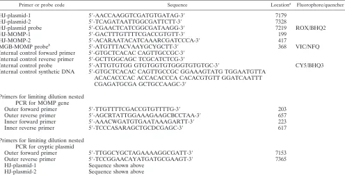
PCR.Real-time PCR was performed with a Rotor-Gene 3000 (Corbett Ro-botics, Australia). Rotor-Gene has four light-emitting diodes that allow the detection of up to four targets in a single amplification reaction. This character-istic of the system was used to develop the MTR-PCR for the detection ofC. trachomatis. A pair of primers and a labeled probe in the TaqMan format were used for each component of the assay, i.e., CP, MOMP, and IC. Except for the probe for the MOMP gene, the IC DNA and all primers and probes were synthesized by Metabion International AGi, Germany. The probe for the MOMP gene was obtained from Applied Biosystems, Cheshire, United King-dom. The sequences of the primers, probes, and the internal control are shown in Table 3. PCR was performed in a 25-l reaction mixture containing 10l of DNA from a clinical sample or a bacterial isolate, 5 mM MgCl2, 12.5l of
TABLE 1. Chlamydia strains
Strains ATCC code
LGV I strain 440 ...VR-901B LGV II strain 434...VR-902B LGV III strain 404 ...VR-903 A strain HAR-13 ...VR-571B B strain HAR-36 ...VR-573 C strain TW-3 ...VR-1477 D strainUW-36/Cx...VR-885 E strain BOUR...VR-348B F strain IC-Cal-3 ...VR-346 G strain UW-57/Cx ...VR-878 H strain UW-43/Cx ...VR-879 I strain UW-12/Ur ...VR-880 J strain UW-36/Cx...VR-886 K strain UW-31/Cx...VR-887
C. suisstrain S45 ...VR-1474
[image:2.585.43.542.440.725.2]C. pneumoniae...CDC-CWL-011
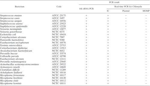
TABLE 2. Specificity of real-time PCR for cryptic plasmid and MOMP components of the assay
Bacterium Code
PCR result
16S rRNA PCR Real-time PCR forChlamydia
IC Plasmid MOMP
Streptococcus mutans ATCC 25175 ⫹ ⫹ ⫺ ⫺
Streptococcus canis ATCC 3497 ⫹ ⫹ ⫺ ⫺
Streptococcus sanguis ATCC 10556 ⫹ ⫹ ⫺ ⫺
Staphylococcus aureus ATCC 25923 ⫹ ⫹ ⫺ ⫺
Staphylococcus epidermidis ATCC 12228 ⫹ ⫹ ⫺ ⫺
Neisseria meningitidis ATCC 13077 ⫹ ⫹ ⫺ ⫺
Neisseria gonorrhoeae NCTC 8375 ⫹ ⫹ ⫺ ⫺
Escherichia coli NCTC 10418 ⫹ ⫹ ⫺ ⫺
Cornyebacterium ulcerans NCTC 7907 ⫹ ⫹ ⫺ ⫺
Pasteurella haemolytica NCTC 9380 ⫹ ⫹ ⫺ ⫺
Fusobacterium necrophorum NCTC 10576 ⫹ ⫹ ⫺ ⫺
Yersinia enterocolitica ATCC 23715 ⫹ ⫹ ⫺ ⫺
Cornyebacterium diptheriae ATCC 11913 ⫹ ⫹ ⫺ ⫺
Arcanobacterium haemolyticum NCTC 8452 ⫹ ⫹ ⫺ ⫺
Prevotella buccae ATCC 33574 ⫹ ⫹ ⫺ ⫺
Veillonella parvula ATCC 10790 ⫹ ⫹ ⫺ ⫺
Fusobacterium ulcerans NCTC 12111 ⫹ ⫹ ⫺ ⫺
Prevotella melaninogenica ATCC 25845 ⫹ ⫹ ⫺ ⫺
Actinobacillus actinomycetemcomitans ATCC 29522 ⫹ ⫹ ⫺ ⫺
Actinomyces israelii ATCC 10049 ⫹ ⫹ ⫺ ⫺
Clostridium difficile NCTC 11209 ⫹ ⫹ ⫺ ⫺
Acholeplasm laidlawii NCTC 10116 ⫹ ⫹ ⫺ ⫺
Mycoplasma fermentans NCTC 10117 ⫹ ⫹ ⫺ ⫺
Mycoplasma hyorhinis NCTC 10130 ⫹ ⫹ ⫺ ⫺
Mycoplasma orale NCTC 10112 ⫹ ⫹ ⫺ ⫺
Mycoplasma hominis NCTC 10111 ⫹ ⫹ ⫺ ⫺
VOL. 44, 2006 REAL-TIME PCR FOR DETECTION OF C. TRACHOMATIS 207
on May 16, 2020 by guest
http://jcm.asm.org/
Platinum Quantitative PCR SuperMix-UDG (Invitrogen Life Technologies, Paisley, United Kingdom), 6.25 pmol of each primer, and 2.5 pmol of each probe for amplification of DNA from the CP and the MOMP gene ofC. trachomatis
and six molecules of the IC and 2.5 pmol of each primer and probe for ampli-fication of the IC. The IC was an artificial single-stranded DNA molecule that was designed by joining A, C, G, and T in a random order. The nucleotide-nucleotide BLAST program at www.ncbi.nlm.nih.gov/BLAST/BLAST.cgi dem-onstrated the unique sequence of this molecule. Metabion quantified it by spec-trophotometery, and the number of molecules/l was calculated by using Avogadro’s number. The amplification reaction profile included heating at 50°C for 2 min and 95°C for 2 min, followed by 40 cycles of 95°C for 1 s and 60°C for 60 s. The acquisition of a signal was preformed at 60°C during each cycle.
Quantification standards.Cryptic plasmid and MOMP gene copy numbers in a stock of purified DNA fromC. trachomatisstrain E (BOUR) were estimated by the limited-dilution, Poisson distribution method by a nested PCR (22). The nested PCRs for the CP and MOMP genes were performed as described previ-ously (12). The sequences of the primers are shown in Table 3. An external calibration curve was prepared by using serial 10-fold dilutions from the stock DNA in nuclease-free water containing 1 ng/l poly(A) RNA (Amersham Bio-science United Kingdom Ltd., Chalfont St. Giles, United Kingdom). The Rotor-Gene software calculated the number ofC. trachomatisgenome copies in am-plification reactions with reference to the external calibration curve. The parameters used for quantification analysis are shown in Table 4.
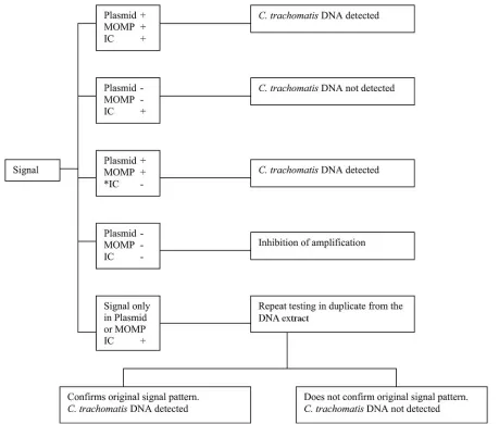
Criteria for a positive result by real-time PCR.A sample was considered positive forC. trachomatisDNA if it produced signals in the channels for the CP and the MOMP components of the assay. If the signal was produced in only one of these channels, the test was repeated, in duplicate, with the DNA extract. The sample was designated positive if repeat testing confirmed the earlier finding. Otherwise, it was designated a false-positive result. The algorithm for interpre-tation of the assay results is shown in Fig. 1.
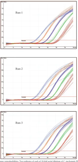
Design of experiments.The lower limits of detection for the MOMP and the CP components of the assay were determined by testing a doubling dilution series of the positive control with from 25 to 0.19 genome copies per reaction mixture. The CP DNA copy number was four times greater than the MOMP gene copy number in each reaction mixture. Each concentration was tested in 40 replicates, and probit analysis of the results was carried out by using “Stats-Direct,” version 2.4.1 (StatsDirect, Ltd., Cheshire, United Kingdom). The ana-lytical specificity of the assay was investigated by performing PCR with DNA from the chlamydial and bacterial strains stated in Tables 1 and 2. Intra- and interassay reproducibilities were investigated in three different runs by using DNA from the positive control. Ten replicates of five serial 10-fold dilutions ranging from 25 to 250,000 genome copies per reaction mixture were included in each run. Intra- and interassay reproducibilities were also investigated for 14 clinical samples. A total of six replicates of each sample were included in two different runs. The results from the intra- and interassay reproducibility experi-ments were used to investigate the linearity and the dynamic range of the MOMP component of the assay.
RESULTS
Analytical sensitivity and specificity. According to probit
[image:3.585.45.541.81.336.2]analysis, the assay demonstrated a 95% probability of detec-tion with three MOMP gene copies and one CP copy per reaction mixture. The assay was able to detect DNA from all strains ofC. trachomatis listed in Table 1. No amplicon was generated in the C. trachomatis-specific components of the assay from DNA ofC. suis, C. pneumoniae, or the bacterial
TABLE 3. Nucleotide sequences of primers and probes
Primer or probe code Sequence Locationa Fluorophore/quencher
HJ-plasmid-1 5⬘-AACCAAGGTCGATGTGATAG-3⬘ 7179
HJ-plasmid-2 5⬘-TCAGATAATTGGCGATTCTT-3⬘ 7328
HJ-plasmid probe 5⬘-CGAACTCATCGGCGATAAGG-3⬘ 7219 ROX/BHQ2
HJ-MOMP-1 5⬘-GACTTTGTTTTCGACCGTGTT-3⬘ 199
HJ-MOMP-2 5⬘-ACARAATACATCAAARCGATCCCA-3⬘ 417
MGB-MOMP probeb 5⬘-ATGTTTACVAAYGCYGCTT-3⬘ 368 VIC/NFQ
Internal control forward primer 5⬘-GTGCTCACAC CAGTTGCCGC-3⬘ Internal control reverse primer 5⬘-GCTTGGCAGC TCGCATCTCG-3⬘
Internal control probe 5⬘-ATTGTGTGG GTGTGGTGTGGGTGTGTGC-3⬘ CY5/BHQ3
Internal control synthetic DNA 5⬘-GTGCTCACAC CAGTTGCCGC GGAAAGTATG TGGAATGTTA ACACACCCAC ACCACACCCA CACACGTGTT GGATCAATTT CGAGATGCGA GCTGCCAAGC-3⬘
Primers for limiting dilution nested PCR for MOMP gene
Outer forward primer 5⬘-TTGTTTTCGACCGTGTTTTG-3⬘ 203
Outer reverse primer 5⬘-AGCRTATTGGAAAGAAGCBCCTAA-3⬘ 657
Inner forward primer 5⬘-AAACWGATGTGAATAAAGARTT-3⬘ 223
Inner reverse primer 5⬘-TCCCASARAGCTGCDCGAGC-3⬘ 617
Primers for limiting dilution nested PCR for cryptic plasmid
Outer forward primer 5⬘-TTGGCYGCTAGAAAAGGCGATT-3⬘ 7153
Outer reverse primer 5⬘-TCCGGAACAYATGATGCGAAGT-3⬘ 7365
HJ-plasmid-1 Sequence shown above HJ-plasmid-2 Sequence shown above
aThe positions of the primers and the probes for MOMP and cryptic plasmid components of the assay are according to GenBank accession nos. AF202457 and
X06707, respectively.
bAdditional labeling of the probe with the MGB molecule at the 3⬘and to increase the melting temperature of the DNA duplex.
TABLE 4. Parameters of Rotor-Gene 3000 for quantification analysis
Parameter Value
Threshold ...0.03 Left threshold ...10
Start normalizing...Variable (generally from cycle 5) Reaction efficiency threshold ...Disabled
Normalization method ...Dynamic tube normalization No-template control threshold...10%
on May 16, 2020 by guest
http://jcm.asm.org/
[image:3.585.44.285.648.724.2]isolates (Table 2). The 16S rRNA PCR amplified DNA from all bacteria shown in Table 2.
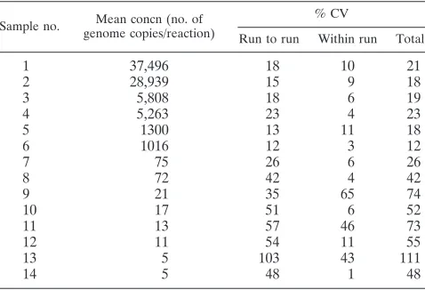
Intra- and interassay reproducibilities and dynamic range.
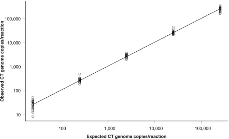
The results of the intra- and interassay reproducibilities for the positive controls are shown in Fig. 2. The coefficient of varia-tion (CV) values for across run, within run, and total variavaria-tion generally increased with decreasing genome copy numbers in the reaction mixture for both the positive control and the clinical samples. The CV values for the positive control and the clinical samples are shown in Table 5 and Table 6, respectively. The data from the intra- and interassay reproducibility exper-iments were used to investigate the linearity of the assay. The MOMP component of the assay exhibited a log-linear re-sponse, characteristic of levels ofC. trachomatisgenome input between 25 and 250,000 copies per reaction mixture. The re-gression line of the observed concentration to the expected concentration on a log10scale is shown in Fig. 3. For this set of
data, the adjusted r2 value was 0.99 and the deviation from
linearity was 0.3 log10.
Validation of the assay.According to the criteria stated in
the Materials and Methods section, the assay detectedC. tra-chomatis DNA from all of the 146 Amplicor PCR-positive samples. It did not detectC. trachomatisDNA from any of the 122 Amplicor PCR-negative samples. Of 146 (96%) samples, 140 generated signals in bothC. trachomatis-specific compo-nents of the assay. Six samples generated a signal only in the CP component of the assay. Repeat testing confirmed the presence of CP DNA and absence ofC. trachomatisgenomic DNA in these samples. The assay was able to quantify theC. trachomatisload in 95 (69%) of 140 samples. The medianC. trachomatisload in these samples was 321 genome copies per reaction mixture (range, 26 to 40,137 genome copies per reac-tion mixture). The IC was detected in all of the 268 samples and the controls. A weak signal was observed after⬎35 cycles of amplification in the MOMP component of the assay for one Amplicor PCR-negative sample. A similar signal was also ob-served in the CP component of the assay for two of the Am-plicor PCR-negative samples. The signals in these three
sam-FIG. 1. Algorithm for interpretation of assay results. Plasmid, MOMP, and IC represent the three components of the assay.ⴱ, high chlamydia genome copy number in the reaction may suppress amplification of the IC.
VOL. 44, 2006 REAL-TIME PCR FOR DETECTION OF C. TRACHOMATIS 209
on May 16, 2020 by guest
http://jcm.asm.org/
[image:4.585.65.524.68.458.2]FIG. 2. Intra- and interassay reproducibilities. Ten replicates of each of 10-fold serial dilutions ofC. trachomatisDNA were included per run. Color code: blue, 250,000 genome copies per reaction mixture; orange, 25,000 genome copies per reaction mixture; purple, 2,500 genome copies per reaction mixture; green, 250 genome copies per reaction mixture; red, 25 genome copies per reaction mixture.
210
on May 16, 2020 by guest
ples were not detected on repeat testing and were considered nonspecific reactivity due to autohydrolysis of the probe. These samples were designated negative for C. trachomatis DNA after repeat testing.
DISCUSSION
The two previously reported in-house real-time PCR assays for the detection ofC. trachomatiswere qualitative in nature (8, 13). This is the first assay that can be used for both the qualitative and the quantitative detection ofC. trachomatisin clinical samples. At present, quantification ofC. trachomatisis not considered valuable for the clinical management of an infection. However, it may be helpful in providing an under-standing of the pathogenesis and the dynamics of the infection, i.e., the degree of infectivity, the severity of disease, the risk of developing sequelae, and the response to therapy.
There are multiple copies of CP in C. trachomatis. The majority of NAATs use it as the target for amplification to enhance the sensitivity of detection. CP is detected among all genotypes of C. trachomatis, and its nucleotide sequence is highly conserved (16). However, a number of studies have described clinical (9, 18, 20, 24) and laboratory (14) isolates of
C. trachomatisthat lack CP, suggesting that it is not essential for growth of the organism. In assays targeting CP alone, there is a risk of producing false-negative results (16), the magnitude of which is not known at present. Furthermore, CP is an inap-propriate target for the quantification of C. trachomatis in
clinical samples because of its variable number in the organ-ism. Assays targeting chromosomal genes ofC. trachomatiscan overcome these two problems, albeit with a slightly reduced sensitivity. Six (4%) of 146 positive samples were detected by identifying the CP component of the assay only, proving its value as a target in NAATs. Thus, CP was chosen as a target for the assay described here. The MOMP gene was used to rule out the possibility of a false-negative result due to the absence of CP inC. trachomatisand to quantify the infection.
A number of studies have reported reproducibility problems with commercial NAATs for the diagnosis ofC. trachomatis
infection (3, 5, 6, 7, 13, 17), and a repeat of the same assay with the initial extract or DNA newly extracted from the same sample did not improve the accuracy of diagnosis (5, 6, 13). Since our new assay targeted two different genes of chlamydia, the presence or absence of an amplification signal in one chla-mydia-specific component of the assay was confirmed by the presence or the absence of a signal in the corresponding chla-mydia-specific component of the assay. Of 268 results, 259 (97%) were confirmed without repeat testing. Repeat testing was performed with nine samples due to the generation of a very weak signal in only one chlamydia-specific component of the assay. There could be two possible explanations for this phenomenon: a very low quantity of target (true weak-positive result) or autohydrolysis of the probe (false-positive result). Transposition errors and cross contamination were unlikely due to the automated extraction of the DNA in a closed sys-tem. Repeat testing of the already extracted DNA identified six of nine samples as having true-positive results and three sam-ples as having false-positive results. Since the Amplicor assay, the reference test, confirmed these results, repeat testing was not done with the original samples. However, reextraction of DNA from the original samples may be needed if repeat test-ing with DNA that was already extracted is unable to resolve the true nature of a weak signal in one of the chlamydia-specific components of the assay. All samples in the validation panel were correctly identified by the assay, which demon-strated a 100% correlation with the Amplicor PCR results. The data presented in this study have demonstrated the excellent analytical performance of the assay. However, the number of samples in the validation panel was relatively small, and vali-dation of the assay was performed in research settings. Further work is needed to investigate the clinical performance of the assay with a much larger number of samples in a routine clinical diagnostic laboratory.
[image:6.585.43.283.90.170.2]The assay detected all genovars ofC. trachomatis, including genovars for lymphogranuloma venereum and trachoma. No cross-reactivity of the primers and the probes was observed for the pathogenic bacteria or the commensal organisms of the oropharynx and the genital tract. The assay demonstrated a wide dynamic range for quantification. The within-run CV was less than 6% for all dilutions with from 25 to 250,000 genome copies per reaction mixture. While the run-to-run CV was less than 19% for dilutions with from 250 to 250,000 genome copies per reaction mixture, this value was 38% for 25 genome copies per reaction mixture. The high CV for a low copy number is a common feature of all quantitative assays. Simultaneous am-plification of three targets in a single reaction may have some effect on the reproducibility of the assay for quantification. No
TABLE 5. Intra- and interassay reproducibilities of the assay with DNA fromC. trachomatisstrain E (BOUR)a
Concn (no. of genome copies/reaction)
% CV
Run to run Within run Total
250,000 19 2 19
25,000 16 5 17
2,500 13 6 14
250 18 1 18
25 38 4 39
a
Thirty replicates were performed.
TABLE 6. Intra- and interassay reproducibilities of the assay for 14 clinical samplesa
Sample no. Mean concn (no. of genome copies/reaction)
% CV
Run to run Within run Total
1 37,496 18 10 21
2 28,939 15 9 18
3 5,808 18 6 19
4 5,263 23 4 23
5 1300 13 11 18
6 1016 12 3 12
7 75 26 6 26
8 72 42 4 42
9 21 35 65 74
10 17 51 6 52
11 13 57 46 73
12 11 54 11 55
13 5 103 43 111
14 5 48 1 48
aSix replicates were performed.
VOL. 44, 2006 REAL-TIME PCR FOR DETECTION OF C. TRACHOMATIS 211
on May 16, 2020 by guest
http://jcm.asm.org/
[image:6.585.44.283.553.716.2]such effect was observed for qualitative detection ofC. tracho-matisDNA.
Due to the detection and confirmation of C. trachomatis
DNA in a single reaction, this assay is ideally suited for labo-ratories with high workloads. DNA extraction and amplifica-tion are already automated in this assay. We are currently testing different platforms to automate this assay fully. Inves-tigations are under way to evaluate its performance in com-parison with that of Amplicor PCR for 1,000 consecutive pa-tients attending a genitourinary medicine clinic. Further studies are planned to investigate the effectiveness of this assay in detectingC. trachomatisDNA in urine and rectal, pharyn-geal, and eye swab specimens. Only three of the four channels available in the Rotor-Gene were used for this assay. The fourth channel could potentially be used for the detection of
Neisseria gonorrhoeae, along withC. trachomatis, in the same reaction at a negligible cost. Although quality control for the DNA extraction step was performed by the inclusion of posi-tive and negaposi-tive controls, the efficiency of extraction was not monitored for individual samples. We plan to genetically en-gineer anEscherichia colistrain containing the unique internal control used in this assay. This strain will be used to monitor the efficiency of all steps in the assay for every individual sample.
ACKNOWLEDGMENTS
This work was supported by a grant from the Health Protection Agency of the United Kingdom.
We are grateful to Ian Silver and Maria Erecinska for their sugges-tions for improving the presentation of this article.
REFERENCES
1.Aliyu, S. H., R. K. Marriott, M. D. Curran, S. Parmar, N. Bentley, N. M. Brown, J. S. Brazier, and H. Ludlam.2004. Real-time PCR investigation into the importance ofFusobacterium necrophorumas a cause of acute pharyn-gitis in general practice. J. Med. Microbiol.53:1029–1035.
2.An, Q., J. Liu, W. O’Brien, G. Radcliffe, D. Buxton, S. Popoff, W. King, M.
Vera-Garcia, L. Lu, J. Shah, J. Klinger, and D. M. Olive.1995. Comparison of characteristics of Q replicase-amplified assay with competitive PCR assay forChlamydia trachomatis. J. Clin. Microbiol.33:58–63.
3.Bauwens, J. E., A. M. Clark, and E. Stamm.1993. Diagnosis ofChlamydia trachomatisendocervical infections by a commercial polymerase chain reac-tion. J. Clin. Microbiol.31:3023–3027.
4.Bobo, L., F. Coultee, R. Yolken, T. Quinn, and R. P. Viscidi.1990. Diagnosis ofChlamydia trachomatiscervical infection by detection of amplified DNA, with an enzyme immunoassay. J. Clin. Microbiol.28:1968–1973.
5.Castriciano, S., K. Lunistra, D. Jang, J. Patel, J. Mahony, J. Kapala, and M. Chernesky.2002. Accuracy of results obtained by performing a second ligase chain reaction assay and PCR analysis on urine samples with positive or near-cut-off results in the LCx test forChlamydia trachomatis. J. Clin. Mi-crobiol.40:2632–2634.
6.Culler, E. E., A. M. Caliendo, and F. S. Nolte.2003. Reproducibility of positive test results in the BDProbeTec ET System for detection of Chla-mydia trachomatisandNeisseria gonorrhoeae. J. Clin. Microbiol.41:3911– 3914.
7.de Barbeyrac, B., P. Rodriguez, B. Dutilh, P. Le Roux, and C. Bebear.1995. Detection ofChlamydia trachomatisby ligase chain reaction compared with polymerase chain reaction and cell culture in urogenital specimens. Genito-urin. Med.71:382–386.
8.Eickhoff, M., T. Laue, T. Rukes, S. O. Cramer, G. Krupp, and C. Tiemann.
2003. Ultra-rapid detection ofChlamydia trachomatisby real-time PCR in the LightCycler using SYBER green technology or 5⬘-nuclease probes. Clin. Lab.49:217–225.
9.Farencena, A., M. Comanducci, M. Donati, G. Ratti, and R. Cevenini.1997. Charaterization of a new isolate ofChlamydia trachomatiswhich lacks the common plasmid and has properties of biovar trachoma. Infect. Immun.
65:2965–2969.
10.Gerbase, A. C., J. T. Rowley, D. H. Heymann, S. F. Berkley, and P. Piot.1998. Global prevalence and incidence estimates of selected curable STDs. Sex. Transm. Infect.74(Suppl. 1):S12–S16.
11.Holland, S. M., A. P. Hudson, L. Bobo, J. A. Whittum-Hudson, R. P. Viscidi, T. C. Quinn, and H. R. Taylor.1992. Demonstration of chlamydial RNA and DNA during a culture negative state. Infect. Immun.60:2040–2047. 12.Jalal, H., H. Stephen, A. Al-Suwaine, C. Sonnex, and C. Carne.The
supe-riority of polymerase chain reaction over an amplified enzyme immunoassay for the detection of genital chlamydial infections. Sex. Transm. Infect., in press.
13.Koenig, M. G., S. L. Kosha, B. L. Doty, and D. G. Heath.2004. Direct comparison of the BD ProbeTec ET system with in-house LightCycler PCR assay for detection ofChlamydia trachomatisandNeisseria gonorrhoeaefrom clinical specimens. J. Clin. Microbiol.42:5751–5756.
[image:7.585.111.477.69.291.2]14.Matsumoto, A., H. Izutsu, N. Miyashita, and M. Ohuchi. 1998. Plaque formation by and plaque cloning ofChlamydia trachomatisbiovar trachoma. J. Clin. Microbiol.36:3013–3019.
FIG. 3. Linearity of the assay. Data are from three 10-fold dilution panels ofC. trachomatis(CT) DNA (10 replicates each) ranging from 25 to 250,000 genome copies per reaction mixture.
on May 16, 2020 by guest
http://jcm.asm.org/
15.Millar, M. R., C. J. Linton, A. Cade, D. Glancy, M. Hall, and H. Jalal.1996. Application of 16S rRNA gene PCR to study bowel flora of preterm infants with and without necrotizing enterocolitis. J. Clin. Microbiol.34:2506–2510. 16.Østergaard, L.1999. Diagnosis of urogenitalChlamydia trachomatis
infec-tion by use of DNA amplificainfec-tion. APMIS Suppl.89:5–36.
17.Peterson, E. M., V. Darrow, J. Blanding, S. Aarnaes, and L. M. De La Maza.
1997. Reproducibility problems with the Amplicor PCRChlamydia tracho-matistest. J. Clin. Microbiol.35:957–959.
18.Peterson, E. M., B. A. Markoff, J. Schachter, and L. M. De La Maza.1990. The 7.5-kb plasmid present inChlamydia trachomatisis not essential for the growth of this microorganism. Plasmid23:144–148.
19.Picket, M. A., J. S. Everson, P. J. Pead, and I. N. Clarke.2005. The plasmids ofChlamydia trachomatisandChalmydophila pneumoniae(N16): accurate determination of copy number and the paradoxical effect of plasmid-curing agents. Microbiology151:893–903.
20.Qi, A. N., G. Radcliffe, R. Vassallo, D. Buxton, W. J. O’Brien, D. A. Pelletier, W. G. Weisburg, J. D. Klinger, and M. Olive.1990. Infection with a
plasmid-free variant chlamydia related toChlamydia trachomatisidentified by using multiple assays for nucleic acid detection. J. Clin. Microbiol.30:2814–2821. 21.Roymans, R. T. J. M., G. Onland, and B. H. Postma.1996. One-day detec-tion of PCR amplified Chlamydia trachomatisDNA in clinical samples: ELISA versus Southern blot hybridisation. J. Clin. Pathol.49:581–583. 22.Simmonds, P., L. Q. Zhang, H. G. Watson, S. Rebus, E. D. Ferguson, P.
Balfe, G. H. Leadbetter, P. L. Yap, J. F. Peutherer, and C. A. Ludlam.1990. Hepatitis C quantification and sequencing in blood products, haemophiliacs, and drug users. Lancet336:1469–1472.
23.Storm, M., I. Gustafsson, B. Herrmann, and L. Engstrand.2005. Real-time PCR for pharmacodynamic studies ofChlamydia trachomatis. J. Microbiol. Methods61:361–367.
24.Stothard, D. A., J. A. Williams, B. Van Der Pol, and R. B. Jones.1998. Identification of aChlamydia trachomatisserovar E urogenital isolate which lacks the cryptic plasmid. Infect. Immun.66:6010–6013.
25.Woodland, R. M., R. P. Kirton, and S. Darougar.1987. Sensitivity of mito-mycin-C treated McCoy cells for isolation ofChlamydia trachomatisfrom genital specimens. Eur. J. Clin. Microbiol.6:653–656.
VOL. 44, 2006 REAL-TIME PCR FOR DETECTION OF C. TRACHOMATIS 213