Use of a Genus- and Species-Specific Multiplex PCR for
Identification of Enterococci
Charlene R. Jackson,* Paula J. Fedorka-Cray, and John B. Barrett
Antimicrobial Resistance Research Unit, U.S. Department of Agriculture Agricultural Research Service, Athens, Georgia 30605
Received 5 November 2003/Returned for modification 7 January 2004/Accepted 23 April 2004
The 16S rRNA gene has previously been used to develop genus-specific PCR primers for identification of enterococci. In addition, the superoxide dismutase gene (sodA) has been identified as a potential target for species differentiation of enterococci. In this study,Enterococcusgenus-specific primers developed by Deasy et al. (E1/E2) were incorporated with species-specific primers based upon the superoxide dismutase (sodA) gene for development of a multiplex PCR. This assay provides simultaneous genus and species identification of 23 species of enterococci using seven different reaction mixtures. Accuracy of identification of the multiplex PCR was determined by comparisons to standard biochemical testing, the BBL Crystal kit, VITEK, and API Rapid ID 32 Strep. Isolates from swine feces, poultry carcasses, environmental sources, and retail food were evaluated and, overall, results for 90% of the isolates tested by PCR agreed with results obtained using standard biochemical testing and VITEK. Eighty-five percent and 82% of PCR results agreed with results from the API Rapid ID 32 Strep and BBL Crystal tests, respectively. With the exception of concurrence between identifica-tion using standard biochemical testing and VITEK (85%) and between BBL Crystal and VITEK (83%), the percent agreement for PCR was higher than or equal to any other pairwise comparison. Multiplex PCR for genus and species determination of enterococci provides an improved, rapid method for identification of this group of bacteria.
Enterococci are important not only because they are a lead-ing cause of nosocomial infections, but also because they may have a significant role in dissemination and persistence of antimicrobial resistance (10, 11). Correct identification is nec-essary in order to monitor which species are causing disease, for treatment purposes. Presently, the standard method for identification of enterococci is phenotypic characterization, primarily using biochemical tests (3, 4). Tests are usually per-formed in test tubes and may require significant amounts of time for preparation and interpretation of results. Further-more, processing of large numbers of samples is inhibited by phenotypic characterization, as 10 or more tests may be nec-essary for differentiation of the species.
Commercial identification kits, such as the API Rapid ID 32 Strep and BBL Crystal identification gram-positive ID kits, and automated identification systems, such as the VITEK gram-positive identification system, are available for identifying en-terococci to the species level (5, 7, 18). These methods have been developed to allow rapid identification of enterococci based upon reactions to panels of biochemicals. Although the kits are cost-effective and results can be obtained in less than 24 h, there are concerns about the reliability of the kits (2, 5). Possible reasons for these observations include atypical species that do not conform to the present biochemical testing scheme or newer species that have not been routinely encountered using the kits. Also, the majority of commercial kits and the traditional phenotypic characterization have been evaluated
using clinical enterococcal strains and not isolates from envi-ronmental, agricultural, or animal sources. Enterococcal strains from these sources have not been extensively studied using commercial kits and therefore may also not be correctly iden-tified.
In order to overcome problems associated with biochemical testing, molecular methods for identification have been devel-oped. Genus-specific PCR primers to 16S rRNA have already been designed and found useful for distinguishing strains of Enterococcus (2). PCR amplification followed by sequencing and sequence comparison of target genes has also allowed differentiation of species of enterococci. To date, several genes, such as heat shock protein 60, elongation factor EF-Tu, D-Ala:D-Ala ligase, and manganese-dependent superoxide dis-mutase appear to have species-specific variable regions which may be useful for further development of methods for species identification (6, 9, 13, 16). Although procedures for genus and species identifications of enterococci have been developed sep-arately, a single technique that would identify both the genus and species simultaneously has yet to be reported. In order to develop this procedure, genus-specific primers were combined with species-specific primers in several different reactions by using multiplex PCR. Because variations in sequences of man-ganese-dependent superoxide dismutase (sodA) genes ap-peared to be greater between species and less within species, this gene was used for designing species-specific primers. In addition, since genus primers were included in the reaction, it was not necessary to test species primers against numerous sodAsequences from other gram-positive bacteria. This novel multiplex PCR identifies 23 species of enterococci and greatly simplifies the identification procedure, allowing its use in the most basic laboratories.
* Corresponding author: Antimicrobial Resistance Research Unit, USDA Agricultural Research Service, Richard B. Russell Research Center, 950 College Station Rd., Athens, GA 30605. Phone: (706) 546-3604. Fax: (706) 546-3616. E-mail: cjackson@saa.ars.usda.gov.
3558
on May 15, 2020 by guest
http://jcm.asm.org/
MATERIALS AND METHODS
Bacterial strains, isolation, and identification.Twenty-three enterococcal type
strains andEnterococcus ratti(ATCC 700914) obtained from the American Type Culture Collection were used as controls in this study (Table 1).Lactococcus garvieae(ATCC 43921) was used as a negative control. One hundred additional enterococci used in this study were randomly selected from a group of isolates collected during 2000 to 2003 to ensure diversity of source. These enterococci were isolated from poultry carcass rinsates, fruits, vegetables, retail meats, and environmental rinsates or from swine fecal samples collected on-farm. All media used in this study were purchased from Becton Dickinson (Sparks, Md.). One-milliliter aliquots of rinsates were inoculated into BBL Enterococcosel broth and incubated for 24 h at 37°C to enrich for enterococci. Presumptive positive cultures were transferred onto BBL Enterococcosel agar and incubated for 24 h at 37°C. For swine fecal samples, 1 g of fecal sample was diluted 1:10 in phos-phate-buffered saline (pH 7.4) and vortexed. One hundred microliters of the diluted sample was inoculated onto BBL Enterococcosel agar and incubated for 24 h at 37°C. A single colony of presumptive enterococcal isolates was subcul-tured onto slants of brain heart infusion agar (BHIA) for initial storage. From slants, isolates were then subcultured twice onto blood agar (Trypticase soy agar containing 5% defibrinated sheep blood) or blood agar followed by Columbia
agar for identification. A single colony of each positive culture was frozen in glycerol medium at⫺70°C.
Identification using standard biochemical testing and commercial kits.
Stan-dard biochemical testing for species identification was performed at the Centers for Disease Control and Prevention (CDC) as previously described (3, 4). En-terococcal species identification was performed in duplicate on isolates from blood agar using the BBL Crystal kit and the BBL Crystal AutoReader (Becton Dickinson) and the API Rapid ID 32 Strep kit (bioMerieux, Durham, N.C.). Duplicate isolates from Columbia agar were identified using the automated VITEK system (bioMerieux). Manufacturers’ instructions were followed for all procedures. No additional tests were necessary for determination of species using VITEK.
Primers.Enterococcal genus primers were as previously published (2).
En-terococcal superoxide dismutase (sodA) gene sequences were acquired from the National Center for Biotechnology Information public databases. Additional sequences were generated by amplification of a portion ofsodAby using degen-erate primers and then sequencing the PCR products at the ARS Regional Sequencing Facility, Southeastern Poultry Research Laboratory, Athens, Ga. (16). Sequences were compared to othersodAgene sequences using NCBI-BLAST analysis and aligned using Align Plus (Scientific and Educational
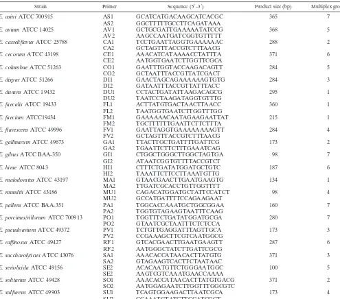
Soft-TABLE 1. PCR primers, products, and reference strains
Strain Primer Sequence (5⬘–3⬘) Product size (bp) Multiplex group
E. asiniATCC 700915 AS1 GCATCATGACAAGCATCACGC 365 7
AS2 GGCTTTTTGCCTTCAGATAAA
E. aviumATCC 14025 AV1 GCTGCGATTGAAAAATATCCG 368 5
AV2 AAGCCAATGATCGGTGTTTTT
E. casseliflavusATCC 25788 CA1 TCCTGAATTAGGTGAAAAAAC 288 2
CA2 GCTAGTTTACCGTCTTTAACG
E. cecorumATCC 43198 CE1 AAACATCATAAAACCTATTTA 371 6
CE2 AATGGTGAATCTTGGTTCGCA
E. columbaeATCC 51263 CO1 GAATTTGGTACCAAGACAGTT 284 5
CO2 GCTAATTTACCGTTATCGACT
E. disparATCC 51266 DI1 GAACTAGCAGAAAAAAGTGTG 284 3
DI2 GATAATTTACCGTTATTTACC
E. duransATCC 19432 DU1 CCTACTGATATTAAGACAGCG 295 1
DU2 TAATCCTAAGATAGGTGTTTG
E. faecalisATCC 19433 FL1 ACTTATGTGACTAACTTAACC 360 1
FL2 TAATGGTGAATCTTGGTTTGG
E. faeciumATCC19434 FM1 GAAAAAACAATAGAAGAATTAT 215 1
FM2 TGCTTTTTTGAATTCTTCTTTA
E. flavescensATCC 49996 FV1 GAATTAGGTGAAAAAAAAGTT 284 4
FV2 GCTAGTTTACCGTCTTTAACG
E. gallinarumATCC 49673 GA1 TTACTTGCTGATTTTGATTCG 173 2
GA2 TGAATTCTTCTTTGAAATCAG
E. gilvusATCC BAA-350 GI1 CTGGCTGGGCTTGGCTAGTGA 98 7
GI2 ATAATCGGTGTTTTACCGTCT
E. hiraeATCC 8043 HI1 CTTTCTGATATGGATGCTGTC 187 6
HI2 TAAATTCTTCCTTAAATGTTG
E. malodoratusATCC 43197 MA1 GTAACGAACTTGAATGAAGTG 134 1
MA2 TTGATCGCACCTGTTGGTTTT
E. mundtiiATCC 43186 MU1 CAGACATGGATGCTATTCCATCT 98 4
MU2 GCCATGATTTTCCAGAAGAAT
E. pallensATCC BAA-351 PA1 TGGCACCAAATGCTGGCGGAA 160 7
PA2 TGGTGTAGAAGTAATTTCAAG
E. porcinus/villorumATCC 700913 PO1 TGGTTTCTGATATGGATGCGA 280 7
PO2 GTAATCGCTAATTTCTCTCCA
E. pseudoaviumATCC 49372 PV1 TCTGTTGAGGATTTAGTTGCA 173 3
PV2 CCGAAAGCTTCGTCAATGGCG
E. raffinosusATCC 49427 RF1 GTCACGAACTTGAATGAAGTT 287 6
RF2 AATGGGCTATCTTGATTCGCG
E. saccharolyticusATCC 43076 SA1 AAACACCATAACACTTATGTG 371 3
SA2 GTAGAAGTCACTTCTAATAAC
E. seriolicidaATCC 49156 SE2 ACACAATGTTCTGGGAATGGC 100 5
SE2 AAGTCGTCAAATGAACCAAAA
E. solitariusATCC 49428 SO1 AAACACCATAACACTTATGTGACG 371 2
SO2 AATGGAGAATCTTGGTTTGGCGTC
E. sulfureusATCC 49903 SU1 TCAGTGGAAGACTTAATCGCA 173 4
SU2 CCAAATGTATCTTCGATCGCT
on May 15, 2020 by guest
http://jcm.asm.org/
[image:2.603.46.541.80.516.2]ware, Durham, N.C.). Conserved sequences within species and degenerate re-gions between species were used to design species-specific primers with the Oligo primer analysis software (Molecular Biology Insights, Inc., Cascade, Colo.). All primers were synthesized by Operon (Alameda, Calif.).
PCR.Template for PCR was prepared by suspending a single isolated bacterial colony in 100l of sterile deionized water. Seven PCR master mixes consisting of different primer sets were prepared. Group 1 wasE. durans,E. faecalis,E. faecium, andE. malodoratus; group 2 wasE. casseliflavus,E. gallinarum, and
E. solitarius; group 3 wasE. dispar,E. pseudoavium, andE. saccharolyticus; group 4 wasE. flavescens,E. mundtii, andE. sulfureus; group 5 wasE. avium,E. colum-bae, andE. seriolicida; group 6 wasE. cecorum,E. hirae, andE. raffinosus; and group 7 wasE. asini,E. gilvus,E. pallens, andE. porcinus/villorum. The base master mix consisted of 3 mM MgCl2(with Ficoll and tartrazine; Idaho
Tech-nology, Salt Lake City, Utah),0.2 mM deoxynucleoside triphosphate mix (Roche,
Indianapolis, Ind.), 16 mM (10⫻) NH4, 3.5 U of Expand high-fidelity PCR
system (Roche), and 1.25l of each genus primer (16M). With the exception ofE. faecalis,E. malodoratus,E. gallinarum,E. saccharolyticus, andE. dispar, 1.25
l of each species primer (16M) was added to the base mix as indicated (Table 1). For primers FL1, FL2, MA1, MA2, GA1, GA2, SA1, SA2, DI1, and DI2, 2.5
l of each primer was used. PCRs were performed in a final volume of 22.5l consisting of 20l of master mix and 2.5l of whole-cell template. Following an initial denaturation at 95°C for 4 min, products were amplified by 30 cycles of denaturation at 95°C for 30 s, annealing at 55°C (groups 1, 2, 5, and 6) or 60°C (groups 3, 4, and 7) for 1 min, and elongation at 72°C for 1 min. Amplification was followed by a final extension at 72°C for 7 min. Ten microliters of product was electrophoresed on a 2% 1⫻Tris-acetate-EDTA agarose gel containing 2g of ethidium bromide/ml. DNA molecular weight marker XIV (100 bp; Roche) was used as the standard.
RESULTS
Primer design.Using previously designedsodAdegenerate primers, a 438-bp internal fragment of the gene was amplified from enterococcal type strains (Table 1). Both strands of the PCR product were sequenced and analyzed to form one con-tiguous sequence. Multiple sequences were aligned using gen-eratedsodAsequences and those available in public databases, allowing conserved regions within species and variable regions between species to be identified (data not shown). Due to the small region sequenced, the number of isolates analyzed, and the close relatedness of some species, some species primers differed from other primers by only a few base pairs, primarily on the 3⬘end (Table 1). Differences in only 1 bp were enough to amplify sodA from multiple target species, but not from other enterococcal species (data not shown). For other species, one forward or reverse primer was identical in sequence to another species primer, but the remaining primer in the set was different. These manipulations in conjunction with a
proof-reading polymerase mix allowed only the correct target se-quence to be amplified. Selected sodA enterococcal species primer sequences were not compared to other bacterial species sequences, because genus-specific primers were included in the PCR, verifying the genus of the isolate. Groups of species primers were developed due to the small region ofsodA se-quenced (Table 1). Each group contained primer sets that would amplify a portion of DNA in four size ranges: 95 to 135 bp, 170 to 215 bp, 280 to 300 bp, orⱖ360 bp. These size ranges allowed separation of amplified product while also standard-izing amplicon sizes across groups. Repeated attempts to am-plify a fragment fromE. rattiwere unsuccessful, resulting in no primer sets for this species.
Amplification using enterococcal genus and species prim-ers.The specificity of the genus and species PCR was deter-mined by testing all species shown in Table 1 against all groups of the multiplex primer sets. All strains reacted with the en-terococcal genus primer, indicating that they were members of Enterococcus (Fig. 1). A very weak band was produced with E. seriolicidadespite repeated attempts (Fig. 1, lane 19). Only positive control strains of group 1 (E. faecalis,E. durans,E. fae-cium, andE. malodoratus) reacted with the appropriate group 1 species primers, producing products of 360, 295, 215, and 134 bp, respectively (Fig. 1). This process was repeated for all groups to verify specificity and rule out cross-reactivity.L. gar-vieae, used as a negative DNA control, did not react with the genus primers or any of the species primers, and no product was produced in control samples in which target DNA was not provided (Fig. 1).
[image:3.603.50.536.70.172.2]Ease of use of the multiplexing PCR was achieved by first circumventing the need for purifying template DNA. Although the PCR performed well with pure DNA, isolated colonies suspended in sterile water worked equally as well and required little processing. The DNA was released from the cells during the initial 95°C denaturation step, and negative results with strains that produced no product with genus and species prim-ers were not due to heat-resistant strains. Results using colo-nies isolated on blood agar were somewhat problematic in the PCR, but all colonies from BHIA produced the expected am-plicons. This suggests that some components present in blood agar may have interfered with the PCR, yielding negative re-sults. An essential requirement for the multiplex PCR was a FIG. 1. Group 1 genus and species multiplex PCR of enterococci. All species of enterococci were tested against group 1 multiplex primers in order to confirm specificity. Genus-specific bands are indicated by the arrow, and species-specific bands are indicated by asterisks. Species positive controls are in lanes 4 to 7 as follows:E. faecalis(360 bp),E. durans(295 bp),E. faecium(215 bp), andE. malodoratus(134 bp). Negative controls in lanes 2 and 3 contained no DNA andL. garvieae, respectively. Lanes 8 to 26:E. solitarius,E. casseliflavus,E. gallinarum,E. mundtii,E. sac-charolyticus,E. dispar,E. pseudoavium,E. gilvus,E. flavescens,E. sulfureus,E. raffinosus,E. seriolicida,E. avium,E. columbae,E. cecorum,E. hirae, E. asini,E. porcinus, andE. pallens. Lanes 1 and 27, DNA standard.
on May 15, 2020 by guest
http://jcm.asm.org/
pure starting culture. Strains streaked less than twice appeared mixed, producing signals in multiple PCR groups. For exam-ple, weak bands in group 4 representing E. flavescens and intense bands in group 1 (E. faecalis) were apparent for some isolates from poultry and retail foods. This problem was solved when the isolates were restreaked onto BHIA and re-tested (data not shown). Therefore, in order to ensure purity of culture, isolates were routinely streaked onto nonselective me-dium at least twice before selection of colonies.
Another important feature of the multiplex PCR was the ability to amplify all PCR groups by using either a 55 or 60°C annealing temperature. Higher annealing temperatures (great-er than 60°C) w(great-ere tested but resulted in reduced product yield or negative results. FM1 and FM2 primers appeared to be sensitive to increased annealing temperatures, as the E. fae-ciumpositive control did not amplify well at 60°C annealing. Group 2 species primers were the most sensitive to changes in annealing temperature and in-lab variations. Care was taken with reaction components (primers, etc.) to ensure successful amplification of controls. Considerable variation in PCR re-sults was also observed when the supplier of PCR primers was changed. In addition to annealing temperature, all other com-ponents of the PCR were standardized between reactions, ex-cept primer concentrations. Primer quantities for some species were less than for other species due to primer interference. For example, higher concentrations of E. faecalis and E. durans primers reduced the intensity of theE. faeciumband. Reduc-tion in intensity or no product was also observed when all positive controls for a PCR group were mixed in one tube for multiplexing. This could be due to overload of the reaction mixture with target DNA or impurities in the target DNA. The best results were obtained when controls were used in individ-ual reactions.
Identification of amplified products.In rare cases, noncific products were observed when using the genus- and spe-cies-specific multiplex PCR. In order to be identified as an Enterococcusspecies, amplicons produced by unknown strains were required to be the same size as the genus and species PCR product. If the bands were not the expected size, then they were not identified as a particular species. An example of an errant band present in several multiplexing groups is shown in Fig. 2. Two unknown enterococcal isolates were tested
against all seven groups of species primers. A band was ob-tained for both isolates when using groups 7, 6, and 4 (Fig. 2). Although an intense similar-sized band (300 to 400 bp) was obtained using group 7 primers, this band was slightly larger than the 280-bpE. porcinus/villorumband and slightly smaller than the 365-bp E. asini band (Fig. 2A). When tested with group 6 primers, amplicons for the two isolates were located between the 371-bp band representing E. cecorum and the 287-bp band representingE. raffinosus, indicating that the iso-lates were not E. cecorumorE. raffinosus(data not shown). But, when tested against group 4 primers, the same-sized band for both isolates andE. mundtiiwas observed, indicating that the unknown isolates wereE. mundtii(Fig. 2B).
Comparison of PCR to standard biochemical testing and commercial identification methods. One hundred isolates from four different sources (swine, poultry, environmental, and retail food) were identified using standard biochemical tests, the BBL Crystal Gram-Positive ID kit, VITEK, Rapid ID 32 Strep, and multiplex PCR in order to determine the accuracy of the PCR (Table 2). Standard biochemical tests can identify all enterococcal species, whereas the three kits allowed iden-tification of seven primary species (E. avium,E. casseliflavus, E. durans,E. faecalis,E. faecium,E. gallinarum, andE. hirae). In addition to these seven species, Rapid ID 32 Strep would also identify E. saccharolyticus, whereas BBL Crystal would also identifyE. raffinosusandE. solitarius. E. casseliflavusand E. gallinarum were grouped into one species with the BBL Crystal kit, requiring additional tests for differentiation. In contrast to the commercial kits, the multiplex PCR identified 23 enterococcal species.
All five methods identified the isolates as belonging to the genus Enterococcus, but not all species identifications agreed (Table 2). Consensus identification was determined by match-ing results from at least three of the five methods. From swine, poultry, environmental, and retail food samples, five consensus species (E. faecalis,E. faecium, E. casseliflavus, E. hirae, and E. gallinarum) were identified. The predominant species was E. faecalis(n⫽53), followed byE. casseliflavus(n⫽16) and E. hirae(n⫽14). Although some isolates were identified asE. duransorE. mundtiiin preliminary biochemical analyses, these species were not the consensus species when results from all identification methods were combined.
FIG. 2. Nonspecific amplicons of genus and species multiplex PCR. Group 7 primers (A) and group 4 primers (B) are shown. Arrows indicate nonspecific bands, and asterisks indicate specific bands. (A) Lanes 2 and 3, no DNA control andL. garvieae, respectively; lanes 4 to 9,E. asini, E. porcinus/villorum,E. pallens,E. gilvus, unknown species A, and unknown species B. (B) Lanes 2 to 8,E. flavescens,E. sulfureus,E. mundtii, unknown species A, unknown species B,E. faecalis, andE. faecium. Lanes 1 and 10 (A) and lanes 1 and 9 (B) are DNA standards.
on May 15, 2020 by guest
http://jcm.asm.org/
TABLE 2. Identification of enterococci by different identification methods
Source Sample Species determination
Standard PCR BBL Vitek ID 32 Strep Consensus ID
Swine feces 110 E. hirae E. hirae E. durans E. hirae E. hirae E. hirae
113 E. hirae E. hirae E. durans E. hirae E. hirae E. hirae
124 E. faecalis E. faecalis E. faecalis E. faecalis E. faecalis E. faecalis
126 E. faecalis E. faecalis E. faecalis E. faecalis E. faecalis E. faecalis
127 E. faecalis E. faecalis E. faecium E. faecalis E. faecalis E. faecalis
188 E. faecalis E. faecalis E. faecalis E. faecalis E. faecalis E. faecalis
190 E. faecalis E. faecalis E. faecalis E. faecalis E. hirae E. faecalis
186 E. hirae E. hirae E. durans E. hirae E. gallinarum E. hirae
165 E. durans E. hirae E. durans E. hirae E. hirae E. hirae
166 E. hirae E. hirae E. durans E. hirae E. hirae E. hirae
170 E. faecalis E. faecalis E. faecalis E. faecalis E. faecalis E. faecalis
135 E. durans E. hirae E. hirae E. hirae E. hirae E. hirae
134 E. faecium E. faecium E. faecium E. faecium E. gallinarum E. faecium
133 E. faecium E. faecium E. faecium E. faecalis E. gallinarum E. faecium
131 E. hirae E. hirae E. faecium E. hirae E. hirae E. hirae
130 E. faecium E. casseliflavus E. casseliflavus E. casseliflavus E. gallinarum E. casseliflavus
125 E. faecalis E. faecalis E. faecalis E. faecalis E. faecalis E. faecalis
128 E. faecium E. faecalis E. faecalis E. faecalis E. faecalis E. faecalis
129 E. faecalis E. faecalis E. faecalis E. faecalis E. faecalis E. faecalis
141 E. faecalis E. faecalis E. faecalis E. faecalis E. hirae E. faecalis
107 E. faecium E. faecium E. faecium E. faecium E. faecium E. faecium
108 E. faecium E. faecalis E. faecalis E. faecalis E. faecalis E. faecalis
109 E. faecium E. faecium E. faecium E. faecium E. gallinarum E. faecium
151 E. faecalis E. faecalis E. faecalis E. faecalis E. faecalis E. faecalis
152 E. faecalis E. faecalis E. faecalis E. faecalis E. faecalis E. faecalis
Poultry carcass A1 E. faecalis E. faecalis E. faecalis E. faecalis E. faecalis E. faecalis
A2 E. faecalis E. faecalis E. faecalis E. faecalis E. faecalis E. faecalis
A3 E. faecium E. faecium E. faecium E. faecium E. faecium E. faecium
A4 E. faecium E. faecium E. hirae E. faecium E. faecalis E. faecium
A5 E. faecalis E. faecalis E. faecalis E. faecalis E. faecalis E. faecalis
A6 E. faecalis E. faecalis E. faecalis E. faecalis E. faecalis E. faecalis
A7 E. faecalis E. faecalis E. faecalis E. faecalis E. faecalis E. faecalis
A8 E. faecalis E. faecalis E. faecalis E. faecalis E. faecalis E. faecalis
A9 E. faecalis E. faecalis E. faecalis E. faecalis E. faecalis E. faecalis
A10 E. faecalis E. faecalis E. faecalis E. faecalis E. faecalis E. faecalis
A11 E. hirae E. hirae E. durans E. hirae E. hirae E. hirae
A12 E. faecalis E. faecalis E. faecalis E. faecalis E. faecalis E. faecalis
B1 E. faecium E. faecium E. durans E. faecium E. gallinarum E. faecium
B2 E. hirae E. hirae E. durans E. hirae E. hirae E. hirae
B3 E. faecium E. faecalis E. faecium E. faecium E. gallinarum E. faecium
B4 E. faecium E. casseliflavus E. casseliflavus E. casseliflavus E. casseliflavus E. casseliflavus
B5 E. faecalis E. faecalis E. faecalis E. faecalis E. faecalis E. faecalis
B6 E. casseliflavus E. casseliflavus E. casseliflavus E. casseliflavus E. casseliflavus E. casseliflavus
B7 E. faecalis E. faecalis E. faecalis E. faecalis E. faecalis E. faecalis
B8 E. faecalis E. faecalis E. faecalis E. faecalis E. faecalis E. faecalis
B9 E. hirae E. hirae E. durans E. hirae E. hirae E. hirae
B10 E. faecalis E. faecalis E. faecalis E. faecalis E. faecalis E. faecalis
B11 E. faecalis E. faecalis E. faecalis E. faecalis E. faecalis E. faecalis
B12 E. faecalis E. faecalis E. faecalis E. gallinarum E. faecalis E. faecalis
C1 E. faecalis E. faecalis E. faecalis E. faecalis E. faecalis E. faecalis
Park
Sand Mem-9 E. casseliflavus E. casseliflavus/flav E. casseliflavus E. casseliflavus E. casseliflavus E. casseliflavus
Fly strip Mem-18 E. casseliflavus E. faecalis E. faecalis E. faecalis E. faecalis E. faecalis
Metal picnic table Bish-1 E. mundtii E. mundtii E. faecium E. faecium E. casseliflavus NDa
Metal picnic table Bish-2 E. faecalis E. faecalis E. faecalis E. faecalis E. faecalis E. faecalis
Metal trash can Bish-3 E. faecalis E. faecalis E. faecalis E. faecalis E. faecalis E. faecalis
Rubber swing Bish-4 E. faecalis E. faecalis E. faecalis E. faecalis E. faecalis E. faecalis
Ladder bar Bish-10 E. faecalis E. faecalis E. faecalis E. faecalis E. faecalis E. faecalis
Plastic slide Bish-12 E. faecalis E. faecalis E. faecalis E. faecalis E. faecalis E. faecalis
Steering wheel Bish-13 E. gallinarum E. gallinarum E. gallinarum E. gallinarum E. gallinarum E. gallinarum
Plastic slide Bish-14 E. hirae E. hirae E. hirae E. hirae E. hirae E. hirae
Plastic slide Bish-15 E. faecalis E. faecalis E. faecalis E. faecalis E. faecalis E. faecalis
Metal picnic table Bish-17 E. casseliflavus E. casseliflavus E. casseliflavus E. casseliflavus E. casseliflavus E. casseliflavus
Plastic slide Bish-19 E. faecalis E. faecalis E. faecalis E. faecalis E. faecalis E. faecalis
Plastic slide Bish-20 E. casseliflavus E. casseliflavus E. casseliflavus E. casseliflavus E. casseliflavus E. casseliflavus
Sand Bish-21 E. casseliflavus E. casseliflavus E. casseliflavus E. casseliflavus E. casseliflavus E. casseliflavus
Sand Bish-22 E. hirae E. hirae E. hirae E. hirae E. hirae E. hirae
Sand Bish-23 E. hirae E. hirae E. hirae E. hirae E. hirae E. hirae
Continued on following page
on May 15, 2020 by guest
http://jcm.asm.org/
Overall, 69% (69 of 100) of the isolates tested agreed in species determination for all five methods, whereas 19% agreed with four of five methods. Consensus identification could not be confidently determined in 5% of the isolates. For these isolates, only two of the five methods agreed in species identification. This was due to certain tendencies of some of the commercial kits. For example, when standard testing, PCR, VITEK, and ID 32 Strep identified an isolate asE. hirae, BBL Crystal identified the same isolate asE. durans. This accounted for 6 of 19 (31.5%) of the isolates for which one test method differed. In addition, ID 32 Strep appeared to identify moreE. gallinarumthan any other commercial method (Table 2). Two isolates identified asE. gallinarumby ID 32 Strep were iden-tified as E. faecium using the other methods. This kit also identified two isolates asE. hiraewhile the other four methods identified them asE. faecalis(Table 2). Standard biochemical testing also differed from the other four tests for 5 of 19 (26%) of the isolates. Three isolates identified asE. faeciumby stan-dard testing were identified asE. faecalisandE. casseliflavusby the other four methods. Differences in identification between the multiplex PCR and the commercial identification kits also resulted from identification of a species by PCR that the com-mercial kits could not identify. For example, the multiplex PCR identified fiveE. mundtiiisolates, whereas those isolates were identified asE. faecium,E. casseliflavus, orE. gallinarum
by the commercial kits. All four species share similar biochem-ical traits and belong in group II of the classbiochem-ical phenotypic characterization table, suggesting a close relationship (3). Standard biochemical testing results concurred with results from PCR, as those five isolates were also identified as E. mundtii(Table 2). For PCR, there was only one isolate (B3) for which results did not agree with results from any other test. Since outcomes of the traditional phenotypic tests can be vari-able, some misidentification of the isolates can occur using those methods. However, no distinct pattern could be dis-cerned from the isolates for which two groups gave matching identifications but the remaining three groups did not.
[image:6.603.42.542.80.419.2]When all five identification methods were compared against each other, discrepancies were apparent (Table 3). The mul-tiplex PCR agreed with BBL Crystal for 82% of the total samples. Although percent agreement increased when results from PCR were compared with those from ID 32 Strep (85%), the highest percent agreements were observed between PCR and VITEK (90%) and PCR and standard testing (93%). With the exception of the BBL-VITEK comparison (83%) and the standard testing-VITEK comparison (85%), the percent agree-ment between PCR and all other methods was higher than for any other combination of identification methods. The lowest percent agreements in identification were 79% for standard testing and ID 32 Strep, 78% for standard testing and BBL TABLE 2—Continued
Source Sample Species determination
Standard PCR BBL Vitek ID 32 Strep Consensus ID
Sand Bish-24 E. hirae E. hirae E. hirae E. hirae E. hirae E. hirae
Fly strip Bish-25 E. faecalis E. faecalis E. faecalis E. faecalis E. faecalis E. faecalis
Fly strip Bish-26 E. faecium E. faecium E. faecium E. faecium E. faecium E. faecium
Plastic slide SC-5 E. faecalis E. faecalis E. faecalis E. faecalis E. faecalis E. faecalis
Plastic slide SC-6 E. faecalis E. faecalis E. faecalis E. faecalis E. faecalis E. faecalis
Tic-tac-toe wheels SC-9 E. faecalis E. faecalis E. faecalis E. faecalis E. faecalis E. faecalis
Step vine climber SC-10 E. mundtii E. mundtii E. faecium E. faecium E. casseliflavus ND
Plastic slide SC-11 E. casseliflavus E. casseliflavus E. casseliflavus E. gallinarum E. casseliflavus E. casseliflavus
Retail foods
Red potato BSM 36 E. casseliflavus E. casseliflavus/flav E. casseliflavus E. casseliflavus E. casseliflavus E. casseliflavus
Chicken BSM 35 E. casseliflavus E. casseliflavus E. casseliflavus E. casseliflavus E. casseliflavus E. casseliflavus
Red potato BSM 37 E. casseliflavus E. casseliflavus E. casseliflavus E. casseliflavus E. casseliflavus E. casseliflavus
Beef BSM 29 E. faecalis E. faecalis E. faecalis E. faecalis E. faecalis E. faecalis
Red potato BSM 34 E. casseliflavus E. casseliflavus E. casseliflavus E. casseliflavus E. casseliflavus E. casseliflavus
Lettuce KSM 24 E. mundtii E. mundtii E. casseliflavus/gallinarum E. faecium E. casseliflavus ND
Turkey KSM 28 E. faecium E. faecium E. faecium E. faecium E. faecium E. faecium
Turkey KSM 29 E. faecalis E. faecalis E. faecalis E. faecalis E. faecalis E. faecalis
Lettuce KSM 25 E. mundtii E. mundtii E. faecium E. faecium E. casseliflavus ND
Chicken KSM 26 E. faecalis E. faecalis E. faecalis E. faecalis E. faecalis E. faecalis
Cucumber WSM 17 E. faecalis E. faecalis E. faecalis E. faecalis E. faecalis E. faecalis
Beef WSM 29 E. faecalis E. faecalis E. faecalis E. faecalis E. faecalis E. faecalis
Pork WSM 32 E. faecalis E. faecalis E. faecalis E. faecalis E. faecalis E. faecalis
Chicken WSM 26 E. faecalis E. faecalis E. faecalis E. faecalis E. faecalis E. faecalis
White potato ISM 42 E. mundtii E. mundtii E. faecium E. faecium E. casseliflavus ND
Pork ISM 30 E. faecalis E. faecalis E. faecalis E. gallinarum E. faecalis E. faecalis
Red potato ISM 32 E. casseliflavus E. casseliflavus E. casseliflavus E. casseliflavus E. casseliflavus E. casseliflavus
Red potato ISM 36 E. casseliflavus E. casseliflavus E. casseliflavus E. casseliflavus E. casseliflavus E. casseliflavus
Apple ISM 1 E. casseliflavus E. casseliflavus E. casseliflavus E. casseliflavus E. casseliflavus E. casseliflavus
Turkey ISM 26 E. faecalis E. faecalis E. faecalis E. faecalis E. faecalis E. faecalis
Pork FSM 29 E. faecalis E. faecalis E. faecalis E. faecalis E. faecalis E. faecalis
White potato FSM 38 E. faecalis E. faecalis E. faecalis E. faecalis E. faecalis E. faecalis
Beef FSM 26 E. faecium E. faecium E. faecium E. faecium E. faecium E. faecium
Red potato FSM 32 E. casseliflavus E. casseliflavus E. casseliflavus E. casseliflavus E. casseliflavus E. casseliflavus
Chicken FSM 23 E. faecalis E. faecalis E. faecalis E. faecalis E. faecalis E. faecalis
aND, not determined.
on May 15, 2020 by guest
http://jcm.asm.org/
Crystal, and only 77% between BBL Crystal and ID 32 Strep (Table 3). When source of isolates was examined, percent agreement between PCR and VITEK was higher than any other combination for swine, whereas percent agreement be-tween PCR and standard testing was higher for poultry (96%), environmental (96%), and retail food samples (100%) (Table 3). Ninety-six percent agreement between BBL Crystal and VITEK was also observed for environmental samples. This discrepancy in agreement between PCR and VITEK can be explained by the identification ofE. mundtiiby PCR but not by VITEK. TwoE. mundtiiisolates from environmental samples and three from retail food samples were identified by PCR.
DISCUSSION
Characterization and identification of enterococci by using the traditional phenotypic differentiation can be a tedious pro-cess requiring numerous tests. Strains are classified based upon growth in various media, biochemical reactions in those media, motility, and pigmentation. Although more than 20 species can be identified using these methods, tests are typically performed in test tubes and often require long periods of incubation before results can be interpreted (3, 4). In addition, grouping and identification of strains with phenotypic tests have been determined using type strains and strains isolated from human sources (17). Strains isolated from nonhuman sources may be atypical and may not conform to the criteria used for standard phenotypic characterization. Problems such as time constraints and number of samples to be processed can be overcome to some degree by using commercial identification kits. However, their accuracy has also been assessed using type strains and strains from clinical sources. Moreover, they usually only iden-tify a maximum of 10 enterococcal species, which must also conform to the testing scheme. In addition, auto-reading of results should be utilized with commercial kits, as manual interpretation of results can lead to erroneous identification, and aberrant reactions will result in no species identification of the strain. An auto-reader was used for the commercial iden-tification kits in this study and, therefore, factors other than manual reading of results must have accounted for the low overall agreement in identification.
Other methods for identification of enterococci have utilized molecular techniques such as PCR and sequencing (1, 6, 9, 12, 22). A previous report identified the manganese-dependent superoxide dismutase genesodA as an ideal gene for species identification of enterococci (16). The superoxide dismutase gene has been used to distinguish genera and species of my-cobacteria, streptococci, staphylococci, and enterococci (14,
15, 24). For differentiating species of enterococci,sodA gene sequences were used to create a library of sequences. Other unknown isolates were compared to the type strains and sub-sequently identified by percent homology to those reference strains. Although sequencing is becoming more available, it can be expensive and time-consuming if a number of isolates need to be analyzed. In order to overcome these limitations, PCR primers to uniquesodA sequences in each enterococcal species were designed.
When coupled with genus primers, the multiplex PCR pro-vides an accurate and quick method for identification of en-terococci, without the need for extensive phenotypic tests. Ge-nus primers designed by Deasy et al. (2) were used in each reaction to confirm the genus enterococci. Previously, these primers were rigorously tested against a number of gram-pos-itive bacteria and only produced product from bacteria belong-ing to the enterococci. Although those authors acknowledged that the genus primer may also amplify a product from Car-nobacterium, we have not encountered this problem and have found these primers to be specific forEnterococcusand nega-tive for other bacterial genera. Moreover, a genus- and species-specific PCR has been developed forCarnobacterium, but the enterococcal primers were not used for that purpose (19).
The inclusion of certain species in the multiplex PCR was based on previous reports and availability of isolates. A single species primer pair was designed for detection of both E. villorumandE. porcinus, because previous studies have shown that these two species are the same (3). Also of interest was the ability of the PCR, for some isolates, to distinguish E. cas-seliflavus from E. flavescens, even though those two species have been reported to comprise a single species (3). Some isolates were clearly identified as either E. casseliflavus or E. flavescens, while other isolates were positive for both sets of primers. In addition, the species E. haemoperoxidus and E. moraviensiswere not included in the multiplex PCR because sodA sequences from multiple isolates were not available for comparison (20). Moreover, althoughE. seriolicidaandE. soli-tariusmay eventually be reclassified into other genera, they are presently classified as enterococci and were included in the analysis (21, 23).
[image:7.603.42.549.81.178.2]As with every identification system, multiplex PCR has lim-itations and will not identify every isolate. Difficulty with iden-tification was encountered when pure cultures were not used in the analysis. If isolates were not streaked at least twice onto nonselective medium, then mixed signals resulted. This could be due to either contamination of the selected colony with smaller colonies not visible to the naked eye or clumping of TABLE 3. Comparison of enterococci identification methods
Source testedNo.
No. in agreement (%) for the two methods
STD/PCR STD/BBL STD/VITEK 32 StrepSTD/ID PCR/BBL PCR/Vitek PCR/ID32 Strep BBL/VITEK 32 StrepBBL/ID VITEK/ID32 Strep
Swine 25 20 (80) 15 (60) 20 (20) 14 (56) 18 (72) 24 (96) 18 (72) 17 (68) 12 (48) 18 (72)
Poultry 25 24 (96) 19 (76) 23 (92) 21 (84) 19 (76) 23 (92) 22 (88) 19 (76) 19 (76) 21 (84)
Park 25 24 (96) 22 (88) 21 (84) 22 (88) 23 (92) 22 (88) 23 (92) 24 (96) 23 (92) 22 (88)
Retail food 25 25 (100) 22 (88) 21 (84) 22 (88) 22 (88) 21 (84) 22 (88) 23 (92) 23 (92) 21 (84)
Total 100 93 (93) 78 (78) 85 (85) 79 (79) 82 (82) 90 (90) 85 (85) 83 (83) 77 (77) 82 (82)
on May 15, 2020 by guest
http://jcm.asm.org/
cells. Mixed cultures for enterococci have been observed pre-viously when using the BBL Crystal ID kit for identification (8). Errant bands that did not correspond to bands in control samples were regarded as nonspecific and were not used for identification purposes. These bands were often either of lower or higher molecular weights than control bands and were easily recognized as being different from the expected size. It could be argued that the lower- or higher-molecular-weight bands could have resulted from the addition or loss of DNA between the primers, but when all seven groups of primers were tested a perfect match to the control strains was always made, sug-gesting that the errant bands were artifacts. Sequencing of errant products also revealed that it was possible for one primer from one species and another primer from another species to form a product, albeit of the incorrect size. Although the multiplex PCR was designed to amplify specific regions of enterococcal species, this property can be both a positive and a negative feature. Species-specific primers amplified only the designated species; however, while major variations in sodA sequences would not be expected, minor sequence variations among strains of the same species could result in the absence of expected species amplicons. Isolates from environmental and retail food samples were found to be more difficult to identify to the species level and sometimes to the genus level. These isolates have not been extensively characterized, and it would not be surprising if their sodA sequences were more variable than those from other sources or if those sources contained previously unidentified species. Presently, approxi-mately 15% of isolates from environmental and retail food samples remain unidentified at the species level when using multiplex PCR. Studies are ongoing in order to determine significant differences, if any, in the sodA gene sequence of such isolates.
ACKNOWLEDGMENTS
We thank Lari Hiott, Carolina Hall, Sandra House, and Tiffanie Woodley for technical assistance and Katie Gay and Katie Lewis (CDC) for standard biochemical testing. We also thank Mark Englen for critical review of the manuscript.
REFERENCES
1. Baele, M., P. Baele, M. Vaneechoutte, V. Storms, P. Butaye, L. A. Devriese,
G. Verschraegen, M. Gillis, and F. Haesebrouck.2000. Application of tRNA
intergenic spacer PCR for identification ofEnterococcusspecies. J. Clin. Microbiol.38:4201–4207.
2. Deasy, B. M., M. C. Rea, G. F. Fitzgerald, T. M. Cogan, and T. P. Beresford.
2000. A rapid PCR based method to distinguish betweenLactococcusand
Enterococcus.Syst. Appl. Microbiol.23:510–522.
3. Facklam, R. R., M. G. S. Carvalho, and L. M. Teixeiram.2002. History,
taxonomy, biochemical characteristics, and antibiotic susceptibility testing of enterococci, p. 1–54.InM. S. Gilmore, D. B. Clewell, P. Courvalin, G. M. Dunny, B. E. Murray, and L. B. Rice (ed.), The enterococci: pathogenesis, molecular biology, and antibiotic resistance. ASM Press, Washington, D.C.
4. Facklam, R. R., and M. D. Collins.1989. Identification ofEnterococcus
species isolated from human infections by a conventional test scheme. J. Clin. Microbiol.27:731–734.
5. Garcia-Garrote, F., E. Cercenado, and E. Bouza.2000. Evaluation of a new
system, VITEK 2, for identification and antimicrobial susceptibility testing of enterococci. J. Clin. Microbiol.38:2108–2111.
6. Goh, S. H., R. R. Facklam, M. Chang, J. E. Hill, G. J. Tyrrell, E. C. Burns,
D. Chan, C. He, T. Rahim, C. Shaw, and S. M. Hemmingsen.2000.
Identi-fication ofEnterococcusspecies and phenotypically similarLactococcusand
Vagococcusspecies by reverse checkerboard hybridization to chaperonin 60 gene sequences. J. Clin. Microbiol.38:3953–3959.
7. Hamilton-Miller, J. M., and S. Shah.1999. Identification of clinically
iso-lated vancomycin-resistant enterococci: comparison of API and BBL Crystal systems. J. Med. Microbiol.48:695–696.
8. Hudson, C. R., P. J. Fedorka-Cray, M. C. Jackson-Hall, and L. M. Hiott.
2003. Anomalies in species identification of enterococci from veterinary sources using a commercial biochemical identification system. Lett. Appl. Microbiol.36:245–250.
9. Ke, D., F. J. Picard, F. Martineau, C. Menard, P. H. Roy, M. Ouellette, and
M. G. Bergeron.1999. Development of a PCR assay for rapid detection of
enterococci. J. Clin. Microbiol.37:3497–3503.
10. Moellering, R. C., Jr.1992. Emergence ofEnterococcus as a significant
pathogen. Clin. Infect. Dis.14:1173–1176.
11. Murray, B. E.1990. The life and times of theEnterococcus.Clin. Microbiol.
Rev.3:46–65.
12. Naimi, A., G. Beck, and C. Branlant.1997. Primary and secondary structures
of rRNA spacer regions in enterococci. Microbiology143:823–834.
13. Ozawa, Y., P. Courvalin, and M. Gaiimand.2000. Identification of
entero-cocci at the species level by sequencing of the genes forD-alanine:D-alanine
ligases. Syst. Appl. Microbiol.23:230–237.
14. Poyart, C., G. Quesne, C. Boumaila, and P. Trieu-Cuot.2001. Rapid and
accurate species-level identification of coagulase-negative staphylococci by using thesodAgene as a target. J. Clin. Microbiol.39:4296–4301.
15. Poyart, C., G. Quesne, S. Coulon, P. Berche, and P. Trieu-Cuot.1998.
Identification of streptococci to species level by sequencing the gene encod-ing the manganese-dependent superoxide dismutase. J. Clin. Microbiol.36:
41–47.
16. Poyart, C., G. Quesnes, and P. Trieu-Cuot.2000. Sequencing the gene
encoding manganese-dependent superoxide dismutase for rapid species identification of enterococci. J. Clin. Microbiol.38:415–418.
17. Ruoff, K. L., L. de la Maza, M. J. Murtagh, J. D. Spargo, and M. J. Ferraro.
1990. Species identities of enterococci isolated from clinical specimens. J. Clin. Microbiol.28:435–437.
18. Sader, H. S., D. Biedenbach, and R. N. Jones.1995. Evaluation of Vitek and
API 20S for species identification of enterococci. Diagn. Microbiol. Infect. Dis.22:315–319.
19. Scarpellini, M., D. Mora, S. Colombo, and L. Franzetti.2002. Development
of genus/species-specific PCR analysis for identification ofCarnobacterium
strains. Curr. Microbiol.45:24–29.
20. Svec, P., L. A. Devriese, I. Sedlacek, M. Baele, M. Vancanneyt, F.
Haese-brouck, J. Swings, and J. Doskar.2001.Enterococcus haemoperoxidussp.
nov. andEnterococcus moraviensissp. nov., isolated from water. Int. J. Syst. Evol. Microbiol.51:1567–1574.
21. Teixeira, L. M., V. L. Merquior, M. C. Vianni, M. G. Carvalho, S. E.
Fracalanzza, A. G. Steigerwalt, D. J. Brenner, and R. R. Facklam.1996.
Phenotypic and genotypic characterization of atypicalLactococcus garvieae
strains isolated from water buffalos with subclinical mastitis and confirmation ofL. garvieaeas a senior subjective synonym ofEnterococcus seriolicida.Int. J. Syst. Bacteriol.46:664–668.
22. Tyrrell, G. J., R. N. Bethune, B. Willey, and D. E. Low.1997. Species
identification of enterococci via intergenic ribosomal PCR. J. Clin. Micro-biol.35:1054–1060.
23. Williams, A. M., U. M. Rodrigues, and M. D. Collins.1991. Intrageneric
relationships of enterococci as determined by reverse transcriptase sequenc-ing of small-subunit rRNA. Res. Microbiol.142:67–74.
24. Zolg, J. W., and S. Philippi-Schulz.1994. The superoxide dismutase gene, a
target for detection and identification of mycobacteria by PCR. J. Clin. Microbiol.32:2801–2812.