Capture Enzyme-Linked Immunosorbent Assay for Diagnosis of
Dengue Virus Infection
Madhuri Namekar,a,bEsther M. Ellis,a,bMaile O’Connell,a,bJoe Elm,cAlexandra Gurary,a,bSarah Y. Park,cAllison Imrie,a,b,d Vivek R. Nerurkara,b
Department of Tropical Medicine, Medical Microbiology and Pharmacology, John A. Burns School of Medicine, University of Hawaii at Manoa, Honolulu, Hawaii, USAa;
Pacific Center for Emerging Infectious Diseases Research, John A. Burns School of Medicine, University of Hawaii at Manoa, Honolulu, Hawaii, USAb; State of Hawaii
Department of Health, Honolulu, Hawaii, USAc; University of Western Australia, Perth, Western Australia, Australiad
We evaluated the FDA-cleared InBios dengue virus (DENV) IgM capture enzyme-linked immunosorbent assay (ELISA) for qual-itative detection of anti-DENV IgM antibodies from 79 serum samples obtained from dengue virus-infected patients or sus-pected dengue cases. The agreement, sensitivity, and specificity of the InBios assay compared to the gold standard in-house DENV IgM capture ELISA were 94, 92, and 94%, respectively. We conclude that the InBios DENV IgM capture ELISA can be ef-fectively used for rapid diagnosis of acute or recent DENV infection.
D
engue virus (DENV) a mosquito-borne flavivirus, is asignif-icant human pathogen of global importance (1). Dengue, an
acute viral disease, is caused by any one of the four DENV
sero-types, DENV-1, -2, -3, and -4 (2). Although most of the reported
dengue cases in the United States are acquired by travelers or
immigrants (3), autochthonous dengue fever outbreaks have
oc-curred in Brownsville, TX (2005), and southern Florida (2009 to
2011) and Hawaii (2011) (4). To date, there is no vaccine or
spe-cific antiviral treatment for dengue virus infection in humans, and effective management of severe dengue virus disease can be aug-mented by rapid diagnosis during the acute stage of infection
(5,6).
In the majority of DENV infections, immunoglobulin M (IgM) antibodies can be detected within 3 to 5 days following the onset of
fever (7). In secondary DENV infection, IgM antibody titers are
usually lower than those in primary DENV infection but follow
similar kinetics (8). An ideal IgM serologic test should have
suffi-cient sensitivity to detect low DENV IgM antibody titers and be specific enough to discriminate DENV infection in areas where
multiple flaviviruses and other pathogens cocirculate (9). Several
rapid diagnostic tests are commercially available for detection of
anti-DENV IgM antibodies (9). Therefore, it is important to
eval-uate the performance characteristics of these kits in terms of sen-sitivity and specificity in order to ensure accurate and rapid
diag-nosis of dengue virus infection (5). Recently, the U.S. Food and
Drug Administration (FDA) cleared the InBios DENV Detect IgM capture enzyme-linked immunosorbent assay (ELISA) (InBios International, Inc., Seattle, WA) for qualitative detection of
anti-DENV IgM antibodies (4). This test can detect acute or recent
DENV infections and can be used by public health laboratories for
rapid confirmation of dengue cases during dengue outbreaks (4).
(These research data are part of the master’s thesis of M.N. submitted to the University of Hawaii.)
In this study, we evaluated the InBios DENV IgM capture ELISA in comparison with the in-house DENV IgM antibody cap-ture (MAC) ELISA using 79 well-characterized clinical serum samples collected from Hawaii, Vietnam, Niue, Singapore, and American Samoa, where dengue outbreaks have occurred in the past. Samples were coded and collected in compliance with the
University of Hawaii Institutional Review Board guidelines (CHS
16857 and 16873). All serum samples were frozen at⫺70°C prior
to assay.
The InBios DENV IgM capture ELISA was conducted accord-ing to the manufacturer’s instructions. Briefly, serum samples were diluted 1:100, using DENV sample dilution buffer, and were incubated in microtiter wells coated with human IgM anti-bodies for 1 h at 37°C followed by separate incubation with either dengue virus-derived recombinant antigens (DENRA) or normal cell antigen (NCA). NCA was derived from culture supernatant of the COS-1 cell line. After incubation and washing, the wells were treated with a DENV-specific monoclonal antibody labeled with the enzyme horseradish peroxidase (HRP). After a second incu-bation of 1 h at 37°C and a washing step, the wells were incubated with tetramethylbenzidine (TMB) substrate. After addition of stopping solution, absorbance was read at 450 nm. The ratio of the DENRA and the control antigen wells (NCA), designated immune status ratio (ISR), was used to determine the presence of DENV antibodies in the serum sample. All serum samples with an ISR below 1.65 were considered negative for anti-DENV IgM antibod-ies, whereas samples with an ISR above 2.84 were considered pos-itive for anti-DENV IgM antibodies. Serum samples with ISRs between 1.65 and 2.84 were considered equivocal. Equivocal se-rum samples were retested in duplicate. Sese-rum samples that re-mained equivocal after repeat testing were tested using the plaque reduction neutralization test (PRNT), if sufficient serum was available.
An in-house MAC-ELISA for detection of anti-DENV IgM an-tibodies was conducted based on a Centers for Disease Control
and Prevention (CDC) protocol as described previously (10,11).
Received2 April 2013Returned for modification3 May 2013
Accepted27 June 2013
Published ahead of print3 July 2013
Address correspondence to Vivek R. Nerurkar, nerurkar@hawaii.edu.
Copyright © 2013, American Society for Microbiology. All Rights Reserved.
doi:10.1128/JCM.00351-13
on May 16, 2020 by guest
http://jcm.asm.org/
Briefly, the inner 60 wells of Immulon II plates (Dynatech Labo-ratories, Inc., Alexandria, VA) were coated with goat anti-human IgM antibodies (Kirkegaard & Perry Laboratories Inc., Gaithers-burg, MD) diluted 1:2,000 in carbonate-bicarbonate buffer (pH 9.6), and the plate was incubated overnight at 4°C. The fluid in
plate wells was aspirated and blocked with 200l/well of 1⫻
phos-phate-buffered saline (PBS), 0.05% Tween 20, and 5% milk for 30 min at room temperature. Further, plates were then washed five times with PBS containing 0.05% Tween 20 using an automated plate washer. Fifty microliters of 1:40-diluted serum samples was added in triplicate for the virus antigen wells and for the normal antigen wells, and the plates were incubated for 1 h at 37°C. Each plate included one positive- and one negative-control human se-rum. Plates were washed five times as described above. DENV antigens used were sucrose-acetone extracts from DENV-infected suckling mouse brain, obtained from the Division of Vector-Borne Diseases, CDC. DENV-1, -2, -3, and -4 antigens were di-luted 1:80, 1:160, 1:80, and 1:80, respectively, using PBS. One hun-dred microliters of DENV antigen dilutions was added to each
respective virus well, and 50l of 1:20 normal antigen was added
to each control well. Plates were incubated overnight at 4°C and then washed as described above. Fifty microliters of flavivirus group-reactive monoclonal antibody 6B6C-1 conjugated to HRP (Hennessy Research, Shawnee, KS) diluted 1:2,500 in blocking buffer was added to each well. Plates were then incubated for 1 h at 37°C, washed five times as described above, rotated 180 degrees, and washed an additional five times. After washing, the wells were incubated with tetramethylbenzidine (TMB) substrate at room temperature for 10 min. The reaction was then stopped by the
addition of 50l of TMB stop solution (Kirkegaard & Perry
Lab-oratories Inc., Gaithersburg, MD), and absorbance values were read at 450 nm using a Biotek fluorescence plate reader (Biotek, Winooski, VT). Mean absorbance values for triplicate wells of
each test serum (P) were divided by the mean values of the
corre-sponding negative-control serum (N). AP/Nratio ofⱖ3.0 was
considered positive whereas aP/Nratio ofⱖ2 and⬍3 was
con-sidered presumptive positive and aP/Nratio of⬍2 was
consid-ered negative. One of the limitations of the MAC-ELISA is cross-reactivity between circulating flaviviruses. This limitation must be considered when working in geographic regions where multiple flaviviruses cocirculate.
PRNT was conducted to detect DENV neutralizing anti-bodies using 90% plaque reduction for all four DENV serotypes,
according to the protocol recommended by the CDC (12).
DENV-1 (1943), DENV-2 (Dakara), DENV-3 (H87), and DENV-4 (H241) grown in Vero cells were employed for conduct-ing PRNT.
In this study, for evaluation of the sensitivity and specificity of the InBios DENV IgM capture ELISA, the in-house DENV MAC-ELISA was used as a gold standard. In addition to sensitivity and specificity, percent agreement, 95% confidence intervals (95% CI), and kappa coefficients were also determined as measures of agreement. Levels of agreement as defined by kappa values were categorized as nearly perfect (0.81 to 1.0), substantial (0.61 to 0.8), moderate (0.41 to 0.6), fair (0.21 to 0.4), slight (0 to 0.2), or poor
(⬍0) (13). Equivocal results by the InBios DENV IgM capture
ELISA were considered negative for calculating percent sensitivity and positive for calculating percent specificity. Data analysis was conducted using Microsoft Excel and SAS software.
Out of 79 serum samples tested using the InBios assay and MAC-ELISA, 22 samples were positive and 50 samples were
neg-ative by using both methods (Table 1). PRNT endpoint dilution
was conducted for only six positive samples due to limited sample volume. PRNT results based on endpoint serum dilution coupled with patient clinical history indicated that five out of the six (83%) serum samples were from patients with recent primary DENV infection and one sample (17%) was from a patient with recent secondary DENV infection. Out of the 50 dengue virus IgM-neg-ative serum samples by both assays, PRNT was conducted at 1:20 and 1:40 serum dilutions on 39 samples to confirm the presence or absence of DENV infection, out of which 18 samples (46%) were PRNT positive and thus indicated the presence of anti-DENV neutralizing antibodies or past DENV infection. The PRNT assay does not differentiate between anti-DENV IgM and IgG antibodies.
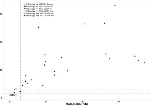
We observed a significant correlation in detecting anti-DENV IgM antibodies between the InBios IgM capture ELISA and the
in-house MAC-ELISA (Pearson’sr⫽0.80,P⬍0.0001) (Fig. 1).
The agreement, sensitivity, and specificity of InBios IgM capture ELISA were 94% (95% CI, 86 to 98%), 92% (95% CI, 73 to 99%),
and 94% (95% CI, 84 to 99%), respectively (Table 1). In addition,
the InBios assay showed near-perfect agreement ( ⫽0.87) (13)
with the in-house MAC-ELISA (Table 1). Out of 79 serum
sam-ples, two serum samples (samples 3 and 4) were MAC-ELISA
pre-sumptive positive and InBios equivocal (Table 2). Serum samples
[image:2.585.41.547.87.190.2]3 and 4 were obtained from DENV-infected patients in Singapore. These two serum samples were DENV-NS1 positive as well as anti-DENV IgM positive using an independent IgM assay (data
TABLE 1Sensitivity, specificity, percent agreement, and kappa value for InBios DENV detect IgM capture ELISA compared to the in-house MAC-ELISA
Result by InBios DENV Detect IgM capture ELISA (n⫽79)b
No. of samples with result by in-house
MAC-ELISAc % (95% CI)
Kappa value Positive Negative
Presumptive
positive Sensitivity Specificity Agreement
Positive 22 2 0
Negative 0 50 0
Equivocala 2 1 2
Total 24 53 2 92 (73–99) 94 (84–99) 94 (86–98) 0.87
a
Equivocal samples were considered false negative for sensitivity and false positive for specificity.
bInBios IgM positive, ISR of⬎2.84; InBios IgM equivocal, ISR of 1.65 to 2.84; InBios IgM negative, ISR of⬍1.65. c
MAC-ELISA positive,P/Nofⱖ3; MAC-ELISA presumptive positive,P/Nofⱖ2 and⬍3; MAC-ELISA negative,P/Nof⬍2.
on May 16, 2020 by guest
http://jcm.asm.org/
not shown). In addition, serum sample 2, obtained from a DENV-infected patient in Singapore, was MAC-ELISA positive and InBios equivocal; however, it was anti-DENV IgM positive by in-dependent IgM assay. Although it is not certain, these three sam-ples may have been collected from patients with secondary DENV
infection or during the early phase of primary DENV infection, as the levels of IgM antibodies were low.
Out of 79 serum samples, only five serum samples (samples 1,
2, 5, 6, and 7) exhibited discordant results (Table 2). Dengue
an-tigen (DENRA) and NCA optical density (OD) values obtained
[image:3.585.52.537.66.410.2]FIG 1Correlation between the in-house DENV MAC-ELISA and the InBios DENV IgM capture ELISA. Scatter plot depicting in-house DENV MAC-ELISA versus InBios DENV IgM capture ELISA determined using 79 human serum samples. Left of the vertical solid line are MAC-ELISA negatives,P/Nof⬍2; right of the vertical dashed line are MAC-ELISA positives,P/Nofⱖ3; and all values between the vertical solid and dashed lines are MAC-ELISA presumptive positive samples,P/Nofⱖ2 and⬍3. Below the horizontal solid line are InBios IgM ELISA negatives, ISR of⬍1.65; above the dotted horizontal line are InBios IgM ELISA positives, ISR of⬎2.84; all samples between these two lines are InBios IgM capture ELISA equivocal (ISR of 1.65 to 2.84).
TABLE 2Results of MAC-ELISA and InBios DENV IgM capture ELISA equivocal and discordant samplesa
Sample no.
MAC-ELISAb InBios DENV IgM capture ELISAc
PRNT result
P/N Result DENRA (OD) NCA (OD) ISR Result
1 7.6 Pos 0.27 0.11 2.4 Equivocal QNS
2 4.3 Pos 0.22 0.08 2.6 Equivocal QNS
3 2.9 Presumptive Pos 0.13 0.06 2.2 Equivocal QNS 4 2.8 Presumptive Pos 0.16 0.07 2.4 Equivocal QNS
5 1.2 Neg 0.21 0.11 1.9 Equivocal DENV-2 Pos
6 1.7 Neg 0.21 0.06 3.4 Pos DENV-1 Pos
7 1.2 Neg 0.19 0.06 3.0 Pos DENV Pos
a
Abbreviations: DENRA, dengue virus-derived recombinant antigens; NCA, normal cell antigen; ISR, immune status ratio;P/N, positive-to-negative ratio; QNS, quantity not sufficient; Pos, positive; Neg, negative; OD, optical density.
b
MAC-ELISA positive,P/Nofⱖ3; MAC-ELISA presumptive positive,P/Nofⱖ2 and⬍3; MAC-ELISA negative,P/Nof⬍2.
cInBios IgM positive, ISR of⬎2.84; InBios IgM equivocal, ISR of 1.65 to 2.84; InBios IgM negative, ISR of⬍1.65.
on May 16, 2020 by guest
http://jcm.asm.org/
[image:3.585.39.548.585.686.2]for 79 serum samples are given inTable 3. The NCA used in the InBios IgM capture ELISA detects the nonspecific reactivity of test
serum samples (14). Out of the five samples that exhibited
discor-dant results, three MAC-ELISA-negative samples (samples 5, 6,
and 7) were InBios equivocal or positive (Table 2). These three
samples exhibited DENRA OD values in the range of 0.19 to 0.21 (Table 2), which are closer to the highest DENRA OD value (0.14)
obtained for negative serum samples using the InBios assay (Table
3). These data support the true negativity of these serum samples
by the MAC-ELISA. In addition, sample 1 depicted high back-ground as indicated by an NCA OD value of 0.11 and was thus interpreted as equivocal even though the DENRA OD value was
0.27 (Table 2). Thus, inclusion of positive as well as negative
an-tigens is critical for interpretation of the test results as the test
detects background nonspecific reactivity of serum samples (14).
Due to unavailability of sufficient sample volume, PRNT end-point dilution was conducted on only three discordant samples, 5,
6, and 7 (Table 2), to identify the infecting DENV serotype and to
confirm primary or secondary DENV infection. Out of three dis-cordant samples, sample 5 from Vietnam was from a patient with past DENV-2 infection, sample 6 from Hawaii was collected in 2010 from a patient infected with DENV-1 in 2001, and for sample 7 from Vietnam, the DENV serotype could not be determined
because of possible multiple past DENV infections (Table 2).
Se-rum samples 6 and 7 from Hawaii and Vietnam, respectively, from patients with previous DENV infection as determined by PRNT, produced false-positive reactions using the InBios DENV IgM
capture ELISA (Table 2). However, ISR values for these two
sam-ples ranged from 3.0 to 3.4 (Table 2) and were closer to the ISR
cutoff of 2.84 for InBios positives. Out of these two serum samples,
serum sample 6 from Hawaii (Table 2), collected in 2010 from a
patient infected with DENV-1 in 2001 (15), was previously
stud-ied in our laboratory for long-term T cell memory responses (A. Gurary, unpublished data). In humans, dengue virus
epitope-spe-cific tetramer-positive CD8⫹T cells can be detectedex vivoup to
a year after natural primary DENV infection and even longer after
secondary DENV infection (16). In sample 6, we were unable to
detect dengue virus epitope-specific tetramer-positive CD8⫹T
cellsex vivo. In addition, even after stimulation with cognate
den-gue virus epitope, sample 6 demonstrated a lower denden-gue
virus-specific CD8⫹T cell proliferative response than did samples from
recently DENV-infected patients (A. Gurary, unpublished data). These data combined with MAC-ELISA data confirm the absence of recent secondary DENV infection and thus anti-DENV IgM antibodies in sample 6.
The sensitivity of other commercially available anti-DENV IgM tests for detection of anti-DENV IgM antibodies varies from 21 to 99%, whereas the specificities are 77 to 98% compared with the
ref-erence solid-phase MAC-ELISA used by the CDC (9). Recently, in
one study by Blacksell et al., sensitivities and specificities of the two commercial IgM antibody ELISAs for detection of acute dengue virus
infection ranged from 85 to 89% and 88 to 100%, respectively (17).
This study has several limitations. First, cross-reactivity with IgM from West Nile virus and other flaviviruses may occur with the InBios dengue virus IgM assay. Second, we did not have access to detailed clinical history or vaccination records for other flavi-viruses such as yellow fever virus (YFV) for the 79 serum samples tested in this study. Third, the number of samples used in this evaluation study is small, and fourth, we did not have PRNT data for all 79 serum samples due to insufficient sample volume. De-spite these limitations, the InBios DENV IgM capture ELISA is advantageous compared to the MAC-ELISA since the results can
be obtained in 1 day (⬃5 h), whereas the latter requires 2 to 3 days.
This study for the first time evaluated the only U.S. Food and Drug Administration-cleared InBios DENV IgM capture ELISA.
In summary, this study indicates that the InBios DENV Detect IgM capture ELISA is a reliable, rapid, sensitive, and specific sero-logical test for detection of acute or recent dengue virus infections and thus can be employed by public health laboratories for rapid detection of dengue virus infections during dengue epidemics.
ACKNOWLEDGMENTS
This study was supported in part by grant P20GM103516 from the Cen-ters of Biomedical Research Excellence, National Institute of General Medical Sciences; grant U01AI078213 from the National Institute of Al-lergy and Infectious Diseases, National Institutes of Health (NIH); and grant W8IXWH0720073 from the Department of Defense and by institu-tional funds.
We thank James Davis of the Biostatistics and Data Management Core supported by the RMATRIX grant (U54MD007584) from the National Institute on Minority Health and Health Disparities, NIH, for conducting statistical data analysis.
The funders had no role in the study design, data collection and anal-ysis, decision to publish, or preparation of the manuscript.
REFERENCES
1.Guzman MG, Halstead SB, Artsob H, Buchy P, Farrar J, Gubler DJ, Hunsperger E, Kroeger A, Margolis HS, Martinez E, Nathan MB, Pelegrino JL, Simmons C, Yoksan S, Peeling RW. 2010. Dengue: a continuing global threat. Nat. Rev. Microbiol.8:S7–S16.
2.Solomon T, Mallewa M.2001. Dengue and other emerging flaviviruses. J. Infect.42:104 –115.
3.Hynes NA.2012. Dengue: a reemerging concern for travelers. Cleve. Clin. J. Med.79:474 – 482.
4.Adalja AA, Sell TK, Bouri N, Franco C.2012. Lessons learned during dengue outbreaks in the United States, 2001–2011. Emerg. Infect. Dis. 18:608 – 614.
5.Peeling RW, Artsob H, Pelegrino JL, Buchy P, Cardosa MJ, Devi S, Enria DA, Farrar J, Gubler DJ, Guzman MG, Halstead SB, Hunsperger E, Kliks S, Margolis HS, Nathanson CM, Nguyen VC, Rizzo N, Vazquez S, Yoksan S.2010. Evaluation of diagnostic tests: dengue. Nat. Rev. Mi-crobiol.8:S30 –S38.
6.Thomas SJ, Endy TP.2011. Critical issues in dengue vaccine develop-ment. Curr. Opin. Infect. Dis.24:442– 450.
7.Simmons CP, Farrar JJ, Nguyen VV, Wills B.2012. Dengue. N. Engl. J. Med.366:1423–1432.
8.Halstead SB.2007. Dengue. Lancet370:1644 –1652.
9.Hunsperger EA, Yoksan S, Buchy P, Nguyen VC, Sekaran SD, Enria DA, Pelegrino JL, Vazquez S, Artsob H, Drebot M, Gubler DJ, Halstead SB, Guzman MG, Margolis HS, Nathanson CM, Lic NRR, Bessoff KE, Kliks S, Peeling RW.2009. Evaluation of commercially available anti-dengue virus immunoglobulin M tests. Emerg. Infect. Dis.15:436 – 440. 10. Innis BL, Nisalak A, Nimmannitya S, Kusalerdchariya S, Chongswasdi
V, Suntayakorn S, Puttisri P, Hoke CH.1989. An enzyme-linked im-TABLE 3Detection of anti-DENV IgM antibodies using InBios DENV
IgM capture ELISA
InBios DENV IgM capture ELISA result typea No. of samples Avg (range)
ISR DENRA (OD) NCA (OD)
Positive 24 12.1 (3.0–33.0) 0.83 (0.19–2.11) 0.07 (0.06–0.14) Equivocal 5 2.4 (1.9–2.6) 0.21 (0.13–0.27) 0.08 (0.06–0.11) Negative 50 1.0 (0.9–1.3) 0.07 (0.06–0.14) 0.07 (0.06–0.13)
aInBios IgM positive, ISR of⬎2.84; InBios IgM equivocal, ISR of 1.65 to 2.84; InBios
IgM negative, ISR of⬍1.65.
on May 16, 2020 by guest
http://jcm.asm.org/
[image:4.585.40.286.86.154.2]munosorbent assay to characterize dengue infections where dengue and Japanese encephalitis co-circulate. Am. J. Trop. Med. Hyg.40:418 – 427. 11. Martin DA, Muth DA, Brown T, Johnson AJ, Karabatsos N, Roehrig JT.
2000. Standardization of immunoglobulin M capture enzyme-linked im-munosorbent assays for routine diagnosis of arboviral infections. J. Clin. Microbiol.38:1823–1826.
12. Roehrig JT, Hombach J, Barrett AD. 2008. Guidelines for plaque-reduction neutralization testing of human antibodies to dengue viruses. Viral Immunol.21:123–132.
13. Landis JR, Koch GG.1977. The measurement of observer agreement for categorical data. Biometrics33:159 –174.
14. Welch RJ, Anderson BL, Litwin CM.2008. Evaluation of a new commer-cial enzyme immunoassay for the detection of IgM antibodies to West Nile virus using a ratio method to eliminate nonspecific reactivity. J. Clin. Lab. Anal.22:362–366.
15. Imrie A, Meeks J, Gurary A, Sukhbataar M, Kitsutani P, Effler P, Zhao Z.2007. Differential functional avidity of dengue virus-specific T-cell clones for variant peptides representing heterologous and previously en-countered serotypes. J. Virol.81:10081–10091.
16. Friberg H, Bashyam H, Toyosaki-Maeda T, Potts JA, Greenough T, Kalayanarooj S, Gibbons RV, Nisalak A, Srikiatkhachorn A, Green S, Stephens HA, Rothman AL, Mathew A.2011. Cross-reactivity and ex-pansion of dengue-specific T cells during acute primary and secondary infections in humans. Sci. Rep.1:51. doi:10.1038/srep00051.
17. Blacksell SD, Jarman RG, Gibbons RV, Tanganuchitcharnchai A, Mammen MP, Jr, Nisalak A, Kalayanarooj S, Bailey MS, Premaratna R, de Silva HJ, Day NP, Lalloo DG. 2012. Comparison of seven commercial antigen and antibody enzyme-linked immunosorbent as-says for detection of acute dengue infection. Clin. Vaccine Immunol. 19:804 – 810.