Infections: a Retrospective Analysis of Prolonged 14-Day Incubation
Nora Schwotzer,a* Peter Wahl,bDominique Fracheboud,cEmanuel Gautier,bChristian Chuarda,d
‹Department of Internal Medicine,aDepartment of Orthopedic Surgery and Traumatology,bMicrobiology Laboratory,cand Division of Infectious Diseases,dHôpital Cantonal de Fribourg, Fribourg, Switzerland
Accurate diagnosis of orthopedic device-associated infections can be challenging. Culture of tissue biopsy specimens is
often considered the gold standard; however, there is currently no consensus on the ideal incubation time for specimens.
The aim of our study was to assess the yield of a 14-day incubation protocol for tissue biopsy specimens from revision
sur-gery (joint replacements and internal fixation devices) in a general orthopedic and trauma sursur-gery setting. Medical records
were reviewed retrospectively in order to identify cases of infection according to predefined diagnostic criteria. From
August 2009 to March 2012, 499 tissue biopsy specimens were sampled from 117 cases. In 70 cases (59.8%), at least one
sample showed microbiological growth. Among them, 58 cases (82.9%) were considered infections and 12 cases (17.1%)
were classified as contaminations. The median time to positivity in the cases of infection was 1 day (range, 1 to 10 days),
compared to 6 days (range, 1 to 11 days) in the cases of contamination (P
<
0.001). Fifty-six (96.6%) of the infection cases
were diagnosed within 7 days of incubation. In conclusion, the results of our study show that the incubation of tissue
biopsy specimens beyond 7 days is not productive in a general orthopedic and trauma surgery setting. Prolonged 14-day
incubation might be of interest in particular situations, however, in which the prevalence of slow-growing microorganisms
and anaerobes is higher.
S
urgical implants play a major role in orthopedic trauma
sur-gery and in the management of degenerative and
inflamma-tory joint diseases. However, the rising number of indwelling
de-vices is associated with increases in related complications. Along
with device loosening or malfunctions and foreign-material
reac-tions, infection remains one of the most serious problems
en-countered with surgical implants. Although orthopedic
device-associated infections (ODAI) are uncommon, occurring in only 1
to 2% of patients with hip and knee replacements and up to 6% of
patients after internal fixation of closed fractures, their
manage-ment is difficult (
1
). Management can require multiple revision
surgeries and prolonged antibiotic treatment, may result in
per-manent disabilities, and is associated with high costs (
2
,
3
).
Despite the promising results reported with newer techniques,
such as sonication cultures and molecular testing, the diagnosis of
ODAI remains a medical challenge, as routinely used methods
lack sensitivity and specificity (
4–7
). Synovial fluid sample
cul-ture, tissue biopsy specimen culcul-ture, and histopathological
exam-ination show high sensitivities and are frequently considered the
gold standard. A reliable microbiological diagnosis is crucial for
determining appropriate treatment (
8
).
There is currently no consensus regarding the appropriate
in-cubation time for ODAI tissue biopsy specimens. The duration of
incubation is not specified in most studies, but a 5-day period has
often been reported (
9–11
). Recently, some authors have
pro-posed prolonging the incubation period to 7 or 14 days in order to
reveal microorganisms with low virulence, such as
Propionibacte-rium acnes
,
Peptostreptococcus
spp., and
Corynebacterium
spp.
(
12–15
). Low-virulence, foreign-material-adherent bacteria are
typically in a dormant starved state with a slow replicating rate
(
16
). This particular behavior may require a longer culture
incu-bation time (
16–19
). However, prolonging the incubation time is
costly and labor-intensive and could increase the likelihood of
detecting organisms that are not clinically relevant. Thus, the aim
of our study was to determine if an incubation time of 14 days for
tissue biopsy specimens is useful in the diagnosis of ODAI.
MATERIALS AND METHODS
Study design.Microbiological samples of tissue biopsy specimens that were taken from orthopedic device revision surgery (joint replacement and internal fixation devices) between August 2009 and March 2012 and were incubated for 14 days were analyzed. At our institution, 14-day in-cubation is standard for implant-associated samples and is performed on request for other bone and joint infections. In this study, case identifica-tion was prospective and continuous, while the study was retrospective. When there were several interventions for the same joint, only the first revision surgery was considered. The time until microbial growth was recorded. In cases of polymicrobial growth, infection was diagnosed if at least one microorganism fulfilled the diagnostic criteria (see below). The day of growth of the most slowly growing microorganism was used to avoid overlooking late-growing bacteria.
The study was performed in a hospital acting as a primary care and referral center for a population of about 280,000 inhabitants. Elective orthopedic surgery and trauma surgery each account for about one-half of the activity of the Department of Orthopedic Surgery and Traumatology at this hospital. Medical records were reviewed in order to determine if infection was present. Infection was diagnosed according to predefined
Received19 July 2013 Returned for modification15 August 2013 Accepted12 October 2013
Published ahead of print23 October 2013
Editor:R. Patel
Address correspondence to Nora Schwotzer, nora.schwotzer@chuv.ch, or Christian Chuard, christian.chuard@h-fr.ch.
* Present address: Nora Schwotzer, Department of Internal Medicine, Centre Hospitalier Universitaire Vaudois, Lausanne, Switzerland.
Copyright © 2014, American Society for Microbiology. All Rights Reserved.
doi:10.1128/JCM.01766-13
on May 16, 2020 by guest
http://jcm.asm.org/
diagnostic criteria (see below). Cases were reviewed by an infectious dis-ease specialist and an orthopedic surgeon. Patient files were scanned for indications of clinical signs of infection (fever, erythema, edema, local hyperthermia, wound discharge, and/or the presence of a sinus tract). A temperature above 38.5°C was considered fever, and fracture nonunion was taken as a potential sign of infection. Preoperative antimicrobial treat-ment was defined as the administration of any type of antibiotic for more than 24 h during the 14 days preceding surgery. The histopathological findings were divided into 3 categories depending on the average number
of polymorphonuclear cells (PMN) per high-power field (HPF) (⫻400
magnification) on microscopic analysis, as a mean value of at least 10
fields examined, i.e.,⬍1 PMN/HPF, 1 to 5 PMN/HPF, or⬎5 PMN/HPF.
Definition of infection.Infection was diagnosed if one of the
follow-ing criteria was fulfilled: (i) positive culture withⱖ3 positive samples
showing identical microorganisms (20) (microbiological criterion), (ii)
positive culture with any number of positive samples and
histopatholog-ical examination showing⬎5 PMN/HPF not explained by an acute
frac-ture (21–23) (histopathological criterion), or (iii) positive culture with
any number of positive samples and clinical signs of infection, i.e., ery-thema, edema, local hyperthermia, wound discharge, presence of a sinus
tract, or fracture nonunion (8,24) (clinical criterion). Patients who had
not been treated postoperatively with antibiotics and who showed no signs of infection after 12 months of follow-up were not considered to be infected, independent of the diagnostic criteria. Cases with positive cul-tures that did not fulfill the criteria for infection were classified as contam-ination.
Culture methods.Tissue sampling was performed in the operating room according to usual surgical methods. The standard procedure was to obtain 3 to 6 samples, with priority given to tissue biopsy specimens if not
limited by anatomical restrictions (as in the finger, hand, and foot) (20,
25–27). In order, tissues were sampled from the inflammatory membrane around the implant, the joint capsule, and any macroscopically suspect
tissue (28,29). Each biopsy specimen was stored in transportation
me-dium (BBCPort-A-Cul; Becton, Dickinson, and Company, Sparks, MD) to ensure the survival of all bacteria, including anaerobic microorganisms. Homogenization of the tissue biopsy specimens was carried out using a disposable closed tissue homogenization system (gentleMACS dissocia-tor; Miltenyi Biotec GmbH, Bergisch Gladbach, Germany), with the ad-dition of normal saline solution as necessary to obtain a heavy suspension. All manipulations were performed under sterile conditions and under laminar airflow. One hundred microliters of this suspension was inocu-lated on each of the following agar plates: (i) blood agar, i.e., Columbia-D agar base (bioMérieux, Marcy l’Etoile, France) with 5% sheep blood; (ii) chocolate agar, i.e., Columbia-D agar base (bioMérieux, Marcy l’Etoile, France) with 5% sheep blood (heated to lyse blood cells) supplemented with Vitox SR0090 growth factors (Oxoid-Thermo Fisher Scientific,
Bas-ingstoke, United Kingdom); or (iii) prereducedBrucellaagar with 5%
sheep blood, hemin, and vitamin K1(bioMérieux, Marcy l’Etoile, France).
After inoculation, the plates were sealed with Parafilm laboratory film (Bemis Company, Inc., Oshkosh, WI) to avoid desiccation. The first 2
media were incubated at 35°C in a 5% CO2atmosphere for cultivation of
aerobic and facultative organisms. The third plate was incubated at 35°C in an anaerobic atmosphere for the cultivation of anaerobic and faculta-tive organisms. The remainder of the suspension was inoculated into thio-glycolate broth medium CM0173 (Oxoid-Thermo Fisher Scientific, Bas-ingstoke, United Kingdom) and incubated at 35°C for enrichment of aerobic, anaerobic, and facultative microorganisms. Quality control as-sessments showed the presence of adequate anaerobic conditions in the lower part of the broth. Each medium was inspected for signs of growth every day for a period of 14 days.
Statistical methods.Continuous variables are presented as medians and ranges and categorical variables as rates. Statistical significance was assessed using the chi-square test or Fisher’s exact test for categorical
variables and the Mann-WhitneyUtest (Kruskal-Wallis test) for
contin-uous variables. All tests were performed using SPSS version 21 (SPSS Inc.,
Chicago, IL).Pvalues of⬍0.05 were considered statistically significant.
For graphical representation, Microsoft Excel 2008 (Microsoft Corp., Redmond, WA) was used.
RESULTS
Study population.
During the study period, 499 tissue biopsy
specimens were collected from 117 cases of revision surgery,
cor-responding to a median number of 4.0 samples per case (range, 1
to 12 samples per case). In 70 cases (59.8%), a minimum of one
sample was positive for microbiological growth, leaving 47 cases
(40.2%) as sterile during the incubation period.
The study population consisted of 50 women (42.7%) and 67
men (57.3%), with a median age of 68.0 years (range, 14 to 94
years). The time between index surgery and revision surgery was
⬍
1 month in 31 cases (26.5%), 1 to 12 months in 32 cases (27.4%),
and
⬎
12 months in 54 cases (46.2%). Orthopedic devices
in-cluded 62 cases (53.0%) of joint prostheses and 55 cases (47.0%)
of internal fixation devices. Localization of the devices varied from
the hip in 51 cases (43.6%), the knee in 29 cases (24.8%), the lower
extremity in 22 cases (18.8%), and the upper limb in 9 cases
(7.7%) to the spine in 6 cases (5.1%). Articular devices were
la-beled according to the joint region involved. Histopathological
analysis results were available for 85 cases (72.6%). Among the 70
cases with positive culture results, 58 cases (82.9%) were classified
as infections and 12 cases (17.1%) as contaminations. The
propor-tion of infecpropor-tions among the 117 cases of revision surgery was
49.6%, with 41.9% for joint replacement and 58.2% for internal
fixation devices.
[image:2.585.299.545.79.194.2]Microbiology.
The majority of the isolated microorganisms
were Gram-positive bacteria, mainly
Staphylococcus aureus
in 22
cases (31.4%) and coagulase-negative staphylococci in 18 cases
(25.7%).
Streptococcus
spp. accounted for 2 cases (2.9%),
Entero-coccus
spp. for 2 cases (2.9%),
P. acnes
for 3 cases (4.3%),
Gram-negative bacteria for 6 cases (8.6%), and polymicrobial culture
results for 17 cases (24.3%). The full spectrum of bacteria
accord-ing to case classification is illustrated in
Table 1
. The two types of
TABLE 1Spectrum of microorganisms in tissue biopsy specimens
Microorganism(s)
Infection
(n[%])
Contamination
(n[%])
Staphylococcus aureus 22 (37.9)
Coagulase-negative staphylococci 10 (17.2) 8 (66.7)
Streptococcusspp.a 2 (3.4)
Enterococcusspp.b 2 (3.4)
Propionibacterium acnes 1 (1.7) 2 (16.7)
Gram-negative bacillic 6 (10.3)
Polymicrobial cultured 15 (25.9) 2 (16.7)
Total 58 (100) 12 (100)
a
Streptococcusspp. includedStreptococcusgroupmitis(n⫽1) andStreptococcus pyogenes(n⫽1).
b
Enterococcusspp. includedEnterococcus faecalis(n⫽1) andEnterococcus faecium (n⫽1).
c
Gram-negative bacilli includedEscherichia coli(n⫽3),Enterobacter cloacae(n⫽2), andPseudomonas aeruginosa(n⫽1).
d
Polymicrobial cultures classified as infections included coagulase-negative staphylococci (n⫽11),S. aureus(n⫽5),E. cloacae(n⫽3),Bacillusspp. (n⫽2),E. coli(n⫽2),Streptococcus agalactiae(n⫽1),E. faecalis(n⫽1),Corynebacteriumsp. (n⫽1),Dermatobia hominis(n⫽1),P. acnes(n⫽1),Morganella morganii(n⫽1),P. aeruginosa(n⫽1), and Gram-positive rods (n⫽1). Polymicrobial cultures classified as contaminations included coagulase-negative staphylococci (n⫽1),E. faecalis(n⫽1), E. coli(n⫽1), and Gram-negative bacilli (n⫽1).
Schwotzer et al.
on May 16, 2020 by guest
http://jcm.asm.org/
orthopedic devices showed similar spectra of microorganisms
ex-cept for
S. aureus
, which was significantly more frequent for
pros-theses than for internal fixation devices (15 cases versus 7 cases;
P
⫽
0.04).
Diagnostic criteria.
Of the 58 cases of infection, most were
diagnosed by at least 2 diagnostic criteria, leaving only 9 cases
diagnosed with a single criterion (the microbiological criterion in
3 cases and the clinical criterion in 6 cases). Discrepancy between
the study’s definition of infection and the treating medical team’s
diagnosis occurred in one case, which was treated as an infection
based on one of 8 samples showing
P. acnes
on day 7. This case was
classified as contamination according to our criteria.
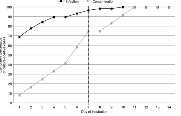
Time to culture positivity.
The median time to culture
posi-tivity for the 70 cases with positive results for tissue biopsy
speci-mens was 1 day (range, 1 to 11 days). A total of 47 cases (67.1%)
became positive within the first 2 days of incubation, 57 cases
(81.4%) within 5 days, and 65 cases (92.9%) within 7 days.
The median time to positivity in cases classified as infections
was 1 day (range, 1 to 10 days), compared to 6 days (range, 1 to 11
days) for cases considered contaminations (
P
⬍
0.001). A total of
52 cases (89.7%) of infections were diagnosed within 5 days of
incubation and 56 cases (96.6%) within 7 days (
Fig. 1
). No
infec-tion was diagnosed beyond 10 days. Twenty-five percent of
con-taminants grew after 7 days, representing the majority (60%) of
late-growing microorganisms. Because the absolute numbers of
cases of infections or contaminations are also relevant, graphical
representation of the results is shown in a histogram in
Fig. 2
.
Only 2 cases of infection were detected after 7 days of
incuba-tion, at day 8 and day 10 (
Table 2
). The first case was a late
post-operative infection, which showed a large amount of
Corynebac-FIG 1Time to tissue culture positivity for cases of infection versus contamination.
FIG 2Absolute numbers of cases with positive tissue culture results at each day of incubation for infection versus contamination.
on May 16, 2020 by guest
http://jcm.asm.org/
[image:3.585.136.452.67.270.2] [image:3.585.136.451.510.709.2]terium
sp. (4 of 5 positive tissue biopsy specimens) growing within
4 days. This was associated with an
Escherichia coli
infection (one
of 5 positive tissue biopsy specimens) growing at day 8, and both
bacteria were considered pathogens. The second case involved an
early postoperative infection and demands special consideration.
In this case, sampling occurred during antibiotic treatment, which
had been initiated after a superficial wound swab showed
S. aureus
(not recorded in our database) in the presence of clinical signs of
acute infection (erythema, edema, and wound discharge).
Coag-ulase-negative staphylococci found to be growing in the tissue
biopsy specimen after 10 days were recorded as the etiological
agent, according to our study definition (histopathological and
clinical criteria). However, considering the clinical picture, it is
more probable that
S. aureus
was responsible for the infection. As
a consequence, the late growth of coagulase-negative
staphylo-cocci is probably not relevant.
Our sample showed no significant difference in the median
time to culture positivity (
P
⫽
0.84) with regard to the type of
orthopedic device (joint replacement or internal fixation device).
DISCUSSION
Few studies have focused on the optimal incubation time for
or-thopedic surgical specimens. Most commonly, incubations of 5 to
7 days are used for ODAI (
10
,
11
,
30–32
). In recent studies (
12
,
13
,
15
), however, prolonging the incubation time for up to 14 days has
been proposed. Schaefer et al. (
13
) addressed the infectious
com-ponent in aseptic loosening and considered mostly elective
sur-gery. Zappe et al. (
15
) and Butler-Wu et al. (
12
) specifically
ex-plored an extended culture protocol for
P. acnes
. In studies on
alternative diagnostic procedures, such as 16S rRNA PCR or
son-ication, 14-day incubations have sometimes been reported,
al-though without assessment of their benefits (
33
,
34
). The aim of
our study was to explore the relevance of 14-day tissue biopsy
specimen culture incubations for the diagnosis of overall ODAI in
the setting of general orthopedic and trauma surgery, where both
acute and chronic infections are encountered.
We found that an incubation period of 7 days was sufficient to
identify 56 of 58 cases (96.6%) of infection. The major difference
between our data and those of Schaefer et al. (
13
) is the
pro-portion of low-virulence microorganisms such as
Propionibacte-rium
spp., coryneform bacteria, and coagulase-negative
staphylo-cocci, which accounted for 80% of infections in their study
population. These microorganisms are known to have slow
growth rates. In our study, only 54% of the cases showed
low-virulence microorganisms. In particular,
P. acnes
accounted for
4.3% of isolated bacteria, which corresponds to reports from other
general orthopedic and trauma surgery departments (
8
,
35
,
36
).
The diagnosis of ODAI is a well-known challenge. In our study,
we have tried to provide clear reproducible diagnostic criteria that
are applicable to retrospective analysis, which has well-established
limitations. For the microbiological criterion, our threshold of at
least 3 positive samples with identical microorganisms could be
viewed as stringent, in comparison with other studies in which 2
culture-positive specimens are considered sufficient for the
diag-nosis of infections (
10
,
37
,
38
). However, none of our
contamina-tion cases had more than one positive sample, meaning that our
results would not have been different if we had adopted a lower
threshold. The same is true for the histopathological criterion, as
no contaminant showed an intermediate result of 1 to 5 PMN/
HPF. Although the clinical diagnostic criterion is somewhat
sub-jective, all of the cases classified as infections on this basis were
quite evident; 10% of infections would have been missed without
this strategy.
Overall, we identified a large proportion of infections (
22
,
39
,
40
). This can be explained by the inclusion of trauma cases, for
which early revision surgery was indicated on the basis of a high
preoperative suspicion of infection, whereas the systematic
sam-pling of loose prostheses used in other studies obviously had lower
yields (
39
,
40
).
Most reports focus on hip and knee prostheses (
1
,
13
,
24
,
37
).
We believe that including both prostheses and internal fixation
devices in our study makes sense because similar pathogens have
been described in the two settings (
41–43
) and biofilm formation
is common to all types of foreign-body infections (
44–46
). We
found that slow-growing microorganisms such as coagulase
neg-ative-staphylococci and
P. acnes
were equally represented in the
two groups.
In conclusion, the results of our study show that extension of
culture incubation times beyond 7 days has a low yield in a general
orthopedic and trauma setting, where virulent bacteria
predomi-nate and posttraumatic infections are frequent. However, based
on the current literature, prolonging incubations to 14 days or
using molecular techniques might be useful in particular
situa-tions in which the prevalence of slow-growing bacteria and
anaer-obes is higher.
REFERENCES
1.Darouiche RO.2004. Treatment of infections associated with surgical
implants. N. Engl. J. Med. 350:1422–1429. http://dx.doi.org/10.1056
/NEJMra035415.
2.Kurtz SM, Lau E, Watson H, Schmier JK, Parvizi J. 2012. Economic burden of periprosthetic joint infection in the United States. J.
Arthro-plasty27:61– 65.e1.http://dx.doi.org/10.1016/j.arth.2012.02.022.
3.Vanhegan IS, Malik AK, Jayakumar P, Ul Islam S, Haddad FS.2012. A financial analysis of revision hip arthroplasty: the economic burden in
relation to the national tariff. J. Bone Joint Surg. Br.94:619 – 623.http://dx
.doi.org/10.1302/0301-620X.94B5.27073.
4.Zimmerli W, Moser C.2012. Pathogenesis and treatment concepts of
orthopaedic biofilm infections. FEMS Immunol. Med. Microbiol.65:
158 –168.http://dx.doi.org/10.1111/j.1574-695X.2012.00938.x.
5.Matthews PC, Berendt AR, McNally MA, Byren I.2009. Diagnosis and
management of prosthetic joint infection. BMJ338:b1773.http://dx.doi
[image:4.585.35.546.79.144.2].org/10.1136/bmj.b1773.
TABLE 2Characteristics of 2 cases of infection with times to tissue culture positivity beyond 7 days of incubation Case
no. Microorganism(s)
No. of culture-positive samples/no. tested
No. of
PMN/HPFa
Type of
implant Clinical signs
Day of growth
1 Corynebacteriumsp. 4/5 0 Hip prosthesis Ongoing pain 4
E. coli 1/5 0 8
2 Coagulase-negative staphylococci 1/3 ⬎5 Knee prosthesis Erythema, wound discharge 10
a
Number of polymorphonuclear cells (PMN) per high-power field (HPF) (⫻400 magnification);⬎5 PMN/HPF is highly suggestive of infection.
Schwotzer et al.
on May 16, 2020 by guest
http://jcm.asm.org/
6.Berbari E, Mabry T, Tsaras G, Spangehl M, Erwin PJ, Murad MH, Steckelberg J, Osmon D.2010. Inflammatory blood laboratory levels as markers of prosthetic joint infection: a systematic review and
meta-analysis. J. Bone Joint Surg. Am.92:2102–2109.http://dx.doi.org/10.2106
/JBJS.I.01199.
7.Parvizi J, Zmistowski B, Berbari EF, Bauer TW, Springer BD, Della Valle CJ.2011. New definition for periprosthetic joint infection from the workgroup of the Musculoskeletal Infection Society. Clin. Orthop. Relat.
Res.469:2992–2994.http://dx.doi.org/10.1007/s11999-011-2102-9.
8.Zimmerli W, Trampuz A, Ochsner PE.2004. Prosthetic-joint infections. N.
Engl. J. Med.351:1645–1654.http://dx.doi.org/10.1056/NEJMra040181.
9.Larsen LH, Lange J, Xu Y, Schonheyder HC.2012. Optimizing culture methods for diagnosis of prosthetic joint infections: a summary of
modi-fications and improvements reported since 1995. J. Med. Microbiol.61:
309 –316.http://dx.doi.org/10.1099/jmm.0.035303-0.
10. Piper KE, Jacobson MJ, Cofield RH, Sperling JW, Sanchez-Sotelo J, Osmon DR, McDowell A, Patrick S, Steckelberg JM, Mandrekar JN, Fernandez Sampedro M, Patel R.2009. Microbiologic diagnosis of pros-thetic shoulder infection by use of implant sonication. J. Clin. Microbiol.
47:1878 –1884.http://dx.doi.org/10.1128/JCM.01686-08.
11. Font-Vizcarra L, Garcia S, Martinez-Pastor JC, Sierra JM, Soriano A.
2010. Blood culture flasks for culturing synovial fluid in prosthetic joint
infections. Clin. Orthop. Relat. Res.468:2238 –2243.http://dx.doi.org/10
.1007/s11999-010-1254-3.
12. Butler-Wu SM, Burns EM, Pottinger PS, Magaret AS, Rakeman JL, Matsen FA, III, Cookson BT.2011. Optimization of periprosthetic culture for
diag-nosis ofPropionibacterium acnesprosthetic joint infection. J. Clin. Microbiol.
49:2490 –2495.http://dx.doi.org/10.1128/JCM.00450-11.
13. Schaefer P, Fink B, Sandow D, Margull A, Berger I, Frommelt L.2008. Prolonged bacterial culture to identify late periprosthetic joint infection: a
promising strategy. Clin. Infect. Dis.47:1403–1409.http://dx.doi.org/10
.1086/592973.
14. Gollwitzer H, Diehl P, Gerdesmeyer L, Mittelmeier W.2006. Diagnostic strategies in cases of suspected periprosthetic infection of the knee: a
re-view of the literature and current recommendations. Orthopade35:904 –
916. (In German.)http://dx.doi.org/10.1007/s00132-006-0977-z.
15. Zappe B, Graf S, Ochsner PE, Zimmerli W, Sendi P.2008. Propionibac-teriumspp in prosthetic joint infections: a diagnostic challenge. Arch.
Orthop. Trauma Surg.128:1039 –1046.http://dx.doi.org/10.1007/s00402
-007-0454-0.
16. Costerton JW, Cheng KJ, Geesey GG, Ladd TI, Nickel JC, Dasgupta M, Marrie TJ.1987. Bacterial biofilms in nature and disease. Annu. Rev.
Microbiol.41:435– 464.
17. Ceri H, Olson ME, Stremick C, Read RR, Morck D, Buret A.1999. The Calgary Biofilm Device: new technology for rapid determination of
anti-biotic susceptibilities of bacterial biofilms. J. Clin. Microbiol.37:1771–
1776.
18. Anderl JN, Zahller J, Roe F, Stewart PS.2003. Role of nutrient limitation
and stationary-phase existence inKlebsiella pneumoniaebiofilm resistance
to ampicillin and ciprofloxacin. Antimicrob. Agents Chemother.47:
1251–1256.http://dx.doi.org/10.1128/AAC.47.4.1251-1256.2003.
19. Chuard C, Lucet JC, Rohner P, Herrmann M, Auckenthaler R, Wald-vogel FA, Lew DP.1991. Resistance ofStaphylococcus aureusrecovered
from infected foreign bodyin vivoto killing by antimicrobials. J. Infect.
Dis.163:1369 –1373.
20. Atkins BL, Athanasou N, Deeks JJ, Crook DWM, Simpson H, Peto TEA, McLardy-Smith P, Berendt AR, OSIRIS Collaborative Study Group.1998. Prospective evaluation of criteria for microbiological diag-nosis of prosthetic-joint infection at revision arthroplasty. J. Clin.
Micro-biol.36:2932–2939.
21. Pandey R, Drakoulakis E, Athanasou NA.1999. An assessment of the histological criteria used to diagnose infection in hip revision arthroplasty
tissues. J. Clin. Pathol.52:118 –123.
22. Lonner JH, Desai P, Dicesare PE, Steiner G, Zuckerman JD.1996. The reliability of analysis of intraoperative frozen sections for identifying ac-tive infection during revision hip or knee arthroplasty. J. Bone Joint Surg.
Am.78:1553–1558.
23. Mirra JM, Amstutz HC, Matos M, Gold R.1976. The pathology of the joint tissues and its clinical relevance in prosthesis failure. Clin. Orthop.
Relat. Res. (117):221–240.
24. Widmer AF.2001. New developments in diagnosis and treatment of
in-fection in orthopedic implants. Clin. Infect. Dis.33(Suppl 2):S94 –S106.
http://dx.doi.org/10.1086/321863.
25. Senneville E, Savage C, Nallet I, Yazdanpanah Y, Giraud F, Migaud H, Dubreuil L, Courcol R, Mouton Y.2006. Improved aero-anaerobe re-covery from infected prosthetic joint samples taken from 72 patients and
collected intraoperatively in Rosenow’s broth. Acta Orthop.77:120 –124.
http://dx.doi.org/10.1080/17453670610045795.
26. Zuluaga AF, Galvis W, Saldarriaga JG, Agudelo M, Salazar BE, Vesga O.
2006. Etiologic diagnosis of chronic osteomyelitis: a prospective study.
Arch. Intern. Med.166:95–100.http://dx.doi.org/10.1001/archinte.166.1
.95.
27. Aggarwal VK, Higuera C, Deirmengian G, Parvizi J, Austin MS.2013. Swab cultures are not as effective as tissue cultures for diagnosis of
periprosthetic joint infection. Clin. Orthop. Relat. Res.471:3196 –3203.
http://dx.doi.org/10.1007/s11999-013-2974-y.
28. Bori G, Munoz-Mahamud E, Garcia S, Mallofre C, Gallart X.2011. Interface membrane is the best sample for histological study to diagnose
prosthetic joint infection. Mod. Pathol.24:579 –584.http://dx.doi.org/10
.1038/modpathol.2010.219.
29. Bjerkan G, Witsoe E, Nor A, Viset T, Loeseth K, Lydersen S.2012. A comprehensive microbiological evaluation of fifty-four patients undergo-ing revision surgery due to prosthetic joint loosenundergo-ing. J. Med. Microbiol.
61:572–581.http://dx.doi.org/10.1099/jmm.0.036087-0.
30. Virolainen P, Lahteenmaki H, Hiltunen A, Sipola E, Meurman O, Nelimarkka O.2002. The reliability of diagnosis of infection during
revi-sion arthroplasties. Scand. J. Surg.91:178 –181.
31. Trampuz A, Piper KE, Jacobson MJ, Hanssen AD, Unni KK, Osmon DR, Mandrekar JN, Cockerill FR, Steckelberg JM, Greenleaf JF, Patel R.
2007. Sonication of removed hip and knee prostheses for diagnosis of
infection. N. Engl. J. Med. 357:654 – 663. http://dx.doi.org/10.1056
/NEJMoa061588.
32. Dempsey KE, Riggio MP, Lennon A, Hannah VE, Ramage G, Allan D, Bagg J.2007. Identification of bacteria on the surface of clinically infected and non-infected prosthetic hip joints removed during revision arthro-plasties by 16S rRNA gene sequencing and by microbiological culture.
Arthritis Res. Ther.9:R46.http://dx.doi.org/10.1186/ar2201.
33. Ince A, Rupp J, Frommelt L, Katzer A, Gille J, Lohr JF.2004. Is “aseptic” loosening of the prosthetic cup after total hip replacement due to noncul-turable bacterial pathogens in patients with low-grade infection? Clin.
Infect. Dis.39:1599 –1603.http://dx.doi.org/10.1086/425303.
34. Sendi P, Frei R, Maurer TB, Trampuz A, Zimmerli W, Graber P.2010.
Escherichia colivariants in periprosthetic joint infection: diagnostic
chal-lenges with sessile bacteria and sonication. J. Clin. Microbiol.48:1720 –
1725.http://dx.doi.org/10.1128/JCM.01562-09.
35. Moran E, Masters S, Berendt AR, McLardy-Smith P, Byren I, Atkins BL.
2007. Guiding empirical antibiotic therapy in orthopaedics: the microbi-ology of prosthetic joint infection managed by debridement, irrigation
and prosthesis retention. J. Infect.55:1–7.http://dx.doi.org/10.1016/j.jinf
.2007.01.007.
36. Fulkerson E, Valle CJ, Wise B, Walsh M, Preston C, Di Cesare PE.2006. Antibiotic susceptibility of bacteria infecting total joint arthroplasty sites.
J. Bone Joint Surg. Am.88:1231–1237.http://dx.doi.org/10.2106/JBJS.E
.00004.
37. Osmon DR, Berbari EF, Berendt AR, Lew D, Zimmerli W, Steckelberg JM, Rao N, Hanssen A, Wilson WR.2013. Diagnosis and management of prosthetic joint infection: clinical practice guidelines by the Infectious
Diseases Society of America. Clin. Infect. Dis.56:e1– e25.http://dx.doi.org
/10.1093/cid/cis803.
38. Workgroup Convened by the Musculoskeletal Infection Society.2011.
New definition for periprosthetic joint infection. J. Arthroplasty26:1136 –
1138.http://dx.doi.org/10.1016/j.arth.2011.09.026.
39. Zimmerli W, Widmer AF, Blatter M, Frei R, Ochsner PE.1998. Role of rifampin for treatment of orthopedic implant-related staphylococcal
in-fections: a randomized controlled trial. JAMA279:1537–1541.
40. Padgett DE, Silverman A, Sachjowicz F, Simpson RB, Rosenberg AG, Galante JO.1995. Efficacy of intraoperative cultures obtained during
revision total hip arthroplasty. J. Arthroplasty10:420 – 426.
41. Prasarn ML, Ouellette EA, Miller DR. 2010. Infected nonunions of
diaphyseal fractures of the forearm. Arch. Orthop. Trauma Surg.130:867–
873.http://dx.doi.org/10.1007/s00402-009-1016-4.
42. Trampuz A, Widmer AF.2006. Infections associated with orthopedic
implants. Curr. Opin. Infect. Dis.19:349 –356.
43. Esteban J, Sandoval E, Cordero-Ampuero J, Molina-Manso D, Ortiz-Perez A, Fernandez-Roblas R, Gomez-Barrena E.2012. Sonication of
on May 16, 2020 by guest
http://jcm.asm.org/
ullary nails: clinically-related infection and contamination. Open Orthop. J.
6:255–260.http://dx.doi.org/10.2174/1874325001206010255.
44. Stoodley P, Nistico L, Johnson S, Lasko LA, Baratz M, Gahlot V, Ehrlich GD, Kathju S.2008. Direct demonstration of viableStaphylococcus aureus
biofilms in an infected total joint arthroplasty: a case report. J. Bone Joint
Surg. Am.90:1751–1758.http://dx.doi.org/10.2106/JBJS.G.00838.
45. Palmer M, Costerton W, Sewecke J, Altman D.2011. Molecular
tech-niques to detect biofilm bacteria in long bone nonunion: a case report.
Clin. Orthop. Relat. Res. 469:3037–3042. http://dx.doi.org/10.1007
/s11999-011-1843-9.
46. Moriarty TF, Schlegel U, Perren S, Richards RG. 2010. Infection in fracture fixation: can we influence infection rates through implant design?
J. Mater. Sci. Mater. Med. 21:1031–1035. http://dx.doi.org/10.1007
/s10856-009-3907-x.
Schwotzer et al.