Response of Functional Mitral Regurgitation during Dobutamine
Infusion in Relation to Changes in Left Ventricular Dyssynchrony
and Mitral Valve Geometry
Woong Gil Choi,
1Soo Hyun Kim,
1Soo Han Kim,
2Sang Don Park,
2Young Soo Baek,
2Sung Hee Shin,
2Sung Il Woo,
2Dae Hyeok Kim,
2Keum Soo Park,
2and Jun Kwan
21Division of Cardiology, Department of Internal Medicine, Konkuk University College of Medicine, Chungju;
2Division of Cardiology, Department of Internal Medicine, Inha University College of Medicine, Incheon, Korea.
Received: February 5, 2013 Revised: June 20, 2013 Accepted: July 15, 2013
Corresponding author: Dr. Jun Kwan, Division of Cardiology, Department of Internal Medicine, Inha University College of Medicine, 27 Inhang-ro, Jung-gu, Incheon 400-711, Korea. Tel: 82-32-890-2440, Fax: 82-32-890-2447 E-mail: kuonmd@inha.ac.kr
∙ The authors have no financial conflicts of interest.
© Copyright:
Yonsei University College of Medicine 2014 This is an Open Access article distributed under the terms of the Creative Commons Attribution Non-Commercial License (http://creativecommons.org/ licenses/by-nc/3.0) which permits unrestricted non-commercial use, distribution, and reproduction in any medium, provided the original work is properly cited.
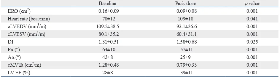
Purpose: Functional mitral regurgitation (FMR) and myocardial dyssynchrony commonly occur in patients with dilated cardiomyopathy (DCM). The aim of this study was to elucidate changes in FMR in relation to those in left ventricular (LV) dyssynchrony as well as geometric parameters of the mitral valve (MV) in DCM patients during dobutamine infusion. Materials and Methods: Twenty-nine DCM patients (M:F=15:14; age: 62±15 yrs) with FMR underwent echocardiography at baseline and during peak dose (30 or 40 ug/min) of dobutamine infusion. Using 2D echocardiography, LV end-diastolic volume, end-systolic volume (LVESV), ejec-tion fracejec-tion (EF), and effective regurgitant orifice area (ERO) were estimated. Dys-synchrony indices (DIs), defined as the standard deviation of time interval-to-peak myocardial systolic contraction of eight LV segments, were measured. Using the multi-planar reconstructive mode from commercially available 3D image analysis software, MV tenting area (MVTa) was measured. All geometrical measurements were corrected (c) by the height of each patient. Results: During dobutamine infu-sion, EF (28±8% vs. 39±11%, p=0.001) improved along with significant decrease in cLVESV (80.1±35.2 mm3/m vs. 60.4±31.1 mm3/m, p=0.001); cMVTa (1.28±0.48
cm2/m vs. 0.79±0.33 cm2/m, p=0.001) was significantly reduced; and DI (1.31±0.51
vs. 1.58±0.68, p=0.025) showed significant increase. Despite significant deteriora-tion of LV dyssynchrony during dobutamine infusion, ERO (0.16±0.09 cm2 vs.
0.09±0.08 cm2, p=0.001) significantly improved. On multivariate analysis, ΔcMVTa
and ΔEF were found to be the strongest independent determinants of ΔERO (R2=0.443, p=0.001). Conclusion: Rather than LV dyssynchrony, MV geometry
determined by LV geometry and systolic pressure, which represents the MV closing force, may be the primary determinant of MR severity.
Key Words: Mitral regurgitation, dyssynchrony, dobutamine
INTRODUCTION
tems (Vivid 7; GE Medical Systems, Milwaukee, WI, USA) equipped with 3S phased-array transducers. Standard 2D echocardiographic study was performed according to the rec-ommendations of the American Society of Echocardiography using conventional views and measurements.10 Dobutamine
stress echocardiography was performed on the same day. LV end systolic volume (LVESV) and LV end diastolic volume (LVEDV) were measured by the biplane Simpson’s disk method. LVEDV and LVESV were corrected (c) by the height of each patient (expressed as cLVEDV, cLVESV).6,7
ΔcLVEDV was defined as [baseline cLVEDV-cLVEDV at peak dobutamine infusion] and ΔcLVESV was defined as [baseline cLVESV-cLVESV at peak dobutamine infusion]. EF was calculated by the equation 100×(EDV-ESV)/EDV. ΔEF was defined as [EF at peak dobutamine infusion-base-line EF]. Degree of MR was quantified by effective regurgi-tant orifice (ERO) by the PISA method.11 ΔERO was defined
as [baseline ERO-ERO at peak dobutamine infusion].
Analysis of dyssynchrony
Color tissue Doppler images were acquired at rest and the final 90 seconds of each dobutamine stress. Images were adjusted to optimize pulse repetition frequency, color satu-ration, and depth to allow for high frame rates. The images were stored and analyzed off-line using customized soft-ware (Echopac 6.3, GE, Milwaukee, WI, USA). Sample cursors were placed at the midpoint of each of the 8 non-apical segments of the anterior, inferior lateral, and septal walls in the 2, 4 chamber apical views, and myocardial ve-locity curves were reconstituted. The time interval to-peak systolic velocity (Ts) was measured from the onset of the QRS complex to the peak of the myocardial systolic veloci-ty during ejection period in each of the eight segments at rest and at peak stress. Ts was corrected for heart rate (Ts-cor) using Bazett’s formula (Tscor=Ts/√R-R interval) to al-low for comparison between the Ts of any segment at rest and peak stress.12,13 Dyssynchrony index (DI) was measured
as the standard deviation of Tscor of all eight segments. All time interval were calculated as the average of at least three consecutive cardiac cycles. All images acquisition and dys-synchrony analyses were performed by a single investigator blinded to the clinical characteristics of the patients.
Real-time 3D echocardiography
Volumetric image acquisition and 3D measurements
A real time 3D echocardiography imaging system (Vivid 7, and is a marker for adverse clinical outcomes.1-3 Increased
mitral valve tenting area (MVTa) has been identified as a determinant of the severity of FMR in patients with dilated cardiomyopathy (DCM).4-7 Recent data have revealed the
potential role of intra-ventricular dyssynchrony in relation to dynamic changes in mitral regurgitation (MR) in failing hearts.8,9 Although previous studies have evaluated
dynam-ic changes in FMR, the precise mechanism involved there-in remathere-ins uncertathere-in.
The purpose of this study was to elucidate the changes in FMR in relation to those in left ventricular (LV) dyssyn-chrony and mitral valve (MV) geometry as well as LV sys-tolic function in DCM patients during dobutamine infusion.
MATERIALS AND METHODS
Study population
Twenty-nine consecutive DCM patients with an LV ejection fraction (EF) of less than 40% and significant MR (≥grade I) were included in this study. All patients underwent coronary angiography to exclude those with any significant coronary artery disease. Exclusion criteria were 1) structural abnor-malities of the mitral apparatus, such as chordae rupture, pro-lapse, or restricted leaflet due to degenerative calcification 2) atrial fibrillation. 3) Inadequate echocardiographic window of the LV structure and the mitral apparatus to not allow anal-ysis of 3D geometry, and 4) inadequate Doppler color flow image for proximal isovelocity surface area (PISA) analysis.
Dobutamine stress protocol
All other drugs except beta-blockers were continued before the study. Dobutamine was infused intravenously in an incre-mental doses of 5, 10, 20 and 40 mcg/kg/min for 3 minute at each stages. End points of the infusion included achievement of target heart rate [85%×(220-age in years)], ST segment el-evation or depression on electrocardiography (ECG), new wall motion abnormality, significant arrhythmia such as atrial fibrillation, ventricular tachycardia, atrioventricular block, se-vere hemodynamic change (systolic blood pressure >200 mm Hg or drop in blood pressure from baseline >20 mm Hg), severe breathlessness. If the target heart rate was not reached with maximal dose of dobutamine, intravenous atro-pine (300 to 1000 mcg) was administered.
Echocardiographic measurements
sys-height of each patient. ΔcMVTa was defined as [baseline cMVTa-cMVTa at peak dobutamine infusion].
Statistical analysis
The data were analyzed using SPSS software, version 12, for Windows (SPSS Inc., Chicago, IL, USA). Data are expressed as mean±SD. Continuous variables were described as means and standard deviations and compared using Student’s paired t-test. The data at rest and peak stress were compared using Student’s paired t-test. Linear regression analysis was applied to study the correlation between change in ERO and various parameters. To determine independent predictors of stress-induced changes in ERO, a stepwise multiple linear regression was performed. p-values <0.05 were considered significant.
RESULTS
Baseline characteristics of population
Twenty-nine patients with heart failure (M:F=15:14, age: 62±15 yrs) were enrolled. All patients were New York Heart GE, Milwaukee, WI, USA) with a 2.5-MHz handheld
transducer was used to image the mitral apparatus. For all patients, transthoracic volumetric images were obtained with an apical view. Care was taken to include the entire MV in the volumetric data set. The volumetric frame rate was 17 to 22 frames/second with an imaging depth of 12 to 16 cm. All volumetric images were transferred to a person-al computer for offline anperson-alysis.
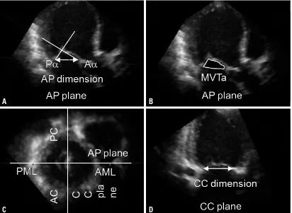
[image:3.595.86.498.388.689.2]We used 3D computer software (4D Cardio-View, Tom-tec Co., Munich, Germany) to define measurement planes. First, the mid-systole of the heart cycle was defined. Then, a cross-sectional plane of the MV that clearly visualized both mitral commissures was used to define the commis-sure-commissure (CC) plane, a plane that passes through both commissures and the LV apex. Finally, anteroposterior (AP) planes perpendicular to the center of the CC axis were defined for imaging of the geometry of the MV. The degree of leaflet tethering was estimated by measuring the angle at which each leaflet met the annular plane (Aα: anterior teth-ering angle, Pα: posterior tethteth-ering angle) in the AP plane. MVTa, the area enclosed by the annular plane, and two leaflets were also measured (Fig. 1). MVTa was c by the
Fig. 1. Illustrations explaining geometric measurements of the mitral tethering angle (A), mitral valve tenting area (B), cross-sectional vol-umetric image at the mitral valve level (C), and CC plane (D) connecting both commissures. CC, commissure-commissure; Pα, posterior tethering angle; Aα, anterior tethering angle; AP, anteroposterior; MVTa, mitral valve tenting area; AML, anterior mitral leaflet; PML, pos-terior mitral leaflet; PC, pospos-terior commissure; AC, anpos-terior commissure.
A
C
B
ΔPα, or ΔAα. Correlations between ΔERO and echocardio-graphic parameters are summarized in Table 3. Based on stepwise multivariate regression analysis, ΔcMVTa and ΔEF (R2=0.443, p=0.001) remained independent
determi-nants of dobutamine-induced changes in ERO (Table 4).
Intra-observer variability
The intra-observer correlation coefficients for anterior papil-lary muscle distance, posterior papilpapil-lary muscle distance, and ERO were all >0.90 (all p<0.001).
DISCUSSION
FMR is the result of incomplete closure of normal leaflets without organic mitral lesion. Mitral valvular tenting has been identified as a main determinant of FMR.6 Mitral
val-vular tenting is characterized by lateral papillary muscle dis-placement and ventricular dysfunction with reduced trans-mitral pressure to close the leaflets.
Association functional class I or II. Mean QRS interval [left bundle branch block (LBBB): n=4] was 112±26 ms. The mean EF and ERO were 28% and 0.16±0.09 cm2,
respec-tively. The demographic and clinical characteristics are summarized in Table 1 and 2.
Changes in echocardiographic parameters during stress echocardiography
All patients were administered the peak dose of dobutamine (up to 40 mcg/kg/min) with exception of 1 case that reached target heart rate at 20 mcg/kg/min of dobutamine. During dobutamine stress echocardiography, heart rate (78±12 beat/ min vs. 109±18 beat/min, p=0.005) and EF (28±8% vs. 39±11%, p=0.001) increased significantly from rest to peak dose. ERO (0.16±0.09 cm2 vs. 0.09±0.08 cm2, p=0.001)
sig-nificantly reduced after dobutamine infusion in all patients with individually variable changes. There were significant changes in cLVEDV (109.5±38.5 mm3/m vs. 92.1±36.6
mm3/m, p=0.001), cLVESV (80.1±35.2 mm3/m vs. 60.4±
31.1 mm3/m, p=0.001), Pα (64±10° vs. 57±11°, p=0.001),
Aα (43±8° vs. 25±9°, p=0.001), and cMVTa (1.28±0.48 cm2/
m vs. 0.79±0.33 cm2/m, p=0.001) during dobutamine
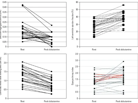
infu-sion. DI significantly increased from 1.31±0.51 at baseline to 1.58±0.68 at peak dose of dobutamine infusion (p=0.0025) regardless of LBBB. The changes in echocardiographic pa-rameters with dobutamine stress echocardiography are sum-marized in Table 2. Individual changes in ERO, EF, MVTa, and DI between rest and dobutamine infusion are illustrated in Fig. 2.
Determinants of dobutamine-induced change in ERO
[image:4.595.315.539.390.533.2]Dobutamine-induced ΔERO were significantly correlated with ΔEF and ΔcMVTa (r=0.491, p=0.011 and r=0.499, p= 0.009, respectively), but not with ΔDI, ΔcLVEDV, ΔcLVESV,
Table 1. Demographic and Clinical Characteristics of the Study Population
n=29
Age (yrs) 62±15
Male/female 15/14
Diabetes mellitus (%) 6 (20)
Hypertension (%) 11 (38)
QRS duration (ms) 112±25.6
LBBB (%) 4 (14)
ACE-inhibitors (%) 22 (76)
Beta-blockers (%) 9 (31)
Loop-diuretics (%) 20 (69)
Spironolactone (%) 15 (52)
LBBB, left bundle branch block; ACE, angiotensin converting enzyme.
Table 2. Comparison of Echocardiographic Parameters: Baseline and Peak Dobutamine Infusion
Baseline Peak dose p value
ERO (cm2) 0.16±0.09 0.09±0.08 0.001
Heart rate (beat/min) 78±12 109±18 0.041
cLVEDV (mm3/m) 109.5±38.5 92.1±36.6 0.001
cLVESV (mm3/m) 80.1±35.2 60.4±31.1 0.001
DI 1.31±0.51 1.58±0.68 0.025
Pα (°) 64±10 57±11 0.001
Aα (°) 43±8 25±9 0.001
cMVTa (cm2/m) 1.28±0.48 0.79±0.33 0.001
LV EF (%) 28±8 39±11 0.001
[image:4.595.69.541.577.707.2]MV tenting area decreased in most of the patients. 2) MR re-duced in most of the patients in association with the changes in LV systolic function and MV tenting area. 3) Despite de-creases in MR severity, LV dyssynchrony showed no sig-nificant improvement in most of the patients.
In our study, ΔMVTa and ΔEF were found to be the stron-gest independent determinants of MR severity change, while LV dyssynchrony exhibited no significant role therein. These results suggested that MV geometry determined by LV ge-ometry and LV systolic pressure, which represents the MV closing force, may be the primary determinant of MR se-verity, rather than LV dyssynchrony in DCM.
However, FMR varies dynamically with loading condi-tions that modulate LV volume and mitral valve geometry.
[image:5.595.59.524.64.414.2]In this study, we attempted to reveal possible factors that contribute to changes in FMR during dobutamine stress. The following are the main findings of our study: during dobutamine infusion, 1) LV systolic function improved and
[image:5.595.300.526.477.515.2]Fig. 2. Individual changes in effective regurgitant orifice, ejection fraction, mitral tenting area, dyssynchrony index between rest, and peak dobutamine infu-sion (red line: LBBB patients). LBBB, left bundle branch block.
Table 3. Correlations between ΔERO and Echocardiographic Parameters
r p value
ΔEF 0.491 0.011
ΔcMVTa 0.499 0.009
ΔcLVEDV -0.200 0.327
ΔAα -0.035 0.864
ΔPα -1.192 0.347
ΔcLVESV 0.175 0.393
ΔDI -0.011 0.959
Δ, difference between baseline and peak dose of dobutamine infusion; ERO, effective regurgitant orifice; LVEDV, left ventricle end diastolic vol-ume; LVESV, left ventricle end systolic volvol-ume; DI, dyssynchrony index; Pα, posterior tethering angle; Aα, anterior tethering angle; MVTa, mitral valve tenting area; EF, ejection fraction; c, corrected.
Table 4. Stepwise Multivariate Regression Analysis for Do-butamine Induced Changes in Mitral ERO
Variables β Std. error p value
ΔcMVTa 0.076 0.026 0.008
ΔEF 0.005 0.002 0.010
Δ, difference between baseline and peak dose of dobutamine infusion; ERO, effective regurgitant orifice; MVTa, Mitral valve tenting area; EF, ejection fraction; c, corrected.
[image:5.595.55.283.477.579.2]tions with more diverse severity of FMR are needed to char-acterize more precise mechanism associated with dynamic changes in FMR. Second, our assessment of dynamic chang-es in LV synchronicity was based on a four-basal-four-mid segmental model to assess significant LV dyssynchrony. DI from a relatively small number (8) of segments, compared to other studies (12 segments), and segments in which pap-illary muscles were attached were not included. Third, the study population was relatively small in the present study. Therefore, further investigation with a larger population is required in the future.
Fourth, we assessed MR severity using the PISA method that assumed the geometry of proximal flow convergence to be hemispherical shape. However, with development of 3D color doppler imaging, PISA, particularly in FMR, has been found to be hemiellipsoidal shape, which suggests that MR severity might be underestimated by the conventional PISA method.26,27
In conclusion, our data showed that dobutamine induced stress reduces MVTa in DCM patients with FMR in associa-tion with improvement in LV systolic funcassocia-tion representa-tive of MV closing force and decrease in LV chamber size. As a result, FMR severity decreased despite the exacerba-tion of LV dyssynchrony. This finding suggests that changes in LV geometry and MV closing force, which consequently determine the MV geometry, rather than LV dyssynchrony, may be the primary determinants of the dynamic changes in FMR severity in DCM during dobutamine stress.
REFERENCES
1. Blondheim DS, Jacobs LE, Kotler MN, Costacurta GA, Parry WR. Dilated cardiomyopathy with mitral regurgitation: decreased survival despite a low frequency of left ventricular thrombus. Am Heart J 1991;122(3 Pt 1):763-71.
2. Tahta SA, Oury JH, Maxwell JM, Hiro SP, Duran CM. Outcome after mitral valve repair for functional ischemic mitral regurgita-tion. J Heart Valve Dis 2002;11:11-8.
3. Tomita T, Nakatani S, Eishi K, Takemura T, Takasawa A, Koy-anagi H, et al. [Effectiveness of surgical repair of mitral regurgita-tion concomitant with dilated cardiomyopathy]. J Cardiol 1998;32: 391-6.
4. Otsuji Y, Handschumacher MD, Schwammenthal E, Jiang L, Song JK, Guerrero JL, et al. Insights from three-dimensional echocardiography into the mechanism of functional mitral regur-gitation: direct in vivo demonstration of altered leaflet tethering geometry. Circulation 1997;96:1999-2008.
5. Otsuji Y, Handschumacher MD, Liel-Cohen N, Tanabe H, Jiang L, Schwammenthal E, et al. Mechanism of ischemic mitral regurgita-tion with segmental left ventricular dysfuncregurgita-tion: three-dimensional
However, according to observations by Ennezat, et al.8
and D’Andrea, et al.,9 LV dyssynchrony is an independent
determinant of changes in FMR during dynamic exercise in patients with congestive heart failure due to LV systolic dysfunction. FMR was aggravated during exercise with de-terioration of LV dyssynchrony in DCM.
These inconsistent results with our results might be ex-plained as follows. First, when comparing dobutamine with exercise, there is less increase in arterial blood pressure, representing afterload during dobutamine stress because of the vasodilatory effect of dobutamine. This difference in re-sponse of the afterload that had a negative impact on FMR could be a factor responsible for the different FMR re-sponses between exercise and dobutamine.14-16 Second,
do-butamine stress to detect myocardial contractile reserve shows a tendency to improve LV systolic function and re-duce LV chamber size more than exercise.17,18 This might
result in more improvement in LV geometry and MV clos-ing force.19 Even though contractile reserve in each segment
was not measured, contractile function in segments attached to the papillary muscles might be especially important in terms of improvement of MV geometry. With relevance to clinical situations, considering improvement of LV systolic function and reverse LV remodeling after cardiac resynchro-nization therapy (CRT), the reverse of the geometry of the mitral apparatus, rather than resynchronization itself, may be regarded as the main reason for the improvement of FMR af-ter CRT.20-22 Third, LV mechanical dyssynchrony trends
to-ward a deteriorated state as heart rate increases in patients with non-ischemic LV systolic dysfunction.23,24 As both
exer-cise and dobutamine stress increase heart rate, the increased response of LV dyssynchrony during both exercise and do-butamine stress in DCM might be associated with increased heart rate.8,9,25 Nevertheless, the maximal heart rate achieved
by dobutamine stress in the present study seemed to be low-er than that by exlow-ercise in previous studies.9,12,14 These three
factors might explain why FMR responded in a different way in the present study from the previous studies. Never-theless, further investigation is needed to clarify the differ-ences in the effect on LV function and LV dyssynchrony be-tween exercise and dobutamine.
Study limitations
investiga-17. Keren G, Laniado S, Sonnenblick EH, Lejemtel TH. Dynamics of functional mitral regurgitation during dobutamine therapy in pa-tients with severe congestive heart failure: a Doppler echocardio-graphic study. Am Heart J 1989;118:748-54.
18. Eichhorn EJ, Grayburn PA, Mayer SA, St John Sutton M, Apple-ton C, Plehn J, et al. Myocardial contractile reserve by dobutamine stress echocardiography predicts improvement in ejection fraction with beta-blockade in patients with heart failure: the Beta-Blocker Evaluation of Survival Trial (BEST). Circulation 2003;108:2336-41.
19. Tatsumi K, Kawai H, Sugiyama D, Norisada K, Kataoka T, Onishi T, et al. Dobutamine-induced improvement in inferior myocardial contractile function predicts reduction in functional mitral regurgi-tation: a study using tissue Doppler strain rate imaging. Circ Car-diovasc Imaging 2010;3:638-46.
20. Breithardt OA, Sinha AM, Schwammenthal E, Bidaoui N, Markus KU, Franke A, et al. Acute effects of cardiac resynchronization therapy on functional mitral regurgitation in advanced systolic heart failure. J Am Coll Cardiol 2003;41:765-70.
21. Kanzaki H, Bazaz R, Schwartzman D, Dohi K, Sade LE, Gorcsan J 3rd. A mechanism for immediate reduction in mitral regurgita-tion after cardiac resynchronizaregurgita-tion therapy: insights from me-chanical activation strain mapping. J Am Coll Cardiol 2004;44: 1619-25.
22. Ypenburg C, Lancellotti P, Tops LF, Boersma E, Bleeker GB, Hol-man ER, et al. Mechanism of improvement in mitral regurgitation after cardiac resynchronization therapy. Eur Heart J 2008;29:757-65.
23. Kurita T, Onishi K, Dohi K, Tanabe M, Fujimoto N, Tanigawa T, et al. Impact of heart rate on mechanical dyssynchrony and left ventricular contractility in patients with heart failure and normal QRS duration. Eur J Heart Fail 2007;9:637-43.
24. Plehn G, Vormbrock J, Butz T, Christ M, Trappe HJ, Meissner A. Different effect of exercise on left ventricular diastolic time and interventricular dyssynchrony in heart failure patients with and without left bundle branch block. Int J Med Sci 2008;5:333-40. 25. Chattopadhyay S, Alamgir MF, Nikitin NP, Fraser AG, Clark AL,
Cleland JG. The effect of pharmacological stress on intraventricu-lar dyssynchrony in left ventricuintraventricu-lar systolic dysfunction. Eur J Heart Fail 2008;10:412-20.
26. Min SY, Song JM, Kim JH, Jang MK, Kim YJ, Song H, et al. Geometric changes after tricuspid annuloplasty and predictors of residual tricuspid regurgitation: a real-time three-dimensional echocardiography study. Eur Heart J 2010;31:2871-80.
27. Matsumura Y, Fukuda S, Tran H, Greenberg NL, Agler DA, Wada N, et al. Geometry of the proximal isovelocity surface area in mi-tral regurgitation by 3-dimensional color Doppler echocardiogra-phy: difference between functional mitral regurgitation and pro-lapse regurgitation. Am Heart J 2008;155:231-8.
echocardiographic studies in models of acute and chronic progres-sive regurgitation. J Am Coll Cardiol 2001;37:641-8.
6. Yiu SF, Enriquez-Sarano M, Tribouilloy C, Seward JB, Tajik AJ. Determinants of the degree of functional mitral regurgitation in patients with systolic left ventricular dysfunction: a quantitative clinical study. Circulation 2000;102:1400-6.
7. Kwan J, Shiota T, Agler DA, Popović ZB, Qin JX, Gillinov MA, et al. Geometric differences of the mitral apparatus between isch-emic and dilated cardiomyopathy with significant mitral regurgita-tion: real-time three-dimensional echocardiography study. Circu-lation 2003;107:1135-40.
8. Ennezat PV, Maréchaux S, Le Tourneau T, Lamblin N, Bauters C, Van Belle E, et al. Myocardial asynchronism is a determinant of changes in functional mitral regurgitation severity during dynamic exercise in patients with chronic heart failure due to severe left ventricular systolic dysfunction. Eur Heart J 2006;27:679-83. 9. D’Andrea A, Caso P, Cuomo S, Scarafile R, Salerno G,
Limon-gelli G, et al. Effect of dynamic myocardial dyssynchrony on mi-tral regurgitation during supine bicycle exercise stress echocar-diography in patients with idiopathic dilated cardiomyopathy and ‘narrow’ QRS. Eur Heart J 2007;28:1004-11.
10. Lang RM, Bierig M, Devereux RB, Flachskampf FA, Foster E, Pellikka PA, et al. Recommendations for chamber quantification: a report from the American Society of Echocardiography’s Guide-lines and Standards Committee and the Chamber Quantification Writing Group, developed in conjunction with the European As-sociation of Echocardiography, a branch of the European Society of Cardiology. J Am Soc Echocardiogr 2005;18:1440-63. 11. Enriquez-Sarano M, Seward JB, Bailey KR, Tajik AJ. Effective
regurgitant orifice area: a noninvasive Doppler development of an old hemodynamic concept. J Am Coll Cardiol 1994;23:443-51. 12. Stoylen A, Wisløff U, Slørdahl S. Left ventricular mechanics
dur-ing exercise: a Doppler and tissue Doppler study. Eur J Echocar-diogr 2003;4:286-91.
13. Thomas VC, Cumbermack KM, Lamphier CK, Phillips CR, Fyfe DA, Fornwalt BK. Measures of dyssynchrony in the left ventricle of healthy children and young patients with dilated cardiomyopa-thy. J Am Soc Echocardiogr 2013;26:142-53.
14. Lapu-Bula R, Robert A, Van Craeynest D, D’Hondt AM, Gerber BL, Pasquet A, et al. Contribution of exercise-induced mitral re-gurgitation to exercise stroke volume and exercise capacity in pa-tients with left ventricular systolic dysfunction. Circulation 2002; 106:1342-8.
15. Keren G, Katz S, Strom J, Sonnenblick EH, LeJemtel TH. Dy-namic mitral regurgitation. An important determinant of the he-modynamic response to load alterations and inotropic therapy in severe heart failure. Circulation 1989;80:306-13.