The Effect of Ablation for Paroxysmal Atrial Fibrillation on Left
Atrial Volume and Function: A One-Year Follow-Up Study
Jung Yeon Chin
1and Ho-Joong Youn
21Division of Cardiology, Eulji University School of Medicine, Daejeon;
2Cardiovascular Center, Division of Cardiology, Department of Internal Medicine, Seoul St. Mary’s Hospital, College of Medicine, The Catholic University of Korea, Seoul, Korea.
Received: September 13, 2013 Revised: November 25, 2013 Accepted: November 26, 2013
Corresponding author: Dr. Ho-Joong Youn, Cardiovascular Center,
Division of Cardiology, Department of Internal Medicine, Seoul St. Maryʼs Hospital, College of Medicine,
The Catholic University of Korea, 222 Banpo-daero, Seocho-gu, Seoul 137-701, Korea.
Tel: 82-2-2258-6029, Fax: 82-2-591-1506 E-mail: younhj@catholic.ac.kr
∙ The authors have no financial conflicts of interest.
© Copyright:
Yonsei University College of Medicine 2014 This is an Open Access article distributed under the terms of the Creative Commons Attribution Non-Commercial License (http://creativecommons.org/ licenses/by-nc/3.0) which permits unrestricted non-commercial use, distribution, and reproduction in any medium, provided the original work is properly cited.
Purpose: The effect of radiofrequency catheter ablation (RFCA) on left atrial (LA) volume and function in patients with paroxysmal atrial fibrillation (PAF) has not been extensively studied. The aim of this study was to evaluate the long-term
impact of RFCA on LA volume and function in patients with PAF. Materials and
Methods: A total of 90 patients with drug-refractory PAF who had sinus rhythm on the initial echocardiogram were examined at baseline, 3 months and 1 year af-ter ablation. We measured LA volume index, LA ejection fraction (LAEF; maxi-mal--minimal LA volume/maximal LA volume), and LA active emptying fraction
(LAAEF; mid-diastolic--minimal LA volume/mid-diastolic LA volume). Results:
After 12±1 months, 78 patients returned, and 61 patients (78%) had sinus rhythm. After 3 months, the LA maximal volume indices decreased (from 33±13 to 28±12 mL/m2; p<0.001). But, LAEF and LAAEF also decreased (from 48±13 to 39±12;
p<0.001, from 27±13 to 19±11; p<0.001). After 1 year, LA volumes, LAEF, and LAAEF remained similar at 3 months. In patients without atrial fibrillation (AF) recurrence, LAEF and LAAEF decreased after 3 months (from 50±12 to 40±11; p<0.001, from 29±13 to 22±11; p<0.001) and did not change after 1 year. Howev-er, in patients with AF recurrence, those who did not have decreased levels after 3 months had significantly decreased after 1 year (from 43±14 to 34±11; p=0.026,
from 22±12 to 15±10; p=0.012). Conclusion: Successful RFCA of PAF decreased
LA volume and function at 3 months. At one year, LA volume and function was remained unchanged in successfully ablated patients whereas LA function in pa-tients with AF recurrence worsened.
Key Words: Atrial fibrillation, ablation, left atrium
INTRODUCTION
Decreases in left atrial (LA) diameters and volumes after successful radiofrequen-cy ablation (RFCA) for atrial fibrillation (AF) have been consistently reported in clinical studies.1-9 However, data on the changes in LA function after RFCA have
od. Patients were then divided into two groups on the basis of recurrence: maintained sinus rhythm (SR group) and re-currence of AF (AF group).
The Ethics Committee of our hospital approved the study (IRB number KC 12RISI0860). All patients gave informed consent to participate in the study and all clinical informa-tion was obtained from medical records.
Echocardiographic studies
Each patient underwent a complete standard echocardio-graphic study using a Vivid 7 apparatus (GE Medical sys-tems, Milwaukee, WI, USA) with a 2.5-MHz phased array transducer, and electrocardiograms were analyzed offline us-ing a customized software package (Echopac, GE Medical Systems, Milwaukee, WI, USA). An experienced observer who was blinded to the study performed all the echocardio-grams, and a second blinded observer analyzed the parame-ters in offline mode. LV volume, LV mass index, and LVEF were measured as recommended by the American Society of Echocardiography (ASE) guidelines.10 LA
anterior-pos-terior, superior-inferior, and medial-lateral diameters were measured on the parasternal long axis, the parasternal short axis, and the apical four-chamber views at end-systole, as based on the ASE guidelines.10 As described by the
guide-lines, LA maximal volume (LAVmax) was measured using Simpson’s rule using the maximal LA volume between the apical four-chamber and apical two-chamber views at the end of ventricular systole and LA minimal volume (LAV-min) was measured at the end of ventricular diastole. LA ejection fraction (LAEF) was calculated as [LAVmax- LAVmin]/LAVmax, and the LA active emptying fraction (LAAEF) was calculated as [LA mid-diastolic volume at onset of the P wave on the surface electrogram
(LAVmid)-LAVmin]/LAVmid.7 Transmitral [early (E), late (A)], and
tissue Doppler myocardial velocity recordings [systolic (S’), diastolic early (E’), and late (A’) velocities] from the septal and lateral mitral annulus were also obtained. The E/E’ ratio was calculated, and the E-wave deceleration time (DT) was measured.
Electrophysiologic procedures
Two 7 Fr decapolar catheters (Woven; Bard Electrophysiol-ogy, Lowell, MA, USA) and one 5 Fr Nogami catheter (EP star His-RC Fixed; Japan Lifeline Co. Ltd., Tokyo, Japan) were positioned for pacing and recording in the coronary si-nus, right atrium, and His bundle, respectively. Endocardial mapping was performed using a three-dimensional electro-preserved LA function and volume before ablation as
com-pared to patients with chronic AF, it is difficult to identify the subtle changes in LA function and reverse remodeling of LA volume after ablation.
Previous studies had limitations by the lack of follow-up data beyond 6 months,2-4,6,7,9 by study groups of less than 50
patients with paroxysmal AF,1-6,8 and by the simultaneous
analysis of the data of patients with paroxysmal AF with those of patients with persistent AF.1-6,8
The purpose of this study was to evaluate the effects of RFCA in patients with paroxysmal AF on LA size, volume, and function at 3 months and at 1 year after ablation.
MATERIALS AND METHODS
Study population
Between March 2009 to May 2011, 105 consecutive patients were referred to our institution for RFCA due to symptomat-ic drug-refractory paroxysmal AF. Paroxysmal AF was de-fined as the occurrence of two or more episodes of AF dur-ing the previous year, all of which terminated spontaneously within one week. We excluded patients with a permanent pacemaker, more than a moderate degree of heart valve dis-ease, a left ventricular (LV) ejection fraction (EF) of less than 50%, and those lacking sinus rhythm during initial echocar-diogram to reduce the impact of other variables on LA func-tion. Finally, 90 patients (65 men; age 57±13 years) with 29.9±4.4 months of mean AF duration were enrolled for RFCA. Echocardiograms of healthy volunteers matched for age and sex were also taken as a control group.
Protocol
peri-cardiogram. The echocardiogram was followed-up with for 88 of the 90 patients (98%) at 3 months, and 78 of the 90 patients (87%) at 1 year. After 12±1 months, all 90 patients remained in follow-up. Among them, 61 patients were in sinus rhythm without any AF recurrence and 29 (32%) pa-tients had AF recurrences. Papa-tients with AF recurrences were treated with antiarrhythmic drugs (n=11), electrical cardioversion (n=9), another ablation (n=7), or a Maze op-eration (n=1).
The clinical characteristics of the patients in the SR and AF groups are listed in Table 1. The mean age was older in the AF group than in the SR group (61±10 years vs. 54±13 years, p<0.013). Both groups were well balanced with re-spect to their comorbidity, except for having a history of hypertension (HTN). Patients with a history of HTN were more frequent in the AF group as compared to the SR group
(p=0.022). The number of patients using antiarrhythmic
drugs before the RFCA was similar in both groups, but fle-cainide was the most commonly used medicine in the SR group, whereas propafenone was the most commonly used medicine in the AF group. The duration of AF was similar in both groups (29.9±39.5 months vs. 30.7±39.3 months, p<0.782).
Echocardiographic parameters
Before the RFCA, the mean LVEF was 62.3±6.4% and the mean E/E’ was 9.8±5.3 without significant difference be-tween the SR group and AF group, as summarized in Table 2. There were also no differences in LVEF or diastolic pa-rameters (E/E’, DT) between patients with paroxysmal AF and healthy volunteers. However, patients with paroxysmal AF had longer LA diameters, larger LA volume indices, and lower LA functions than those of healthy volunteers at base-line. The SR group had significantly shorter LA superior-inferior diameters, smaller LA volume indices, and higher LAEF and LAAEF than those of the AF group. Whereas initial E/E’ had no difference between two groups, follow-up E/E’ was significantly lower in the SR grofollow-up than in the AF group (Table 3).
LA diameters
The LA diameters at baseline, 3 months, and 1 year of fol-low-up are listed in Fig. 1. There was no difference in base-line LA diameters and those at the 3 month follow-up. How-ever, the LA anterior-posterior and superior-inferior diameters were significantly decreased after 1 year. When reviewed on the basis of AF recurrence, decreases in all three LA diame-anatomic mapping system (CARTO, Biosense Webster,
Di-amond Bar, CA, USA). Intracardiac electrograms were
ob-tained using a Prucka CardioLabTM Electrophysiology
system (General Electric Medical Systems, Inc., Milwau-kee, WI, USA). Pulmonary vein isolation was performed through the transeptal approach using a circular decapolar (Lasso; Biosense-Webster, Diamond Bar, CA, USA) cathe-ter and ablated with a 4-mm open irrigated-tip cathecathe-ter (20--30 mL/min; Navistar Thermocool, Johnson & Johnson, Inc., Diamond Bar, CA, USA). RF energy was delivered with a maximal temperature of 48°C, a maximum power of 35 W, and a maximum duration of 60 seconds per point. The proce-dural end-point was disconnection of all pulmonary veins (PVs). Additional lines consisting of LA roof and/or LA infe-rior ablation were performed in cases of persisting arrhyth-mia after electrical isolation of the PVs.
Statistical analysis
Continuous data are expressed as mean±SD. Categorical data are expressed as absolute numbers or percentages. Dif-ferences between the groups (control group, SR group, and AF group) were tested using unpaired Student’s t-tests and chi-square tests. Differences between the echocardiograph-ic data for each patient at different time points of assessment were examined with two-tailed paired Student’s t-tests. The correlations between AF recurrence after RFCA and the pre-dictors were assessed by multivariable logistic regression analysis. Multivariable analyses employed the forward step-wise method, with entry and removal probability values set at 0.5. A value of p<0.05 was considered statistically signifi-cant. A receiver operating characteristic analysis was per-formed to define cutoff values, and the cutoff values were de-fined by minimizing the expression of (1-sensitivity)2
+(1-specificity)2. Data were analyzed using SPSS (version 12.0;
SPSS Inc., Chicago, IL, USA).
RESULTS
Clinical characteristics
echo-ameters of the AF group were similar to those of SR group, but were significantly longer 3 months after RFCA. The difference was maintained at the 1 year follow-up only in the LA medial-lateral diameter. In terms of the LA superior-ters after 1 year remained significant in the SR group but
only decreases in the LA superior-inferior diameter were significant in the AF group.
Initially, the LA anterior-posterior and medial-lateral
di-Table 1. Baseline Characteristics
Total (n=90) SR group (n=61) AF group (n=29) p value
Demographics
Age, yrs (mean±SD) 56±13 54±13 61±10 0.013*
Male (%) 65 (72.2) 43 (70.5) 22 (75.9) 0.802
Comorbidity, n (%)
CHF (EF <45%) 1 1 0 1
Hypertension (%) 34 18 (30) 16 (55) 0.022*
Age ≥75 7 4 3 0.677
Diabetes mellitus 11 5 6 0.165
Stroke 4 3 1 1
Coronary artery disease 8 4 4 0.266
Laboratory findings
MDRD eGFR (mL/min/1.73 m2) 101.5±25.5 103.7±27.8 96.6±19.1 0.216
Antiarrhythmics history, n (%) 0.98±0.13 0.90±0.3 0.121
Flecainide 50 39 (64) 11 (38) 0.013*
Propafenone 30 18 (30) 12 (42) 0.101
Others 6 4 (6) 6 (20) 0.211
Characteristics of AF
Duration (months) 29.9±39.5 30.7±39.8 28.2±39.3 0.782
CHF, congestive heart failure; MDRD eGFR, glomerular filtration rate by modification of diet in renal disease equation; AF, atrial fibrillation; EF, ejection fraction; SR, sinus rhythm.
[image:4.595.57.526.144.387.2]Data are expressed as mean±SD or as number (percentage). *p<0.05.
Table 2. Baseline Echocardiographic Parameters
Normal subject (n=90) SR group (n=61) AF group (n=29) p value
Left ventricular ejection fraction (%) 62±3 63±6 61±5 NS
Left ventricular end-systolic volume (mL) 31±8 32±11 34±14 NS
Left ventricular end-diastolic volume (mL) 84±21 83±25 85±23 NS
Left ventricular mass index (g/m2) 96±19 102±23 107±29 NS
E (cm/s) 63±15 67±20 64±23 NS
A (cm/s) 66±20 58±21* 59±26 NS
Deceleration time (msec) 216±51 210±56 197±45 NS
Medial E’ (cm/s) 8±3 10±14 8±7 NS
Medial A’ (cm/s) 10±8 9±2 10±6 NS
E/E’ 10±3 9±4 11±7 NS
LA anterior-posterior diameter (mm) 37±5 40±7†
41±5†
NS
LA medial-lateral diameter (mm) 38±6 41±7†
44±6†
NS
LA superior-inferior diameter (mm) 52±6 56±7† 61±7† 0.004
Maximal LAVI (mL/m2) 23±8 29±11† 39±13† <0.001
Mid-diastolic LAVI (mL/m2) 14±6 21±8† 30±12† <0.001
Minimal LAVI (mL/m2) 10±4 15±8† 23±11† <0.001
Left atrial ejection fraction (%) 58±8 51±12† 42±14† 0.003
Left atrial active emptying fraction (%) 34±11 29±13* 22±16† 0.019
[image:4.595.59.525.455.698.2]astolic volume indices after 1 year only showed a differ-ence in trend, though without statistical significance.
LA function
LA function at baseline and at 3 months and 1 year of follow-up are listed in Fig. 3. LAEF and LAAEF were decreased at 3 months follow-up (from 48±13 to 39±12; p<0.001, from 27±13 to 19±11; p<0.001), and remained similar at 1 year. When reviewed on the basis of AF recurrence, the initial LA function of the SR group was superior to that of the AF group, but they were similar at 3 months. At the 1 year fol-low-up, the LA function of the SR group was again better than that of the AF group (LAEF 44±13 vs. 34±1, p=0.001;
LAAEF 22±11 vs. 14±10, p=0.011).
As compared to the SR group, the LA function of the AF group showed a consistent decreasing trend at both the short-term and long-term follow-up visits, whereas the LA function of the SR group had a trend of recovery at the inferior diameters, there was an initial difference between
the SR group and the AF group, and this difference was maintained for 1 year.
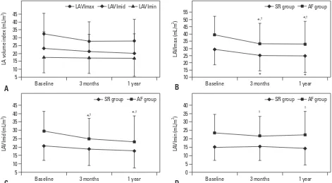
LA volumes
The LA volumes at baseline and the three month and 1 year follow-ups are listed in Fig. 2. After 3 months, the LAVmax indices decreased (from 33±13 to 28±12 mL/m2; p<0.001).
The LA mid-diastolic volume indices decreased at 1 year (from 23±11 to 20±13 mL/m2; p=0.002), but the LAVmin
in-dices remained unchanged. However, when reviewed on the basis of AF recurrence, the LA mid-diastolic volume indi-ces were only decreased in the AF group after 3 months (30±12 to 25±12 mL/m2; p=0.034), but were not decreased
in the SR group until the 1 year follow-up.
The initial left atrial volume index (LAVI) of the AF group was significantly larger than that of the SR group, and re-mained larger throughout follow-up, though the LA
mid-di-Table 3. E/E’ at Baseline and Follow-Up in All Patients and in the SR Group or the AF Group
E/E’ Total (n=90) SR group (n=61) AF group (n=29)
Baseline 9.8±5.3 9.3±4.3 10.8±7.0
3-months follow-up 10.5±5.1 9.8±4.4* 11.9±6.3
1 yr follow-up 10.0±4.9 8.8±3.8* 12.4±6.0
SR, sinus rhythm; AF, atrial fibrillation. *p<0.05 SR group versus AF group.
Fig. 1. (A) LA diameters in all patients, (B) AP diameter, (C) ML diameter, (D) SI diameter in the SR and AF groups at baseline, after three months, and after one year. *p<0.05, baseline versus follow-up parameters, †p<0.05, SR group versus AF group. AP diameter, anterior-posterior diameter; ML diameter, medial-lateral diameter; SI diameter, superior-inferior diameter; LA, left atrial; SR, sinus rhythm; AF, atrial fibrillation.
30 30
35 30
40
50 40
55
45 40
60
50
65 50
LA
d
ia
m
et
er
s (
m
m
)
LA
A
P
di
am
et
er
(m
m
)
LA
S
I d
ia
m
et
er
(m
m
)
LA
M
L d
ia
m
et
er
(m
m
)
Baseline Baseline
Baseline Baseline
3 months 3 months
3 months 3 months
1 year 1 year
1 year 1 year
A B
D C
AP diameter ML diameter SI diameter SR group AF group
SR group AF group SR group AF group
* †
†
*,† †
† †
* *
*
[image:5.595.71.543.81.429.2]max=49.4 mL, the sensitivity of a large LAVmax (>49 mL) for predicting AF recurrence was 89.3% in 26 of 29 pa-tients with AF recurrence and the specificity was 56.7% in 35 of 61 patients. The area under the curve for the predic-tion of AF recurrence was 0.745 for LAVmax.
DISCUSSION
The main findings of this study are that: 1) successful RFCA in patients with paroxysmal AF decreased LAVmax and function in the short-term and showed a trend of recovery of LA function in the long term, whereas the patients with 1-year follow-up, but was still decreased compared to initial
values.
LA volumes and LA function and predicting AF recurrence
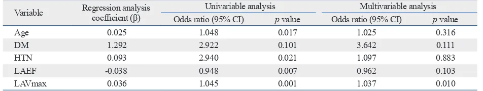
The results of the logistic regression analysis of AF recur-rence are summarized in Table 4. Univariate analysis showed that being more than 75 years old (p=0.017), having a history
of HTN (p=0.021), LAEF (p=0.007), and LAVmax
(p=0.001) were associated with AF recurrence. Multivariate analysis identified LAVmax as an independent predictor of AF recurrence (odds ratio 1.037, 95% confidence interval 1.008 to 1.066; p=0.010). When the cutoff value was
LAV-Fig. 2. (A) LA volume index in whole patients (B) LAVImax (C) LAVImid (D) LAVImin in the SR and AF groups at baseline, after three months, and after one year. *p<0.05 baseline versus follow-up parameters, †p<0.05 SR group versus AF group. LAVImax, left atrial maximal volume index; LAVImid, left atrial mid-di-astolic volume index; LAVImin, left atrial minimal volume index; LA, left atrial; SR, sinus rhythm; AF, atrial fibrillation.
5 10 0 5 20 15 10 25 30 30 25 20 15 25 20 30 15 10 5 25 20 15 10 45 40 35 55 50 45 40 35 35 40 45 40 35 30 LA vo lu m e in de x ( m L/ m 2 ) LA VI m ax (m L/ m 2 ) LA VI m in (m L/ m 2 ) LA VI m id (m L/ m 2 ) Baseline Baseline Baseline Baseline
3 months 3 months
3 months 3 months
1 year 1 year
1 year 1 year
A B
D C
LAVImax LAVImid LAVImin SR group AF group
SR group AF group SR group AF group
[image:6.595.55.526.67.326.2]*,† *,† † † *,† *,† * *
Fig. 3. (A) LAEF and LAAEF of whole patients (B) LAEF (C) LAAEF in the SR and AF groups at baseline, after three months, and after one year. *p<0.05 baseline versus follow-up parameters, †p<0.05 SR group versus AF group. LAEF, left atrial ejection fraction; LAAEF, left atrial active emptying fraction; SR, sinus rhythm; AF, atrial fibrillation.
5 20 0
25 45
40 20
65 60 40
LA fu nc tio n (% ) LA EF (% ) LA AE F ( % )
Baseline 3 months 1 year Baseline 3 months 1 year Baseline 3 months 1 year
A B C
LAEF LAAEF SR group AF group SR group AF group
[image:6.595.57.525.373.505.2]to induce a reduction in LAVmax by three-dimensional echocardiography. The same phenomenon has been pa-tients with repeated RFCAs.
It has been proposed that reduced LA size after AF abla-tion could be secondary to reverse remodeling due to the elimination of the arrhythmia (i.e., only responders would re-model) or because of scarring induced by the radiofrequency lesion (i.e., all patients would remodel).9,11 Recent studies of
LA scars induced by RFCA as assessed by cardiac magnetic resonance imaging (MRI) and by a time course of markers of tissue repair after RFCA (matrix metalloproteinase-9 and transforming growth factor-β1) support reduction of LA vol-ume by scarring, which would support the latter opinion.12,13
Our results, which identified reductions in LA volume in both the SR group and the AF group, favor the theory of scarring by RFCA.
However, continuing differences in LA volume between the two groups after RFCA implies the importance of posi-tive remodeling by reducing AF burden. Therefore, the re-duction of LA volume observed in both groups may be be-cause scarring by RFCA outweighs the effects of positive remodeling by restoring sinus rhythm in patients with only paroxysmal atrial fibrillation due to the presence of relatively lower AF burden than in patients with persistent AF.
LA function
In our study, LAEF and LAAEF remained decreased until 1 year after RFCA. However, there have been conflicting results regarding atrial function after catheter ablation of AF depending on imaging modality, follow-up period, and the type of AF. Choi, et al.3 reported recovery of LAEF and
A velocity at 3 months after ablation, but Rodrigues, et al.6
reported decreases in LAEF at more than 6 months after RFCA by two-dimensional echocardiography. Montserrat, et al.9 reported preserved LAEF and LAAEF even after
re-peated ablation by three-dimensional echocardiography at 6 months after the ablation. Donal, et al.14 noted decreased
recurrence of AF had worse LA function; 2) patients with recurrent AF had significantly larger LA volumes than pa-tients without recurrent AF; and 3) the LAVmax RFCA was an independent predictor of AF recurrence after ablation.
LA diameters
In this study, the LA anterior-posterior and superior-inferior diameter was decreased at 1 year, whereas the LA medial-lateral diameter showed no change. A recent meta-analysis showed significant decreases in LA diameter at follow-up imaging ≥1 month after RFCA.7 However, a recent study in
patients with paroxysmal AF by two-dimensional echocar-diography with a follow-up time of 8±2 months and a study using three-dimensional echocardiography after 6 months of follow-up9 both showed no change in LA diameter after
ablation.2 The above three studies used LA
anterior-posteri-or diameters in their analyses. Although LA anterianterior-posteri-or-poste- anterior-poste-rior diameters represent linear LA measurements, this is of-ten an inaccurate depiction of true LA size. Sometimes the relationships between LA anterior-posterior diameter and other diameters are not consistent because expansion of the LA in the anterior-posterior (AP) direction may be con-strained by the thoracic cavity between the sternum and the spine.10 Predominant enlargement in the superior-inferior
and medial-lateral diameters will alter LA geometry such that the AP dimension may not be representative of the true LA size. This may explain why our LA diameters showed different results by different diameters.
LA volumes
In the present study, LAVImax and LAVImid decreased af-ter the ablation, but LAVImin did not. The changes were identified in both the SR group and the AF group, but the LAVI of the SR group was smaller than that of the AF group. Previous studies demonstrated reverse morphological re-modeling of the LA by ablation for isolated AF by
[image:7.595.68.544.81.171.2]two-di-mensional echocardiogram.2,6 Recently, RFCA was found
Table 4. Risk of AF Recurrence after Radiofrequency Catheter Ablation
Variable Regression analysis coefficient (β) Univariable analysis Multivariable analysis Odds ratio (95% CI) p value Odds ratio (95% CI) p value
Age 0.025 1.048 0.017 1.025 0.316
DM 1.292 2.922 0.101 3.642 0.111
HTN 0.093 2.940 0.021 1.097 0.883
LAEF -0.038 0.948 0.007 0.962 0.103
LAVmax 0.036 1.045 0.001 1.037 0.010
tients with paroxysmal AF. Major limitations of this study are the small sample size, the low recurrence rate, and the relatively wide range of the odds ratios in the multiple lo-gistic regression analysis. In addition, echocardiography has limitations in terms of poor acoustic windows and may un-derestimate true LA size and volume compared to
comput-ed tomography or MRI.7
In addition, the different kinds of antiarrhythmic drugs being taken by the patients may have influenced the results. These drugs may assist in maintaining sinus rhythm and la-tently improve the LA function seen during follow-up. Dif-ferent ablation lesions targeting the additional LA lines and variation in the duration of RFCA may all affect the recur-rence of AF. Due to the above limitations and growing con-cern about AF recurrence after RFCA, a large cohort study is needed in the future.
Conclusion and clinical implications
Successful RFCA in patients with paroxysmal AF signifi-cantly decreases LAVmax and function shortly after ablation and improves LA function gradually with time. Patients with recurrent AF had significantly larger LA volumes than pa-tients with SR. LAVmax before RFCA was an independent predictor of AF recurrence after the ablation.
Because delayed functional improvements could cause thromboembolism and other cardiovascular events, careful anticoagulation during prolonged follow-up is warranted, but this potential management approach requires further study. In addition, the assessment of LA using new echo-cardiographic parameters such as LA strain, three-dimen-sional LA volume, and CT or MRI may lead to more accu-rate assessments in patients with AF.
ACKNOWLEDGEMENTS
The authors are grateful to Miss Byung Sun Song, sonogra-pher, for her assistance in the performance of this study.
REFERENCES
1. Reant P, Lafitte S, Jaïs P, Serri K, Weerasooriya R, Hocini M, et al. Reverse remodeling of the left cardiac chambers after catheter ab-lation after 1 year in a series of patients with isolated atrial fibrilla-tion. Circulation 2005;112:2896-903.
2. Tops LF, Bax JJ, Zeppenfeld K, Jongbloed MR, van der Wall EE, Schalij MJ. Effect of radiofrequency catheter ablation for atrial
fi-LA compliance after 1 year of RFCA by fi-LA strain. Wylie, et al.12 reported reduced atrial systolic function correlated
with LA scars at a mean of 48 days by cardiac MRI. Masu-da, et al.15 demonstrated that AF ablation appears to have a
beneficial effect on LA function, as assessed by cardiac CT at 3 months after RFCA.
The left atrium has three functions: acting as a contractile pump that provides up to 1/3 of the left ventricular volume, as a reservoir that collects pulmonary venous return during ventricular systole, and as a conduit for the passage of stored blood from the LA to the LV just after the isovolumetric re-laxation phase of ventricular diastole, when the mitral valve opens.11 LAEF reflects LA conduit and contractile function
and LAAEF shows only contractile function. In contrast to other reports, the LAEF and LAAEF of the SR group were significantly decreased at 3 months after the RFCA, but not in the AF group in our study. We hypothesized that the cause of significant decreases of LA function in the SR group was because of the differential changes between LAVmax and LAVmin. While the LAVmax of both groups changed signif-icantly in serial follow-ups, LAVmin remained unchanged. In addition, the range of the decreases in LAVmid and LAV-max were larger in the AF group than in the SR group. We believe that these changes in LA volumes resulted in signif-icant decreases in LA function in the SR group at the 3 month follow-up due to the formulae of LAEF and LAAEF.
One of the most persuasive studies done with MRI showed improved LAEF in patients without recurrence and no change in those with recurrence.16 The method of ablation in the
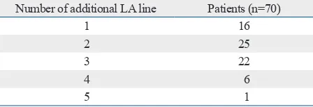
study was only 4 pulmonary vein isolation, whereas we conducted additional LA ablations (anterior, roof, posterior, perimitral and septal line) in most of our patients (Table 5). The additional LA ablation may affect the deterioration of LA function whereas pulmonary vein isolation was not.
Study limitation
Our study was a retrospective echocardiographic evaluation of reverse LA remodeling after RFCA of AF, including mor-phological and functional changes in the LA only in
pa-Table 5. Patients with Additional LA Lines during Ablation
Number of additional LA line Patients (n=70)
1 16
2 25
3 22
4 6
5 1
[image:8.595.57.281.642.719.2]Pellikka PA, et al. Recommendations for chamber quantification: a report from the American Society of Echocardiography’s Guide-lines and Standards Committee and the Chamber Quantification Writing Group, developed in conjunction with the European As-sociation of Echocardiography, a branch of the European Society of Cardiology. J Am Soc Echocardiogr 2005;18:1440-63. 11. Delgado V, Vidal B, Sitges M, Tamborero D, Mont L, Berruezo A,
et al. Fate of left atrial function as determined by real-time three-dimensional echocardiography study after radiofrequency catheter ablation for the treatment of atrial fibrillation. Am J Cardiol 2008; 101:1285-90.
12. Wylie JV Jr, Peters DC, Essebag V, Manning WJ, Josephson ME, Hauser TH. Left atrial function and scar after catheter ablation of atrial fibrillation. Heart Rhythm 2008;5:656-62.
13. Richter B, Gwechenberger M, Socas A, Zorn G, Albinni S, Marx M, et al. Time course of markers of tissue repair after ablation of atrial fibrillation and their relation to left atrial structural changes and clinical ablation outcome. Int J Cardiol 2011;152:231-6. 14. Donal E, Ollivier R, Veillard D, Hamonic S, Pavin D, Daubert JC,
et al. Left atrial function assessed by trans-thoracic echocardiogra-phy in patients treated by ablation for a lone paroxysmal atrial fi-brillation. Eur J Echocardiogr 2010;11:845-52.
15. Masuda M, Inoue K, Iwakura K, Okamura A, Koyama Y, Kimura R, et al. The impact of atrial fibrillation ablation on left atrial func-tion: association with baseline left atrial function. Pacing Clin Electrophysiol 2012;35:327-34.
16. Jahnke C, Fischer J, Gerds-Li JH, Gebker R, Manka R, Fleck E, et al. Serial monitoring of reverse left-atrial remodeling after pulmo-nary vein isolation in patients with atrial fibrillation: a magnetic resonance imaging study. Int J Cardiol 2011;153:42-6.
brillation on left atrial cavity size. Am J Cardiol 2006;97:1220-2. 3. Choi JI, Park SM, Park JS, Hong SJ, Pak HN, Lim do S, et al.
Changes in left atrial structure and function after catheter ablation and electrical cardioversion for atrial fibrillation. Circ J 2008;72: 2051-7.
4. Marsan NA, Tops LF, Holman ER, Van de Veire NR, Zeppenfeld K, Boersma E, et al. Comparison of left atrial volumes and func-tion by real-time three-dimensional echocardiography in patients having catheter ablation for atrial fibrillation with persistence of sinus rhythm versus recurrent atrial fibrillation three months later. Am J Cardiol 2008;102:847-53.
5. Hwang HJ, Choi EY, Rhee SJ, Joung B, Lee BH, Lee SH, et al. Left atrial strain as predictor of successful outcomes in catheter ablation for atrial fibrillation: a two-dimensional myocardial imag-ing study. J Interv Card Electrophysiol 2009;26:127-32.
6. Rodrigues AC, Scannavacca MI, Caldas MA, Hotta VT, Pisani C, Sosa EA, et al. Left atrial function after ablation for paroxysmal atrial fibrillation. Am J Cardiol 2009;103:395-8.
7. Jeevanantham V, Ntim W, Navaneethan SD, Shah S, Johnson AC, Hall B, et al. Meta-analysis of the effect of radiofrequency catheter ablation on left atrial size, volumes and function in patients with atrial fibrillation. Am J Cardiol 2010;105:1317-26.
8. Mirza M, Caracciolo G, Khan U, Mori N, Saha SK, Srivathsan K, et al. Left atrial reservoir function predicts atrial fibrillation recur-rence after catheter ablation: a two-dimensional speckle strain study. J Interv Card Electrophysiol 2011;31:197-206.
9. Montserrat S, Sitges M, Calvo N, Silva E, Tamborero D, Vidal B, et al. Effect of repeated radiofrequency catheter ablation on left atrial function for the treatment of atrial fibrillation. Am J Cardiol 2011;108:1741-6.