R E S E A R C H A R T I C L E
Open Access
Investigation of anti-oxidative stress
in vitro
and
water apparent diffusion coefficient in MRI on rat
after spinal cord injury
in vivo
with
Tithonia
diversifolia
ethanolic extracts treatment
Chi-Long Juang
1†, Fei Shish Yang
2†, Ming Shuang Hsieh
1, Hu-Yi Tseng
3, Su-Chiu Chen
2and Hsiao-Chuan Wen
4*Abstract
Background:Spinal cord injury (SCI)-induced secondary oxidative stress associates with a clinical complication and high mortality. Treatments to improve the neurological outcome of secondary injury are considered as important issues. The objective of the current study is to evaluate the anti-oxidative effect ofTithonia diversifoliaethanolic extracts (TDE) on cells and apply the pharmacological effect to SCI model using a MRI imaging algorism. Methods:The anti-oxidation properties were tested in a 2,2-diphenyl-1-picrylhydrazyl (DPPH) radical scavenging assay. Rat liver cells (clone-9) were treated with various doses of TDE (0 ~ 50μg/ml) before exposed to 250μM H2O2and cell survival was determined by MTT and LDH assays. We performed water apparent diffusion coefficient (ADC) map in MR techniques to investigate the efficacy of TDE treatment on SCI animal model. We performed T5 laminectomy and compression (50 g, 1 min) to induce SCI. PHILIP 3.0 T MRI was used to image 24 male Sprague–Dawley rats weighing 280–320 g. Rats were randomly divided into three groups: sham group, SCI group, SCI treated with TDE group. The MRI images were taken and ADC were acquired before and after of treatment of TDE (50 mg/kg B. W. orally, 5 days) in SCI model.
Results:TDE protected clone-9 cells against H2O2-induced toxicity through DPPH scavenging mechanism. In addition, SCI induced the increase in ADC after 6 hours. TDE treatment slightly decreased the ADC level after 1-week SCI compared with control animals.
Conclusion:Our studies have proved that the cytoprotection effect of TDE, at least in part, is through scavenging ROS to eliminate intracellular oxidative stress and highlight a potential therapeutic consideration of TDE in alternative and complementary medicine.
Keywords:Spinal cord injury, Apparent diffusion coefficient,Tithonia diversifoliaethanolic extracts, DPPH, LDH
Background
Tithonia diversifolia (T. diversifolia), a bushy perennial weed, is commonly found in Nigerian fields, wastelands and roadsides in Taiwan. The plant is primarily used for ornamental purposes, and further studies have demon-strated its effects on treatment of diabetes mellitus, diar-rhea, fever, hematomas, hepatitis, hepatomas, malaria and wounds [1,2]. TheT. diversifolia contains bioactive
compounds which have been proved to possess anti-inflammatory effects [3]. In addition, phytochemical inves-tigations of T. diversifolia have identified the existence other bioactive compounds, such as cadinane, chromene, eudesmane, flavone, germacrane and rearranged eudes-mane derivatives [4-11]. The extracts from aerial and stem of T. diversifolia have demonstrated anti-inflammatory and liver protective effects in rats against paw edema in-duced by carrageenan and acute hepatic damage inin-duced by CCl4, respectively [2]. The extracts fromT. diversifolia
and the major effective compound sesquiterpenoid, the tagitinin C, have been identified as a potential anti-cancer * Correspondence:[email protected]
†Equal contributors
4Department of Optometry, Yuanpei University, Hsinchu 30015, Taiwan
Full list of author information is available at the end of the article
therapeutic reagent both in vitro and in vivo [12-16]. Other than its anti-cancer activity, the sesquiterpenoid that ex-tracted from other species shows its anti-inflammatory ef-fect via inhibition of oxidative stress both in vitro and
in vivoin the treatment of psoriasis [17]. However, whether the anti-inflammatory effect fromT. diversifoliaextracts is mediated by its ability of anti-oxidative stress remains to be elucidated.
Increasing numbers of small animal models are in use in the field of neurological research. To evaluate the functions of soft tissues, Magnetic Resonance Imaging (MRI) can provide an excellent method for non-invasive imaging of nervous tissue. Specialized small-bore animal MRI scanners are available for high-resolution MRI of small rodents’ tissue, but the major drawback of this equipment is its high cost. Several research groups have performed clinical MR scanners for imaging small ani-mal models. To achieve a reasonable spatial resolution at an acceptable signal-to-noise ratio with these scan-ners, several requirements regarding to sequence param-eters need to be matched considerately. Thus, the goal of this study is seeking to successfully apply MRI for examining small rodents’ spinal cord injury using a standard clinical 3 T scanner and to keep instrumental cost low. Our improvement including the use of appar-ent diffusion coefficiappar-ent (ADC), which measures the dif-fusion of water molecules within central nervous system, is applied in this study. A low value of ADC indicates that the nerve fiber tracts are well-organized, while a high value of ADC indicates that these tracts are damaged and disorganized. Therefore, while evaluating an acute ische-mic stroke, ADC images may play a crucial role in identi-fying the degree of damage due to its higher water content in injured area.
The damage that occurs in the central nervous system (CNS) following trauma is due to secondary effects of glutamate excitotoxicity, Ca2+ overload, and oxidative stress [18,19]. Oxidative stress activates mechanisms that result in a neutrophil-mediated inflammation which can also cause secondary damage. Mechanisms of oxidative stress are involved in lipid peroxidation and production of reactive oxidative species production. We suggest that decreasing oxidative stress greatly reduces the amount of secondary damage due to trauma. Aggressive nutri-tional support following CNS trauma can also contribute to maximizing antioxidant defenses. Furthermore, we sug-gest that natural supplements such as herbs have the po-tential to be therapeutically effective because of their free radical quenching, iron chelating, and anti-inflammatory properties.
Decrease in oxidative phosphorylation processes in CNS may induce neurological deficits. Natural antioxi-dants may help the antioxidant capacity of the organism to prevent from disease associated with reactive oxygen
species (ROS) and other oxidative damage inducing agents. In this study, we sought to explore anti-oxidative stress in vitro with T. diversifolia Ethanolic Extracts (TDE) and the pharmacological effect of TDE on ADC in MRI on rat after SCIin vivo.
Methods Plant extraction
The leaves ofT. diversifolia were collected in Hsin-Chu, Taiwan, in January 2011. A voucher specimen was de-posited in the Department of Medical Imaging and Radiological Technology, Yuanpei, University, Hsin-Chu, Taiwan. Collected plant materials were dried and ground into powder using a grinder and screened through a 20-mesh sieve (aperture of 0.94 mm). The powder of T. diversifolialeaves (200 g) was extracted 3 times with 1 L ethanol at room temperature and sonicated for the dur-ation of 5 h (100 min for each time). The plant residue was filtered through a 10 μm cartridge paper, and the ethanolic extracts were combined and concentrated by a rotary vacuum evaporator. The detailed extraction of
T. diversifolia was followed by Liaoet al. [13,14], modi-fied by Tsenget al. [20] and reported in previously paper [13-15,20]. Ming-Shueh Shieh, a Professor in Depart-ment of EnvironDepart-mental Engineering and Health, who major in identity of plant, preceded the identity of the plant material used in our study prior to its use. The ethanolic extracts of the leaves of T. diversifolia were named TDE.
Cell lines and cultures
Clone-9 (normal rat liver cell) were cultured in Dulbecco modified Eagle medium (DMEM) (Gibco, NY, USA) sup-plemented with 10% fetal bovine serum (FBS), 2 mM glu-tamine, 100 U/mL penicillin,100 mg/mL streptomycin sulfate and 1 mM sodium pyruvate then incubated at 37°C at an atmosphere of 5% CO2/O2..
Scavenging ability on DPPH radical
The 2,2-diphenyl-1- picrylhydrazyl (DPPH) test was used to evaluate the free radical scavenging capacity (RSC) of TDE in cell free systems according to a published pro-cedure [21] using ascorbic acid as positive control sub-stance. Different concentrations of TDE were tested and absorbance was measured at 490 nm after 30 min of re-action with a stock solution of DPPH.
Cell viability assay
washed once before adding 50 mL of FBS-free medium containing MTT (0.5 mg/mL). After 3 hr of incubation at 37°C, the medium was discarded, and the formazan blue in the cells was dissolved in DMSO. The optical density (O. D.) was measured at 540 nm. The experiments were repeated three times.
Determination of protection of H2O2-induced damage in normal liver cells by the MTT and LDH Assays
The protective effect of TDE against toxicity induced by H2O2in normal rat liver cells (clone 9) was carried out
using MTT and lactate dehydrogenase (LDH) assays. Cells were seeded as described above and treated with TDE (10, 30, 50 μg/ml) for 24 hr. Clone 9 cells after treatment were then exposed to phosphate buffered sa-line (PBS) free medium containing 250 μM H2O2 for
1 hr. The MTT assay and LDH assay was performed after hydrogen peroxide exposure. The assay of LDH re-lease was determined by a commercial kit from Roche® following the manufacturer’s instruction.
Compression SCI model
All animal experimental manipulations were under taken in accordance with the National Institutes of Health Guide for the Care and Use of Laboratory Animals, with the ap-proval of the Experimental Animal Ethical Committee of Taipei Mackay Memorial Hospital. All animals were anesthetized with an overdose of sodium pentobarbital and sacrificed immediately after finishing experiment. Sprague-Dawley rats weighing between 280 and 320 g were anesthetized by intramuscular (inraperitoneal) ad-ministration of Zoletil 50 and Ropum (2:1). A laminec-tomy was performed at vertebral level T5, leaving the dura undisturbed. The T4 and T6 transverse processes were clamped in a spinal compression frame and the compression was applied by a modified aneurysm clip which produced a compression force of 50 grams and lasted for 1 min. After being induced spinal cord injury or sham control, all rats were placed in warmed cages to recover from anaesthesia. The rats were given free ac-cess to food and tap water, and were maintained on a 12 hr light/dark cycle at 21 ± 1°C.
Animal MRI
The rats were examined in a 3 T whole-body MR system (Achieva X-series; Philips, Eindhoven, Netherland). A wrist coil (SENSE-wrist-8) was used to obtain all MR im-ages, allowing parallel imaging. To avoid movement, rats were placed into a plastic holder. Animals imaged with T1-, T2-, and diffusion-weighted sequences. ADC maps were constructed from the diffusion-weighted data. A fast T2- weighted spin echo sequence was used as localizer. A transverse T1-weighted spin-echo [3000 msec/100 msec (repetition time/echo time), field of view (FOV) of
81.3 mm2] and a T2-weighted turbo spin-echo sequence TR 2000 msec/TE40 msec were performed with a section area of 1 mm2. A DW-MRI echo-planar sequence was performed with a b-values = 800 sec/mm2. The following parameters were used for this sequence: TR = 2870 milli-seconds, TE = 111 milliseconds and a total time of acquisi-tion of 2:35 minutes. Gradient strength was adjusted for
b-value. ADC maps were calculated from the native diffu-sion images with the built-in software tools of the MRI scanner. The measured fractional anisotropy (FA) direc-tional diffusivities derived from diffusion weighted image (DWI) and the diffusion tensor imaging (DTI) was applied in capture details of the anatomy for tractography.
Statistical analysis
All data are expressed as mean ± SEM of three inde-pendent experiments in in vitro study. Every independ-ent experimindepend-ent included three wells of cells. Data were processed by one-way analysis of variance (ANOVA) tests using SPSS. Intergroup comparisons were made using paired t-test or students’ t -test. Differences were considered significant whenp< 0.05.
Results
We first examined the antioxidative effect of TDE by using an in vitrocell free system. We performed DPPH to test the free RSC of TDE. The use of vitamin C was as a positive control. Free radical scavenging profile of TDE was expressed as % of DPPH inhibition compared to ascorbic acid. The result showed that the IC50 values for TDE and ascorbic acid were 0.93 ± 0.20 and 0.48 ± 0.10 μg/ml, respectively (see Figure 1), suggesting a free radical scavenging ability of TDE.
We then tested the anti-oxidative effects of TDE in cell culture system. The clone 9 was pretreated with TDE of different doses (0-50 μg/ml), followed by exposing the cells in 250μM H2O2. Cell survival was detected by MTT
assay and LDH release assay. Vitamin C treatment was used as a positive control (Figure 2). We found that treat-ment of H2O2alone resulted in significant decrease of cell
viability. Pretreatment of TDE protected H2O2-induced
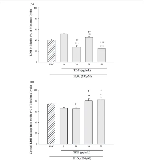
decrease of cell viability (Figure 2). Similar results were found in LDH release assay, where H2O2-induced release
of LDH into medium from cytosol was significantly re-duced by TDE protection (Figure 3A) and cytosolic LDH was significantly higher with TDE treatment than without TDE treatment (Figure 3B). These results suggest that TDE possesses anti-oxidative effect against H2O2-induced
cytotoxicity.
Given that TDE has demonstrated a potential ant-oxidative and free radical scavenging effect in thein vitro
system, we are seeking whether this plant extract can be performed to anin vivosystem. One of the knownin vivo
To assist the data evaluation, we also performed ADC to analyze MRI images and examined the SCI grading. At different time points (0, 2, 4, 6 hr) after SCI, rats were anesthetized and analyze the ADC value using MRI. The results showed SCI resulted in significant in-crease in ADC after 6 hr (Figure 4), suggesting an
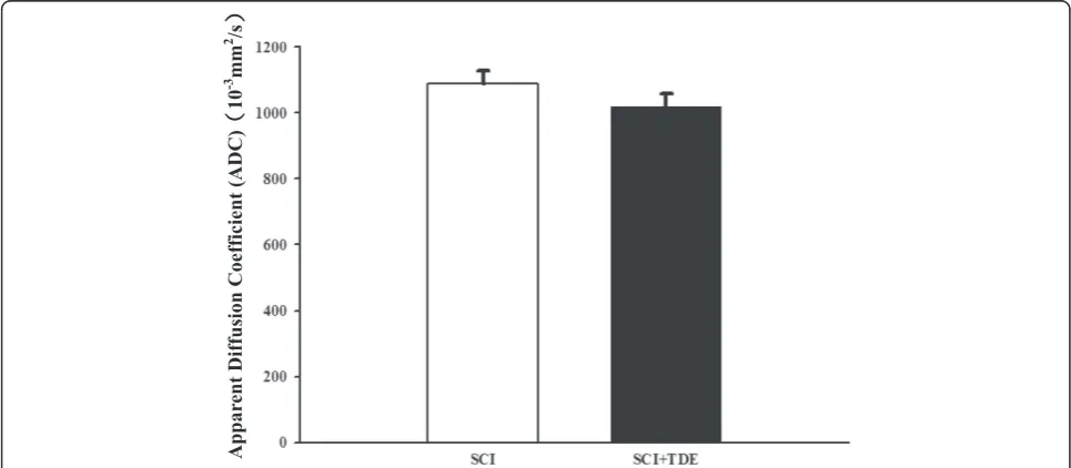
[image:4.595.59.540.87.324.2]increase of cell permeability after SCI. To further con-firm the SCI injury model was successfully executed, we then performed DTI to evaluate the damage level of fiber tracts after SCI [20]. TDE treatment slightly de-creased the ADC level in 1 week after SCI compared with no TDE treatment in animal study (Figure 5). Figure 1Free radical scavenging profile of TDE expressed as % of DPPH scavenging compared to ascorbic acid.IC50values for TDE and
ascorbic acid were 0.93 ± 0.2 and 0.48 ± 0.1μg/ml respectively.
[image:4.595.55.539.464.704.2]Discussion
Development of early detectors of treatment response can provide information which would be of significant benefit for both experimental and clinical trials. These issues underscore the need for using noninvasive imaging to
[image:5.595.58.538.87.619.2]therapeutic protocol planning. Serial study of such MR pa-rameters as DWI, ADC, and T2-weighted imaging may provide information on the pathophysiological mecha-nisms of acute ischemic stroke [19,22-25]. Our studies showed ADC was elevated in the acute stage after SCI 6 hr because of increased extracellular water permeability
viadecrease of Na-K pump activity, influx of Ca2+, water
and efflux of K+. The information provides us that the time to improve the neurological outcome of SCI is very important.
Compression models of rat SCI mainly use either a modified aneurysm clip or a static compression plat-form, and the aneurysm clip has been described in detail [26-28]. A static compression platform has been used in
Appar
ent Diffusion Coefficient
(ADC)
10
[image:6.595.64.537.88.317.2]-3 mm 2 /s
Figure 4Time course of spinal cord injury (SCI) on apparent diffusion coefficient (ADC).Rats were induced SCI by T5 laminectomy and compression (50 g, 1 min). ADC map was used to assessment the injured level. Data are presented as mean ± S.E.M of six rats. Significance for the time-dependent effect after SCI treatment was calculated by ANOVA. *P< 0.05, significantly different from untreated control samples.
Appar
ent Diffusion Coefficient
(ADC)
10
-3 mm 2 /s
[image:6.595.56.539.495.706.2]several studies [16,29] but has not been characterized so fully [30]. In this paper we have therefore used a T5 static compression model of SCI using diffusion tensor imaging (DTI) to evaluate the damage level of the fiber tracts and this data has successfully published [20]. Huang et al. found that static compression SCI results in a prolonged profile of cell loss [31]. In contrast, the temporal profile of cell loss in the rat contusion SCI model is very different. Both neuronal and oligodendrocyte loss after contusion injury are acute events. The differences between the com-pression and contusion models are important to study the pathophysiology of SCI. The delayed cell death in our compression models provides a good opportunity to assess the efficacy of neuroprotective interventions.
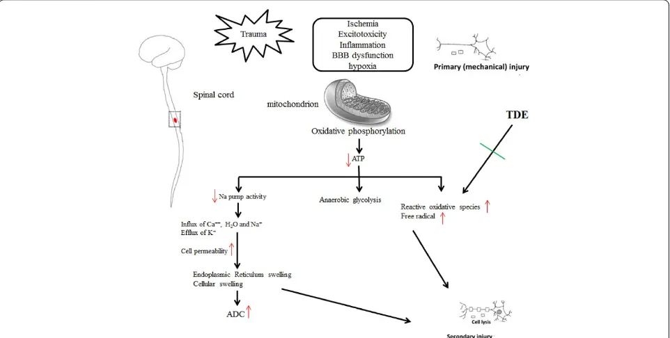
T. diversifolia has been shown to possess anti-inflammatory and analgesic activity [2]; however, the neuroprotective or antioxidant effect has never been established. In the present study, we have evaluated the free radical scavenging effect, anti-oxidative stress of TDE in the in vitromodel system and the results are summa-rized in Figure 6. Furthermore, ooxidative stress in SCI is known to induce neuronal damage and results in serial dysfunctions. In current study, our study has also sug-gested a potential therapeutic effect of TDE in SCI.
Consequently, the consumption of plants with neuro-protecive effects in the complementary therapy may repre-sent a strategy to prevent from a wide range of disease. Given the composition of TDE is complex, there has been marked variability in animal study. That is the reason that the data showed only 4 of 6 in SCI decrement assayed by
ADC after TDE treatment. It has been reported that con-tain sesquiterpenoids and flavonoids [12] which may have neuroprotective effect. In our phytochemical screening, TDE reveals flavonoids (350.46 rutin equivalent μg/mg), and phenols (1273.01 galic acid equivalentμg/mg). Isolated constituents from TDE have previously been demonstrated to exert their effect in neuronal cells [13-15]. For example, studies regarding toT. diversifoliareveal anti-inflammatory effect [2]. In this context, TDE may act through ROS scav-enging in part in response to reduce intracellular oxidative stress to exert its neuroprotective effect.
Spinal cord injury such as ischemia, excitotoxicity, blood brain barrier dysfunction and hypoxia cause primary or mechanical injury. Previous reports about CNS trauma can induce oxidative stress and produce free radical and then cause secondary injury which induces paraspinal edema, stroke and other neurological damages. Trauma-induced edema formation in SCI is related with clinical complication and high mortality. In this animal study, we used Diffusion Tensor Imaging (DTI) to confirm the consistency effect of damage level of fiber tracts after SCI [20] and ADC map in MR techniques for studying the efficacy of TDE treatment.
Conclusion
[image:7.595.58.539.459.702.2]These findings demonstrate the protective effect of TDE against oxidative stress may act through free radical scav-enging properties. TDE may be considerate as an interest in complementary medicine in the future. Further re-search is needed to define health benefits and clinical ef-fects of TDE.
Competing interests
The authors declare that they have no competing interests.
Authors’contributions
HMS preceded all cellular experiments. JCL, YFS, THY, and CSC were responsible for MR technique assistance and image processing. WHC was responsible for pharmacological aspects of the experiments. All authors read and approved the final manuscript.
Acknowledgements
We thank Taipei Mackay Memorial Hospital for the assistance of Magnetic Resonance Imaging and for the financial support (MMH 9988) and Dr. Wang YK (Taipei Medical University, Taipei, Taiwan) for the critical review of the manuscript.
Author details 1
Department of Medical Imaging and Radiological Technology, Yuanpei University, Hsinchu 30015, Taiwan.2Mackay Memorial Hospital, Department of Radiological Technology, Taipei, Taiwan.3Taipei City Hospital, Yangming Branch, Department of Radiological Technology, Taipei, Taiwan.4Department of Optometry, Yuanpei University, Hsinchu 30015, Taiwan.
Received: 4 July 2013 Accepted: 18 August 2014 Published: 18 November 2014
References
1. Takanashi M:U.S. Patent 5,773,004.1998.
2. Lin CC, Lin ML, Lin JM:The antiinflammatory and liver protective effect of
Tithonia diversifolia (Hemsl.) grayandDicliptera chinensis Juss.Extracts in
rats.Phytotherapy Res1993,7:305–309.
3. Rungeler P, Lyss G, Castro V, Mora G, Pahl HL, Merfort I:Study of three sesquiterpene lactones from Tithonia diversifolia ontheir
anti-inflammatory activity using the transcription factor NF-κB and enzymes
of the arachidonic acid pathway as targets.Planta Med1998,64:588–593.
4. Kuo YH, Lin BY:Five new taraxastane-type triterpenes from the aerial
roots ofFicus microcarpa.Chem Pharm Bull1999,47:498–500.
5. Kuo YH, Chen CH:Sesquiterpenes from the leaves ofTithonia diversifolia.
J Nat Prod1998,61:827–828.
6. Kuo YH, Chen CH:Diversifolol sesquiterpenes from the leaves ofTithonia diversifolia.Chem Pharm Bull1997,45:1223–1224.
7. Bordoloi M, Barua NC, Ghosh AC:An artemisinic acid analogue from
Tithonia diversifolia.Phytochemistry1996,1:557–559.
8. Baruah NC, Sarma JC, Barua NC, Sarma S, Sharma RP:Germination and
growth inhibitory sesquiterpene lactones and aflavone fromTithonia
diversifolia.Phytochemistry1994,36:29–36.
9. Schuster A, Stokes S, Papastergiou F, Castro V, Poveda L, Jakupovic J:
Sesquiterpene lactones from twoTithoniaspecies.Phytochemistry1992,
31:3139–3141.
10. Chowdury PK, Barua NC, Sharma RP, Thyagarajan G, Herz W:Structure of
deacetylviguiestenin (tagitinin E). An addendum.J Org Chem1980,
5:535–536.
11. Baruah NC, Sharma RP, Madhusudanan KP, Thyagarajan G, Herz W, Murari R:
Sesquiterpene lactones ofTithonia diversifolia.Stereochemistry of the
tagitinins and related compounds.J Org Chem1979,44:1831–1835.
12. Kuroda M, Yokosuka A, Kobayashi R, Jitsuno M, Kando H, Nosaka K, Ishii H, Yamori T, Mimaki Y:Sesquiterpenoids and flavonoids from the aerial
parts ofTithonia diversifoliaand their cytotoxic activity.Chem Pharm Bull
2007,55:1240–1244.
13. Liao MH, Lin WC, Wen HC, Pu HF:Tithonia diversifoliaand its main active component tagitinin C induce surviving inhibition and G2/M arrest in
human malignant glioblastoma cells.Fitoterapia2011,82:331–341.
14. Lee MY, Liao MH, Tsai YN, Chiu KH, Wen HC:Identification and anti-human
glioblastoma activity of tagitinin C fromTithonia diversifoliamethanolic
extract.J Agr Food Chem2011,59:2347–2355.
15. Liao MH, Tsai YN, Yang CY, Juang CL, Lee MY, Chang LH, Wen HC: Anti-human hepatoma Hep-G2 proliferative, apoptotic, and antimutagenic
activity of tagitinin C fromTithonia diversifolialeaves.J Nat Med2013,
67(1):98–106.
16. Xu W, Chi L, Xu R, Ke Y, Luo C, Cai J, Qiu M, Gozal D, Liu R:Increased production of reactive oxygen species contributes to motor neuron
death in a compression mouse model of spinal cord injury.Spinal Cord
2005,43:204–213.
17. Payá M, De Rosa S, Terencio MC:Antipsoriatic effects of avarol-3′ -thiosali-cylate are mediated by inhibition of TNF-alpha generation and
NF-kappaB activation in mouse skin.Br J Pharmacol2007,152(3):353–365.
18. Juurlink BH, Paterson PG:Review of oxidative stress in brain and spinal cord injury: suggestions for pharmacological and nutritional
management strategies.J Spinal Cord Med1998,21:309–334.
19. Knight RA, Dereski MO, Helpern JA, Ordidge RJ, Chopp M:Magnetic resonance imaging assessment of evolving focal cerebral ischemia:
comparison with histopathology in rats.Stroke1994,25:1252–1261.
20. Tseng HY, Yang FS, Chen SC, Wang SH, Wen HC, Juang CL:Changes of
intramyocellular lipid level in Rat with spinal cord injury afterTithonia
diversifoliaextract treatment.Taipei City Med J2013,10(1):17–25. 21. López V, Akerreta S, Casanova E, García-Mina JM, Cavero RY, Calvo MI:In
vitro antioxidant and anti-rhizopus activities of Lamiaceae herbal
extracts.Plant Foods Hum Nutr2007,62:151–155.
22. Welch KM, Windham J, Knight RA, Nagesh V, Hugg JW, Jacobs M, Peck D, Booker P, Dereski MO, Levine SR:A model to predict the histopathology of human stroke using diffusion and T2-weighted magnetic resonance
imaging.Stroke1995,26:1983–1989.
23. Rordorf G, Koroshetz WJ, Copen WA, Cramer SC, Schaefer PW, Budzik RF Jr, Schwamm LH, Buonanno F, Sorensen AG, Gonzalez G:Regional ischemia and ischemic injury in patients with acute middle cerebral artery stroke as defined by early diffusion-weighted and perfusion-weighted MRI.
Stroke1998,29:939–943.
24. D’Olhaberriague L, Welch KM, Nagesh V, Gymnopoulos C, Mansbach HH, Hugg JW, Boska MD, Knight RA, Schultz LR, Levine SR, Chopp M: Preliminary clinical-radiological assessment of a MR tissue signature
model in human stroke.J Neurol Sci1998,156:158–166.
25. Schlaug G, Siewert B, Benfield A, Edelman RR, Warach S:Time course of the apparent diffusio n coefficient (ADC) abnormality in human stroke.
Neurology1997,49(1):113–119.
26. Joshi M, Fehlings MG:Development and characterization of a novel, graded model of clip compressive spinal cord injury in the mouse: part
1. Clip design, behavioral outcomes, and histopathology.J Neurotrauma
2002,19(2):175–190.
27. Joshi M, Fehlings MG:Development and characterization of a novel, graded model of clip compressive spinal cord injury in the mouse: Part 2. Quantitative neuroanatomical assessment and analysis of the relationships between axonal tracts, residual tissue, and locomotor
recovery.J Neurotrauma2002,19(2):191–203.
28. Marques SA, Garcez VF, Del Bel EA, Martinez AM:A simple, inexpensive and easily reproducible model of spinal cord injury in mice:
morphological and functional assessment.J Neurosci Methods2009,
177(1):183–193.
29. Li GL, Farooque M, Isaksson J, Olsson Y:Changes in synapses and axons demonstrated by synaptophysin immunohistochemistry following spinal
cord compression trauma in the rat and mouse.Biomed Environ Sci2004,
17(3):281–290.
30. Farooque M:Spinal cord compression injury in the mouse: presentation
of a model including assessment of motor dysfunction.Acta Neuropathol
2000,100(1):13–22.
31. Huang WL, George KJ, Ibba V, Liu MC, Averill S, Quartu M, Hamlyn PJ, Priestley JV:The characteristics of neuronal injury in a static compression
model of spinal cord injury in adult rats.Eur J Neurosci2007,25(2):362–372.
doi:10.1186/1472-6882-14-447
Cite this article as:Juanget al.:Investigation of anti-oxidative stress
in vitroand water apparent diffusion coefficient in MRI on rat after spinal cord injuryin vivowithTithonia diversifoliaethanolic extracts