Endotoxin Is Not Essential for the Development of Cockroach
Induced Allergic Airway Inflammation
Yoo Seob Shin,
1* Jung-Ho Sohn,
1,2* Joo-Young Kim,
1Jae Hyun Lee,
1Sang-Heon Cho,
3Soo-Jong Hong,
4Joo-Shil Lee,
5Chein-Soo Hong,
1and Jung-Won Park
11Department of Internal Medicine, Institute of Allergy, Yonsei University College of Medicine, Seoul;
2Department of Life Science, Biomedical Research Institute, Hanyang University, Seoul;
3Department of Internal Medicine, Seoul National University College of Medicine, Seoul;
4Department of Pediatrics, University of Ulsan College of Medicine, Seoul;
5Center for Immunology and Pathology, Korea National Institute of Health, Cheongwon, Korea.
Received: October 21, 2011 Revised: January 4, 2012 Accepted: January 5, 2012
Corresponding author: Dr. Jung-Won Park, Department of Internal Medicine, Yonsei University College of Medicine, 50 Yonsei-ro, Seodaemun-gu, Seoul 120-752, Korea.
Tel: 82-2-2228-1930, Fax: 82-2-393-6884 E-mail: [email protected]
*Yoo Seob Shin and Jung-Ho Sohn contributed equally to this work.
∙ The authors have no financial conflicts of interest.
© Copyright:
Yonsei University College of Medicine 2012
This is an Open Access article distributed under the terms of the Creative Commons Attribution Non-Commercial License (http://creativecommons.org/ licenses/by-nc/3.0) which permits unrestricted non-commercial use, distribution, and reproduction in any medium, provided the original work is properly cited.
Purpose: Cockroach (CR) is an important inhalant allergen and can induce aller-gic asthma. However, the mechanism by which CR induces airway alleraller-gic in-flammation and the role of endotoxin in CR extract are not clearly understood in regards to the development of airway inflammation. In this study, we evaluated whether endotoxin is essential to the development of CR induced airway allergic
inflammation in mice. Materials and Methods: Airway allergic inflammation was
induced by intranasal administration of either CR extract, CR with additional en-dotoxin, or endotoxin depleted CR extract, respectively, in BALB/c wild type mice. CR induced inflammation was also evaluated with toll like receptor-4
(TLR-4) mutant (C3H/HeJ) and wild type (C3H/HeN) mice. Results: Intranasal
admin-istration of CR extracts significantly induced airway hyperresponsiveness (AHR), eosinophilic and neutrophilic airway inflammation, as well as goblet cell hyperpla-sia in a dose-dependent manner. The addition of endotoxin along with CR allergen attenuated eosinophilic inflammation, interleukin (IL)-13 level, and goblet cell hy-perplasia of respiratory epithelium; however, it did not affect the development of AHR. Endotoxin depletion in CR extract did not attenuate eosinophilic inflamma-tion and lymphocytosis in BAL fluid, AHR and IL-13 expression in the lungs com-pared to CR alone. The attenuation of AHR, eosinophilic inflammation, and goblet cell hyperplasia induced by CR extract alone was not different between TLR-4 mu-tant and the wild type mice. In addition, heat inactivated CR extract administration induced attenuated AHR and eosinophilic inflammation. Conclusion: Endotoxin in CR extracts may not be essential to the development of airway inflammation.
Key Words: Cockroach, endotoxin, toll like receptor-4
INTRODUCTION
dotoxin in CR extracts remains controversial.25 Therefore,
we evaluated the role of endotoxin in a CR -induced mouse model of asthma using BALB/c and TLR-4, a crucial mole-cule for the function of endotoxin, mutant mice.
MATERIALS AND METHODS
Animals
Female BALB/c (6-8 weeks old), TLR-4 mutant (C3H/ HeJ), and TLR-4 wild type (C3H/HeN) mice were pur-chased from Jackson Laboratories (Seoul, Korea). All mice were housed under specific pathogen free conditions, the American Association for the Accreditation of Laboratory Animal Care-approved facilities, and maintained on a 12 hour light-dark cycle with food and water ad libitum. All ex-periments described in this study were approved by the Ani-mal Research Ethics Board of Yonsei University (Seoul, Korea).
Allergen preparations
German CR extract
We prepared crude German CR extract as previously men-tioned.26 Thirty grams of live or frozen Blattela germanica
were pulverized in liquid nitrogen. The sample was then de-fatted in 200 mL of 1 : 1 ethyl ether/ethyl acetate and extract-ed overnight with slow stirring at 4°C in phosphate-bufferextract-ed saline (PBS) (pH 7.4) containing 6 mM 2-mercaptoethanol and 1 mg/mL of 1-phenyl-3-(2-thiazolyl)-2-thiourea to pre-vent melanization. The extract was then centrifuged at 9300 g for 30 min at 4°C, and the supernatant was filtered through a 0.22 mm filter, and then lyophilized. Supernatant con-tained 0.11 U of Bla g 1 per mg of CR extract. The level of endotoxin in the lyophilized extract was 5810 EU/mg ac-cording to the QCL-1000 chromogenic LAL kit (Cambrex, East Rutherford, NJ, USA). For the preparation of endotox-in added to CR extract, 2 μg of endotoxendotox-in (10000 EU; Sig-ma-Aldrich, MO, USA) was added to 300 μg of CR extract.
Endotoxin free CR extract
Endotoxin was eliminated from the above crude CR extract by endotoxin removing columns (Thermo, Rockford, IL, USA). Before use, detoxi-gel resin was washed sequential-ly with five resin-bed volumes of 1% sodium deoxycholate, followed by 3-5 resin-bed volumes of pyrogen-free buffer or water to remove the detergent. The crude CR extract was areas appears to be due to increases in allergen exposure. In
particular, Cockroach (CR) allergen exposure has demon-strated a significant correlation with a rise in adolescent and childhood asthma, in more densely crowded inner cities,
where large numbers of CRs can be found.1,2 Busse and
Mitchell3 reported evidence that sensitization to CR allergen
and the level of exposure was associated with hospital admis-sion and emergency room visits due to asthma for both adults and children.
For the last 10 years, mouse models of asthmas has been developed to investigate the mechanisms of asthma.4 Among
them, OVA and indoor allergens, such as CR and dust mite, are commonly used as allergens for mouse models.5-7
How-ever, each model has its own advantages and disadvantag-es, and no model is identical to the conditions found in
hu-man asthma.8 CR is common and has been associated with
the sensitization and severity of asthma in humans.1,2
More-over, CR can sensitize a host via the respiratory system with-out any adjuvant,9,10 and has other initiating factors for
in-nate immune responses, such as endotoxin,11 protease,12,13
and chitin,14 which are certainly present and play important
roles in the sensitization and induction of allergic inflam-mation.15
Endotoxin is an outer layer component of the outer cell membrane of all Gram-negative bacteria, and influences dis-ease severity by acting as a natural adjuvant to allergic in-flammation. A significant amount of data has shown that ex-posure to endotoxin in the environment or via cigarette
smoke leads to exacerbation of pre-existing asthma.16,17
However, other recent studies have claimed that endotoxin has protective effects against developing asthma,18-20 and
high endotoxin exposure in rural houses which kept stable animals prevented the development of asthma.18 These
re-sults suggested not only the existence of an early protective window period, but also that exposure of endotoxin after al-lergen sensitization aggravates allergic disease.21 In this
en-dotoxin-allergy relationship, genetic factors are also involved in host responses.22,23 Further complicating the enigma, the
essential immunostimulant of each indoor allergen may be different. For house dust mites, β-glucan, instead of prote-ase activity or endotoxin, may be essential for activation of respiratory epithelium and recruitment of dendritic cells; however, it is not found in CR extract.24
en-onds during regular ventilation. Two measurements were made at one-minute intervals following each aerosol.
Analysis of Bronchoalveolar lavage (BAL) fluid and lung homogenate
To collect BAL fluid, lungs were lavaged with 1 mL of Hank’s balanced salt solution via an intubated tube. Total cell numbers were counted with a hemocytometer. After that procedure, BAL fluid was centrifuged at 1500 rpm for 3 minutes at 4°C, and then smears of BAL cells were pre-pared by cytocentrifugation (cytospin3, Thermo, MA, USA) at 1000 rpm for 3 minutes. All smears were stained with Diff Quick staining. Differential cell counts in BAL cells were done with at least 200 leukocytes, using the stan-dard hemocytologic procedure to classify the cells as either neutrophils, eosinophils, lymphocytes, or other mononuclear leukocytes (macrophages and monocytes). After collecting BAL fluid, one side of the lungs were removed and homog-enized in 3 mL of lysis buffer containing 0.5% Triton X-100, 150 mM NaCl, 15 mM Tris, 1 mM CaCl, 1 mM MgC1, and protease inhibitor (Sigma-Aldrich, St. Louis, MO, USA) at a pH of 7.40, using a tissue homogenizer (Biospec Products, Bartlesville, OK, USA). Homogenates were incu-bated on ice for 30 min, and then centrifuged at 2500 rpm for 10 min. Supernatants were collected, passed through a 0.45-micron filter (Gelman Sciences, Ann Arbor, MI, USA), and then stored at -20°C for assessment of cytokine levels.
Measurement of cytokine analysis
ELISA kits were purchased from R&D Systems (Minneap-olis, MN, USA). Interleukin (IL)-5, 13, and interferon-gamma (IFN-γ) in the homogenate of lung tissue were de-tected by sandwich ELISA according to the manufacturer’s recommendations.
Pulmonary pathology analysis
After BAL fluid collection, one side of the lungs was inflat-ed with 10% formalin at a standard pressure. Tissues were then embedded in paraffin, and 3 µm thick sections were cut and stained with H&E, trichrome for fibrosis, and Peri-odic Acid-Schiff for goblet cell hyperplasia.
Statistical analysis
Statistical differences in the mean values among groups were determined by using one-way ANOVA. The paired Student’s t-test was used to compare the mean between two groups. Differences in AHR were determined by F-test anal-applied to the column, and the column was incubated for
one hour before collecting the sample. After storing the sample, the level of endotoxin was found to be 0.0876 EU/ mg according to the QCL-1000 chromogenic LAL kit (Cambrex, East Rutherford, NJ, USA).
Study schedule
BALB/c mice were administered with an intranasal saline or CR allergen (18.8 µg, 75 µg, or 300 µg of CR allergen in 40 µL of PBS/mouse, respectively) twice per week during the five-week dose-dependent experiment. The total amount of CR extract administered to each mouse of this experi-ment was 188 µg, 750 µg or 3000 µg, respectively. Also, BALB/c mice were administered twice per week with an intranasal saline, CR extract (300 µg of CR/40 µL of PBS), CR extractwith additional endotoxin (10000 EU) or endo-toxin depleted CR extractfor endoendo-toxin dependent experi-ments without any adjuvant during the five weeks under isoflurane anesthesia. To investigate the function of endo-toxin, TLR-4 mutant mice (C3H/HeJ) and TLR-4 wild type mice (C3H/HeN) were used. C3H/HeJ mice are an inbred strain that are TLR4 defective, and demonstrate endotoxin hypo-responsiveness.27 These mice were administered twice
per week with an intranasal saline or CR allergen (300 µg of CR/40 µL of PBS) without any adjuvant during the five weeks under isoflurane anesthesia. Finally, CR extracts were treated with heat at 65°C for 30 minutes to remove protease activity, which were then given to BALB/c mice to investigate the role of protease in CR extracts. Each group comprised five or six mice, and all experiments were per-formed more than three times.
Measurements of airway hyper-responsiveness (AHR)
Forty-eight hours after the last challenge, AHR to metha-choline (MCh) was measured in mice intubated with an 18 gauge steel tube. Mice were ventilated with a flexiVent 5.1®
small animal ventilator (SCIREQ, Montreal, PQ, Canada) at 120 breaths per minute and a tidal volume of 30 mL/kg. A positive end expiratory pressure was set at 2 cmH2O. The
ventilation rate was set above the normal breathing rate to suppress spontaneous breathing during measurements.
sec-statistically significant.
RESULTS
Dose-dependency for the development of CR extract-induced mouse model of asthma
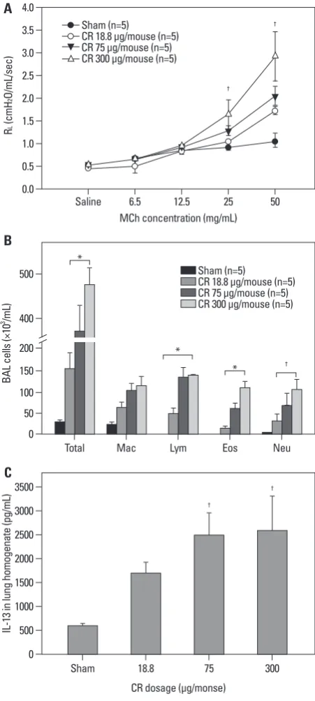
We evaluated dose-dependency for the development of CR extract-induced asthma in BALB/c mice. Mice sensitized with 75 and 300 µg/mL concentrations of CR extract dem-onstrated significant increases in AHR at MCh concentra-tions of 25 and 50 mg/mL, compared to the sham animal (p<0.05), and AHR had a tendency to increase in a dose-de-pendent manner (Fig. 1A).
Analysis of BAL fluid revealed significant differences in the number of total cells, macrophages, lymphocytes, eo-sinophils, and neutrophils between mice sensitized with any concentration of CR extract compared with sham mice. The numbers of these cells in BAL fluid were increased dose-dependently (Fig. 1B). We also observed that the expression of IL-13 in lung homogenate was significantly higher in the mice administered with any concentration of CR extract than sham mice. The levels of IL-13 in lung homogenate were also increased in a dose-dependent manner (Fig. 1C).
The lungs of CR extractadministered mice exhibited re-markable eosinophilic perivascular and peribronchial in-flammation with clear evidence of goblet cell hyperplasia (data not shown).
The effects of endotoxin on the development of CR extract-induced mouse model of asthma
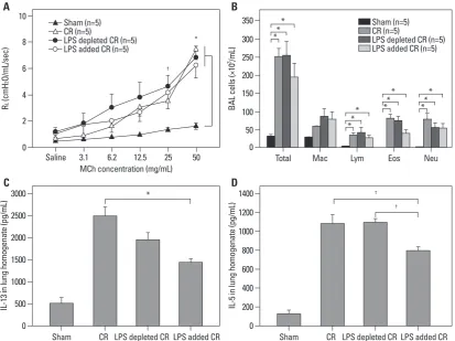
We evaluated the effects of endotoxin on the development of CR extract-induced asthma in BALB/c mice. Endotoxin ad-dition (10000 EU) or endotoxin depletion in CR extract did not affect the development of AHR (Fig. 2A). Analysis of BAL fluid revealed significant differences in the number of eosinophils between mice sensitized with CR, CR without endotoxin, and CR with additional endotoxin. The number of eosinophils in mice administered with CR with additional endotoxin was significantly lower than in those administered with CR alone (p<0.05) (Fig. 2B).
The level of IL-13 and IL-5 in mice administered with CR without endotoxin was not different from mice admin-istered with CR alone, whereas the levels in mice adminis-tered with additional endotoxin were significantly lower than in mice administered with CR alone (Fig. 2C and D). We also observed remarkable perivascular and peribronchi-ysis which compared values over the entire curve between
[image:4.595.56.282.116.619.2]each treatment group. From this F-score, a value for p was generated. In all cases, a value of p<0.05 was considered
Fig. 1. Dose-dependency of CR extract-induced asthma model in BALB/c mice. Developments of AHR (A), airway inflammation (B), and IL-13 level in the lungs (C) showed the dose-dependency of CR extracts. (A) CR 75 and 300 µg/mL administrated mice demonstrated significant increases in AHR at MCh concentrations of 25 and 50 mg/mL compared to the sham animal. (B) Total cell number, lymphocyte, eosinophil and neutrophil counts demon-strated statistical differences between sham mice and CR extractadminis-tered mice of any concentration, and (C) IL-13 levels demonstrated signifi-cant differences at any concentration of CR extract administration compared to sham mice). *p<0.01, †p<0.05. CR, cockroach; AHR, airway hyperrespon-siveness; IL, interleukin; MCh, methacholine.
A
B
C 0.0 0.5 1.0 1.5 2.0 2.5 3.0 3.5 4.0
R
L
(c
m
H
2
O/
m
L/
se
c)
Saline 6.5 12.5 25 50
MCh concentration (mg/mL) Sham (n=5)
CR 18.8 μg/mouse (n=5) CR 75 μg/mouse (n=5) CR 300 μg/mouse (n=5)
†
†
0
0 500 1000 1500 2000 2500 3000 3500 50 100 150 200 400 500
BA
L c
el
ls
(×
10
3 /m
L)
IL
-1
3 i
n
lu
ng
h
om
og
en
at
e
(p
g/
m
L)
Total
Sham 18.8 75 300
Mac Lym Eos Neu
CR dosage (μg/monse) Sham (n=5)
CR 18.8 μg/mouse (n=5) CR 75 μg/mouse (n=5) CR 300 μg/mouse (n=5) *
*
* †
†
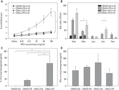
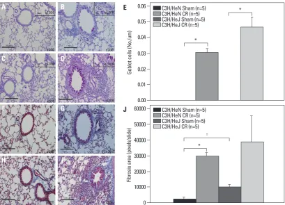
TLR-4 wild type mice (p<0.05). However, the expression of IFN-γ was not affected by TLR-4 mutation and sensiti-zation of CR extract (Fig. 4C and D). We also observed more remarkable perivascular and peribronchial inflamma-tion in CR-sensitized TLR-4 mutant mice than in the wild type, (Fig. 5A-E) but there was no difference in peribron-chial fibrosis between the two groups (Fig. 5F-J).
The effects of protease on the development of CR extract-induced mouse model of asthma
It is known that the protease activity of allergens partici-pates in the development of mucosal allergic sensitization. Thus, we examined whether allergic responses to CR ex-tracts were dependent on protease activity. We removed CR protease activity using heat inactivation, which eliminated protease activity.13 Mice that were administrated with
heat-inactivated CR extractshowed significantly decreased AHR and lower numbers of inflammatory cells in their BAL flu-id, with dramatically lower eosinophilic infiltration, com-pared with non-heat-treated CR mice. These results indicat-ed that the presence of heat-sensitive protease activity in CR al inflammation in all of the CR administered mice,
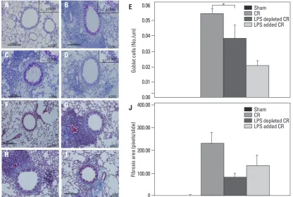
regard-less of endotoxin. However, goblet cell hyperplasia in re-spiratory epithelium was markedly diminished with the addition of endotoxin; nevertheless, perivascular and peri-bronchial inflammatory cell infiltrations were prominent in these mice (Fig. 3).
The effects of TLR-4 on the development of CR extract-induced asthma
[image:5.595.99.513.69.378.2]We evaluated the effects of TLR-4 on the development of CR -induced asthma in C3H/HeN mice and C3H/HeJ mice. The mutant mice developed AHR at all MCh concentra-tions (p<0.05); however, but AHR in wild type mice was not different from that of sham mice (Fig. 4A). There were significantly increased numbers of total cells, macrophages, lymphocytes, eosinophils and neutrophils in the BAL fluid of the C3H/HeN and C3H/HeJ mice sensitized to CR com-pared to sham mice. However, eosinophil count in BAL flu-id was more prominent in the mutant mice than in the wild type mice (p<0.05) (Fig. 4B). IL-13 expression in the TLR-4 mutant mice was significantly higher than that of the
Fig. 2. Effects of endotoxin in CR extract-induced asthma model in BALB/c mice. Development of AHR (A) and airway inflammation (B). Cytokine levels (C and D) in the lungs of CR extract administrated BALB/c mice according to endotoxin level. (A) All CR administrated mice showed significant AHR increase at MCh concentrations of 25 and 50 mg/mL compared to sham mice. *p<0.01, †p<0.05. CR, cock-roach; AHR, airway hyperresponsiveness; MCh, methacholine.
A
C
B
D 0
2 4 6 8 10
R
L
(c
m
H
2
O/
m
L/
se
c)
Saline
MCh concentration (mg/mL) 3.1 6.2 12.5 25 50 Sham (n=5)
CR (n=5)
LPS depleted CR (n=5)
LPS added CR (n=5) *
0 50 100 150 200 250 300 350
BA
L c
el
ls
(×
10
3 /m
L)
Total Mac Lym Eos Neu
*
*
* *
*
*
* *
*
*
* *
Sham (n=5) CR (n=5)
LPS depleted CR (n=5) LPS added CR (n=5)
0 0
500 200
1000 400
600
1500 800
2000 1000
2500 1200
3000 1400
IL
-1
3 i
n
lu
ng
h
om
og
en
at
e
(p
g/
m
L)
IL
-5
in
lu
ng
h
om
og
en
at
e
(p
g/
m
L)
Sham CR LPS depleted CR LPS added CR Sham CR LPS depleted CRLPS added CR *
†
†
mans. Johnson, et al.33 also developed a physiologic asthma
model using house dust mites (HDM). Continuous expo-sure to HDM for up to 7 weeks resulted in sustained AHR and airway inflammation characterized by Th2 effector cell and eosinophil accumulation. These presentations resemble those that have been observed in human asthmatic patients; therefore, we are able to explain a more detail mechanism of the inflammatory process as well as the structural change of the airway following allergen exposure as a result of the development of this model.
CR is not only an important human allergen, but also a potent allergen in mice. Exposure to CR is closely correlat-ed to asthma severity, and it can sensitize a host via the respi-ratory system without any adjuvant.9,10 In the present study,
we administered CR extracts via only intranasal administra-tion, and our results showed that CR extracts induced AHR, eosinophilic and neutrophilic inflammation, and goblet cell hyperplasia of respiratory epithelium in a dose-dependent manner.
Although some asthma animal models using CR have been introduced, there are some critical problems with these models.34,35 These studies did not explain the function of
en-dotoxin in CR extracts: crude CR extractcontains a relative-extracts plays an important role in the development of
aller-gic airway inflammation, particularly in terms of eosinophil infiltration (Fig. 6).
DISCUSSION
Several animal models of allergic asthma, using parasite, OVA, and plant extracts as allergens, have been
devel-oped.28,29 Among these models, the OVA asthma model
us-ing aluminum hydroxide as an adjuvant is the standard ani-mal model and has greatly contributed to the understanding of many important mechanisms of asthma and the basic pro-cesses that occur during allergic airway responses.30
Never-theless, the OVA model is somewhat misrepresentative from that of human asthma: allergic diseases in humans are chronic in nature resulting from intermittent or continuous exposure to allergen, and structural changes such as airway remodeling are characteristic. On the other hand, animal models using OVA are inadequate for evaluating the pro-cesses involved in human airway remodeling.31,32
Therefore, many investigators have attempted to develop a physiologic asthma model more resemblant to that of
hu-Fig. 3. PAS and trichrome staining of CR extract-induced asthma model in BALB/c mice. Representative histology of PAS (A-D) and tri-chrome (F-I) staining in the lung tissues. Sham mice are A and F. CR induced asthma mice are B and G. Endotoxin depleted CR induced asthma mice are C and H. Endotoxin addition CR induced asthma mice are D and I. The results of PAS and trichrome staining were quan-tified in E and J. *p<0.05. CR, cockroach; PAS, Periodic Acid-Schiff.
A
F C
H
B
G
I D
0.00 0.01 0.02 0.03 0.04 0.05 0.06
Go
bl
et
c
el
ls
(N
o.
/u
m
)
Sham CR
LPS depleted CR LPS added CR
0 100.00 200.00 300.00 400.00
Fib
ro
sis
a
re
a
(p
ixe
ls/
sli
de
)
Sham CR
LPS depleted CR LPS added CR *
E
[image:6.595.83.500.70.350.2]ment of CR induced allergic inflammation, and overcomes allergen tolerance in mice.12,13,40 We also showed that
elimi-nation of protease activity in CR extracts can inhibit the de-velopment of AHR and allergic inflammation in mice. Ser-ine protease’s receptor, protease activating receptor-2 (PAR-2), is found on a variety cells, including macrophages and dendritic cells. PAR-2 activation in the airway concurrent with exposure to inhaled OVA induces allergic sensitization via dendritic cell activation, whereas exposure to OVA alone may induce allergen tolerance.12
The lack of a good animal model using endotoxin has so far made it difficult for investigators to explain the relation-ship between endotoxin, allergen and asthmatic responses. In this study, we showed that CR extract induced AHR and air-way inflammation in a dose-dependent manner in BALB/c mice. Moreover, the results obtained with this model showed that the addition of endotoxin did not improve AHR induced by CR allergen extracts, but rather decreased eosinophilic inflammation in BAL fluid and decreased IL-13, IL-5, and goblet cell hyperplasia, compared with those induced by crude CR extracts alone. Also, depletion of endotoxin did ly large amount of endotoxin,10 and the role of endotoxin
contaminated in CR extracts is controversial. Some investi-gators have attempted to explain the function of endotoxin by challenging mice with endotoxin;36,37 however, the
clini-cal relevance of this model is not clear. It has been suggest-ed that exposure to low levels of endotoxin may exacerbate asthmatic responses, whereas exposure to high levels might be protective. This is based on two animal studies with op-posite findings. Wan, et al.38 showed that mice exposed to
40 ng/mL of aerosolized endotoxin prior to OVA challenge resulted in higher specific IgE levels and inhibited tolerance to OVA compared to mice exposed to saline prior to OVA challenge. On the other hand, Tulic, et al.39 found that
expo-sure to 50 μg/mL of endotoxin with OVA challenge showed a protective effect and prevented the IgE response in rats. These studies clearly showed the role of endotoxin in a pure OVA asthma model. In addition to endotoxin and allergens, CR extracts contain several important immunostimulants such as proteases and chitins.14,15 Recent studies have
prov-en that protease activity in CR extracts activates innate im-mune responses during sensitization, leads to the
develop-Fig. 4. CR extractinduced asthma model in TLR-4 wild type and mutant mice. Development of AHR (A), airway inflammation in BAL fluid (B), expression of IL-13 (C), and IFN-γ (D) in the lungs of TLR-4 wild type (C3HeN) and TLR-4 mutant (C3HeJ) mice in the CR induced asthma model. CR administrated C3HeN mice showed significant AHR increase at all MCh concentrations compared to sham mice. *p<0.01, †p<0.05. CR, cockroach; TLR-4, receptor-4; AHR, airway hyperresponsiveness; IL, interleukin; IFN-γ, interferon-gamma; MCh, methacholine.
A
C
B
D 0
1 2 3 4 5 6
R
L
(c
m
H
2
O/
m
L/
se
c)
Saline
MCh concentration (mg/mL) 6.25 12.5 25 50 100 C3HeN-Sal (n=5)
C3HeN-CR (n=5) C3HeJ-Sal (n=5) C3HeJ-CR (n=5)
0 50 100 150 200
BA
L c
el
ls
(×
10
3 /m
L)
Total Mac Lym Eos Neu
C3HeN-Sal (n=5) C3HeN-CR (n=5) C3HeJ-Sal (n=5) C3HeJ-CR (n=5)
0 0
20 40
50 100 150 60
80 200
100 250
IL
-1
3 i
n
lu
ng
h
om
og
en
at
e
(p
g/
m
L)
IF
N
-γ
in
lu
ng
h
om
og
en
at
e
(p
g/
m
L)
C3HeN-Sal C3HeN-CR C3HeJ-Sal C3HeJ-CR C3HeN-Sal C3HeN-CR C3HeJ-Sal C3HeJ-CR *
*
† †
†
[image:7.595.100.513.70.382.2]dotoxin reduced group. We also used TLR-4 mutant (C3H/ HeJ) and TLR-4 wild type (C3H/HeJ) mice to evaluate the function of endotoxin in the CR extract-induced asthma model. TLR-4 functions as the signal transduction receptor for endotoxin in the cells of immune systems including mac-rophages and dendritic cells.42 Our results showed that AHR,
eosinophil count in BAL fluid, IL-13 in lung homogenate, and goblet cell hyperplasia induced in C3H/HeJ mice by CR not demonstrate a difference in AHR, Th2 cytokine (IL-13,
IL-5), and goblet cells in respiratory epithelium. These data agree quite well with previous studies. Natarajan, et al.,41
who administered endotoxin depleted CR extract to mice, reported no significant difference in the infiltration of aller-gic inflammation, and the development of AHR between endotoxin reduced and the other groups. They also demon-strated elevated production of IL-5, IL-13 and IFN-γ in
[image:8.595.87.497.65.360.2]en-Fig. 5. PAS and trichrome staining of the CR extract-induced asthma model in TLR-4 wild type and mutant mice. Representative histology of PAS (A-D) and trichrome (F-I) staining in the lung tissues. No treated wild type mice are A and F. CR treated wild type mice are B and G. No treated TLR-4 mutant mice are C and H. CR treated TLR-4 mutant mice are D and I. The results of PAS and trichrome staining were quantified in E and J. *p<0.01, †p<0.05. CR, cockroach; PAS, Periodic Acid-Schiff; TLR-4, receptor-4.
Fig. 6. Effects of heat inactivation on the CR extract-induced asthma model in BALB/c mice. Development of AHR (A) and airway inflam-mation (B) in BALB/c mice administered with heat inactivated CR or non-heat-treated CR. Heat inactivated CR administrated mice showed significant AHR decrease at MCh concentrations of 25 and 50 mg/mL compared to non-heat-treated CR administrated mice. *p<0.01, †p<0.05. CR, cockroach; AHR, airway hyperresponsiveness; MCh, methacholine.
A
F C
H
B
G
I D
A B
0.00
0 0.01
10000 20000 0.02 0.03
30000 40000 0.04
50000 0.05 0.06
60000
Go
bl
et
c
el
ls
(N
o.
/u
m
)
Fib
ro
sis
a
re
a
(p
ixe
ls/
sli
de
)
*
*
* C3H/HeN Sham (n=5) C3H/HeN CR (n=5) C3H/HeJ Sham (n=5) C3H/HeJ CR (n=5)
C3H/HeN Sham (n=5) C3H/HeN CR (n=5) C3H/HeJ Sham (n=5) C3H/HeJ CR (n=5)
0.0 0.5 1.0 1.5 2.0 2.5 3.0 3.5
R
L
(c
m
H
2
O/
m
L/
se
c)
0
MCh concentration (mg/mL) 3.12 6.25 12.5 25 50 Sham (n=5)
CR (n=5)
Heat inactivated CR (n=5)
0 20 40 60 80
BA
L c
el
ls
(×
10
3 /m
L)
Total
cells Mac Lym Eos Neu
*
*
* Sham (n=5) CR (n=5)
Heat inactivated CR (n=5)
†
E
J
[image:8.595.86.499.408.561.2]tial usefulness for studying health effects of inhaled particles. Inhal Toxicol 2000;12:829-62.
5. de Siqueira AL, Russo M, Steil AA, Facincone S, Mariano M, Jancar S. A new murine model of pulmonary eosinophilic hyper-sensitivity: contribution to experimental asthma. J Allergy Clin Immunol 1997;100:383-8.
6. Clarke AH, Thomas WR, Rolland JM, Dow C, O’Brien RM. Mu-rine allergic respiratory responses to the major house dust mite al-lergen Der p 1. Int Arch Allergy Immunol 1999;120:126-34. 7. Warner RL, Lukacs NW, Shapiro SD, Bhagarvathula N, Nerusu
KC, Varani J, et al. Role of metalloelastase in a model of allergic lung responses induced by cockroach allergen. Am J Pathol 2004;165:1921-30.
8. Smith H. Animal models of asthma. Pulm Pharmacol 1989;2:59-74. 9. Arruda LK, Vailes LD, Ferriani VP, Santos AB, Pomés A, Chap-man MD. Cockroach allergens and asthma. J Allergy Clin Immu-nol 2001;107:419-28.
10. Hong JH, Lee SI, Kim KE, Yong TS, Seo JT, Sohn MH, et al. German cockroach extract activates protease-activated receptor 2 in human airway epithelial cells. J Allergy Clin Immunol 2004; 113:315-9.
11. Kim J, Merry AC, Nemzek JA, Bolgos GL, Siddiqui J, Remick DG. Eotaxin represents the principal eosinophil chemoattractant in a novel murine asthma model induced by house dust containing cockroach allergens. J Immunol 2001;167:2808-15.
12. Ebeling C, Lam T, Gordon JR, Hollenberg MD, Vliagoftis H. Pro-teinase-activated receptor-2 promotes allergic sensitization to an inhaled antigen through a TNF-mediated pathway. J Immunol 2007;179:2910-7.
13. Arizmendi NG, Abel M, Mihara K, Davidson C, Polley D, Nadeem A, et al. Mucosal allergic sensitization to cockroach aller-gens is dependent on proteinase activity and proteinase-activated receptor-2 activation. J Immunol 2011;186:3164-72.
14. Lee CG. Chitin, chitinases and chitinase-like proteins in allergic in-flammation and tissue remodeling. Yonsei Med J 2009;50:22-30. 15. Jeong KY, Hong CS, Lee JS, Park JW. Optimization of allergen
standardization. Yonsei Med J 2011;52:393-400.
16. Hasday JD, Bascom R, Costa JJ, Fitzgerald T, Dubin W. Bacterial endotoxin is an active component of cigarette smoke. Chest 1999; 115:829-35.
17. Reed CE, Milton DK. Endotoxin-stimulated innate immunity: a contributing factor for asthma. J Allergy Clin Immunol 2001;108: 157-66.
18. Braun-Fahrländer C, Riedler J, Herz U, Eder W, Waser M, Grize L, et al. Environmental exposure to endotoxin and its relation to asthma in school-age children. N Engl J Med 2002;347:869-77. 19. Strachan DP. Family size, infection and atopy: the first decade of
the “hygiene hypothesis”. Thorax 2000;55 Suppl 1:S2-10. 20. Matricardi PM, Franzinelli F, Franco A, Caprio G, Murru F, Cioffi
D, et al. Sibship size, birth order, and atopy in 11,371 Italian young men. J Allergy Clin Immunol 1998;101(4 Pt 1):439-44. 21. Michel O, Kips J, Duchateau J, Vertongen F, Robert L, Collet H,
et al. Severity of asthma is related to endotoxin in house dust. Am J Respir Crit Care Med 1996;154(6 Pt 1):1641-6.
22. Eder W, Klimecki W, Yu L, von Mutius E, Riedler J, Braun-Fah-rländer C, et al. Opposite effects of CD 14/-260 on serum IgE lev-els in children raised in different environments. J Allergy Clin Im-munol 2005;116:601-7.
23. Zambelli-Weiner A, Ehrlich E, Stockton ML, Grant AV, Zhang S, Levett PN, et al. Evaluation of the CD14/-260 polymorphism and extractwere similar to that of the wild type mice (C3H/HeN).
These results indicated that endotoxin in CR extracts plays a limited role in inducing allergic inflammation, according to this model. Consistent to our study, Arizmendi, et al.13 also
reported that the protease activity of CR extract is sufficient for CR sensitization and the induction of allergic inflamma-tion even in TLR-4 knock-out mice. These results are also concordant with a human epidemiologic study, which showed that endotoxin levels in samples of dust from a child’s mat-tress were inversely related to the occurrence of hay fever, atopic asthma, and atopic sensitization, and cytokine produc-tion by leukocytes.18
In the present study, we characterized a mouse asthma model in response to CR extract and investigated the roles of endotoxin and protease therein. CR exposure can elicit asthma-like pulmonary inflammation characterized by AHR, eosinophilia, elevation of Th2 cytokine and goblet cell hy-perplasia, which were related to the amount of CR extract exposure. CR extract-induced allergic responses developed not as a result of endotoxin, but rather from protease. Even though high dose endotoxin inhibited eosinophilic inflam-mation, Th2 cytokine levels and goblet cell hyperplasia were induced by CR allergen. The CR extract-induced mouse asthma model presented herein will be a valuable tool and may allow for the discovery of the mechanism of allergen sensitization and its relationship with innate im-mune responses in allergic asthma.
ACKNOWLEDGEMENTS
This study was supported by a grant from the Korea Health-care Technology R&D Project, Ministry for Health, Welfare & Family Affairs, Republic of Korea (A092076).
REFERENCES
1. Sarpong SB, Hamilton RG, Eggleston PA, Adkinson NF Jr. Socio-economic status and race as risk factors for cockroach allergen ex-posure and sensitization in children with asthma. J Allergy Clin Immunol 1996;97:1393-401.
2. Rosenstreich DL, Eggleston P, Kattan M, Baker D, Slavin RG, Gergen P, et al. The role of cockroach allergy and exposure to cockroach allergen in causing morbidity among inner-city children with asthma. N Engl J Med 1997;336:1356-63.
3. Busse WW, Mitchell H. Addressing issues of asthma in inner-city children. J Allergy Clin Immunol 2007;119:43-9.
poten-33. Johnson JR, Wiley RE, Fattouh R, Swirski FK, Gajewska BU, Coyle AJ, et al. Continuous exposure to house dust mite elicits chronic airway inflammation and structural remodeling. Am J Respir Crit Care Med 2004;169:378-85.
34. Zhou D, Chen G, Kim JT, Lee LY, Kang BC. A dose-response re-lationship between exposure to cockroach allergens and induction of sensitization in an experimental asthma in Hartley guinea pigs. J Allergy Clin Immunol 1998;101:653-9.
35. McKinley L, Kim J, Bolgos GL, Siddiqui J, Remick DG. Repro-ducibility of a novel model of murine asthma-like pulmonary in-flammation. Clin Exp Immunol 2004;136:224-31.
36. Eisenbarth SC, Piggott DA, Huleatt JW, Visintin I, Herrick CA, Bottomly K. Lipopolysaccharide-enhanced, toll-like receptor 4-dependent T helper cell type 2 responses to inhaled antigen. J Exp Med 2002;196:1645-51.
37. Piggott DA, Eisenbarth SC, Xu L, Constant SL, Huleatt JW, Her-rick CA, et al. MyD88-dependent induction of allergic Th2 re-sponses to intranasal antigen. J Clin Invest 2005;115:459-67. 38. Wan GH, Li CS, Lin RH. Airborne endotoxin exposure and the
development of airway antigen-specific allergic responses. Clin Exp Allergy 2000;30:426-32.
39. Tulic MK, Holt PG, Sly PD. Modification of acute and late-phase allergic responses to ovalbumin with lipopolysaccharide. Int Arch Allergy Immunol 2002;129:119-28.
40. Kheradmand F, Kiss A, Xu J, Lee SH, Kolattukudy PE, Corry DB. A protease-activated pathway underlying Th cell type 2 acti-vation and allergic lung disease. J Immunol 2002;169:5904-11. 41. Natarajan S, Kim J, Bouchard J, Cruikshank W, Remick DG.
Re-ducing LPS content in cockroach allergens increases pulmonary cytokine production without increasing inflammation: a random-ized laboratory study. BMC Pulm Med 2011;11:12.
42. Medzhitov R. Toll-like receptors and innate immunity. Nat Rev Immunol 2001;1:135-45.
house dust endotoxin exposure in the Barbados Asthma Genetics Study. J Allergy Clin Immunol 2005;115:1203-9.
24. Nathan AT, Peterson EA, Chakir J, Wills-Karp M. Innate immune responses of airway epithelium to house dust mite are mediated through beta-glucan-dependent pathways. J Allergy Clin Immunol 2009;123:612-8.
25. Campbell EM, Kunkel SL, Strieter RM, Lukacs NW. Temporal role of chemokines in a murine model of cockroach allergen-in-duced airway hyperreactivity and eosinophilia. J Immunol 1998; 161:7047-53.
26. Lee KE, Kim JW, Jeong KY, Kim KE, Yong TS, Sohn MH. Regu-lation of German cockroach extract-induced IL-8 expression in human airway epithelial cells. Clin Exp Allergy 2007;37:1364-73. 27. Russo M, Lutton JD. Decreased in vivo and in vitro colony stimu-lating activity responses to bacterial lipopolysaccharide in C3H/ HeJ mice. J Cell Physiol 1977;92:303-7.
28. Pritchard DI, Eady RP, Harper ST, Jackson DM, Orr TS, Richards IM, et al. Laboratory infection of primates with Ascaris suum to provide a model of allergic bronchoconstriction. Clin Exp Immu-nol 1983;54:469-76.
29. Mapp C, Hartiala J, Frick OL, Shields RL, Gold WM. Airway re-sponsiveness to inhaled antigen, histamine, and methacholine in inbred, ragweed-sensitized dogs. Am Rev Respir Dis 1985;132: 292-8.
30. Flach TL, Ng G, Hari A, Desrosiers MD, Zhang P, Ward SM, et al. Alum interaction with dendritic cell membrane lipids is essen-tial for its adjuvanticity. Nat Med 2011;17:479-87.
31. Leigh R, Ellis R, Wattie J, Southam DS, De Hoogh M, Gauldie J, et al. Dysfunction and remodeling of the mouse airway persist af-ter resolution of acute allergen-induced airway inflammation. Am J Respir Cell Mol Biol 2002;27:526-35.