© Associated Asia Research Foundation (AARF)
A Monthly Double-Blind Peer Reviewed Refereed Open Access International e-Journal - Included in the International Serial Directories. Page | 1
TO DETECT AND ANALYSE HEART TUMOR USING PARALLEL
DATA MODELLING
Bhawna
Research Scholar (M.Tech) BCET, Gurdaspur,India
Dr. RC Gangwar (Associate Professor) BCET, Gurdaspur,India
ABSTRACT
Electronic health records (EHRs) contain patient diagnostic records, physician records, and
records of hospital departments. The parallel computing systems are widely used in order to
enhance and analyze complex systems. The Gauss seidel method in order to analyze total
number of iterations which are required in order to determine the abnormal cell growth
within the Heart. The parabolic equations will be used in order to determine the position of
the cells in the Heart and their growth. The progress of the expectation scheme is the
mixtures of the parallel algorithms, open source software on Linux environment and
distributed multiprocessor system. The paper ends with a closing observation on the parallel
performance assessments and mathematical study in decreasing the execution time,
communication cost and computational complexity.
Keywords: Heart Tumours, Classification, Risk Predicion, Fundus Images, USI.
I. INTRODUCTION
The Cardiac or heart comprised of neurons cells or valves; these cells or valves are prone for
usual actions of Cardiac or heart. Usually, the Cardiac or heart makes new cells or valves
only at what time they are required to exchange older or destroyed ones. Mainly cells or
valves re-establish themselves through allocating to formulate additional cells or valves. This International Research Journal of Mathematics, Engineering and IT
ISSN: (2349-0322) Impact Factor- 5.489, Volume 6, Issue 02, February 2019 Website- www.aarf.asia, Email : [email protected] , [email protected]
© Associated Asia Research Foundation (AARF)
A Monthly Double-Blind Peer Reviewed Refereed Open Access International e-Journal - Included in the International Serial Directories. Page | 2 turnover is generally takes place in an ordered and specific way. For any cause, if the growth
happens to be out of control, the cells or valves will continue to segregate, developing into to
an inflammation that is known as a tumor. The cancer treatment has been a most important
objective of researchers of medical for decades; however growth of new treatments acquires
time and money. Medical science may until now discover the basic roots of all cancers and
build up safer techniques for diminish them. Cardiac or heart tumors are benevolent and can
be prior to they comprise a possibility to develop or proliferate. About 40 percent of all major
effectively cured by surgery and, in a few cases, radiation. The number of malignant Cardiac
or heart tumors emerges to be growing excluding for any obvious cause. Cardiac or heart
cancer is a multifaceted ailment, grouped into 120 different kinds. So called Benign (non
malignant) and life-threatening as malignant tumors, as they compress out usual Cardiac or
heart tissue and interrupt task. The glioma family of tumors encompasses 44.4 % of all
cardiac or heart tumors. Glioblastoma kind of Astrocytoma is the majorly general glioma that
encompasses 51.9 %, pursued by other forms of astrocytoma at 21.6 % of all tumors of
Cardiac or heart.
Cardiac or heart is alienated into three different divisions. First division is recognized as
cerebrum. It fills up the largest part of the skull. It involves in problem solving, thinking and
feeling. It is also controls the movement. Second division is recognized as cerebellum that be
seated at the backside of the head. It controls synchronization and stability. Third division is
Cardiac or heart stem that be seated underneath the cerebrum in frontage of the cerebellum. It
joins the Cardiac or heart with the spinal code. The tumor of Cardiac or heart can arise in any
division of the Cardiac or heart. The indications can be dissimilar depending upon the
division of the Cardiac or heart in which tumor arise. Cardiac or heart tumor is developed by
irregular cell expansion inside divisions of the Cardiac or heart. The warning signs can be
dissimilar relying on the division of the Cardiac or heart in which tumor arise. The proposed
work will examine the tumor of Cardiac or heart through taking into consideration the
parametric equations and expanse among the several cells or valves by Gauss Seidel method.
LITERATURE SURVEY
Breward et al. (2004) planned a model that illustrates the vascular tumor development in
which the density of artery or vein is considered noticeably. The model illustrated in this
work is proficient to construct the picture of configuration of tumor that is originated in vivo
in some instances. The planned work of this study can be simply changed to embrace the
© Associated Asia Research Foundation (AARF)
A Monthly Double-Blind Peer Reviewed Refereed Open Access International e-Journal - Included in the International Serial Directories. Page | 3 illustrate the development of a vascular tumor. The researchers assume that the tumor
embrace three phases, specifically tumor cells or valves, blood vessels and extracellular
material. (Wilfred D. Stein et al. (2008)) describing the equation of regression development
on the bases of the model that the PSA level drops off exponentially although there is as well
self-standing exponential redevelopment of the tumor revealed in the considered PSA level.
PSA is the top class model for metastatic tumor, and for analyzing new approaches for the
evaluation of an ailment. The authors explicated utilize of arithmetic to explain tumor
kinetics has been extensively discovered in prostate cancer due to the understanding and
explicitly of the PSA (tumor marker). (Harpold et al. (2007))described an explanation of the
frequent contrasts of hypothesis and actuality that have permitted the moderate enhancement
of a comparatively uncomplicated bio-arithmetical representation. In this study the author
conversed just about gliomas, however there is definitely a significant overlie with latest
arithmetic modeling effort relating to another type of cancers. By the predictable and
associated development in imaging, it is understandable that the development in modeling
will carry on altering our considerate of in vivo tumor actives. (Mahlet Aseefa et al. (2009)
)described arithmetical representations for the development of gliomas surrounded by central
nervous system (CNS). The focus of the model is on two main factors; the net propagation
speed of glioma cells or valves, and the development of glioma cells or valves to tissues
surrounded by the CNS. In accordance of the paper, this model evaluates the site of the tumor
surrounded by the Central Nervous System for the reason that tumor cells or valves are
recognized to spread at a more rapidly rate in white segment in comparison of grey segment.
As an outcome, more correct estimation of patient’s long life and the duration of tumor’s
predictability reappearance can be prepared.
I. RSULT & DISCUSSION
In the heart or heart valves, the irregular growths are known to be Cardiac tumors. In
general, cardiac tumors are rare. Cardiac tumors are classified into various forms. The cardiac
tumors can be benign (noncancerous) or malignant (cancerous). Cardiac tumors that start
© Associated Asia Research Foundation (AARF)
A Monthly Double-Blind Peer Reviewed Refereed Open Access International e-Journal - Included in the International Serial Directories. Page | 4
BENIGN MALIGNANT
Finding Features from the Mri images is the Basic Parameter of Finding the Heart Related
issue in our Project work. The Existing approach using the SVM nearest Neighbor Algorithm
to find the Features of MRI image and detecting the disease.
In Proposed Approach we use meta learning categorization algorithm which is more effective
then SVM and finding more accurate Results efficiently. Tumor is an irregular type of growth
of cells in the body, it can be either benign (non cancerous) or malignant (cancerous). Benign
tumors are deliberating growing and often not dangerous depending on wherever they sited in
the body, whereas malignant tumors are rapid growing and probable to extend to other body
parts rapidly. Assume we have several component classication algorithms. Consider the ith
category. Let eij be the classication error of the training set on the jth algorithm. Classication
errors willrst undergo a logistic alteration to yield the dependent variable or the response
variable, for the metamodel. Accurately, the transformation is given in Equation 1.
Yij = 1𝑛 𝑒ij
1 − 𝑒𝑖𝑗
Whereyij is the response variable. This transformation ensures that the response variable is in
the range of 0 and 1. The response variable, yij is related to the feature characteristics by the
© Associated Asia Research Foundation (AARF)
A Monthly Double-Blind Peer Reviewed Refereed Open Access International e-Journal - Included in the International Serial Directories. Page | 5 Where k j is the parameter estimate for the kth feature, by using the algorithm j. F k i is the
kth feature characteristic in the ith category. ∈ij is assumed to follow a Gaussian distribution
N (0, var(∈ij )). The number of document feature characteristics used in the meta-model is p.
Existing Feature Sigmoid Proposed Feature Sigmoid
80.0024 91.4235
79.8734 90.7299
80.6571 91.0893
80.3559 90.948
80.3138 91.5037
Table 1: Showing Feature Extraction of existing and proposed system
Graph1: Showing the Existing and Proposed Difference of Feature Extraction.
In the heart or heart valves, the irregular growths are known to be Cardiac tumors. In
general, cardiac tumors are rare. Cardiac tumors are classified into various forms. The cardiac
tumors can be benign (noncancerous) or malignant (cancerous). Cardiac tumors that start
growing in the heart and stay in the heart are known to be primary tumors.
74 76 78 80 82 84 86 88 90 92
1 2 3 4 5
Existing Feature Sigmoid
© Associated Asia Research Foundation (AARF)
A Monthly Double-Blind Peer Reviewed Refereed Open Access International e-Journal - Included in the International Serial Directories. Page | 6
BENIGN MALIGNANT
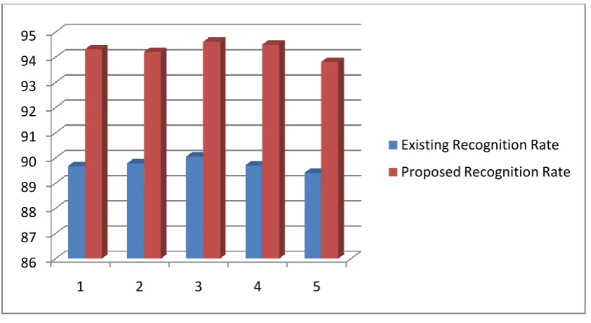
Recognition Rate of MRI images are also a Key Parameter by Which we can find the disease.
Finding Features from the Mri images is the Basic Parameter of Finding the Heart Related
issue in our Project work. The Existing approach using the SVM nearest Neighbor Algorithm
to find the Features of MRI image and detecting the disease. In Proposed Approach we use
meta learning categorization algorithm which is more effective then SVM and finding more
accurate Results efficiently. Tumor is an irregular type of growth of cells in the body, it can
be either benign (non cancerous) or malignant (cancerous). Benign tumors are deliberating
growing and often not dangerous depending on wherever they sited in the body, whereas
malignant tumors are rapid growing and probable to extend to other body parts rapidly.
Existing Recognition Rate Proposed Recognition Rate
89.6811 94.3148
89.7906 94.2127
90.0589 94.615
89.7066 94.506
89.4125 93.8135
© Associated Asia Research Foundation (AARF)
A Monthly Double-Blind Peer Reviewed Refereed Open Access International e-Journal - Included in the International Serial Directories. Page | 7 Table 2: Showing Recognition Rate of existing and proposed system
Graph2: Showing the Existing and Proposed Difference of Recognition Rate.
II. CONCLUSION AND FUTURE SCOPE
The simulations will show that the proposed system will give better results in terms of time
and number of infected cells or valves detected. To develop an efficient approach to detect
heart tumor using parallel data modelling.Through the proposed method it will be easy to
detect the tumour at earliest stage and hence warn the persons about the disease so that cure
can be taken in time. From the created environment it is clear that the proposed system is
producing better results as compared to the existing system.Future work will include
incorporating expert knowledge into our framework and expanding our approach to
additional health care applications.
REFERENCES
[1]Kim E, Shen T, Huang X, “A parallel cellular automata with label priors for interactive Cardiac or heart tumor segmentation”,In: 2010 IEEE 23rd International Symposium on
Computer-Based Medical Systems (CBMS) [Internet]. IEEE; 2010,p. 232–7.
http://ieeexplore.ieee.org/articleDetails.jsp?arnumber=6042647.
[2]Dessai VS, Arakeri MP, Ram Mohana Reddy G, “A parallel segmentation of Cardiac or heart tumor from magnetic resonance images”, In: 2012 Third International Conference on Computing, Communication and Networking Technologies (ICCCNT’12) [Internet].
86 87 88 89 90 91 92 93 94 95
1 2 3 4 5
Existing Recognition Rate
[image:7.595.108.529.64.294.2]© Associated Asia Research Foundation (AARF)
A Monthly Double-Blind Peer Reviewed Refereed Open Access International e-Journal - Included in the International Serial Directories. Page | 8
IEEE; 2012 [cited 2016 Apr 26]. p. 1–6. Available from:
http://ieeexplore.ieee.org/articleDetails.jsp?arnumber=6395880
[3] Shenbagarajan A, Ramalingam V, Balasubramanian C, Palanivel S, “Tumor Diagnosis in MRI Cardiac or heart Image using ACM Segmentation and ANN-LM
Classification Techniques”, Indian J Sci Technol [Internet]. 2016 Feb 1
[4] Teodoro G, Pan T, Kurc TM, Kong J, Cooper LAD, Podhorszki N, et al, “High-throughput Analysis of Large Microscopy Image Datasets on CPU-GPU Cluster Platforms”,
IPDPS [Internet]. 2013 May [cited 2016 Apr 26];2013:103–14. Available
from:http://www.pubmedcentral.nih.gov/articlerender.fcgi?artid=4240318&tool=pmcentrez&
rendertype=abstract
[5]Hooda H, Verma OP, Singhal T, “Cardiac or heart tumor segmentation: A performance analysis using K-Means, Fuzzy C-Means and Region growing algorithm” In: 2014 IEEE
International Conference on Advanced Communications, Control and Computing
Technologies [Internet]. IEEE; 2014 [cited 2016 Apr 27]. p. 1621–6.
http://ieeexplore.ieee.org/articleDetails.jsp?arnumber=7019383.
[6] Goswami S, Bhaiya LKP, “Cardiac or heart Tumour Detection Using Unsupervised Learning Based Neural Network”, In: 2013 International Conference on Communication
Systems and Network Technologies [Internet]. IEEE; 2013
http://ieeexplore.ieee.org/articleDetails.jsp?arnumber=6524466
[7] Cheriyan MM, Michael PA, “Independent component analysis in automated segmentation of Cardiac or heart tumors”, In: 2014 IEEE International Conference on
Advanced Communications, Control and Computing Technologies [Internet]. IEEE; p.1443–
50,2014. http://ieeexplore.ieee.org/articleDetails.jsp?arnumber=7019341
[8] Xuan X, Liao Q, “Statistical Structure Analysis in MRI Cardiac or heart Tumor Segmentation”,In: Fourth International Conference on Image and Graphics (ICIG 2007)
[Internet]. IEEE; 2007 [cited 2016 Apr 27]. p. 421–6. Available from:
http://ieeexplore.ieee.org/articleDetails.jsp?arnumber=4297123
[9] Selvakumar J, Lakshmi A, Arivoli T, “Cardiac or heart tumor segmentation and its area calculation in Cardiac or heart MR images using K-mean clustering and Fuzzy C-mean
algorithm”,IEEE; [cited 2016 Apr 27];186–90. Available from:
© Associated Asia Research Foundation (AARF)
A Monthly Double-Blind Peer Reviewed Refereed Open Access International e-Journal - Included in the International Serial Directories. Page | 9 [10] Logeswari T, Karnan M, “An Enhanced Implementation of Cardiac or heart Tumor Detection Using Segmentation Based on Soft Computing”, IEEE; 2010 p.243–7.
http://ieeexplore.ieee.org/articleDetails.jsp?arnumber=5432723.
[11] Helen Byrne and Luigi Prezios “Modelling solid tumour growth using the theory of mixtures”, Mathematical Medicine and Biology (2003) 20, 341–366
[12] https://en.wikipedia.org/wiki/Parallel_computing
[13] https://www.techopedia.com/definition/12185/parallel-virtual-machine-pvm
[14]Pheng h. s., Norma alias & Norfarizan Mohd Said “High Performance Simulation for Cardiac or heart Tumours Growth Using Parabolic Equation on Heterogeneous Parallel
Computer System”, Journal Technology Maklumat & Multimedia 4(2007): 39-52
[15] Norma Alias,Mohd Ikhwan Safa bin Maseeri,Md.Rajibul Islam and Siti Nurhidayah Khalid “The visualization of three dimensional Cardiac or heart tumour’s growth on distributed parallel computer systems”Journal of Applied Sciences
9(3):505-512,2009.
[16] Anne Talkington and Rick Durrett, “Estimating tumor growth rates in vivo”
[17] Ken C. L. Wong, Ronald M. Summers, Electron Kebebew, and Jianhua Yao “Tumor Growth Prediction with Hyperelastic Biomechanical Model, Physiological Data Fusion, and Nonlinear Optimization”, Med Image Comput Comput Assist Interv. 2014 ;