Thesis by
Douglas Mcintosh Fambrough, Jr.
In Partial Fulfillment of the Requirements For the Degree of
Doctor of Philosophy
California Institute of Technology Pasadena, California
1968
Acknowledgments
To the many people who have been counsellors and generous companions in our quest for knowledge and in the living of our lives,
And to the many others who appear in the jumble of exciting and happy incldents which crowd my memory,
And to the often unrecognized who keep quiet order in our microworld I am totally indebted.
I especially wish to express my gratitude to Professor James Bonner. His breadth of knowledge, contageous enthusiasm and warmth are con-stant example and inspiration to all the members of his research group. I also wish to acknowledge the advice and encouragement of my dear friends Dr. Ru-Chih Huang, Dr. Keiji Marushige, and Dr. Leroy Hood and all the members of Professor Bonner's research group.
For instruction and technical advice I am particularly indebted to Dr. Kenneth Murray, Dro William Gray and Dr. Leroy Hood.
I wish to thank Frank Fujimura, who collaborated in the development of quantitative disc electrophoresis,. Dr. W. J. Dreyer, for the generous loan qf his equipment, Joyce Bullock and John Raes, who performed the amino acid analyses, Ludia Brown, who helped in the preparation of chromatin, and Boo, for skillful technical assistance in many experi-ments.
The histones of the pea plant, Pisum sativum 1..:_, and of calf thymus have been fractionated and further characterized in order to determine the extent of heterogeneity and the main chemical features of these basic nuclear proteinso Histones were fractionated by chromatography on Amberlite CG-50 and by preparative disc electrophoresis. The resulting highly purified histone fractions were futher characterized by analytical disc electrophoresis, amino acid analysis, N-terminal and C-terminal analyses, and the preparati~n of tryptic peptide maps. Calf thymus his tones Ia, Ib, IIbl, III and IV (f 1, fl~ f2a2, f3, and f2al in the nomenclature of Johns, Phillips and Butler) and pea bud histones Ib, IIb, III and IV were obtained ~s electrophoretically pure components and each appears to be a single molecular species on the basis of N-terminal and C-terminal analysis and the number of tryptic peptides. The total number of major histones in calf thymus appears to be six, in pea bud, eight. The apparent heterogeneity of calf thymus histones demonstrated by disc electrophoresis is largely· due to the formation of histone III complexes by disulfide bridges between histone III monomerso While calf thymus histone
Irr
contains two cysteines per molecule pea bud histone III contains but one and thus can form only dimers.acids, and in some cases even peptide maps of corresponding pea and calf histones. Peptide maps of the arginine-rich histone III contain 29 soluble peptides of which 26 are common to calf and pea; maps of histone IV contain 32 peptides of which 27 are common to calf and pea.
By chromatography and electrophoresis the histones of various pea tissues are qualitatively identical to those of pea bud. There are,
however, quantitative differences and these have been accurately measured by a method of quantitative analytical disc electrophoresis. Young
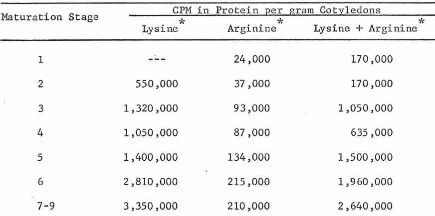
pea cotyledons contain only about a third as much lysine-rich histone as do mature cotyledons. Exploratory experiments on the synthesis of histone in pea cotyledons a~ a function of development and in relation to other macromolecular parameters are described in an appendix.
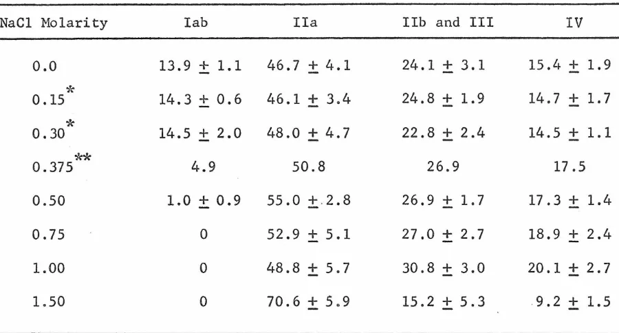
The dissociation of histones from pea bud nucleohistone by NaCl was studied, employing quantitative disc electrophoresis. Histone I
(lysine-rich) is selectively dissociated by 0.5 M NaCl and the remaining histones are non-selectively dissociated primarily over the range
0.5 - 1.5 M NaCl. These data are compared with data for the dissociation of calf thymus histones from nucleohistone by NaCl and the general
CHAPTER
I
Table of Contents
Acknowledgments
Abstract
TITLE
GENERAL INTRODUCTION
FRACTIONATION AND CHARACTERIZATION OF PEA BUD
AND CALF THYMUS HISTONES
Methods
Results
Column chromatography of whole histone
Amino acid composition of chromatographic
fractions
Disc electrophoresis
Fractionation by the method of Johns
Lysine-rich histone components
amino acid compositions
terminal amino acids
peptide maps
Slightly lysine-rich histone components
amino acid compositions
terminal amino acids
peptide maps
Arginine-rich histone components
amino acid compositions
terminal amino acids
.CHAPTER
I I
III
TITLE
properties of histone III related to the presence of cysteine
peptide maps Discussion
References
QUALITATIVE AND QUANTITATIVE DISTRIBUTION OF HISTONE COMPONENTS IN PEA TISSUES
Methods
Quantitative disc electrophoresis Results
Column chromatography Electrophoresis
Discussion References
DISSOCIATION OF PEA BUD NUCLEOHISTONE IN SODIUM CHLORIDE SOLUTIONS
PAGE 89 94 100 112 117 119 124 131 131 141 151 153 156
Methods 157
Results 161
Discussion 174
Comparison of pea bud and calf thymus data 174
Conclusions 178
CHAPTER
APPENDIX
TITLE
SYNTHESIS OF HISTONES IN MATURING PEA COTYLEDONS Methods
Results References
PAGE
General Introduction
Histones are basic proteins which are natively complexed with DNA in the chromosomes of higher organisms. This definition is intentionally vague, for it is not known whether histones are natively complexed with the DNA of cytoplasmic organelles such as mitochondria or only with nuclear DNA, and although it is clear that histones are not found in bacteria (Zubay and Watson, .1959; Butler, 1964; R.aaf, 1966), it is not known whether the protozoa and all the metazoa contain histones or only those organisms which are quite phylogenetically advanced. The defi-nition does, however, embody a very important criterion: histones are native components of the genetic apparatus. Direct evidence for the native association of histones and DNA comes from cytological investi-gations (Alfert and Geschwind, 1953; Alfert, 1957; Horn and Ward, 1957; Bloch and Hew, 1960; Swift, 1964; Holtzman, 1965).
In practice.it is often difficult to establish beyond reasonable .
doubt that a protein isolated as protein-DNA complex is actually assoc-iated with DNA in the living cell. This difficulty has led to the use of such terms as "major histones0 and "minor histones" to distinguish between
Although the histones were discovered around 1880, little was known about their characteristics before Stedman and Stedman (1950, 1951) proposed on the basis of very tenuous evidence that histones might
function as the regulators of genes. T~eir theory of the chemical nature of genes was incorrect, but this proposal together with exciting new insights into the nature of genes and the mechanisms of gene duplication and gene usage (DNA synthesis, RNA synthesis and protein synthesis) pro-vided impetus to the study of the histones.
The biological functions of the histones are still not well under-stood. A large body of data has been amassed to support the hypothesis that histones are directly involved in repression of genetic information (Huang and Bonner, 1962; Bonner and Huang, 1963; Bonner et al., 1963; Allfrey et al., 1963; Barr and Butler, 1963; Hindley, 1963; Hnilica and
Billen, 1964; Johns and Butler, 1964; Huang et al., 1964; Littau~ al., 1965; Marushige and Bonner, 1966; Holoubek, 1966). It is also apparent that at least some components of histone confer special structural pro-perties on native nucleohistone (Zubay and Doty, 1959; Wilkins et al., 1959; Peacocke and Preston, 1962; Giannoni and Peacocke, 1963; Ohba, 1966; Tuan, 1967; also :MlIF Wilkins, S. Pardon and B. Richards, unpublished
It remains quite possible that some histone components may be gene regu-lators while others are purely structural elements of chromatin.
The most central problem of histone chemistry concerns histone hetero-geneity. For many years after their discovery (Kassel, 1884) the histones were treated as a single substance (see Greenstein, 1944). Stedman and Stedman reported a crude fractionation of histones in 1950. Four years later the first clear-cut fractionation of histones into lysine-rich and
arginine-rich was accomplished (Daly and Mirsky, 1954). Subsequently a wide variety of techniques were developed for the analytical and pre-parative fractionation of histones. These included methods of frac-tional precipitation, velocity sedimentation, moving boundary electro-phoresis, zone electroelectro-phoresis, selective extraction from DNA, ion exchange chromatography, and exclusion chromatography (see Phillips, 1961, for a complete review). The great spectrum of amino acid com-positions, terminal amino acids and molecular weights reported for the fractions produced by these methods forms a body of data interpreted by many as being indicative of tremendous molecular heterogeneity
(Murray~ 1964). The histones appeared to constitute a nearly continuous spectrum of molecular species, having lysine/arginine ratios ranging from infinity for the al fraction of Cruft~ al.(1957) to zero for most of the protamines (Felix, 1960).
cross-contamination of fractions (Power and Butler, 1965). Comparative studies of the histones of various organs of a given species and com-parative studies of the histones from different species (Crampton et al., 1957; Hnilica ~
±!.L..,
1962; Neidle and Waelsch, 1964; Laurence.£.!:. al., 1963, 1966; Palau and Butler, 1966; Hnilica, 1966; Fambrough and Bonner, 1966) demonstrate a lack of tissue or even species specificity ofhistones, findings more compatible with limited than with extensive histone heterogeneity.
These comparative studies deal more directly with a second major problem in histone chemistry the problem of determining which chemical and physical characteristics of histones are essential to histone
function. At the molecular level the whole multiplicity of
modern-day organisms is believed to be the result of countless small and seemingly random alterations in the information content of genes (via processes
such as crossing-over, base changes, and the addition and deletion of bases) coupled with natural selection for the resultant phenotypes best suited to various ecological niches. Those alterations which adversely affect information vital to the survival of the organism express themselves generally as "lethal mutations" and are weeded out of the gene pool by death of the organism. By this mechanism, charac-teristics essential to survival are preserved relatively unchanged through great time-spans of evolution (see Handler, 1964). Through chemical
demonstrate a high degree of similarity in chemical characteristics between the histones of calf thymus and pea bud. These common char-acteristics probably include the most essential properties of histones. Such chemical studies also provide a description of histones from which a more refined definition of histones can be drawn.
The research reported in this thesis began as an attempt to char-acterize the histones of the pea plant Pisum sativum L. var Alaska. The availibility of methods for the preparation of highly purified pea plant deoxyribonucleopr~tein (Huang and Bonner, 1962), the very remote phylogenetic relationship of the pea plant to all other
organ-isms whose histones have been studied {all of the latter are vertebrates), and the use of the pea plant in studies of the functions of histones
{reviewed in Bonner~ al.~ 1967) all have made the characterization of pea histones a feasible and timely enterprise. During the course
of this research a number of methods were applied which had not previously been employed in histone research. In order to make meaningful compar-ison of the pea histones with those of vertebrates, it was necessary to simultaneously study the histones of some vertebrate. Calf thymus histone was the material of choice, since the histones of calf thymus have been more thoroughly studied than histones from any other source.
References
Alfert, M. (1957) in The Chemical Basis of Heredity (W.D. McElroy and B. Glass, eds.) Johns Hopkins Press, Baltimore, p. 186.
Alfert, M. and Geschwind, I.I. (1953), Proc. Nat. Acad. Sci. (Wash.)
]2., 991.
Allfrey, V.G., Littau, V.C., and Mirsky, A.E. (1963), Proc. Nat. Acad. Sci. (Wash.) 50, 1026. ·
Barr, G.C. and Butler, J.A.V. (1963), Nature 199, 1170.
Bloch, D.P. and Hew, H.Y.C. (1960), J.Biophys. Biochem. Cytol . .§., 69.
Bonner, J. , Dahmus, M. , Fambrough, D. , Huang, R. C. , Marushige, K. , and Tuan, D. (1967), Science,in press.
Bonner, J. and Huang, R.C. (1963), J. Mol .• Biol..§., 169.
Bonner, J., Huang, R.C., and Gilden, R.V. (1963). Proc. Nat. Acad. Sci. (Wash.) 50, 893.
Busch, H., Hnilica, L.S., Chien, S., Davis, J.R., and Taylor, C.W. (1962), Cancer Res. 22, 637.
Butler, J.A.V. (1964), in The Nucleohistones (J. Bonner and P.O.P. Ts'o eds.) Holden-Day, San Francisco, p 36.
Crampton, C.F. , Stein, W.H., and Moore,.S. (1957), J. Biol. Chem. 225, 363.
Cruft, H.J., Hindley, J., Mauritzen, C.M. and Stedman, E. (1957), Nature 180' 1107.
Daly, M.M. and Mirsky, A.E. (1954-1955), J. Gen. Physiol. 38, 405.
Fambrough, D., and Bonner, J. (1966), Biochemistry .2_, 2563.
Felix, Ko (1960), Advan. Protein Chem.
12.,
1.Frenster, J.H. (1965), Nature 206, 680.
Giannoni, G. and Peacocke, A.R. (1963), Biochim. Biophys. Acta 68, 157.
Greenstein, J.Po (1944), Advan. Protein Chem. 1, 209.
Grogan, D.E., Desjardins, R., and Busch, H. (1966), Cancer Res. 26, 775.
Handler, P. (1964), Fed. Proc. 23, 1229.
Hindley, J. (1963), Biochem. Biophys. Res. Connn.
,11,
175.Hnilica, L.So (1966), Biochim. Biophys. Acta 117, 163.
Hnilica, L.S. and Bess, L.G. (1965), Anal. Biochem •
.!l,
421.Hnilica, L.S. and Billen, D. (1964)) Biochim. Biophys. Acta
21.,
271.Holoubek, V. (1966), J. Cell. Biol •
.ll,
49A.Holtzman, E. (1965), J. Histochem. Cytochem.
J1.,
318.Horn, E.C. and Ward, C.L. (1957), Proc. Nat. Acad. Sci. (Wash) 43, 776. Huang, R.C. and Bonner, J. (1962), Proc. Nat. Acad. Sci. (Wash.) 48, 1216.
Huang, R.C., and Bonner, J. and Murray, K (1964),
J.
Mol. Biol • .§., 54.Johns, E.W. (1964), Biochem. J. 93, 161.
Johns, E.W. and Butler, J.A.V. (1964), Nature 204, 853.
Kassel, A. (1884), Z. Physiol. Chem • .§., 511.
Laurence,
D.J.R.,
Phillips, D.M.P. and Butler, J.A.V. (1966), Arch. Biophys. Biochem. 113, 338.Laurence, D.J.R., Simson, P., and Butler, J.A.V. (1963) Biochem. J •
.§L,
200. Littau, V.C., Burdick, C.Jo, Allfrey, V.G., and Mirsky, A.E. (1965).Proc. Nat. Acad. Sci. (Wash.) 54, 1204.
Maio, J.J. and Schildkraut, C.L. (1967), J. Mol. Biol. 24, 29.
Marushige, K. and Bonner, J. (1966), J. Mol. Biol. 12_, 160.
Murray, K. (1964), in The Nucleohistones (J. Bonner, and P.O.P. Ts'o, eds.) Holden-Day, San Francisco. p. 150
Neidle, A., and Waelsch, H. (1964), Science 145, 1059.
Palau, J., and Butler, J.A.V. (1966), Biochem. J. 100, 779.
Peacocke, A.R. and Preston, B.N. (1961), Nature 192, 228.
Phillips, D.M.P. (1961), Progr. Biophys. Biophys. Chem.~' 211.
Power, D.F. and Butler, J.A.V. (1965) Biochem. J. 2l_, 32p.
Raaf, J. (1966), D. Phil., Oxford University, Oxford.
Stedman, E. and Stedman, E. (1950) Nature 166, 780.
Stedman, E. and Stedman, E. (1951), Phil. Trans. Roy. Soc (London)
Ser. Ba, 235, 565.
Swift, H. (1964), in The Nucleohistones (J. Bonner and P.O.P. Ts'o,
eds.) Holden Day, San Francisco. p 169.
Tuan, D. (1967), Ph.D. Thesis, California Institute of Technology.
Wilkins, M.H.F., Zubay, G., and Wilson, H.R. (1959), J. Mol. Biol.
l,
179.Zubay, G. and Doty, P. (1959), J. Mol. Biol.
l,
1.CHAPTER I
Introduction
Methods
Preparation of chromatin
For the preparation of histones minimally contaminated by
non-~hromosomal protein it is necessary to use purified chromatin as the
starting material for histone extraction. For the preparation of pea
bud chromatin (Bonner,£! al., 1967) approximately 5 kg of pea seeds
were soaked overnight in water, planted in vermiculite, and germinated
0
in the dark for 4 days at 25 C. The apical buds (approximately one cm
of stem plus bud) were then harvested to yield about 600 g fresh weight
of buds. The buds were homogenized with approximately one liter of
grinding medium (0.25 M sucrose, 0.05 M tris buffer pH 8.0, 0.001 M
MgC1
2) for 1.5 minutes at 100 volts in a Waring blender. This and all
b f d 0-5
oc.
su sequent steps were per orme at The homogenate was filtered
through four layers of cheesecloth and then through two layers of
Miracloth (Chicopee Mfg. Co., Miltown, N.J.). The filtrate was next
centrifuged at·4(X)O x g for 30 minutes. The soft pellets were scraped
from the underlying layers of starch, suspended in 300 ml of grinding
medium, and centrifuged at 10,000 x g for 20 minutes. The pellets were
again separated from the starch~ suspended in 300 ml. of 0.05 M (or
O.OlM) tris buffer (pH 8.0), and centrifuged at 10,000 x g for 20
chromatin were suspended in a total of 30 ml of 0.01 M tris buffer
(pH 8.0), homogenized with a Potter-Elvehjem homogenizer (about 20 strokes)
and layered in 5 ml portions on 25 ml aliquots of 1.7 M sucrose in
cellulose nitrate tubes. The upper two-thirds of the contents of each
tube were stirred to form a rough gradient. The tubes were then
centri-fuged at 22,000 rpm for 3 hours in the SW-25 Spinco rotor. The
resul-ting gelatinous pellets (purified chromqtin) were suspended in 0.01 M
tris buffer and dialyzed against 100 volumes of the same buffer
over-night. Recovery of DNA from the tissue homogenate was 70 to 80%.
For the preparation of calf thymus chromatin the method of
Marushige and Bonner (1966) was used without modification~ Except for
the use of saline EDTA (0.075 M NaCl, 0.024 M sodium
ethylene-diamine-tetraacetate, pH 8.0) as grinding medium, this method is very similar
to that described above.
Preparation ~f h~stone
For extraction of histones the dialyzed chromatin was diluted to
a concentration of less than 400 pg DNA/ml with cold 0.01 M tris buffero
This suspension was stirred on ice and one-fourth volume of cold 1 N
sulfuric acid slowly added. After 30 minutes of stirring, the suspension
w~s centrifuged at 17,000 x
z
for 20 minutes. The sediment was brokenup and extracted with 0.4 N sulfuric acid (half the final volume of the
first extract) . To the combined supernatants four volumes of cold absolute
ethanol were added and the histone ·sulfate precipitated at -20°c for
three times with ethanol, and dried in a vacuum desiccator.
It is necessary to use very pure nucleoprotein to obtain suitable
pea histone sulfate preparations. When crude nucleoprotein or whole
nuclei are extracted with acid, histone may constitute less than 10%
of the acid-soluble protein. Contamination, apparently largely by
ribosomal proteins, is extensive. Crude histone preparations made from
such crude chromatin are wholly soluble only in concentrated urea, and
as little as 20% may be again soluble in acid. Densitometric tracings
of the electrophoretic fractionation of such a crude nuclear extract,
of pure histone, and of a mixture of the two are presented in Figure 1.
Extraction of histones with HCl is less satisfactory for preparative
purposes. Histone chlorides are not totally precipitable from acidic
ethanol solution~ are more hygroscopic, and are more difficult to
redissolve than are histone sulfates.
Preparation of histones by the method of Johns
Alternative to the extraction of whole histone from chromatin, a
crude fractionation of histones can be effected by the selective
extrac-tion of histones based upon dissociaextrac-tion of histone from DNA by acids
and acidified ethanol and upon the different solubilities of the several
histone salts in ethanol and acetone (Johns, 1964). This method of
preparation was used for the large scale preparation of histone fractions
from calf thymus nucleoprotein and with partial success for the
Figure 1. Densitometric tracings of the electrophoretic patterns of the acid soluble proteins from a crude preparation of pea cotyledon
nuclei and from pea nucleohistone and from a mixture of the two. The crude nuclear preparation was a 500 x g pellet from a strained homogenate
-0
+
-0
-0
rn
:c
fTl
fTl
l>
-l>
l>
CJ)
:r:
oc
~zz
c
CJ)
zo
( )-1
nir
r
0
en
·rn
rn
z
)>l>
+
rn
en
::0
::0
-0
-0
:::0
;o
0
0
-I
.
-1
(Tl (Tl
-
Purified chromatin at a concentration of less than 5 mg DNA/ml in
5% perchloric acid (PCA) was homogenized at full speed in a Waring
Blender for 2 minutes (4°C). The homogenate was centrifuged for 20
minutes at 17,000 x g and the pellets extracted twice more in ilie same
way, but each time with half the initial volume of 5% PCA. The combined
supernatants were clarified by filtration through a sintered glass
funnel or through Miracloth, and trichloroacetic acid (TCA) was added
to a final concentration of l.lM. The precipitate of lysine rich
0
histone (fraction fl) was aliowed to form overnight at 4 C and then
was collected by centrifugation, washed with acidified acetone (200 ml
acetone: 0.1 ml cone. HCl) and three times with acetone, and then
dried in vacuo.
The pellet from the 5% PCA extraction of the lysine-rich histone
was suspended in ethanol (less than 2.5 mg DNA/ml) and allowed to stand
overnight at 4°C. The suspension was then centrifuged and the resulting
pellet extracted twice wit~ 80% ethanol (less than 2.5 mg DNA/ml). The
combined ethanolic supernatants were clarified by centrifugation and the
histone quantitatively precipitated by the addition of five volumes
of acetone and .005 volumes concentrated HCl. After standing in the
cold overnight this precipitate was collected by centrifugation and
washed once with acetone. The histone was then dissolved in distilled
water, and ethanol and cone. HCl were added to make a final solution
80% ethanol, 0.25N HCl at less than lmg/ml histone. This solution was
dialyzed twice against an equal volume of absolute ethanol, and the
washed twice with acetone, and dried in vacuo. The soluble histone was precipitated from the fraction f3 supernatant by the addition of three volumes of acetone, collected by centrifugation, washed twice with acetone and dried in vacuo (fraction f2a).
The insoluble material which remained after extraction into ethanol of histone fractions f3 and f2a was next extracted with 0.25 N HCl to remove the remaining histone (fraction f2b) by homogenization of the material (5 mg DNA/ml solution) in a blender. The homogenate was cen-trifuged and the pellet extracted with 0.25 N HCl twice more. The com
-bined supernatants were clarified by filtration, and the histone fraction f2b was precipitated overnight by the addition of five volumes of acetone, washed three times with acetone, and then dried in vacuo.
Column Chromatography
For the separation of the major classes of histones, they were applied to and eluted from Amberlite CG-50, using a gradient of
Guanidinium chloride buffered at pH 6.8 (Luck~ al., 1958; Satake ~al., 1960; Rasmussen et al, 1962). Chromatographic procedures are described below.
Preparation of the Resin and of Guanidinium Chloride
followed in each case by filtration (Buchner funnel on suction line,
using two sheets filter paper): 2 N HCl; distilled water; 2 N NaOH
(the filtrate is very yellow and turbid if new Amberlite is used);
distilled water; 2 N HCl; distilled water; 2 N NaCl; 2 N NaCl (the
resin was then titrated to pH 7 with NaOH); 8% guanidinium chloride
(GuCl) buffered with 0.1 M sodium phosphate at pH 6.8 (GuCl-P0 4).
Finally the resin was suspended in 8% GuCl-P0
4 and the slurry used
to pack the columns. The resin may be stored in this form.
Practical grade GuCl (Eastman Organic Chemicals) was purified by
filtration of two liters of 60-80% solution through an 8 x 30 cm
acti-vated charcoal column (Celite (#545 Johns-Minville) and actiacti-vated
charcoal (#655 Matheson Chem. Co.) 2:1 w/w). The concentration of
GuCl in the purified solut~on was determined from its refractive
index, using the relation:
25° 25°
n GuCl - n H 0 2
.00166 weight
%
of GuCl in H2
o
which holds for solutions up to more than 60%.
Operation of a 2.5 x 60 cm column
Approximately 50 mg of histone were powdered with a glass rod and
dissolved in 2.0 ml of 8% GuCl-P0
4 at room temperatureo Pure histone
samples were totally soluble. If a histone solution was turbid, the
for 20 minutes. The clear supernatant was allowed to flow into the column and then washed in with three 1 ml portions of 8% GuCl-P0
4. Ten ml of 8% GuCl-P0
4 were then layered on the resin and continuous flow initiated.
A linear gradient from 8-13% GuCl-P0
4 was used to elute the lysine-rich and slightly-lysine-lysine-rich histone fractions in a total 700 ml of solution. The column was then flushed with 100 ml of 40% GuCl-P0
4 followed by 150 ml 8% GuCl-P0
4. Flow of solution through the column was then discontinued, and the column was ready for reuse.
A flow rate of 30-40 ml per hour was found to be most satisfactory. The first 75 ml of effluent were collected in a graduated cylinder. During this time adjustment of the flow rate was completed. Four ml
fractions were then collected until termination of the run (
,_.zoo
fractions). For the determination of chromatographic profiles on an analyticalscale, a scaled-down fractionation procedure was developed (column
0.6 x 55 cm). A micro-column 0.2 x 20 cm, taking a load of about 0.5 mg and using a 20 ml salt gradient was also developed. In these cases the entire fractions were used for protein assay, but after such assay the precipitated protein can be recovered by centrifugation and used, for example, for electrophoretic analysis.
Protein assay
Guanidinium chloride interferes with colorimetric protein assays and with
on
230 measurements. Because his tones have a low and variable content of aromat,ic amino acids,
on
Two-tenths ml aliquots from every second or every third fraction were transferred by syringe into small test tubes and diluted with 0.6 ml distilled water. Four-tenths ml of 3.3 M TCA were then added by syringe to groups of 12 tubes and the tubes shaken vigorously for a few seconds. After thirteen minutes, measurement of OD at 400 mp was begun. Dev-elopment of turbidity is virtually complete after 13 minutes and
on
400 remains constant within 2% for the next 5 minutes.The measured turbidity at the plateau level is a linear function of protein concentration over practically the entire range of experimental determinations (up to OD 400
mp
0.5 or perhaps greater). Turbidity measurements are independent of the fraction of histone being assayed. Ten ug_/ml of any histone in the final TCA containing solution yields an OD 400 nyi of 0.083.Dil~tion of the samples with water is necessary since GuCl is
only sparingly soluble in 1.1 M TCA. Even with dilution, the GuCl concentration in the final fractions is so high that voluminous pre-cipitation of GuCl crystals occurs when the samples are vigorously shaken. Since the crystals are large, solution free of them can be pipetted off.
Recovery of Histones
-2 was pooled in dialysis tubing (previously boiled in EDTA, (2.5 x 10 M) and washed exhaustively with water and ethanol) and dialyzed against 20 volumes of distilled water or 0.1 M acetic acid, with a change of dialysis medium every 4 hours for six changes. The volume of each dialysate was then reduced to about 6-7 ml by flash evaporation at room temperature. These concentrated solutions were dialyzed against 200 volumes of 0.1 M acetic.acid for several changes, lyophilized, and stored in airtight vialso Recovery for the entire procedure is about 70%. The 0.1 M acetic acid is used to prevent precipitation of the arginine-rich histones during dialysis. It does not interfere with lyophilization and does not affect the protein in any detected way.
Electrophoresis
Disc electrophoresis was performed, using a modification (Bonner et al., 1967) of the method of Rei'sfeld et al. (1962). A pH 4.3 gel which·was 15% in acrylamide and 6 M in urea was prepared by mixing 1 volume Terned solution (48 ml N KOH, 17.2 ml glacial acetic acid, 4 ml
(NNN'N')-tetramethylethylenediamine, deionized water to 100 ml), two volumes of acrylamide solution (60 g acrylamide, 0.4 g NN'methylene bis acrylamide, deionized water to 100 ml), and five volumes of 0.2% (w/v) ammonium persulfate in freshly deionized 10 M aqueous urea solution. For 7.5% gels the acrylamide solution contains 30 g acrylamide, 0.8 g NN'methylene bis acrylamide, deionized water to 100 ml.
poly-merization. After polymerization, this layer was removed. Each histone sample was dissolved at a concentration of 1 mg/ml in 10 M urea and 1-20 pl applied to a gel. The sample .solutions were ovPrlaid with tray
buffer (31.2 g ~-alanine, 8 ml acetic acid, water to 1 liter), and electrophoresis was performed in a standard disc electrophoresis ap-paratus at constant current of 4 milliamperes per tube for 1.5 hours. Currents of greater than 5 milliamperes per gel produce excessive ohmic heating within the gel, which in turn causes curved bands. Lower
currents require longer runs to give acceptable band resolution. Dif-fusion limits resolution if electrophoresis times are greater than 2-3 hourso
Gels were stained for at least 4 hours in
1%
amidoschwarz lOb, 50% ethanol, 7% acetic acid aqueous solution. The gels were then de-stained by electrophoresis and stored in 40% ethanol containing 7%acetic acid. The ethanol in the staining and storage solutions prevents swelling of the gels without affecting staining and fixation of the proteins. The electrophoretic destaining was performed at less than .
2 ma per gel and a trace of stain was added to the destaining solution, both procedures to prevent discoloration of the protein bands.
Fifteen
%
acrylamide gels containing 6 M urea are most useful for the disc electrophoresis of histones. The procedure for non-urea gels is identical to that for urea gels except that the persulfate so-1ution is made in aqueous solution and 'gelpolymerization is carried out for 2 hours under a layer of water rather than under 3M urea. Histone mobilities are similar in urea and urea-free gels. Urea gels, however, have the advantages of shorter polymerization time and sharper band resolution. There is no difference in histone electrophoretic pattern in 15% and 7.5% acrylamide gels (see Fig. 2). Seven and one-half%
gels yield greater band separatio~, but also more band diffusion. Furthermore, the lower concentration of acrylamide produces more fragile gels which undergo noticeable shrinkage during storage.There is reasonable latitude in the solvent used for the histone sample • Solvents of high ionic strength such as 8% GuCl are unsuit-able because they greatly decrease histone mobilities and interfere with stacking. Solvents containing 0.2 ~ HCl, 0.2 N H
2
so
4., or 10 M urea have been used successfully.The ideal amount of whole pea histone for fractionation by disc electrophoresis is 15-20
pg.
Overloading occurs with over 50 pg of whole histone and non-linear staining· with over 20 pg (see Chapter II). The volume of sample solution placed on the gel is not critical when urea solution is used. As little as 1 pl and as much as 0.1 ml were used without affect on band resolution.Figure 2. Fractionation of pea bud histones by disc electrophoresis in
polyacrylamide gels. Electrophoretic migration was from top to bottom;
gels were stained with amidoschwarz. Three different gel conditions are
illustrated. From left to right they are: 15% gel, 15% gel containing
the areas under such tracings are directly proportional to protein concentration. These tracings show, however, somewhat less resolution of histone components than the original gels. An appreciation of the difference in resolution can be obtained by studying Figure 3.
In some experiments radioactively labelled histones were frac-tionated by disc electrophoresis. To measure radioactivity in the fractions, the stained bands were cut from the gels and each band
0 .
was dissolved by incubation at 70 C in 0.5 ml of 30% hydrogen peroxide. Then 10 ml of scintillation counting fluid1 were added to each solution and the solutions were counted in a Beckman scintillation counter.
The counting efficiency in such solution was found to be about 45% (14
c).
Preparative Disc Electrophoresis
For the preparation of electrophoretically pure histone fractions a Canalco Prep-Disc Apparatus was used. By systematic variation of
parameters~ conditions were established for the maximal resolution of histone components in minimal time. It is not feasible to prepare
pure histone fractions from whole histone, so histone fractions prepared by column chromatography were used as starting materials for
prepar-1
The composition of this scintillation counting fluid is: 4 g PPO (Packard Instrument Co.)
Figure 3. Photograph of the electrophoretic pattern of pea bud histones
(above) and densitometric tracing of the same pattern (below). Compare
resolution of histone components in the gel with the densitometric tracing
ative electrophoresis, with the single exception that the fastest electrophoretic component (histone IV) can be prepared from whole histone.
The PD2/320 upper column with central cooling and eluting tube was used exclusively. Eight ml of a 7.5% acrylamide gel solution
con-taining 6 M urea (formula given above) were polymerized in the column under a layer of 3M urea to form a gel 2 cm high. Next the Prep-Disc apparatus was assembled, the urea solution was removed from the gel, and the upper column filled with ~ -alanine buffer. A solution of histone in approximately 1 ml of 8M urea was then applied to the gel surface, using a syringe and long needle, and electrophoresis was per-formed at constant current of 35-40 milliamperes. Tap water was cir-culated through the cooling jacket and the central column. A flow rate of eluting buffer ( ~ -alanine buffer) of about 30 ml/hour was generally used, and a slit width of about 1 mm between the gel surface and the lower electrode slit disc was found satisfactory. Fraction collection was set at 1 ml/tube; a drop counter was used to insure the collection
of equal volume samples. Most preparative electrophoretic runs were complete in two to four hours.
methylene blue tracking dye, which migrates with the salt front; the histone bands can be directly visualized, due to their indices of
~ '.;
refractio~, until they reach the last half centimeter of the gel. As
in analytical disc electrophoresis, no sample gel or stacking gel is required.
The fractions were assayed for protein by the direct addition of
~volume of 3.3 M TCA and subsequent measure of turbidity
(?D
400mp)
after fifteen minutes. The fractions from each peak were then combined and the precipitated histone collected by centrifugation at 10,000 x _g for 20 minutes, washed once with acidified acetone (O.l ml cone. HCl per 200 ml acetone) and twice with acetone and then dissolved in 1 ml of 0.1 M acetic acid and lyophilized. Recovery of histones sometimes'
approached 100%. Lower recovery in other experiments is probably due to the slow elution rate. There is no protein remaining in the gel after completion of electrophoresis.
Amino acid analyses
Histone samples (0.3 - 3 mg) were hydrolyzed in 1-2 ml constant boiling HCl in evacuated, sealed tubes for 22 hours at 105°C. Oxygen was expelled from the hydrolysis tubes by repeatedly flushing with nitrogen followed by evacuation. Amino acid analyses were performed using a Beckman/Spinco Automatic Amino Acid Analyzer.
separated by high voltage paper electrophoresis in 6.7% formic acid at 40-4S°C and the dried electrophoreograms dipped in cadmium ninhydrin reagent (10 ml H
20, 2 ml acetic acid, 100 mg cadmium acetate, 100 ml acetone, 2g ninhydrin) and dried overnight in a dry oven at 37°C to develop the stain. Quantification of the stained amino acids was accomplished by elution of the spots from the paper with anhydrous
methanol and determination of absorbanca at 500 mp. Duplicate standards were simultaneously measured to obtain staining constants. Staining
is virtually linear with amino acid concentration over the range 1 ;to
30 nano.moles.
This method of analysis does not adequately detect praline. Proline was determined by paper chromatography of amino acid mixtures in 70% n-propanol, 30% ammonium hydroxide and staining with isatin reagent (100 mg isatin, 50 ml n-butanol, 5 ml acetic acid). The chro-matograms were dipped.in the stain, air dried, and then heated over a bunsen flame or hot plate to bring out the bright blue praline spots. Accurate quantitative determination of praline is not possible by this method but an estimate can be made by comparison of the staining of the unknown spot with the color produced by known amounts of proline.
N-ter~inal analysis
mg of histone were suspended in 1 ml of coupling buffer (15 ml pyridine, 10 ml water, 1.18 ml dimethylallylamine - this mixture titrated to pH . ·
9.0 with approximately 2 ml of 20% trifluoroacetic acid). To the suspension 50 pl of redistilled phenylisothiocyanate were added, the suspension was flushed with nitrogen, and the coupling reaction allowed to proceed at 40°C for 1 hour. The suspension was next washed with
5 ml of benzene and then four times with 4 ml portions of butyl acetate. One-half ml of water was added to each tube and the mixture lyophilized. The dried material was washed three times with 1 ml portions of ethyl acetateo
The phenylthiocarbamyl derivatives resulting from the above pro-cedures were next cleaved by the addition of 0.2 ml of trifluoroacetic acid and incubation for 15 minutes at 40°C under nitrogen. This
cleavage and the subsequent cyclization produces 5-thiazolinone der-ivatives which were then extracted with successive 2, 2, and 1 ml
portions of dichloroethane. The dichloroethane was removed by flushing with nitrogen. To the dried thiazolinones 0.3 ml of conversion buffer
(30% ethanol titrated to pH 1.0 with 0.15 M HCl) were added, and the
0
14,300 for the molar extinction coefficients at 269 mp of the alanine and proline derivatives (Edman and Sj~quist, 1956).
PTH amino aicds were identified by thin layer chromatography
on Eastman TSC fluorescent sheets, using two solvent systems: m-xylene and 1:2:2 heptane: 75% formic acid: dichloroethane (Systems D and F of Sj~quist (Sj~quist, 1960)). For system D the thin layer sheets were first treated with 1:4 formamide: acetone and dried briefly.
Identifications were confirmed and the existence of minor com-ponents investigated by conversion of the PTH amino acids or thiazo-linones to dansyl amino acids followed by electrophoresis in two buffer systems (Gray, 19~·). To prepare dansyl amino acids, 10 pl of the PTH amino acid-dichloroethane solution or a comparable amount of thiazolinones were combined with 30 pl of 0.1 N NaOH and hydrolyzed for 12 hours at 105° in a sealed tube. The pH of the hydrolysate was adjusted to about 8 by exposure to ~
co
2 atmosphere. Sixty pl of dansyl ·chloride (1 dimethylarninonaphthalene5sulfonyl chloride -3 mg/ml in acetone) were then added and the solution was incubated
0
at 40 for 2 hours. The acetone was evaporated and the remaining solution extracted twice with 60 pl portions of ethyl acetate (water saturated) and twice more at pH
4
(citrate buff er) • The low pH extracts were then dried and dissolved in 10 pl of 1 M ammonium hydroxide.was cut out and sewn onto fresh paper and a second electrophoresis was performed at pH 1.7 (6.7% formic acid) or at pH 12.6.(0.1 M trisodium phosphate, 0.1 M sodium hydroxide). Electrophoreograms were inspected in UV light, the dansyl amino acids appearing as strongly fluorescing spots at characteristic positions (some dansyl amino acids such as dansyl praline also have characteristic hues) •
C-terminal Determinations:
For the determination of the carboxy-terminal amino acids of
the histone fractions two methods were used: cleavage of the c-terminal amino acids from the proteins with carboxypeptidases and hydraz~nolysis of the fractions by the method of Nui and Fraecl<el-Conrat (1955) as modified by Bennettand Dreyer (1967). In the former method, 1 mg samples of histone fractions were dissolved as well as possible in 100 pl of 0.2 M ammonium carbonate solution containing 5 M urea. These solutions were heated to l00°C for 60 seconds, cooled, and after the
addition of 5 pl of carboxypeptidase A or B (Worthington 2x recrystalized) solution (10 mg/ml in ammonium carbonate buffer, DFP treated) the
samples were incubated at 37°C for one to sixty minutes. Each incu-bation mixture was then chromatographed on a calibrated Sephadex G-25 column (10 ml bed volume) to separate the liberated amino acids from the remains of the protein. The amino acids were subsequently identified as described under "Amino Acid Analyses" above.
constant boiling fractions were collected under nitrogen and stored
0
under nitrogen in sealed ampules at -20 C. Histone samples were dried under vacuum with P
2
o
5• Three hundred pl aliquots of hydrazine were added to 0.2 to 1.0 mg histone samples in thick walled tubes, the solutions were frozen, and the tubes were evacuated and flushed with nitrogen several times and then sealed under vacuum. Hydrazinolysis was carried out at 70°C for forty-eight hours. The unreacted hydrazine was removed under vacuum. The remaining free amino acids and hydrazide derivatives were dissolved in 200 pl of distilled water and the pH ad-justed to neutral with acetic acid. The solutions were applied to1 x 10 cm XE-64 columns equilibrated with 0.5 M ammonium acetate buffer, pH 5.6, and chromatographed with the same eluting buffer. Fifteen
1 ml fractions were collected, and these were dried under vacuum sep-arately. The fractions were then redissolved in 10 pl of distilled water and each fraction analyzed for amino acids by the high voltage electrophoresis technique described above.
The principal losses of C-terminal amino acids probably occur during the transfer of sample to the column for chromatography and to the paper for electrophoresis. These losses are probably minor, since a yield of one mole of C-terminal amino acid per 20,000 to 35,000 grams of protein was obtained in nearly every case. The two amides, glutamine and asparagine, are not detected in this method of C-terminal analysis. In only one case, however, was the yield sufficiently low to suggest that some C-terminal group might have been missed in the analysis. Since no amino acid amides are found in the carboxypeptidase digestions of any of the histone fractions,
it is unlikely that these amino acids occur as C-terminal in any histone.
Reduction and Alkylation
For the reductive cleavage of disulfide linkages, histone was dissolved at a concentration of 1 mg/ml in 10 M urea and (3 -mercapto ethanol was added to give a final concentration of 0.1 M. The solution was incubated for one hour at 37°C to effect complete reduction. An aliquot was removed for analytical disc electrophoresis to confirm the completeness of the reduction.
To the reduced histone solution was next added one fifth volume of 1 M iodoacetate in 1 M tris buffer, pH 8, containing an additional measured amount of 14
c
labelled iodoacetate of known specific activity.0
absolute ethanol. The washed protein pellet was dissolved in 8M urea and the protein concentration determined by the method of Lowry~ al.
(1951). Known quantities of histone were then subjected to disc electro-phoresis and the amount of 14
c
label in each histone band was determined as described in the section on disc electrophoresis above. To obtain control values for the non-specific reaction of iodoacetate with histone, histone in the oxidized state and histone fractions lacking cysteine were subjected to identical alkylation conditions, and the value of 14c
labelling used as the level of non-specific reaction.Preparation of N-terminal peptides
Advantage was taken of the fact that in the enzymatic hydrolysates of proteins with blocked N-terminal amino acids the N-terminal peptides are unique in having no positive charge (unless they contain basic amino acids). The N-terminal peptides which have no positive charge can be easily separated from the other peptides by chromatography on a cation exchange resin which will retain positively charged molecules. Histone fractions with blocked end groups were digested with. pronase
(CalBiochem Co.) or subtilysin (Nutritional Biochemical Corp.) in 0.1
0
M ammonium carbonate buffer at 37 C for several hours~ The resulting hydroiysates were passed through 1 x 10 cm Dowex-50 columns in the hydrogen form at neutral pH, using distilled water as solvent for
Analysis for N-terminal Acetyl groups
Several histone fractions contain no demonstrable N-terminal amino acids and are presumed to be blocked by acetylation of the terminal amino group. N-terminal peptides obtained by the method described immediately above were subjected to hydrazinolysis. The hydrazide products were chromatographed on Schleicher
&
Schuell #589 paper intwo solvent systems: pyridine: acetic ,acid: water 10:1:4 and pyridine: analine: water 9:1:4. Acetyl hydrazide was synthesized by refluxing ethyl acetate and hydrazine in ethanol solution for several hours and subsequently removing the unreacted materials by vacuum desiccation (Narita, 1958). The resulting crystalline acetyl hydrazide was used as standard in the paper chromatography. Chromatograms were sprayed with Ehrlich's reagent. Acetyl hydrazide appeared first as a fluores-cent spot which later became a bright visible orange.
Determination of peptide seguence
The amino acid sequence of one peptide was determined by partial acid hydrolysis of the peptide in 2 N HCl for 10 minutes, separation of the resulting peptide fragments by paper electrophoresis, and de-termination of the fragment peptide sequences by the subtractive Edman-dansyl technique of Gray (1967). This method differs from that described for determination of N-terminal amino acids in that portions of the
dansylated pieces (6 N HCl~ 105°C, 12 hours) and identification of the dansyl derivatives.
Preparation of peptide maps
Histones were dissolved at a concentration of 10 mg/ml in 0.2 M ammonium bicarbonate, trypsin (free of chymotryptic activity (Kostka
and Carpenter, 1964)) was added to give a final concentration of 20 ug/ml, and the tryptic hydrolysis was carried out at 37°C for 3 or 4 hours.
Each incubation mixture was then .. applied in small aliquots to a sheet of Whatman #3 paper, and descending chromatography along the short direction was performed, using 70% n-propanol, 30% ammonium hydroxide. About twenty hours were requ~red to move the most mobile peptides near to the bottom of the paper. The chromatograms were then air dried at room temperature, rewet . with 0.55 M pyridine acetate buffer, pH 3.5, and electrophoresis in the second dimension was performed at 20°c,
3300 volts, for 60 minutes. The peptide maps were dried in a ventilated oven at 65°C for 30 minutes, dipped in collidine-ninhydrin stain (600 ml ethanol, 200 ml acetic acid, 80 ml 2-4-6 trimethylpyridine, 1 g ninhydrin) and dried 10 minutes at 65°C.
To selectively stain peptides containing tyrosine and histidine residuesj some maps were sprayed with Pauly reagent(0.025 M sulfanilic acid,0.05 M NaN0
2 in 0.5 M HCl, followed by a spray of 10% Na2
co
3),Peptide maps were dipped in 0.0125% alpha-naphthol in 8bsolute ethanol
and dried at room temperature. The maps were then sprayed with a fresh
solution of 6 parts 1.5 N sodium hypochlorite, 94 parts 10% NaOH.
After one minute papers were lightly sprayed with a solution of 12
parts iodine - potassium iodide (30g KI, 22.4 g
r
2, 400 ml H2
o)'
and100 parts 10% NaOH. Arginine containing peptides appear as bright
Results
Column chromatography
Pea bud and calf thymus histones chromatographed on Amberlite CG-50 with a guanidinium chloride gradient yielded chromatographic patterns presented in Figure 4ab. Protein in the first peak (A) is not retained by the resin and appears inunediately after one hold-up volume of effluent. Three major histone fractions are separated by column chromatography. These fractions are labeled I, II, and III-IV to correspond to existing nomenclature for calf thymus histone frac-tions similarly separated (Rasmussen et al., 1962). Amino acid analyses confirm that in both cases the three major fractions are, in order of elution, lysine-rich, slightly lysine-rich, and arginine-rich.
The finer detail of column fractionation became understood only after analysis of the fractions by several other methods, as will be discussed below. However, a more detailed description of the column fractionation at this time will serve to clarify the nomenclature used to denote the different histones.
Figure 4. Fractionation of histones by column chromatogr~phy on Amberlite CG-50. a) Calf thymus histones. b) Pea bud histones. Protein was
0.6
0.5
0 0.4
0 .;:-ci b 0.3 0.2 0.1
g.
0.20 0 v
>-...
u; z w 0 ..J ~ u ~& 0.1
COLUMN CHROMATOGRAPHY OF CALF THYMUS HISTONES
COLUMN SIZE 0.6 x 55 cm
RESIN: AMBERLITE CG50
20 40 60
PEA BUD HISTONE
30 50 70 90
Ila Ilb
80 100 120
FRACTION NUMBER
(0.25ml/ FRACTION)
110 130 150
FRACTION 140 170 160 .400 190 180 .641 .292 210 200 14 12
10 ~
C>
named, in order of elution, Ia and lb. These two subfractions have been shown to have very similar amino acid compositions (Rasmussen et al., 1962), and to differ primarily in that histone Ia contains slightly more arginine than does histone Ib. Further, these fractions have been shown to possess very similar primary structures (Kinkade, 1966; and see below).
Calf thymus histone Ila is apparently identical with histone III-IV: both are arginine-rich histone, containing the rare amino acid e-methylysine. Calf thymus fraction Ila is also apparently responsible for the N-terminal alanine found to contaminate the otherwise proline N-terminal group of the slightly lysine-rich histone (Luck~ al., 1958). Histone IIb, which superficially appears to be rather chromatographically homogeneous, is actually composed of two electrophoretic components, termed IIbl and IIb2 by Rasmussen ~ al. , (1962). Both are slightly lysine-rich.
histones, based principally on the chemical fractionation of Johns
(see Methods) has assigned to the arginine-rich histones the names
f2al and f3. The present choice of nomenclature equates III with ~3.
For a complete summary of the relation between the two systems of
nomenclature see "Fractionation of Histones by the Method of Johns"
(page 62) and table V.
A degree of complexity similar to that of the calf thymus profile
is found in the chromatographic fractionation profile of pea bud histones.
The lysine-rich histone I peak is rarely symmetrical and is composed
of three components termed Ia, lb and le in order of increasing
electro-phoretic mobility. Chromatographically, the order of elution is lb,
Ia and le, the first two comprising the bulk of the histone I peak,
the le component eluting principally in the ~rough between histone I
and II (see Figure 5). These three components appear to bear the same
structural relation to each other as do calf thymus histones Ia and lb.
Pea bud histone includes three slightly lysine-rich components.
The first two are practically superimposed in elution profile, but can
be distinguished by different C-terminal amino acids and by their
electrophoretic properties. They bear the names Ilal and Ila2 in order
of increasing electrophoretic mobility. Component Ilb appears as a
shoulder on the descending edge of the histone Ila peak. Histone !lb
is closely followed in elution by an unnamed material which contains
all of the/histone components, but which is usually richest in the
arginine-rich histones III and IV. This peak is quite variable in size
and poses a major obstacle to the use of column chromatography for
Figure 5. Chromatographic fractionation of pea bud lysine-rich histones on Amberlite CG-50. The histone I peak was divided into three cuts (A, B and C) and the composition of each cut in terms of histone I com-ponents was determined by quantitative disc electrophoresis (see
COMPOSITION
( O/o
HI STONE
I)FRACTION
Ia
Ib
le
A
17
83
0
B
67
30
3
c
54
7
39
:l
0.2
E
0 0¢
>-~Cf)
z
w
0
_J
<(
0.1
u
-
~Q..
0
B
c
40
50
60
70
80
90
The arginine-rich histones constitute the last peak of the elution profile. Again, there may be a partial separation of two components in this peak, but these subfractions appear to be alike. I have there-fore again designated the two molecular species of this material, by homology with the calf thymus arginine-rich histones, "III" and "IV".
Amino acid analyses of the column fractions
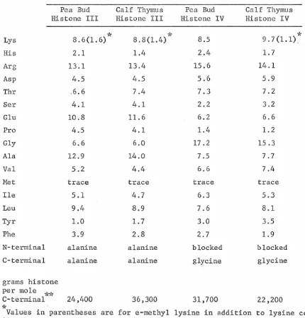
The amino acid compositions of pea bud histone fractions are pre-sented in table I. These compositions are expressed as moles of each amino acid per 100 moles of. total amino acids recovered. Although serine and threonine are partially degraded during hydrolysis, the losses are relatively small (approximately 10% and 5% respectively
(Rees, 1946)), and no correction has been made for them. For comparison with the pea bud histone fractions, amino acid compositions of calf
thymus histone fractions {taken from Rasmussen~ al., 1962) are also given in table I. The calf thymus histone amino acid compositions are corrected for serine and threonine decomposition. The amino acid analyses were performed on histone fractions comparable to those, the electrophoretic patterns of which are given in Figures 7 and 8. Thus there is slight contamination of each fraction by material of other fractions, and amino acid compositions are composites of the amino acid compositions of the different molecular species present in each fraction.
Table I.
The Amino Acid Compositions of Chromatographic Histone Fractions
Amino Pea Pea Calf a Pea Pea Calf a Pea Calf a Acid A I Thymus- Ila IIb Thymus- III-IV
Thymus-lb' IIb III-IV
Lys 8.2 22.9 26.2 16.8 14.1 13.5 9.7 9.7
His 1.5 0.9 0.2 1.6 1.1 2.8 1.9 1.9
Arg 3.2 2.7 2.6 7.2 7.6 7.9 10.8 11.9
Asp 7.0 3.0 2.5 6.7 6.2 5.6 6.1
s..
0Thr 5.2 4.6 5.4 4.7 4.8 5.2 6.1 6.7
Ser 7.8 5.6 6.5 7.3 6.2 7.0 4.4 4.6
Glu 9.7 7.8 4.3 8.6 7.7 8.7 8.8 10.4
Pro 6.8 10.0 9.1 6.9 5.3
4.7
3.9 4.2Gly 11.8 3.7 7.3 10.5 10.0 8.2 9.8 8.6
Ala 12.9 22.9 24.2 7.9 11.8 11.5 9.7 11.6
Val 7.3 6.2 4.1 4.5 6.9 6.7 6.7 5.9
Met 1.0 Trace 0.1 0.7 0.6 0.8 0.4 1.3
Ile 4.8 2.9 1.2 3.8 5.0 4.5 5.9 5.3
Leu 8.5 4.6 5.0 7.9 8.4 8.6 10.5 8.9
Tyr 2.3 0.9 0.7 2.4 1.8 3.0 2.4 2.2
Phe 2.2 1. 3 0.6 2.7 2.4 1. 3 3.1 2.5
a
[image:57.617.118.549.164.619.2]pea bud and calf thymus histone fractions. The lysine-rich histones of pea bud and calf thymus show a particularly striking resemblance due to their unusual compositions. Their arginine/lysine ratios are approximately 0.1. In addition to their high lysine content, these histones are almost equally rich in alanine, the two amino acids ac-counting for about half of the total compositions. Praline content is also remarkably high -- about 9-10 mole per cent. Aromatic amino acids are present in very small amounts and methionine is practically lacking. Of the two pea bud slightly lysine-rich histone components, IIb par-ticularly resemble·s calf thymus his tone IIb, both possessing arginine/ lysine ratios of about 0.5. The III-IV fractions are also extremely similar, differing in content by more than one mole per cent in only six amino acids.
Disc electrophoresis
Electrophoresis of histones in polyacrylamide gels was first used in the course of this work as a means of analysis and identification of the histone fractions produced by column chromatography. Although disc electrophoresis has proved to be a far superior tool for the res-olution of histone components, especially for the separation of pea histones, little additional heterogeneity of histone fractions is revealed by it.
Figure 6 shows densitometric tracings of the electrophoretic
L ___
fi _ _
0c
0
-Ia and Ib is evident. These two are, however, not reproducibly re-solved by disc electrophoresis. The patterns reveal no indication of separation of components IIbl, IIb2 and III. After longer electrophor-esis times III does, however, often emerge as a band of greater
mo-")'(
bility than that of !Ib. The electrophoretic pattern of calf thymus histone IIa is identical with that of III-IV. Histones III and IV are quite different in electrophoretic properties, histone IV having
the greatest mobility of all the histones. Note the presence of some material of lesser mobility in the III-IV fraction. The nature of this material is discussed under "Arginine-rich Histones" below.
Densitometric tracings of the electrophoretic patterns of whole pea bud histone and of pea bud histone fractions Iab, IIa, IIb and III-IV are presented in Figures 7 and 8. Here the identification of column chromatography fractions with electrophoretic components is quite apparent. The tracings also give some indication of the degree of cross-contamination between fractions. Histone lab contains about 20% histone Ila, histone Ila contains a trace of histone I, and histone III-IV contains some histone II, e-specially IIb. The degree of purity of the chromatographic fractions here illustrated is typical with the exception that histone Ilb is rarely obtained in such pure form. More often the best cut of Ilb contains some IIa as well as appreciable
III-IV. That the bands identified as contaminant histones in the column fractions are indeed such can be demonstrated by purification of
compo-*
Histones IIbl and IIb2 can be separated (for example) by electrophoresisA
8
D