R E S E A R C H
Open Access
The contribution of
Alu
exons to the
human proteome
Lan Lin
1*†, Peng Jiang
2†, Juw Won Park
1,3,4†, Jinkai Wang
1†, Zhi-xiang Lu
1, Maggie P. Y. Lam
5, Peipei Ping
5,6and Yi Xing
1*Abstract
Background:Aluelements are major contributors to lineage-specific new exons in primate and human genomes.
Recent studies indicate that someAluexons have high transcript inclusion levels or tissue-specific splicing profiles, and may play important regulatory roles in modulating mRNA degradation or translational efficiency. However, the contribution ofAluexons to the human proteome remains unclear and controversial. The prevailing view is that exons derived from young repetitive elements, such asAluelements, are restricted to regulatory functions and have not had adequate evolutionary time to be incorporated into stable, functional proteins.
Results:We adopt a proteotranscriptomics approach to systematically assess the contribution ofAluexons to the human proteome. Using RNA sequencing, ribosome profiling, and proteomics data from human tissues and cell lines, we provide evidence for the translational activities ofAluexons and the presence ofAluexon derived peptides in human proteins. TheseAluexon peptides represent species-specific protein differences between primates and other mammals, and in certain instances between humans and closely related primates. In the case of the RNA editing enzyme ADARB1, which contains anAluexon peptide in its catalytic domain, RNA sequencing analyses of A-to-I editing demonstrate that both theAluexon skipping and inclusion isoforms encode active enzymes. TheAluexon derived peptide may fine tune the overall editing activity and, in limited cases, the site selectivity of ADARB1 protein products.
Conclusions:Our data indicate thatAluelements have contributed to the acquisition of novel protein sequences during primate and human evolution.
Keywords:Alu, Transposable element, Exon, Alternative splicing, RNA editing, RNA-seq, Ribo-seq, Transcriptome, Proteome, Evolution, Primate
Background
Transposable elements have played important roles in the evolution of gene regulation and function [1]. The
primate-specific Alu retrotransposon is the most
abundant class of transposable elements in the human genome, with over 1 million copies occupying over 10 %
of the human genomic DNA [2]. AlthoughAluelements
were historically considered as‘junk DNA’, extensive re-search in the past two decades has revealed significant
contributions of Alu to the evolution of the human
genome and gene regulatory networks [1–3].
Alu is a major contributor to de novo origination of lineage-specific exons in primates [4]. Because the Alu element contains multiple sites that resemble the consensus donor and acceptor splice site signals, the in-sertion ofAlu elements into intronic regions of existing genes produces preferable substrates for subsequent mu-tations that create and establish new exons [4, 5]. The exonization of Alu elements is frequent during primate
and human evolution –thousands of human genes
con-tain Alu-derived exon segments in their mRNA
tran-scripts [6, 7]. Although the majority ofAluexons have low splicing activities and probably represent non-functional evolutionary intermediates, a subset of Alu exons have acquired ubiquitously strong or tissue-specific splicing ac-tivities in human tissues, as demonstrated by recent tran-scriptome studies using splicing-sensitive exon microarray * Correspondence:[email protected];[email protected]
†Equal contributors 1
Department of Microbiology, Immunology & Molecular Genetics, University of California, Los Angeles, Los Angeles, CA 90095, USA
Full list of author information is available at the end of the article
© 2016 Lin et al.Open AccessThis article is distributed under the terms of the Creative Commons Attribution 4.0 International License (http://creativecommons.org/licenses/by/4.0/), which permits unrestricted use, distribution, and reproduction in any medium, provided you give appropriate credit to the original author(s) and the source, provide a link to the Creative Commons license, and indicate if changes were made. The Creative Commons Public Domain Dedication waiver (http://creativecommons.org/publicdomain/zero/1.0/) applies to the data made available in this article, unless otherwise stated. Linet al. Genome Biology (2016) 17:15
and deep RNA sequencing [8, 9]. These ‘established’
Alu exons are preferentially located in the mRNA 5’
untranslated regions (5’-UTR) and may play a role in regulating mRNA translational efficiency [6, 8, 9]. Alu exons inserted into coding regions of protein-coding genes frequently contain premature termination codons (PTCs) and may provide a mechanism for fine-tuning steady-state mRNA levels by inducing mRNA nonsense-mediated decay (NMD) [10]. Together, these data have established the regulatory roles of Alu exons in multiple aspects of RNA metabolism including translation and degradation.
Despite the prevalence of Alu exons in the human
transcriptome, the contribution of Alu exons to the
human proteome remains unclear and controversial [11–15]. In the 1990s and early 2000s, Makałowski and others carried out large-scale discoveries ofAluexons in human genes using cDNA sequences and expressed sequence tags (ESTs) [11, 12, 16]. They identified nu-merous instances of ‘in-frame’Alu exons in the coding region of human mRNAs that are predicted to addAlu -derived peptides to the protein products. However, the
presence of an in-frame Aluexon in the mRNA coding
sequence does not guarantee its translation and incorp-oration into stable protein products. Additionally, since most Aluexons have low splicing activities [6, 16], even a‘translatable’coding-region Alu exon may only be in-corporated into a small fraction of the gene’s transcript products and consequently become largely undetectable and negligible on the protein level. In fact, in these stud-ies there was little experimental evidence to support the existence of Alu-derived peptides in vitro or in vivo. In 2006, Makalowski and colleagues revisited this question by searching for transposable elements derived peptides in non-redundant protein entries in the Protein Databank (PDB) [13]. Since all proteins in PDB have solved 3D structures, Makalowski and colleagues reasoned that PDB provides a high-confidence collection of stable, functional proteins. They did not identify any Alu derived pep-tides in PDB protein entries. On the basis of this re-sult, Makalowski and colleagues concluded that exons
derived from Alu or other young repetitive elements
do not have adequate evolutionary time to be incor-porated into stable protein products, and the role of Alu exons should be almost entirely regulatory [13]. Since then, this has become the prevailing view on
the contribution of Alu exons to the human proteome
[3, 14, 15, 17]. However, this PDB-based study also has major drawbacks. Most importantly, PDB has very limited coverage of alternatively spliced protein isoforms, and less than 10 protein isoforms had structures deposited in PDB by the time of this study [18]. When structural bi-ologists select proteins for structural characterization, there is an inherent bias towards selecting evolutionarily
conserved protein isoforms and against protein isoforms with non-conserved (lineage-specific) exon segments such
as Alu exons. Therefore, the presence of Alu exon
peptides in the human proteome may be significantly underestimated in the PDB-based analysis. Indeed, in gene-specific studies, researchers have found evidence for the expression ofAluexon peptides in vitroor in vivo– two examples being theAluexons in genes encoding the RNA editing enzyme ADARB1 [19] and DNA methyl-transferase DNMT1 [20]. Neither case was recapitulated in the PDB study [13]. In summary, despite substantial interest in this topic, the real contribution ofAluexons to the human proteome remains unclear, and past studies may have reached dramatically different conclusions due to inherent limitations of their data sources.
In this work, we adopted a proteotranscriptomics ap-proach to systematically assess the contribution of Alu exons to the human proteome. Using RNA sequencing (RNA-seq), ribosome profiling (Ribo-seq), and mass spectrometry data of diverse human tissues and cell lines, we evaluated the evidence for the translational activities of Aluexons and the presence ofAluexon derived peptides in human proteins.
Results
RNA-seq discovery of putative codingAluexons with high splicing activities
RNA-seq has become a powerful approach for quantita-tive analyses of exon splicing [21]. We previously used an RNA-seq dataset of human cerebellum to characterize the splicing profiles ofAluexons [9]. AmongAluexons with high splicing levels (>50 % exon inclusion) in the cerebel-lum according to this RNA-seq analysis, the vast majority (over 80 %) were located in the mRNA 5’-UTR, and only a handful were located within the protein-coding region [9]. In the present work, to comprehensively identify putative codingAluexons and characterize their splicing activities, we used a much larger RNA-seq dataset with approxi-mately 1.8 billion RNA-seq reads covering 19 human tis-sues, including 16 tissues from the Illumina Human Body Map 2.0 dataset and three tissues from different anatom-ical compartments of the human placenta (see Materials and Methods).
We implemented a computational pipeline to identify putative coding Alu exons with high splicing activities (Fig. 1a and Materials and Methods). Briefly, from Ensembl transcript annotations, we extracted 1,996 Alu-derived internal (spliced) exons. Of these, 911
Alu exons were located within the coding sequences
(CDS). To removeAluexons that would trigger transcript degradation via mRNA nonsense-mediated decay (NMD), we further identified 262 coding-region Alu exons that would not introduce premature termination codons
included into the transcripts. Specifically, PTCs were de-fined as stop codons located more than 50 bp upstream of the last exon-exon junction of the mRNA [22]. These 262 non-PTC exons were considered as candidate protein-coding Alu exons that had the potential to addAlu -de-rived peptides to the protein products. Of note, 69 % (182/262) of these candidate protein-coding Alu exons were divisible by 3 in exon length while only 24 %
(159/649) of the putative NMD-inducing Alu exons
were divisible by 3 (Fisher’s exact test P= 2e-36). Next, we performed detailed RNA-seq analyses on these exons to identify those with moderate-to-high levels of splicing or differential splicing activities among human tissues. For each exon, we calculated its transcript inclusion level (denoted as percent-spliced-in, or Ψ [23]) in a given tissue using RNA-seq reads uniquely mapped to the upstream, downstream, and exon-skipping splice junctions (see Materials and Methods). To ensure accurate estimation of the transcript inclusion level, we required that at least one splice junc-tion (upstream, downstream, or exon-skipping) had at least 10 reads. We identified 48Aluexons with transcript inclusion levels of at least 33 % in one of the human tis-sues, indicating moderate-to-high levels of exon splicing. In parallel, we performed pairwise tissue comparisons using the MATS algorithm for differential splicing analysis [24] and identified 27Aluexons with at least 10 % change in transcript inclusion levels (that is, |ΔΨ| ≥10 %) be-tween at least one pair of tissues (false discovery rate
(FDR) <10 %), indicating differential splicing among human tissues. The vast majority of these differen-tially spliced Alu exons (23/27) were also among the 48 exons with at least 33 % transcript inclusion levels in at least one of the human tissues. In total, we
ob-tained 52 Alu exons by combining these two exon
lists (Fig. 1b and Additional file 1: Table S1). These
results indicate that although Alu exons with high
splicing levels are preferentially located within the 5’-UTR [6, 8, 9], there is a reasonably large number of putative protein-coding Alu exons with high splicing activities in human tissues.
Translational and proteomic evidence for protein-coding
Aluexons
To systematically assess the proteomic evidence for Alu exon derived peptides in human proteins, we queried
the 262 putative coding Alu exons in Ensembl
tran-scripts (Fig. 1a) against the PRoteomics IDEntifications Database (PRIDE) [25]. PRIDE is a comprehensive re-pository for proteomics data, containing protein and peptide sequences as well as their supporting spectral evidence from mass spectrometry experiments. We searched PRIDE peptide sequences against the putative
open reading frames (ORFs) of Alu exon inclusion
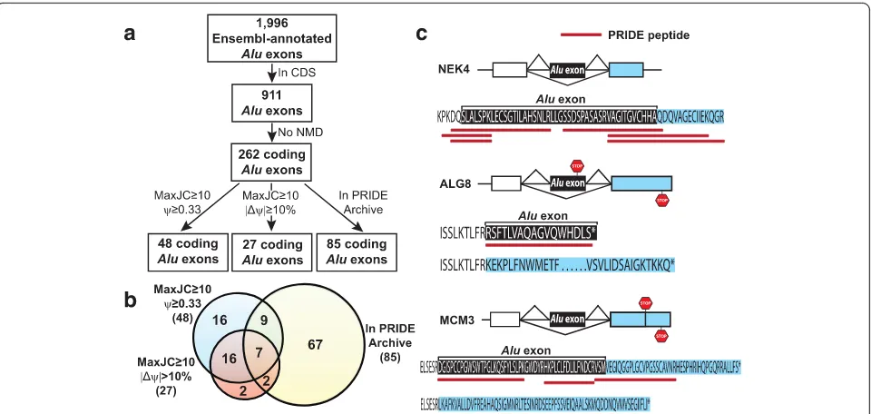
transcripts as well as the ORFs of all other human transcripts. We excluded peptide sequences mapped to multiple locations in human ORFs. Then we defined a putative protein-coding Alu exon as having a peptide Fig. 1Identification and analysis of putative codingAluexons in human genes.aThe bioinformatics work flow to identify and characterize putative codingAluexons using RNA-seq and proteomics data.bThe Venn diagram illustrating the number of putative codingAluexons with different types of RNA-seq or proteomic evidence.cThe splicing patterns and deduced peptide sequences of putative codingAluexons with supporting peptide sequences from the PRIDE database. The stop codon of each isoform is indicated by a STOP sign
[image:3.595.59.539.87.315.2]match in PRIDE if there was a peptide sequence uniquely mapped to the translated peptide sequence from the exon body or the splice junctions spanning the Aluexon and its upstream or downstream flanking exon. Of the 262 putative coding Alu exons, 85 had peptide evidence in the PRIDE database (Fig. 1a and Additional file 2: Table S2). Of note, 69 of these 85 (81 %) Alu exons had peptide sequences crossing the upstream or downstream splice junctions (Additional file 2: Table S2). Because the flanking exons of most Alu exons are not derived from repetitive elements, these splice junction spanning peptide sequences provide stronger evidence that these putative coding Alu exons are translated into protein sequences. Among the 52 putative protein-coding Aluexons with RNA-seq evidence for high splicing activ-ities in human tissues, 18 had peptide matches in PRIDE (Fig. 1b and Table 1). A few examples are shown in Fig. 1c. In NEK4 (ENSG00000114904) we found six peptide sequences in PRIDE that mapped to the upstream or
downstream splice junction of the Alu exon. In ALG8
(ENSG00000159063), the Alu exon was predicted to
encode a different C-terminal peptide of the protein –
this Alu exon peptide was supported by a peptide
sequence in PRIDE. Another example was MCM3
(ENSG00000112118). In this gene, Alu exon inclusion
caused a frameshift in the downstream 3’ terminal
exon, which resulted in a distinct C-terminal peptide by using a different reading frame within the same ter-minal exon. We found peptide sequences in PRIDE that
mapped to either the Alu exon or the downstream
splice junction. In addition to the PRIDE analysis, we also manually searched the 52 putative protein-coding Aluexons with high splicing activities against a second mass spectrometry database PeptideAtlas [26]. Differ-ent from PRIDE, PeptideAtlas reprocessed the original protein mass spectrometry data using stringent FDR criteria [26]. Therefore, PeptideAtlas has a very low false positive rate at the expense of a higher false nega-tive rate. Our manual search against PeptideAtlas
iden-tified the peptide evidence for Alu exons in SUGT1
(ENSG00000165416) and DNMT1 (ENSG00000130816), both were also supported by PRIDE (Table 1). Overall, among the 52 putative codingAlu exons with high spli-cing activities, 18 (35 %) had peptide evidence in either PRIDE alone or both PRIDE and PeptideAtlas. It should be noted that this percentage is expected to be an underestimate, since peptide identification from prote-omics experiments is biased towards highly expressed proteins [27].
To obtain an independent line of evidence for the translation of these putative codingAlu exons into pro-tein products, we analyzed the footprint of ribosomes on
Alu exons using ribosome profiling (Ribo-seq) data.
Ribo-seq has recently been developed as a powerful
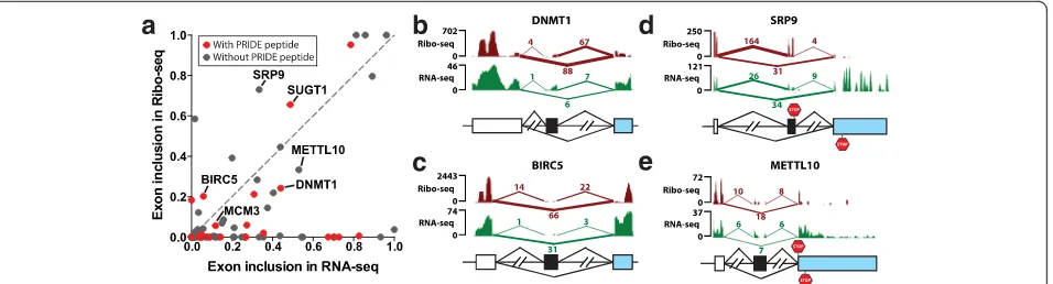
approach for high-throughput analyses of protein trans-lation [28]. Here we jointly analyzed the Ribo-seq and RNA-seq data of HeLa cells [29]. Specifically, we mapped the Ribo-seq and RNA-seq reads to the up-stream, downup-stream, and exon-skipping splice junctions of Alu exons, and computed each Alu exon’s transcript inclusion levels in the Ribo-seq and RNA-seq data, re-spectively (Materials and Methods). To ensure reliable estimation of transcript inclusion levels, we restricted this analysis toAluexons with at least 10 reads mapped to one of three splice junctions in both Ribo-seq and RNA-seq data. As shown in Fig. 2a, among the 76 putative coding Alu exons that met this criterion, 38 exons had non-zero transcript inclusion levels in the Ribo-seq data, indicating ribosome footprints translat-ing through the Alu exons. Seventeen exons had tran-script inclusion levels of at least 15 % in the Ribo-seq data. Of note, 15 Alu exons had higher transcript in-clusion levels in the Ribo-seq data than in the RNA-seq data (Fig. 2a and Additional file 3: Table S3),
sug-gesting that the Alu exon inclusion mRNA isoform
was translated at a comparable or even higher rate as compared to the ancestral mRNA isoform lacking the Aluexon. The Ribo-seq and RNA-seq signals of fourAlu
exons were shown in Fig. 2b-e. These include Alu
exons with PRIDE or PeptideAtlas evidence (DNMT1 (ENSG00000130816), BIRC5 (ENSG00000089685)), as well as those without (SRP9 (ENSG00000143742), METTL10 (ENSG00000203791)). Of note, in both SRP9
and METTL10, the Aluexon was the penultimate exon,
and the stop codon of theAluexon inclusion mRNA iso-form was located either within theAluexon (SRP9) or in the immediate downstream terminal exon (METTL10). In both cases the Ribo-seq signal diminished beyond the stop codon (Fig. 2d, e). We note that nine Alu exons with PRIDE peptide sequences had no Ribo-seq reads mapped to the exon inclusion splice junctions, includ-ing three exons with RNA-seq transcript inclusion levels of over 50 % (Additional file 3: Table S3). This may be attributed to cell-type-specific differences in protein translation between the HeLa cells and other tissues and cell types.
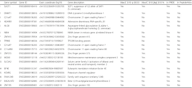
Table 1Putative codingAluexons with significant splicing activities and supporting peptide evidence in proteomics databases
Gene symbol Gene ID Exon coordinate (hg19) Gene description MaxJC≥10ψ≥0.33 MaxJC≥10 |Δψ|≥10 % In PRIDE In PeptideAtlas 1 SUGT1 ENSG00000165416 chr13:53235609-53235705 SGT1, suppressor of G2 allele of SKP1
(S. cerevisiae)
Yes Yes Yes Yes 2 DNMT1 ENSG00000130816 chr19:10290862-10290910 DNA (cytosine-5-)-methyltransferase 1 Yes Yes Yes Yes 3 C21orf7 ENSG00000156265 chr21:30463988-30464082 Chromosome 21 open reading frame 7 Yes No Yes No 4 ADARB1 ENSG00000197381 chr21:46604388-46604508 Adenosine deaminase, RNA-specific, B1 Yes Yes Yes No 5 ALG8 ENSG00000159063 chr11:77813919-77813994 Asparagine-linked glycosylation 8, alpha-1,
3-glucosyltransferase homolog (S. cerevisiae)
Yes No Yes No 6 NEK4 ENSG00000114904 chr3:52783707-52783845 NIMA (never in mitosis gene a)-related kinase 4 Yes No Yes No 7 ZNF415 ENSG00000170954 chr19:53618462-53618560 Zinc finger protein 415 Yes Yes Yes No 8 TPRKB ENSG00000144034 chr2:73959710-73959827 TP53RK binding protein Yes No Yes No 9 C21orf7 ENSG00000156265 chr21:30463821-30463897 Chromosome 21 open reading frame 7 Yes No Yes No 10 C11orf80 ENSG00000173715 chr11:66523823-66523976 Chromosome 11 open reading frame 80 Yes No Yes No 11 ZNF573 ENSG00000189144 chr19:38249115-38249236 Zinc finger protein 573 No Yes Yes No 12 MCM3 ENSG00000112118 chr6:52130032-52130183 Minichromosome maintenance complex component 3 No Yes Yes No 13 SLC3A2 ENSG00000168003 chr11:62639048-62639141 Solute carrier family 3 (activators of dibasic and
neutral amino acid transport), member 2
Yes No Yes No 14 EIF4E ENSG00000151247 chr4:99807604-99807697 Eukaryotic translation initiation factor 4E Yes Yes Yes No 15 KCNRG ENSG00000198553 chr13:50592958-50593056 Potassium channel regulator Yes No Yes No 16 FAM124B ENSG00000124019 chr2:225265097-225265222 Family with sequence similarity 124B Yes Yes Yes No 17 B3GALNT2 ENSG00000162885 chr1:235659495-235659618 Beta-1,3-N-acetylgalactosaminyltransferase 2 Yes No Yes No 18 ZNF195 ENSG00000005801 chr11:3382972-3383119 Zinc finger protein 195 Yes Yes Yes No
Lin
et
al.
Genome
Biology
(2016) 17:15
Page
5
of
strengthen splicing signals and develop protein-coding capabilities.
Assessment of transcriptomic and proteomic evidence for protein-codingAluexons
We performed a series of analyses to further assess the transcriptomic and proteomic evidence for protein-coding Aluexons. First, to validate the splicing of these
exons, we randomly selected 20 protein-coding Alu
exons, and used fluorescently labeled RT-PCR to verify their exon inclusion patterns and quantify their splicing levels in a diverse panel of 21 human tissues (Additional file 4: Table S4). Nineteen out of the 20 exons were validated to be spliced into the human transcriptome (Additional file 5: Figure S1). This high validation rate (19 out of 20) was consistent with our previous
RT-PCR and sequencing confirmation of UTR Alu exons
identified by seq [9], confirming that our RNA-seq identification of Alu exon splicing had a very high accuracy. Second, we further assessed the Ribo-seq
signal of protein-coding Alu exons in HeLa cells,
using putative NMD-inducing Alu exons in
protein-coding genes as the control. Seventeen out of 76
protein-coding Alu exons with sufficient Ribo-seq
coverage had transcript inclusion levels of at least 15 % in the Ribo-seq data, as compared to 24 out of
180 for putative NMD-inducing Alu exons. We
ob-served an increase in Ribo-seq signals of protein-coding Alu exons (22 % vs. 13 %), although the statis-tical significance was marginal (P= 0.056, one-sided Fisher’s exact test), probably due to the small sample size and possibly the effect of the pioneering round of translation in NMD transcripts [31]. These data suggest that Ribo-seq provides a discriminative feature of protein-coding Alu exons but cannot be used alone as their sole evidence.
As the most central and direct evidence for protein-coding Aluexons in this work came from the analysis of PRIDE peptide data, we used multiple negative controls to assess the reliability of our PRIDE search. First, we used mouse peptides as a negative control and searched mouse peptides in PRIDE and in our own COPaKB database of cardiac proteins [32] against humanAluexons. We did not find any mouse peptide hit of longer than four amino acids in either database, which was below the length cutoff we required for human PRIDE peptide hits (≥6 amino acids).
Second, we used putative NMD-inducing Alu exons in
protein-coding genes as another negative control to assess the FDR of our PRIDE analysis using human peptide data. Specifically, we performed PRIDE search on all NMD-inducingAluexons and identified PRIDE hits for 47 out of 649 exons (7.2 %). This percentage is significantly lower than that of the putative protein-codingAluexons (32.4 %, 85 out of 262; P= 1.8e-20, Fisher’s exact test). Even if we take a very conservative estimate that all of these 649 NMD-inducing exons represent true negatives and none of them can be protein-coding via mRNA isoforms with alter-native reading frames or A-to-I editing of the STOP codon (a possible scenario supported by the literature; see [33]), we would estimate that 19 of the 262 putative protein-codingAluexons would generate false positive PRIDE hits (that is, 262 × 7.2 %), yielding a low upper-bound estimate of FDR of 22 % (that is, 19 out of 85). In summary, using putative NMD-inducingAluexons as our negative control, we demonstrate that the FDR of our PRIDE search is low, and that the vast majority of the 85 PRIDE hits reflectbona fideproteomic evidence for protein-codingAluexons.
Human-specific increase in the splicing activity of a protein-codingAluexon in SUGT1
We studied an Alu exon derived peptide in SUGT1,
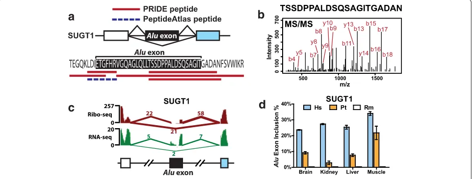
[image:6.595.63.541.88.217.2]proteomics (PRIDE and PeptideAtlas) and Ribo-seq data (Fig. 3a-c). SUGT1 is an assembly factor for the kineto-chore and is required for the G1/S and G2/M transitions [34]. TheAluexon encodes a peptide of 33 amino acids inserted at the end of the third tetratricopeptide repeat (TPR) in the SUGT1 protein product. We identified five peptide sequences in PRIDE that matched to the up-stream or downup-stream splice junction of this Alu exon, and additional peptide evidence was found in the highly stringent PeptideAtlas (Fig. 3a). This exon had a sig-nificant splicing activity in HeLa cells with a tran-script inclusion level of 49 % according to RNA-seq data, while its transcript inclusion level in the Ribo-seq data was even higher (66 %; see Figs. 2a and 3c), suggesting active translation of the Alu exon inclusion mRNA isoform. Given the strong evidence for this
Alu exon derived peptide, we next asked whether the
expression of this Alu exon inclusion protein isoform has been detected in previous studies. A literature search showed that the SUGT1 protein isoform con-taining the 33 amino acids encoded by exon 6 (that
is, the Alu exon) was first identified as a doublet
band in Hela cell extracts, as well as in human tissues including brain, liver, lung, and testis, and termed as SUGT1B, while the exon 6 skipping protein isoform
was called SUGT1A [35]. The authors thought
SUGT1B (that is, the exon inclusion isoform) was the ancestral full-length isoform while SUGT1A (that is, the exon skipping isoform) represented an alternative isoform of the gene. Later studies also detected SUGT1B protein in HeLa, THP-1, and 293T cell ex-tracts [34, 36]. However, it was not recognized that
exon 6 was derived from a primate specific Alu retro-transposon [34–36].
The recent origin of Alu elements suggests that the Aluexon derived peptide in SUGT1 could contribute to the evolutionary divergence of the human and non-human primate protein products. To investigate the
evo-lution of this Alu exon further, we designed RT-PCR
primers for its flanking constitutive exons in human, chimpanzee, and rhesus macaque genomes and quanti-fied the exon splicing levels in four tissues (brain, kid-ney, liver, and muscle) (Fig. 3d; also see Additional file 5: Figure S2 for the fluorescently labeled RT-PCR gel im-ages). Our RT-PCR analyses showed this exon had the highest levels of splicing (12–35 %) in human tissues. By contrast, the orthologous exon region was completely absent from the rhesus macaque transcripts. Chimpan-zee transcripts showed intermediate transcript inclusion levels (0–24 %).
Next we aligned the human, chimpanzee, and rhesus genomic sequences with the consensus sequence of the corresponding Alu subfamily (AluSx) (Additional file 5: Figure S3). The resulting alignment suggested that this Aluexonization event occurred prior to the most recent common ancestor of humans and chimpanzees. Specific-ally, a C to T substitution created the 5’splice site GT dinucleotide, which, when combined with the pre-existing 3’ splice site AG dinucleotide in AluSx, led to Alu exonization. Additionally, a G to A substitution at the +3 intronic position of the 5’splice site strengthened the splice site score from 5.29 to 9.46 as calculated by MAXENT [37], representing an over 16-fold increase in the likelihood of matching to the consensus MAXENT
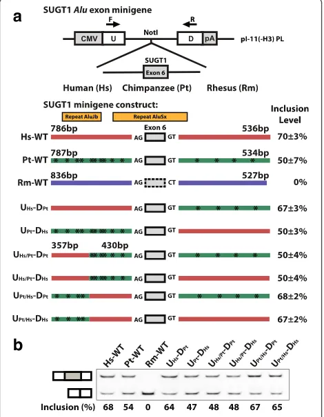
Fig. 3A protein-codingAluexon in SUGT1 supported by multiple lines of proteomics and Ribo-seq evidence.aThe splicing pattern and deduced peptide sequence of a putative codingAluexon in SUGT1 and its corresponding peptide evidence from PRIDE and PeptideAtlas.bTandem mass spectrometry (MS/MS) spectrum of the peptide TSSDPPALDSQSAGITGADAN from PRIDE (experiment ID: 26855, spectrum ID: 7275).cThe UCSC genome browser view of the Ribo-seq and RNA-seq data of the SUGT1Aluexon.dRT-PCR analysis of the SUGT1Aluexon in four different tissues in human (Hs), chimpanzee (Pt), and rhesus macaque (Rm). Error bars show standard error of the mean from at least three replicate experiments
[image:7.595.58.540.490.673.2]5’splice site model. The rhesus sequence lacked the es-sential GT dinucleotide at the 5’ splice site, consistent with the observation that the exon was completely skipped in rhesus transcripts. We did not observe any nucleotide difference between the human and chimpan-zee sequences within the exon or the 5’and 3’splice site regions (Additional file 5: Figure S3). However, beyond the splice sites there were a number of nucleotide differ-ences in the upstream and the downstream intronic re-gions, which may be responsible for the difference in splicing levels between the human and chimpanzee exons.
We carried out a comprehensive investigation of the genomic sequence changes that strengthened the
SUGT1 Alu exon in the human lineage (Fig. 4). There
was no obvious candidate for causal cis sequence
change(s) based on sequence analysis of splicing regula-tory elements. Between human and chimpanzee, no se-quence divergence was found within the Alu exon or in the 9 bp 5’ splice site and 23 bp 3’ splice site regions.
Therefore, the causalcis sequence change(s) must reside in the upstream or downstream intronic region further away from the splice sites. These intronic regions con-tained a fairly large number of sequence changes between human and chimpanzee. We cloned a large genomic
frag-ment surrounding the SUGT1Aluexon into a minigene
splicing reporter (see Materials and Methods) and gener-ated three splicing reporter constructs corresponding to the wild-type human, chimpanzee, and rhesus genomic se-quence (Hs-WT, Pt-WT, and Rm-WT; see Fig. 4a). When expressed in Hela cells, the chimpanzee minigene con-struct had an exon inclusion level of 50 ± 7 %, while the human minigene construct had a higher exon inclusion level of 70 ± 3 %, and no exon inclusion was observed for the rhesus minigene construct. The splicing difference be-tween the human and chimpanzee wild-type minigene constructs was consistent with the difference of en-dogenous splicing levels in human and chimpanzee tis-sues (12–35 % vs. 0–24 %). The overall higher baseline exon inclusion levels in the minigene constructs may indi-cate deeper intronic splicing silencers that were not cloned into the minigene reporter, but this should not affect our human vs chimpanzee comparative analysis. Then we used a sequence swapping strategy [38] to make six additional splicing reporter constructs in which gen-omic segments from different species were swapped in order to narrow down the genomic region responsible for the human-specific splicing pattern (Fig. 4a). The analysis of these minigene splicing reporters indicates that a prox-imal 430 bp upstream intronic region is responsible for the human-specific increase in splicing compared to chimpanzee. All minigene constructs containing the chim-panzee version of this 430 bp intronic region showed ap-proximately 50 % exon inclusion, while all minigene constructs containing the human version of this region showed close to 70 % exon inclusion, despite being placed within different surrounding sequence context (Fig. 4a, b). We obtained similar results when transfecting these re-porters to a chimpanzee fibroblast cell line (data not shown), further supporting our hypothesis that the spli-cing divergence of this SUGT1Aluexon was driven bycis sequence changes. Within this 430 bp upstream intronic region, there were six individual nucleotide substitutions between human and chimpanzee. We were unable to suc-cessfully perform further point mutation analyses of this region, due to the sequence homology between this region (which was AluSx derived) with an adjacent upstream AluJb element (see Fig. 4a). Nonetheless, our minigene splicing reporter data have established the role of specific cisgenomic sequences in shaping the evolution of thisAlu exon in the human lineage.
Collectively, our data indicate a gradually strengthened splicing activity of the SUGT1 Alu exon during recent primate and human evolution. It is possible that the Fig. 4Minigene splicing reporter analysis of the human-specific
[image:8.595.57.292.344.647.2]acquisition and increased expression of thisAlu-derived peptide in human tissues have certain adaptive benefits and are driven by positive selection.
TheAluexon inclusion isoform of ADARB1 encodes an active RNA editing enzyme with an altered editing activity
To investigate whether Alu-derived peptides can be
part of functional proteins, we selected a protein
cod-ing Alu exon in the RNA editing enzyme ADARB1
(ENSG00000197381, also known as ADAR2) for de-tailed studies. The crystal structure of the catalytic domain of human ADARB1 has been solved [39]. The
Alu exon encodes a 40 amino acid peptide inserted
into the catalytic deaminase domain, which is sup-ported by two peptide sequences in PRIDE (Fig. 5a). This exon displayed moderate to high levels of
spli-cing (25–100 %) across human tissues according to
the RNA-seq data, including eight out of 19 tissues with over 75 % exon inclusion levels. Our RT-PCR analyses of human, chimpanzee, and rhesus tissues
showed that the Alu exon was consistently spliced
into transcripts with comparable splicing levels in all three species, while there was variation in its splicing
levels across different tissues and individuals (Additional file 5: Figure S4). In previousin vitrostudies where puri-fied recombinant ADARB1 protein isoforms were incu-bated with an artificial dsRNA substrate, the Alu exon inclusion protein isoform showed a lower catalytic activity than theAluexon skipping isoform [19, 40]. However, the functional activity of the Alu exon inclusion ADARB1 protein isoform on endogenous mRNA transcripts has not been examined on a genome-scale in a live cell setting.
To test the effect of the ADARB1 protein isoforms on RNA editing, we selected the HEK293 cell line which had a low endogenous ADARB1 level, and ectopically
expressed either the Alu exon inclusion or skipping
ADARB1 protein isoform. Real-time qRT-PCR and west-ern blot analyses indicated that both isoforms were expressed at similar levels (Additional file 5: Figure S5). To characterize the transcriptome-wide effect on RNA editing, we then performed strand-specific RNA-seq of cells transfected with one of the two ADARB1 protein isoforms or an empty vector control (Materials and Methods). We collected annotated A-to-I editing sites in human genes from the RADAR database of RNA editing
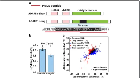
Fig. 5ADARB1Aluexon inclusion isoform encodes an active RNA editing enzyme with altered editing activity.aThe schematic diagram of the protein domain structure of ADARB1 isoforms and supporting peptide sequences from the PRIDE database.bThe change in overall RNA editing levels of 7,618 RNA editing sites in HEK293 cells upon ectopic expression of the exon skipping (Short) or the exon inclusion (Long) ADARB1 isoform as compared to the empty vector (EV) control. Error bars show standard errors calculated based on the 7,618 known RNA editing sites used in this analysis.cCommon and isoform-specific differentially edited sites upon ectopic expression of theAluexon inclusion (Long) or skipping (Short) ADARB1 isoform. Isoform-specific differentially edited sites are further classified (low-confidence, high-confidence) based on the strength of the RNA-seq evidence
[image:9.595.58.540.384.653.2]sites [41], and restricted our analysis to a set of 7,618 sites with sufficient sequencing coverage in our data set (Materials and Methods). We then compared the overall RNA editing levels in the cells across different
experi-mental conditions. Both the Alu exon inclusion and
skipping protein isoforms significantly enhanced the overall editing levels in the HEK293 cells as compared to the empty vector control (Wilcoxon test, Short vs. EV: P= 6.5e-68, Long vs. EV: P= 2.1e-50; Fig. 5b), indi-cating the Alu exon inclusion protein isoform is an ac-tive RNA editing enzyme with a global impact on RNA editing. We also noted that the overall RNA editing level was lower in cells expressing theAluexon inclusion
iso-form as compared to cells expressing the Alu exon
skipping isoform (Wilcoxon test, P= 4.7e-10; Fig. 5b), consistent with thein vitroassay results on the synthetic RNA editing substrate [19, 40].
To investigate potential differences in the site selectiv-ity of these two ADARB1 isoforms, we sought to identify differentially edited sites upon ectopic expression of the Alu exon inclusion (long) or skipping (short) isoform. Specifically, for each annotated RNA editing site in the RADAR database [41], we counted and compared their edited and unedited RNA-seq reads with those of the empty vector (EV) transfected cells. For each individual replicate, we used Fisher’s exact test to calculate the P value, then combined the P values of three replicates using Fisher’s method to generate a combined P value for differential editing, followed by correction of multiple testing to generate FDR. The editing sites with FDR≤10 % were called differential RNA editing sites. We then com-pared the identified differential RNA editing sites for the Alu exon inclusion (long) or skipping (short) ADARB1 isoform to identify common and isoform-specific differen-tial RNA editing sites. To guard against potendifferen-tial spurious calls of isoform-specific differential editing sites due to RNA-seq noise and statistical fluctuation, we also gener-ated a high-confidence list of isoform-specific differential RNA editing sites, defined as those with FDR≤ 10 % for one isoform and FDR ≥ 90 % for the other isoform. We identified a total of 360 differential RNA editing sites in the RNA-seq data after ectopic expression of one of the two isoforms (Fig. 5c). As seen in Fig. 5c, most of them were either common to the two isoforms, or were clas-sified as isoform-specific but the difference between the two isoforms was minor and could be attributed to ran-dom RNA-seq noise and statistical fluctuation (that is, those classified as ‘low-confidence’ isoform specific sites). We did identify six high-confidence long isoform specific and 14 high-confidence short isoform specific differential RNA editing sites, but they represented a fairly small percentage of all identified differential RNA editing sites (Fig. 5c). It is interesting to note that in general, we identified almost twice as many short
isoform specific sites than long isoform specific sites, at various confidence level cutoffs (Fig. 5c). Collectively, these data suggest that regulated alternative splicing of thisAlu exon may fine tune the editing activities and in limited cases the target site selectivity of the ADARB1 protein products.
Discussion
The creation and establishment of new exons provide an important evolutionary strategy for generating genetic novelties in existing genes [42, 43]. A large body of work has investigated the exonization of Alu elements as a major source for new exons during primate and human evolution [5, 7–9, 44–47]. Recent studies have shown that a subset ofAluexons in human genes have acquired strong splicing activities [8, 9], and that they play a var-iety of regulatory roles at the RNA level such as the con-trol of mRNA degradation and translation [8–10]. On the other hand, the contribution of Alu exonization to the human proteome has been considered to be minimal [13]. In 2006, a survey of protein sequence entries in the PDB did not identify any peptide segment derived from Aluor other young transposable elements (TEs), leading the authors to conclude that ‘functional proteins are unlikely to contain TE cassettes derived from young TEs, the role of which is probably limited to regulatory functions’[13].
In this work, we revisited the role of Alu exonization in human proteome evolution and adopted a proteotran-scriptomics approach [48] to systematically evaluate the evidence for Alu exon derived peptides in human pro-teins. We identified 262 putative coding Alu exons in Ensembl human transcripts, among which 85 exons had proteomic evidence in the PRIDE peptide database. Using multiple negative controls, we demonstrated that our proteomic identification of protein-codingAluexons based on the PRIDE search had a low FDR. We also performed detailed analyses of RNA-seq and Ribo-seq data of human tissues and cell lines to provide more fine-grained information on the splicing and
transla-tional profiles of these protein-coding Alu exons.
Using RNA-seq data of 19 human tissues, we identi-fied 52 protein-coding Alu exons with high transcript inclusion levels and/or tissue-specific splicing profiles, significantly expanding the catalog of coding-region
Alu exons with strong splicing activities in normal
human tissues, which were considered rare in previ-ous work [8, 9]. Collectively, our data challenge the
conventional view on the proteomic impact of Alu
We studied a protein coding Alu exon in SUGT1, a gene encoding a cell cycle regulator [34]. The translation of this SUGT1 Alu exon is supported by multiple lines of evidence (Ribo-seq, PRIDE, PeptideAtlas) as well as previous literature [34–36]. Although the specific func-tion of this SUGT1Alu exon inclusion isoform remains to be elucidated, existing data suggest that the splicing of thisAlu exon is under dynamic regulation in human cells. SUGT1 is reported to be a member of the
pro-inflammatory complex ‘inflammasome’ and its protein
level, especially the level of the Alu exon inclusion iso-form (known as ‘SUGT1B’ in the literature), increases after heat shock [49]. SUGT1B also appears to be prefer-entially translocated to and accumulate in the nucleus under heat shock [50]. Additionally, there is anecdotal evidence showing SUGT1B is expressed at a much higher level than SUGT1A (that is, the Alu exon skip-ping isoform) in a human malignant glioblastoma cell line U-251 MG [51], as compared to the near or lower than 1:1 ratio in liver and tonsil tissues tested in the same experiment as well as various tissues and cell lines analyzed in other reports [34–36, 49]. Interestingly, our RT-PCR analyses indicate a significant gradient in the splicing levels of this SUGT1 Aluexon between human, chimpanzee, and rhesus macaque tissues, with the exon spliced at the highest levels in human tissues and com-pletely skipped in rhesus macaque tissues (Fig. 3d). Therefore, thisAluexonization event has contributed to the acquisition and increased expression of a novel pep-tide segment during very recent human evolution. Of note, using a moderate-coverage six-tissue RNA-seq dataset of human, chimpanzee, and rhesus macaque [52], we identified two additional protein-coding Alu exons in SRP9 and ZNF468 (see Additional file 1: Table S1 for their annotations) with more than 15 % increase in splicing levels in at least one human tissue compared to the corresponding chimpanzee and rhesus tissue. Given the limited RNA-seq depth of this dataset, this list is expected to be quite incomplete.
Conclusions
Our study has revealed a large list of Alu exons that may be translated and incorporated into primate-specific or even human-primate-specific protein isoforms.
These Alu exons are created in genes involved in a
wide range of biological functions and molecular pro-cesses (Table 1). We selected an Alu exon in a tran-scriptome regulator ADARB1 and performed RNA-seq experiments to read out the activity of the Alu exon inclusion protein isoform on transcriptome-wide con-trol of A-to-I RNA editing. For other protein-coding Alu exons identified in this work, future experiments tailored towards their genes’specific cellular functions are needed to elucidate the evolutionary significance
of the novel protein isoforms arising from Alu
exonization.
Materials and methods
RNA-seq analysis of putative coding Alu exons
The locations of Alu elements in the human genome
(hg19) were downloaded from the UCSC Genome Browser database [53]. The locations of internal cassette or constitutive exons were taken from Ensembl gene annotations (release 57) [54]. We defined an exon as Alu-derived if theAlu element covered at least 25 bp of the exon and more than 50 % of the total exon length.
We downloaded the Human Body Map 2.0 (HBM2.0) paired-end RNA-seq data from Gene Expression Omnibus (GEO) (accession number GSE30611). HBM2.0 RNA-seq data have a read length of 50 bp. They cover 16 tissues: adipose, adrenal, brain, breast, colon, heart, kidney, liver, lung, lymph node, ovary, prostate, skeletal muscle, testes, thyroid, and white blood cells. We also used paired-end RNA-seq data of three anatomical compartments of the human placenta (amnion, chorion, and decidua) generated in our previous work [55]. We used only 50 bp of each end for mapping and analysis based on the sequencing error profile. We used the same RNA-seq mapping method as previously described [55] to obtain the splice junction read counts and calculate each exon’s tran-script inclusion level. We used MATS to identify dif-ferential alternative splicing events in pairwise tissue comparisons [24].
Search of the PRIDE database
We searched the PRIDE (PRoteomics IDEntifications) database [25] for peptide evidence for putative coding
Alu exons. We downloaded the peptide sequences in
June 2014 with more than 1.5 million unique peptide sequences in PRIDE. Of these 1.5 million peptide se-quences, approximately 900,000 were uniquely mappable to the coding regions of the human Ensembl transcripts (release 57). With these uniquely mappable peptide se-quences, we examined the ORFs (open reading frames) containing putative coding Alu exons and identified 85 Aluexons with PRIDE peptide evidence.
Ribo-seq analysis of putative coding Alu exons
transcript inclusion levels were estimated as described previously [55].
Construction of ADARB1 isoform expression vectors
The ORF for the human ADARB1 short isoform
(without the Alu exon) in the pCMV6-AC vector was
purchased from OriGene, Inc. (catalog no. SC321955; reference transcript NM_001112). Mutageneses using the QuikChange method (Stratagene) were carried out to convert the ORF to encode the ADARB1 reference protein sequence NP_001103. Six nucleotides in pCMV6-AC en-coding two additional amino acids at the C-terminus were removed. A minor allele SNP (rs199697177) ‘C’ (allele frequency <1 %) was mutated back to the major allele
‘T’ in the ADARB1 ORF. Then the Alu exon was
inserted using the same mutagenesis method. Final
pCMV6-ADARB1-short (without the Alu exon) and
pCMV6-ADARB1-long (with the Alu exon) constructs
were confirmed by sequencing.
Ectopic expression of ADARB1 isoforms in HEK293 cells
HEK293 cell line was grown in DMEM with 10 % FBS. Cells were transiently transfected with Lipofectamine 2000 reagent according to the manufacturer’s protocol. Forty-eight hours after transfection, cells were collected for RNA extraction using TRIzol reagent (Life Tech-nologies) and protein lysates with RIPA buffer. Trans-fection experiments were replicated in three different cell passages.
Total RNA samples were treated with DNaseI (Fermentas) and reverse transcribed using the High-Capacity cDNA RT kit (Applied Biosystems). Quantitative real-time PCR (qRT-PCR) was performed using Fast SYBR green Master Mix (Applied Biosystems). Total gene ex-pression level and isoform-specific exex-pression level of ADARB1 were measured using GAPDH as the reference gene. Relative expression level was measured by the com-parative Ct (2-ΔΔCt) method [57]. Primers used are:
GAPDH_F: 5′-TGGTATCGTGGAAGGACTCA-3′,
GAPDH_R: 5′-ACAGTCTTCTGGGTGGCAGT-3′, AD
ARB1_Gene_F: 5′-AGTCTCCGCCAGTCAAGAAA-3′,
ADARB1_Gene_R: 5′-GTTGTCCAGATTGCGGTTTT-3′, ADARB1_short_F: 5′-AGGCTGAAGGAGAATGTCCA-3′, ADARB1_short_R: 5′-TGTCTATCTGCTGGTTCTTC-3′, ADARB1_long_F: 5′-CTCAACCTTCCAAGGAGCTG-3′, ADARB1_long_R: 5′-GTCCGTAGCTGTCCTCTTGC-3′.
Total protein extract in RIPA buffer was used to assay for protein level. ADARB1 (sc-10012, Santa Cruz Biotechnology) and ACTB (A5441, Sigma) anti-bodies were used for western blot following standard
protocol. Signals were detected by ChemiDoc™ MP
imaging system (Bio-Rad). RNA-seq libraries were prepared using the TruSeq Stranded mRNA Sample Prep Kit (Illumina) and sequenced on an Illumina
HiSeq 2000 (100 cycles, paired-end). RNA-seq data were deposited in the Gene Expression Omnibus database (http://www.ncbi.nlm.nih.gov/geo/) under the accession number GSE65999.
RNA-seq analysis of A-to-I editing
Strand-specific paired-end RNA-seq reads were first mapped to Ensembl transcripts and the unmapped reads were then mapped to the human genome (hg19) using Tophat (version 1.4.1) [58]. We used REDItools [59] to calculate the edited and unedited read counts on the corresponding strand of each known RNA editing site collected in the RADAR database [41]. We removed the editing sites that had less than five edited counts in all the nine RNA-seq libraries to avoid using unreliable editing sites or sites not edited in the samples of interest. The sites that had no read coverage (edited counts and unedited counts) for all three replicates of either empty vector (EV), ADARB1 long form, or ADARB1 short form transfected cells were also removed from further analyses because they were uninformative for compari-sons. In the end, 7,618 known editing sites were used in the analyses.
To compare the global editing levels between the three sample groups, we merged the RNA-seq reads of three replicates in each group, and the editing levels of each editing site were calculated as the fraction of edited counts over the total counts in the merged data. To avoid using sites with unreliable editing levels due to low read coverage, we used only the editing sites that had total read coverage≥50. Then we calculated the sig-nificance for the editing level differences between two groups using two-tailed Wilcoxon test.
Total RNA preparation
Postmortem tissue samples of three adult chimpanzees and three adult rhesus macaques were generously pro-vided by the Southwest National Primate Research Center (San Antonio, TX, USA). Total RNAs were extracted using TRIzol (Invitrogen, Carlsbad, CA, USA) according to the manufacturer’s instructions. Total RNAs from vari-ous human tissues were purchased from Clontech (Moun-tain View, CA, USA), BioChain (Newark, CA, USA), and Ambion (now part of Thermo Fisher, Grand Island, NY, USA), or prepared as previously described [55].
Fluorescently labeled RT-PCR analysis of exon splicing
Single-pass cDNA was synthesized using the High-Capacity cDNA Reverse Transcription Kit (Applied Biosystems, Foster City, CA, USA) according to manufacturer's instructions. Two micrograms of total
RNA were used for each 20 μL cDNA synthesis
are listed in Additional file 4: Table S4. Each gel picture shown was a representation of at least three replicates.
Minigene analysis of SUGT1 protein-codingAluexon splicing
Alu exon (Exon 6) of SUGT1 and its adjacent flanking intronic regions were amplified from the human, chim-panzee and rhesus genomic DNAs using KAPA HiFi HotStart ReadyMix PCR Kit (Kapa Biosystems, Inc., Wilmington, MA, USA). PCR products were subcloned into the NotI site of the pI-11-H3 minigene vector [61] (kindly provided by Dr. Russ P. Carstens, University of Pennsylvania, Philadelphia, PA, USA) using the In-Fusion Advantage PCR Cloning Kit (Clontech, Mountain View, CA, USA). Sequence swapping mutagenesis [38] was done using KAPA HiFi HotStart ReadyMix PCR Kit. All se-quences and mutations were verified by DNA sequencing.
In vitrominigene splicing reporter assay
HeLa cells and chimpanzee fibroblast cells (S008861, Coriell Institute, Camden, NJ, USA) were grown in DMEM (Invitrogen, Thermo Fisher, Grand Island, NY, USA) with 10 % FBS (Invitrogen). Cells were plated in 12-well plates and transfected using Lipofectamine LTX and Lipofectamine 2000 (Invitrogen), respectively, ac-cording to the manufacturer’s protocol. RNA was puri-fied 16 h after transfection and reverse-transcribed into single-pass cDNA. Fluorescently labeled RT–PCR was performed as described above. The pI-11-H3 minigene-specific primer sequences were pI11-F: 5′-GCTGTCT GCGAGGTACCCTA-3′; pI11-R: 5′-CGTCGCCGTCCA GCTCGACCAGCGTTCGGAGGATGCATAGAG-3′.
Ethics statement
This work used post-mortem tissues obtained from the Southwest National Primate Research Center. The pri-mate tissue repository was approved by the Institutional Animal Care and Use Committee at the Southwest National Primate Research Center at the Texas Biomedical Research Institute (Animal Welfare Assurance Number A3082-01; IACUC protocol number 525 PT for chimpan-zees, and 525 MM for rhesus macaques).
Additional files
Additional file 1: Table S1.Fifty-two putative codingAluexons with moderate-to-high levels of exon splicing or differential splicing activities among human tissues. (XLSX 16 kb)
Additional file 2: Table S2.Eighty-five putative codingAluexons with peptide evidence from the PRIDE database. (XLSX 18 kb)
Additional file 3: Table S3.Seventy-six putative codingAluexons with at least 10 reads mapped to one of the three splice junctions in both Ribo-seq and RNA-seq data from HeLa cells. (XLSX 18 kb)
Additional file 4: Table S4.List of RT-PCR primers. (XLSX 10 kb)
Additional file 5:Supplementary figures S1-5.(DOCX 2892 kb)
Abbreviations
cDNA:complementary DNA; CDS: coding DNA sequence; DJC: downstream junction read count; EST: expressed sequence tags; FDR: false discovery rate; GEO: Gene Expression Omnibus; mRNA: messenger RNA; NMD: nonsense-mediated decay; PDB: Protein Databank; PRIDE: PRoteomics IDEntifications Database; PSI (Ψ): percent-spliced-in; PTC: premature termination codon; qRT-PCR: quantitative real-time PCR; Ribo-seq: ribosome profiling; RNA-seq: RNA sequencing; SJC: skipping junction read count; TE: transposable element; TPR: tetratricopeptide repeat; UJC: upstream junction read count; UTR: untranslated region.
Competing interests
The authors declare that they have no competing interests.
Authors’contributions
LL, PJ, and YX conceived the study; LL and ZXL carried out the molecular biology experiments; PJ, JWP, JW, MPYL, and PP carried out the bioinformatics analyses; LL, PJ, and YX wrote the manuscript. All authors have read and approved the final manuscript.
Acknowledgements
We wish to thank Peter Stoilov for discussions and comments on this work, and Jerilyn Pecotte for assistance.
Funding
This work is supported by National Institutes of Health grants R01GM088342 and R01GM105431 (YX). YX is supported by an Alfred Sloan Research Fellowship. This work used biological materials obtained from the Southwest National Primate Research Center, which was supported by NIH-NCRR grant P51RR013986.
Author details
1Department of Microbiology, Immunology & Molecular Genetics, University of California, Los Angeles, Los Angeles, CA 90095, USA.2Regenerative Biology, Morgridge Institute for Research, Madison, WI 53707, USA. 3
Department of Computer Engineering and Computer Science, University of Louisville, Louisville, KY 40292, USA.4KBRIN Bioinformatics Core, University of Louisville, Louisville, KY 40202, USA.5Department of Physiology, University of California, Los Angeles, Los Angeles, CA 90095, USA.6Department of Medicine, University of California, Los Angeles, Los Angeles, CA 90095, USA.
Received: 9 November 2015 Accepted: 8 January 2016
References
1. Feschotte C. Transposable elements and the evolution of regulatory networks. Nat Rev Genet. 2008;9:397–405.
2. Hasler J, Strub K. Alu elements as regulators of gene expression. Nucleic Acids Res. 2006;34:5491–7.
3. Cordaux R, Batzer MA. The impact of retrotransposons on human genome evolution. Nat Rev Genet. 2009;10:691–703.
4. Sorek R. The birth of new exons: mechanisms and evolutionary consequences. RNA. 2007;13:1603–8.
5. Lev-Maor G, Sorek R, Shomron N, Ast G. The birth of an alternatively spliced exon: 3′splice-site selection in Alu exons. Science. 2003;300:1288–91. 6. Zhang XH, Chasin LA. Comparison of multiple vertebrate genomes reveals
the birth and evolution of human exons. Proc Natl Acad Sci U S A. 2006;103:13427–32.
7. Sela N, Mersch B, Gal-Mark N, Lev-Maor G, Hotz-Wagenblatt A, Ast G. Comparative analysis of transposed element insertion within human and mouse genomes reveals Alu’s unique role in shaping the human transcriptome. Genome Biol. 2007;8:R127.
8. Lin L, Shen S, Tye A, Cai JJ, Jiang P, Davidson BL, et al. Diverse splicing patterns of exonized Alu elements in human tissues. PLoS Genet. 2008;4:e1000225.
9. Shen S, Lin L, Cai JJ, Jiang P, Kenkel EJ, Stroik MR, et al. Widespread establishment and regulatory impact of Alu exons in human genes. Proc Natl Acad Sci U S A. 2011;108:2837–42.
10. Zarnack K, Konig J, Tajnik M, Martincorena I, Eustermann S, Stevant I, et al. Direct competition between hnRNP C and U2AF65 protects the transcriptome from the exonization of Alu elements. Cell. 2013;152:453–66.
11. Makalowski W, Mitchell GA, Labuda D. Alu sequences in the coding regions of mRNA: a source of protein variability. Trends Genet. 1994;10:188–93. 12. Nekrutenko A, Li WH. Transposable elements are found in a large number
of human protein-coding genes. Trends Genet. 2001;17:619–21. 13. Gotea V, Makalowski W. Do transposable elements really contribute to
proteomes? Trends Genet. 2006;22:260–7.
14. Piriyapongsa J, Rutledge MT, Patel S, Borodovsky M, Jordan IK. Evaluating the protein coding potential of exonized transposable element sequences. Biol Direct. 2007;2:31.
15. Goodier JL, Kazazian Jr HH. Retrotransposons revisited: the restraint and rehabilitation of parasites. Cell. 2008;135:23–35.
16. Sorek R, Ast G, Graur D. Alu-containing exons are alternatively spliced. Genome Res. 2002;12:1060–7.
17. Feschotte C, Pritham EJ. DNA transposons and the evolution of eukaryotic genomes. Annu Rev Genet. 2007;41:331–68.
18. Birzele F, Csaba G, Zimmer R. Alternative splicing and protein structure evolution. Nucleic Acids Res. 2008;36:550–8.
19. Gerber A, O’Connell MA, Keller W. Two forms of human double-stranded RNA-specific editase 1 (hRED1) generated by the insertion of an Alu cassette. RNA. 1997;3:453–63.
20. Hsu DW, Lin MJ, Lee TL, Wen SC, Chen X, Shen CK. Two major forms of DNA (cytosine-5) methyltransferase in human somatic tissues. Proc Natl Acad Sci U S A. 1999;96:9751–6.
21. Wang Z, Gerstein M, Snyder M. RNA-Seq: a revolutionary tool for transcriptomics. Nat Rev Genet. 2009;10:57–63.
22. Hillman RT, Green RE, Brenner SE. An unappreciated role for RNA surveillance. Genome Biol. 2004;5:R8.
23. Katz Y, Wang ET, Airoldi EM, Burge CB. Analysis and design of RNA sequencing experiments for identifying isoform regulation. Nat Methods. 2010;7:1009–15.
24. Shen S, Park JW, Huang J, Dittmar KA, Lu ZX, Zhou Q, et al. MATS: a Bayesian framework for flexible detection of differential alternative splicing from RNA-Seq data. Nucleic Acids Res. 2012;40:e61.
25. Vizcaino JA, Cote RG, Csordas A, Dianes JA, Fabregat A, Foster JM, et al. The PRoteomics IDEntifications (PRIDE) database and associated tools: status in 2013. Nucleic Acids Res. 2013;41:D1063–9.
26. Farrah T, Deutsch EW, Omenn GS, Sun Z, Watts JD, Yamamoto T, et al. State of the human proteome in 2013 as viewed through PeptideAtlas: comparing the kidney, urine, and plasma proteomes for the biology- and disease-driven Human Proteome Project. J Proteome Res. 2014;13:60–75.
27. Fonslow BR, Stein BD, Webb KJ, Xu T, Choi J, Park SK, et al. Digestion and depletion of abundant proteins improves proteomic coverage. Nat Methods. 2013;10:54–6.
28. Ingolia NT, Ghaemmaghami S, Newman JR, Weissman JS. Genome-wide analysis in vivo of translation with nucleotide resolution using ribosome profiling. Science. 2009;324:218–23.
29. Guo H, Ingolia NT, Weissman JS, Bartel DP. Mammalian microRNAs predominantly act to decrease target mRNA levels. Nature. 2010;466:835–40. 30. Price AL, Eskin E, Pevzner PA. Whole-genome analysis of Alu repeat
elements reveals complex evolutionary history. Genome Res. 2004;14:2245–52.
31. Isken O, Maquat LE. Quality control of eukaryotic mRNA: safeguarding cells from abnormal mRNA function. Genes Dev. 2007;21:1833–56.
32. Zong NC, Li H, Li H, Lam MP, Jimenez RC, Kim CS, et al. Integration of cardiac proteome biology and medicine by a specialized knowledgebase. Circ Res. 2013;113:1043–53.
33. Lev-Maor G, Sorek R, Levanon EY, Paz N, Eisenberg E, Ast G. RNA-editing-mediated exon evolution. Genome Biol. 2007;8:R29.
34. Steensgaard P, Garre M, Muradore I, Transidico P, Nigg EA, Kitagawa K, et al. Sgt1 is required for human kinetochore assembly. EMBO Rep. 2004;5:626–31. 35. Niikura Y, Kitagawa K. Identification of a novel splice variant: human SGT1B
(SUGT1B). DNA Seq. 2003;14:436–41.
36. Mayor A, Martinon F, De Smedt T, Petrilli V, Tschopp J. A crucial function of SGT1 and HSP90 in inflammasome activity links mammalian and plant innate immune responses. Nat Immunol. 2007;8:497–503.
37. Yeo G, Burge CB. Maximum entropy modeling of short sequence motifs with applications to RNA splicing signals. J Comput Biol. 2004;11:377–94. 38. Kirsch RD, Joly E. An improved PCR-mutagenesis strategy for two-site
mutagenesis or sequence swapping between related genes. Nucleic Acids Res. 1998;26:1848–50.
39. Macbeth MR, Schubert HL, Vandemark AP, Lingam AT, Hill CP, Bass BL. Inositol hexakisphosphate is bound in the ADAR2 core and required for RNA editing. Science. 2005;309:1534–9.
40. O’Connell MA, Keller W. Purification and properties of double-stranded RNA-specific adenosine deaminase from calf thymus. Proc Natl Acad Sci U S A. 1994;91:10596–600.
41. Ramaswami G, Li JB. RADAR: a rigorously annotated database of A-to-I RNA editing. Nucleic Acids Res. 2014;42:D109–13.
42. Boue S, Letunic I, Bork P. Alternative splicing and evolution. Bioessays. 2003;25:1031–4.
43. Xing Y, Lee C. Alternative splicing and RNA selection pressure–evolutionary consequences for eukaryotic genomes. Nat Rev Genet. 2006;7:499–509. 44. Gal-Mark N, Schwartz S, Ast G. Alternative splicing of Alu exons–two arms
are better than one. Nucleic Acids Res. 2008;36:2012–23.
45. Schwartz S, Gal-Mark N, Kfir N, Oren R, Kim E, Ast G. Alu exonization events reveal features required for precise recognition of exons by the splicing machinery. PLoS Comput Biol. 2009;5:e1000300.
46. Sorek R, Lev-Maor G, Reznik M, Dagan T, Belinky F, Graur D, et al. Minimal conditions for exonization of intronic sequences: 5′splice site formation in alu exons. Mol Cell. 2004;14:221–31.
47. Corvelo A, Eyras E. Exon creation and establishment in human genes. Genome Biol. 2008;9:R141.
48. Nesvizhskii AI. Proteogenomics: concepts, applications and computational strategies. Nat Methods. 2014;11:1114–25.
49. Zabka M, Lesniak W, Prus W, Kuznicki J, Filipek A. Sgt1 has co-chaperone properties and is up-regulated by heat shock. Biochem Biophys Res Commun. 2008;370:179–83.
50. Prus W, Zabka M, Bieganowski P, Filipek A. Nuclear translocation of Sgt1 depends on its phosphorylation state. Int J Biochem Cell Biol. 2011;43:1747–53.
51. Uhlen M, Oksvold P, Fagerberg L, Lundberg E, Jonasson K, Forsberg M, et al. Towards a knowledge-based Human Protein Atlas. Nat Biotechnol. 2010;28:1248–50.
52. Brawand D, Soumillon M, Necsulea A, Julien P, Csardi G, Harrigan P, et al. The evolution of gene expression levels in mammalian organs. Nature. 2011;478:343–8.
53. Rosenbloom KR, Armstrong J, Barber GP, Casper J, Clawson H, Diekhans M, et al. The UCSC Genome Browser database: 2015 update. Nucleic Acids Res. 2015;43:D670–81.
54. Cunningham F, Amode MR, Barrell D, Beal K, Billis K, Brent S, et al. Ensembl 2015. Nucleic Acids Res. 2015;43:D662–9.
55. Kim J, Zhao K, Jiang P, Lu ZX, Wang J, Murray JC, et al. Transcriptome landscape of the human placenta. BMC Genomics. 2012;13:115.
56. Langmead B, Trapnell C, Pop M, Salzberg SL. Ultrafast and memory-efficient alignment of short DNA sequences to the human genome. Genome Biol. 2009;10:R25.
57. Livak KJ, Schmittgen TD. Analysis of relative gene expression data using real-time quantitative PCR and the 2(−Delta Delta C(T)) Method. Methods. 2001;25:402–8.
58. Trapnell C, Pachter L, Salzberg SL. TopHat: discovering splice junctions with RNA-Seq. Bioinformatics. 2009;25:1105–11.
59. Picardi E, Pesole G. REDItools: high-throughput RNA editing detection made easy. Bioinformatics. 2013;29:1813–4.
60. Zhao K, Lu Z-x, Park JW, Zhou Q, Xing Y. GLiMMPS: Robust statistical model for regulatory variation of alternative splicing using RNA-Seq data. Genome Biol. 2013;14:R74.