RESEARCH ARTICLE STEM CELLS AND REGENERATION
Differentiation of human embryonic stem cells into cone
photoreceptors through simultaneous inhibition of BMP, TGF
β
and
Wnt signaling
Shufeng Zhou1,*, Anthony Flamier1,*, Mohamed Abdouh1, Nicolas Tétreault1, Andrea Barabino1, Shashi Wadhwa2and Gilbert Bernier1,3,4,‡
ABSTRACT
Cone photoreceptors are required for color discrimination and high-resolution central vision and are lost in macular degenerations, cone and cone/rod dystrophies. Cone transplantation could represent a therapeutic solution. However, an abundant source of human cones remains difficult to obtain. Work performed in model organisms suggests that anterior neural cell fate is induced‘by default’if BMP, TGFβand Wnt activities are blocked, and that photoreceptor genesis operates through an S-cone default pathway. We report here that Coco(Dand5), a member of the Cerberus gene family, is expressed in the developing and adult mouse retina. Upon exposure to recombinant COCO, human embryonic stem cells (hESCs) differentiated into S-cone photoreceptors, developed an inner segment-like protrusion, and could degrade cGMP when exposed to light. Addition of thyroid hormone resulted in a transition from a unique S-cone population toward a mixed M/S-cone population. When cultured at confluence for a prolonged period of time, COCO-exposed hESCs spontaneously developed into a cellular sheet composed of polarized cone photoreceptors. COCO showed dose-dependent and synergistic activity with IGF1 at blocking BMP/TGFβ/ Wnt signaling, while its cone-inducing activity was blocked in a dose-dependent manner by exposure to BMP, TGFβ or Wnt-related proteins. Our work thus provides a unique platform to produce human cones for developmental, biochemical and therapeutic studies and supports the hypothesis that photoreceptor differentiation operates through an S-cone default pathway during human retinal development.
KEY WORDS: Coco, Dand5, Cerl2, Differentiation, Retina, Cone photoreceptors, hES cells, Mouse, Human
INTRODUCTION
Macular degenerations, retinitis pigmentosa and retinal dystrophies affect millions of people worldwide. In most cases, loss of visual function results from death of photoreceptors, the specialized cells involved in phototransduction (Pacione et al., 2003). Macular degenerations and cone dystrophies preferentially target the macula, a cone photoreceptor-rich retinal structure involved in color discrimination and high-resolution central vision. Cell
replacement therapy may stop disease progression or restore visual function. However, a reliable and abundant source of human cone photoreceptors is not currently available. This limitation may be overcome using embryonic stem cells (ESCs). ESCs originate from the inner cell mass of the blastocyst and represent the most primitive stem cells. Human ESCs (hESCs) can develop into cells and tissues of the three primary germ layers and be expanded indefinitely (Reubinoff et al., 2000; Thomson et al., 1998).
Work performed in amphibians and chick suggests that primordial cells adopt a neural fate in the absence of alternative cues (Muñoz-Sanjuán and Brivanlou, 2002). The default model of neural induction has paved the way to the differentiation of ESCs into neurons (Tropepe et al., 2001). The retina and cerebral cortex originate from the anterior portion of the neural plate, and hESCs spontaneously adopt an anterior positional identity when induced to differentiate into neurons (Banin et al., 2006; Couly and Le Douarin, 1988). However, only a fraction of these cells actually differentiate into retinal neurons, possibly because active inhibition of bone
morphogenetic protein (BMP), transforming growth factor β
(TGFβ) superfamily (including Nodal and Activin), and Wingless
(Wnt) signaling is normally required (Liu et al., 2010). In principle, this can be partially achieved by expressing noggin and chordin, two BMP antagonists, and dickkopf 1 (Dkk1), a Wnt antagonist. Furthermore, retinal fate can be promoted using insulin like growth factor 1 (IGF1) (Pera et al., 2001; Rorick et al., 2006). Application of this rationale has led to the differentiation of hESCs into retinal
progenitor cells, where∼12% of cells express CRX (Lamba et al.,
2006). CRX is expressed by photoreceptor progenitors, mature photoreceptors and a subset of bipolar neurons (Chen et al., 1997;
Freund et al., 1997; Furukawa et al., 1997). Although ∼4% of
differentiated hESCs were reported to express rhodopsin, a marker of
rod photoreceptors, fewer than 0.01% express S-opsin (OPN1SW–
HUGO), a marker of cone photoreceptors. Notably, transplanted cells could adopt rod and cone photoreceptor phenotypes when grafted
into the retina of normal andCrx-deficient mice (Lamba et al., 2009).
The differentiation of hESCs into cone and rod photoreceptors at a frequency of 12-20% over a 150-200 day period was also achieved using Dkk1 and LEFTY, a Nodal antagonist, as well as retinoic acid (RA) and taurine, two factors that can promote the terminal differentiation of photoreceptors (Osakada et al., 2008). In addition, directed differentiation of hESCs into retinal pigment epithelium was performed using nicotinamide (Idelson et al., 2009). More recently, differentiation of human pluripotent stem cells into retinal organoids containing rod and cone photoreceptors was also achieved (Mellough et al., 2015; Nakano et al., 2012; Tucker et al., 2013).
We report here that ∼60-80% of hESCs can be differentiated
within 4-5 weeks into S-cone photoreceptors using human
recombinant COCO, a multifunctional BMP, TGFβ and Wnt
Received 13 April 2015; Accepted 6 August 2015 1
Stem Cell and Developmental Biology Laboratory, Maisonneuve-Rosemont Hospital, 5415 Boul. l’Assomption, Montréal, Canada H1T 2M4.2Department of Anatomy, All India Institute of Medical Sciences, New Delhi 110029, India.
3
Department of Neuroscience, University of Montréal, Montréal H3T 1J4, Canada.
4
Department of Ophthalmology, University of Montréal, Montréal H3T 1J4, Canada. *These authors contributed equally to this work
‡
Author for correspondence (gbernier.hmr@ssss.gouv.qc.ca)
DEVEL
O
antagonist, supporting the hypothesis that photoreceptor development operates through an S-cone default pathway (Jadhav et al., 2006; Mears et al., 2001; Ng et al., 2001; Swaroop et al., 2010; Yanagi et al., 2002; Yaron et al., 2006). The short wave (S) and medium wave (M) cone subtype ratio can also be manipulated by the addition of thyroid hormone and the procedure is achieved
without using the embryonic body (EB) induction step.
Alternatively, a self-organized and polarized 3D cone
photoreceptor sheet can be derived that shows evidence of connecting cilium and outer segment formation. This work might contribute to progress toward the use of hESCs for the treatment of macular degenerations and cone dystrophies, and provides a framework with which to study the biochemistry and developmental genetics of human cones in a culture dish.
RESULTS
MouseCocois expressed in the developing CNS and retina
Members of the Cerberus/Dan family of secreted inhibitors can
block BMP, Nodal/TGFβ and Wnt activities simultaneously
(Bouwmeester et al., 1996; Piccolo et al., 1999). In a search for a
putative‘retinal promoting’factor of this family, we analyzed the
expression pattern of the mammalian ortholog ofCoco(also known
asDand5orCerl2in mammals). InXenopus,Cocois expressed maternally and ubiquitously within the ectoderm prior to neural induction, but expression is not detected after gastrulation (Bell et al., 2003). However, a search of the UNIGENE and EST
databases reveals that mouseCocoand humanCOCOtranscripts are
present in the eye, brain and testis cDNA libraries at embryonic and adult stages.
Coco encodes two transcripts, with one lacking the 5′coding
sequence (supplementary material Fig. S1A). We analyzed mouse
Cocoexpression by RNAin situhybridization. Using the full-length
mouseCococDNA (which recognizes both isoforms), we observed
Cocoexpression in the optic vesicle and CNS at E9.5 (Fig. 1A), in
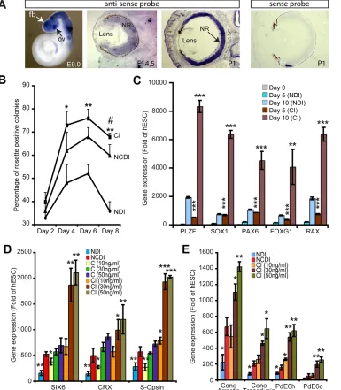
[image:2.612.52.437.291.732.2]the retina and hair follicles at E14.5 and P1 (Fig. 1A), and in the photoreceptor nuclear layer at P60 (supplementary material Fig. S1B). These observations were confirmed using quantitative RT-PCR (qPCR) analysis on tissues from neonates at P3 using
Fig. 1. COCO is a potent retinal and photoreceptor inducer.(A)In situ hybridization on E9.0 mouse embryos (whole mount) revealedCoco expression in the optic vesicle (ov), forebrain (fb) and neural tube, and in the neural retina (NR) at E14.5 and P1 (sections). The sense probe provides a negative control. (B) Analysis of neural rosette induction kinetics in NDI, NCDI, or COCO (30 ng/ml)+IGF1 (10 ng/ml) (CI) culture medium. *P<0.05 and **P<0.01 when compared with NDI-treated culture and#P<0.05 when compared with NCDI-treated culture. (C) hESCs were induced to differentiate in the presence of NDI or CI media for 5 and 10 days and analyzed by qPCR. Data are expressed as fold change over gene expression in undifferentiated hESCs. **P<0.01 and ***P<0.001 when comparing CI with NDI at both time points analyzed. (D,E) hESCs were induced to differentiate in the presence of NDI, NCDI, COCO alone or CI for 3 weeks. Differentiated hESCs were analyzed by qPCR. Data are expressed as fold change over gene expression in undifferentiated hESCs. *P<0.05, **P<0.01, ***P<0.001 as compared with gene expression levels in NCDI-treated cells. (B-E) Results are mean±s.d. (n=3).
RESEARCH ARTICLE Development (2015) 142, 3294-3306 doi:10.1242/dev.125385
DEVEL
O
oligonucleotide pairs that recognize either the 5′coding isoform or both isoforms (supplementary material Fig. S1C).
COCO is a potent neural and photoreceptor inducer
Neural induction of hESCs can be achieved through the formation of floating EBs and subculture on a laminin or Matrigel substrate in serum-free media (supplementary material Fig. S2). Using the hESC line H9, we tested the putative neural/retinal-inducing activity of human recombinant COCO by morphological analysis of EBs maintained in suspension for 4-5 days and then subcultured on Matrigel. We quantified the frequency and kinetics of neural rosette formation (Fig. 1B). In these experiments, COCO (30 ng/ml) was used in combination with IGF1 and FGF2 (referred to as CI medium) or added at 10 ng/ml to the previously described NDI medium (referred to as NCDI medium), which contains noggin, Dkk1, IGF1 and FGF2. The original NDI medium was used as a positive control (Lamba et al., 2006). We found that although more
colonies presented neural rosettes with the NCDI (∼67%) than with
the NDI (∼50%) medium at day 6 of the differentiation protocol,
this process was further improved with the CI medium (∼76%).
To directly compare the neural induction activity of the NDI and CI media, we analyzed the differentiated cells at early time points
for the expression of neural rosette (PLZF; ZBTB16 – HUGO),
pan-neural (SOX1, PAX6), anterior neural/forebrain (FOXG1)
and anterior neural/ventral forebrain/early retinal (RAX) cell fate
markers using qPCR (Chambers et al., 2009; Mathers et al., 1997; Walther and Gruss, 1991). These results revealed that the CI medium provided robust neural and anterior neural/early retinal induction activity (Fig. 1C).
To evaluate the putative retinal-inducing activity of COCO, we
tested whether COCO+FGF2 possessed early retinal (SIX6) and
photoreceptor [CRX, rhodopsin (RHO),NRL, M-opsin (OPN1MW
–HUGO) and S-opsin] inducing activity using qPCR analysis on
colonies isolated at day 21 of the differentiation protocol (Gallardo et al., 1999; Jean et al., 1999). We found that COCO+FGF2 was
very efficient at inducingSIX6,CRXand S-opsin gene expression,
and that this effect was dose dependent (Fig. 1D). The retinal-inducing activity of COCO (30 ng/ml)+FGF2 was comparable to that of NCDI medium. When this was combined with 10 ng/ml IGF1 (the same concentration as in the NDI and NCDI media),
the induction of SIX6, CRX and S-opsin was further enhanced.
This effect was dose dependent, and maximal CI activity was obtained when COCO concentrations ranged between 30 and 50 ng/ml (Fig. 1D). Taken as a whole, the retinal- and
photoreceptor-inducing activity of CI was ∼10-fold more
effective than the NDI medium and∼3- to 4-fold more effective
than the NCDI medium. Expression ofRHOandNRL(rods) and of
M-opsin (M-cones) was not detected after 21 days of hESC differentiation in CI (supplementary material Fig. S3). However, timecourse analysis revealed weak induction (about 4-fold the
hESC level) of theNRLand M-opsin genes at 1 week, and ofRHO
at week 2, suggesting that retinal progenitor cells might be potent to generate all types of photoreceptor cells at these early stages (supplementary material Fig. S3). Robust expression of the
cone-specific genes cone arrestin [also known as arrestin 3 (ARR3) and
X-arrestin], cone transducin (GNAT2), phosphodiesterase 6H
(PDE6H) and phosphodiesterase 6C (PDE6C) was also observed in cells exposed to CI for 21 days (Fig. 1E) (Corbo et al., 2007), altogether suggesting a predominant S-cone photoreceptor cell population. Comparable results, but with variable efficiencies, were obtained with the hESC lines HUES1, HUES8 and HUES9 (supplementary material Fig. S4).
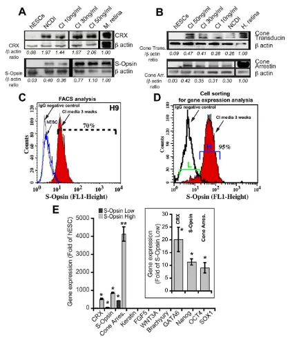
Generation of a highly enriched S-opsin-positive cell population
To further characterize the differentiated cell populations, we performed western blot analyses using extracts from hESCs exposed to NCDI or CI for 21 days. We observed robust expression of CRX and of the phototransduction proteins S-opsin, cone transducin and cone arrestin (Fig. 2A,B). Expression of rhodopsin and of M-opsin proteins was, however, not detected, even when thyroid hormone (T3) was added to the culture media (supplementary material Fig. S6A).
We performed intracellular labeling using an anti-S-opsin antibody combined with fluorescence activated cell sorting (FACS) analysis on cells exposed to CI for 21 days (Chatoo et al.,
2010). We found that∼70±9% (n=5 independent cell differentiation
experiments) of differentiated cells from the H9 cell line highly expressed S-opsin (Fig. 2C). Comparable results were obtained with the HUES1, HUES8 and HUES9 cell lines (supplementary material Fig. S6B). In a separate set of experiments, S-opsin-labeled cell populations were sorted and analyzed by qPCR for the expression of retinal, mesendodermal, ectodermal and ESC markers (Fig. 2D,E).
We found that S-opsinhigh cells (95% in experiment #1, 80% in
experiment #2) expressed photoreceptor genes at levels several orders of magnitude higher than undifferentiated hESCs (Fig. 2E).
The S-opsinlowcells (5% in experiment #1, 20% in experiment #2)
were also positive for CRX, cone arrestin and S-opsin gene
expression but at levels corresponding to 4-10% of those found in
the S-opsinhighpopulation (Fig. 2E, inset). Both populations were,
however, negative for non-retinal lineage-specific genes (Fig. 2E).
Developmental kinetics of the differentiated cells
To study the developmental kinetics of the cell differentiation process, hESCs exposed to CI for 5, 10 and 21 days were analyzed
by immunofluorescence (IF) microscopy for CHX10 (VSX1 –
HUGO), SOX2, RAX and CRX expression (Burmeister et al., 1996; Ellis et al., 2004; Ferda Percin et al., 2000; Graham et al., 2003; Livne-Bar et al., 2006; Mathers et al., 1997; Taranova et al., 2006). At day 5, we observed that the large majority of the cells were positive for the retinal progenitor markers CHX10 (90%) and SOX2 (100%) (Fig. 3A,F,G). This proportion slightly declined at day 10, when numerous RAX-positive and CRX-positive cells also appeared (Fig. 3B,F,G). Yet, almost all CHX10-expressing cells also expressed SOX2 (Fig. 3D). At day 21, few cells positive for CHX10, SOX2 or RAX remained, being replaced by prospective photoreceptor cells expressing CRX (Fig. 3C,F).
Cells expressing S-opsin at the highest levels (40% of S-opsinhigh
cells) were generally located within neural rosettes (Fig. 3E). Most
cells within rosettes also expressed βIII-tubulin (a marker of
immature neurons) at low levels (Fig. 3E). Furthermore,∼3% of
cells present in our cultures had a typical neuronal morphology and
expressedβIII-tubulin at high levels (Fig. 3E). The subtype identity
of these neurons remains to be established. These analyses revealed that, upon exposure to CI, the large majority of hESCs rapidly adopt a retinal progenitor cell identity, which is also rapidly lost toward a cone photoreceptor precursor cell identity.
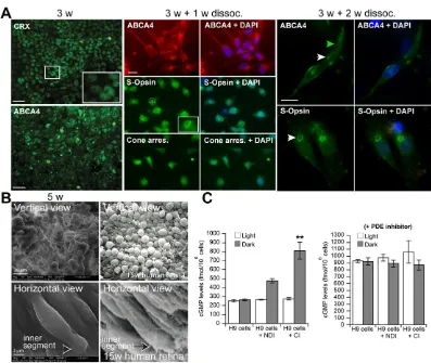
Cone phenotype and cGMP degradationin vitro
In all experiments, hESCs were induced to differentiate into photoreceptors when at confluence, which in our hands improved cell survival and neural/photoreceptor induction. However, this procedure prevented the analysis of single cells. At first, we analyzed cells exposed to CI by high-resolution IF at day 21, which
confirmed immunoreactivity for CRX (mean 72±7%) and ABCA4
DEVEL
O
(mean 67±12%) in densely packed cellular aggregates (Fig. 4A).
ABCA4is mutated in Stargardt disease and is expressed by cone and rod photoreceptors (Allikmets et al., 1997; Molday et al., 2000). Cells differentiated for 21 days were next dissociated to single cells and plated on glass coverslips at low density. After an additional 7 days, they were analyzed by IF for the expression of cone markers. Under these conditions, most cells presented neurites and were positive for ABCA4 (mean 76±11%), S-opsin (mean 79±9%) and
cone arrestin (mean 84.9±5%) (Fig. 4A). Although S-opsin immunoreactivity was diffused throughout the cell at this early stage, it was enriched in the proximal soma of the cells, suggesting possible S-opsin compartmentalization (Fig. 4A, inset).
[image:4.612.96.516.52.541.2]Using the same experimental procedure, single cells were allowed to differentiate for 15 days. On rare occasions, we could observe the formation of more mature cones having an outer segment-like structure (Fig. 4A). These cells were immunoreactive
Fig. 2. COCO induces the generation of a highly enriched S-cone photoreceptor population.(A,B) hESCs cultured in NCDI or CI medium for 3 weeks were subjected to western blot analyses for the expression of CRX and S-opsin (A), or cone transducin and cone arrestin (B).β-actin was used to quantify protein loading. CRX, S-opsin, cone transducin or cone arrestin levels are expressed as a ratio overβ-actin levels. Mouse or human retinas were used as positive controls. (C) Undifferentiated hESCs (blue line) or hESCs cultured in CI for 3 weeks (red) were analyzed by FACS for S-opsin expression. The black line represents the IgG isotypic control serum. (D) hESCs cultured in CI for 3 weeks were sorted by FACS for subsequent gene expression analysis on the basis of S-opsin expression levels [S-opsinlow(L) versus S-opsinhigh(H)]. (E) Sorted cells were analyzed for gene expression by qPCR. Data are expressed as fold change over gene expression in undifferentiated hESCs. In the inset, a parallel analysis was performed in which gene expression in the S-opsinhighcell fraction is represented as fold change over that in the S-opsinlowcell fraction, which was set at 1. Results are mean±s.d. (n=2 cell sortings); *P<0.05 and **P<0.01 as compared with gene expression levels in undifferentiated hESCs and the S-opsinlowcell fraction, respectively.
RESEARCH ARTICLE Development (2015) 142, 3294-3306 doi:10.1242/dev.125385
DEVEL
O
for ABCA4 at the presumptive junction between the inner and outer segments, a localization possibly corresponding to the nascent disk (Fig. 4A). We also observed polarized accumulation of S-opsin at one side of the cells, also suggesting cone maturation (Fig. 4A). Using scanning electron microscopy at day 35 of the differentiation protocol, we observed that cells within these aggregates displayed a buttonhead-like morphology reminiscent of the head-like morphology of week 15 human embryonic cones when viewed vertically (Fig. 4B). In immature cones, this structure corresponds to the apex of the inner segment (Narayanan and Wadhwa, 1998). The morphological similarity was also noticeable when cells were viewed in the horizontal plane, revealing the presence of an inner
segment-like protrusion in thein vitrogenerated cones (Fig. 4B).
One of the unique properties of photoreceptors is to degrade cGMP in response to the activation of photosensitive opsin pigments
by light. This process occurs through release of the α-transducin
[image:5.612.52.567.57.508.2]subunit, which can activate the cGMP phosphodiesterase, ultimately resulting in membrane hyperpolarization (Michaelides et al., 2006). To establish if cells could degrade cGMP in response to light exposure, we measured cGMP levels by immunoassay in cells differentiated for 35 days in NDI or CI medium (Jomary and Jones, 2008). Extracts were isolated from cells exposed to a bright light for 1 min or maintained in the dark for 2 days. No difference in cGMP levels was observed in undifferentiated hESCs between light and dark conditions, in contrast to hESCs differentiated with NDI or CI medium (Fig. 4C). To evaluate the total amount of cGMP
Fig. 3. Developmental kinetics of hESC differentiation into photoreceptors.(A-C) Representative immunofluorescence images of hESCs exposed to CI for 5 (A), 10 (B) or 21 (C) days using antibodies against CHX10, RAX, CRX and SOX2. (D) Co-expression of CHX10 and SOX2 after 10 days of differentiation, as revealed by confocal IF analysis. (E) Expression ofβIII-tubulin and S-opsin after 21 days of differentiation. Arrowheads indicate neurons expressingβIII-tubulin at high levels. The dashed line delineates a neural rosette. Values indicate the percentage of cells expressing high levels ofβIII-tubulin or S-opsin. (F) Quantitative analysis of cells positive for CHX10, SOX2, RAX or CRX. (G) Quantitative analysis of CHX10/SOX2 double-positive cells after 5, 10 and 21 days of differentiation. Results are mean±s.d. (n=3 experiments). Arrows (F,G) indicate the absence of positive cells for the corresponding antibody. Scale bars: 40 µm.
DEVEL
O
hydrolyzed by all phosphodiesterases (PDEs) present in the cells, we applied a non-specific PDE inhibitor (IBMX). The addition of IBMX abolished the difference of cGMP levels between light and dark conditions (Fig. 4C). The level of cGMP hydrolyzed by light-sensitive PDE corresponds to the difference between the levels in
light and dark conditions and represented 240 femtomoles/106cells
in NDI medium and 560 femtomoles/106cells in CI medium. The
total amount of cGMP hydrolyzed by all PDEs (the difference between the cGMP levels in the presence and absence of IBMX
in the light) corresponds to 650 femtomoles/106cells. Therefore,
light-sensitive PDEs account for 36% (240/650) of the total PDE activity in NDI-differentiated cells and for 86% (560/650) in CI-differentiated cells.
Cone phenotype upon cell transplantation in mouse eyes
To test the capacity of the cells to migrate and adopt a cone
phenotypein vivo, we performed cell transplantation experiments
by injecting hESCs differentiated with CI for 2, 3 or 4 weeks into the vitreous of wild-type mouse pups (Lamba et al., 2009). A fraction of the cells that underwent differentiation for 2 weeks, but not for 3 or 4 weeks, could migrate into various layers of the host
retina, as detected using a human-specific mitochondrial antigen antibody (1.4±0.5% from the 10,000 cells injected) (supplementary material Fig. S5A,B). Notably, rare human cells present in the photoreceptor nuclear layer were positive for S-opsin and adopted a morphology that was similar to that of endogenous photoreceptors
(supplementary material Fig. S5B,B′). The non-rodent identity of
the double-positive cells was further confirmed by the unique pattern of chromatin condensation and the larger nuclei of human
cells, as visualized with DAPI (supplementary material Fig. S5B′).
These features were not present in PBS-injected eyes
(supplementary material Fig. S5C). These results suggested that a fraction of the immature human cone progenitors or precursors differentiated with CI could migrate into the mouse retina outer
nuclear layer and adopt an S-cone photoreceptor fatein vivo.
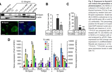
Thyroid hormone signaling allows M-cone genesis
During mouse retinal development, cone differentiation into the M-cone subtype is induced by T3 through activation of the thyroid
hormone receptor β2 (Thrβ2). Thrβ2 can repress the S-opsin
[image:6.612.107.504.54.389.2]promoter while activating the M-opsin gene (Roberts et al., 2006; Swaroop et al., 2010). In previous assays using the EB induction
Fig. 4. Differentiated cells develop neurites and an outer segment-like structure and can degrade cGMP upon light exposure.(A) hESCs differentiated for 21 days (3 w) in CI medium were analyzed by IF. hESCs were differentiated for 3 weeks at confluence in CI medium and then dissociated to single cells and plated on glass coverslips at low density. Cells were analyzed 7 days later (3 w+1 w) by IF, revealing the broad cellular distribution of S-opsin at this early maturation stage but with stronger accumulation in the cell soma (inset), and expression of ABCA4 and cone arrestin. Note the presence of neurites. Cells were further analyzed 15 days later (3 w+2 w) by IF. Note the localization of ABCA4 at the presumptive inner/outer segment junction (white arrowhead) and the presence of an outer segment-like structure (green arrowhead). Note also the polarized accumulation of S-opsin (arrowhead). Scale bars: 5 µm, except 50 µm in 3 w. (B) hESCs differentiated for 35 days were analyzed by scanning electron microscopy. 15-week-old human embryonic retina is shown for comparison. (C) hESCs were cultured in NDI or CI medium for 35 days and cGMP concentrations were measured. (Left) Cells were kept in the dark or exposed to light. (Right) cGMP concentrations were measured in cells cultured in the presence of the PDE inhibitor IBMX. cGMP levels are presented as mean±s.d. (n=3); **P<0.01 as compared with cGMP levels in undifferentiated hESCs cultured in NDI medium.
RESEARCH ARTICLE Development (2015) 142, 3294-3306 doi:10.1242/dev.125385
DEVEL
O
protocol and CI, we were unable to detect M-opsin and Thrb2
expression by qPCR or western blot, even when cells were exposed to various concentrations of T3 (supplementary material Fig. S3 and Fig. S6A). To test if this limitation could be overcome, we induced hESCs predifferentiation by plating them directly on reduced growth factor Matrigel for 5-7 days in hESC medium until they reached confluence, and then added CI medium (thus bypassing the EB induction step) (Chambers et al., 2009). With this modification, addition of T3 to the CI medium resulted in a dose-dependent
activation of M-opsin andThrb2expression (Fig. 5A-D). M-opsin
gene expression reached maximum levels at T3 concentrations of 4-5 nM, decreasing dramatically at higher concentrations (Fig. 5A-D). This result was expected since T3 is toxic for cones
at high concentrations (Ng et al., 2010). In contrast toThrb2, S-opsin
gene expression decreased steadily with the addition of T3, which is suggestive of a binary cell fate choice between S- and M-cones (Fig. 5D). However, because the maximal proportion of M-cones generated (6%) never compensated for the observed reduction in S-cones at 4-5 nM T3 (Fig. 5B,C), S-cone differentiation might also be substantially inhibited by the addition of T3.
Spontaneous development of polarized cellular sheets containing cone photoreceptors
To test for self-organization of retinal tissue, we cultured COCO-exposed hESCs at confluence for 60 days, without additional manipulations (Fig. 6A). This generated a whitish and uniform cellular sheet (or tissue) that could be manipulated (see supplementary material Movie 1). The sheet could be grown to cover an entire well of a 6-well plate or cell culture dish, totaling
∼6×10⁶cells. Pigmented cells were not observed on either side of
the sheet, suggesting the absence of retinal pigment epithelium.
Quantitative analyses revealed that∼80% of the cells were positive
for CRX (Fig. 6B). Confocal IF combined with 3D reconstruction analyses revealed that the sheet was polarized and that peanut agglutinin (PNA) staining, which labels the inner and outer segment membrane of cones, was located at the opposite side of the DAPI-stained nuclear layer (Fig. 6C) (Blanks and Johnson, 1983). The
presumptive PNA+outer segment of cones thus connected with the
Matrigel-coated Petri dish surface (Fig. 6A). On average, the sheet
was 150 µm thick and the nuclear layer was composed of∼5 nuclei,
with additional sparse nuclei randomly distributed (Fig. 6C). Notably, immunolabeling for S-opsin was predominantly observed at the opposite side of the nuclei-rich layer (Fig. 6C).
Formation of the photoreceptor outer segment requires the presence of a connecting cilium (Novarino et al., 2011; Rachel et al., 2012). To test for this, we used three antibodies against proteins located at the connecting cilium, namely RP2, RPGR and acetylated
α-tubulin (Ghosh et al., 2010; Hurd et al., 2010; Rachel et al., 2012).
[image:7.612.55.527.56.369.2]These antibodies, and especially RPGR, decorated a unique rod-shaped structure located in between the S-opsin-labeled outer segments and CRX-labeled nuclei (Fig. 6D). IF analyses of flat-mount P17 mouse retinas and human retinal sections were used to validate the specificity of all antibodies (supplementary material Fig. S7A-C). We used transmission electron microscopy to analyze cellular morphology and observed in longitudinal sections the presence of cells having a well-developed inner segment-like structure containing numerous mitochondria and a large Golgi apparatus (Fig. 6E). These cells had an additional protrusion resembling an immature outer segment, since membrane stacks were not present (Fig. 6E). The presence of a connecting cilium was not observed in the limited number of samples analyzed. In transverse sections and at the level of the mitochondria-rich inner segment, we also observed groups of cells with a rosette-like organization (Fig. 6E).
Fig. 5. Exposure to thyroid hormone can induce the generation of M-cone photoreceptors.(A) hESCs were cultured in CI medium with or without thyroid hormone (T3) for 3 weeks and analyzed by western blot and IF for M-opsin expression. Scale bar: 40 µm. (B,C) hESCs cultured as in A were labeled with anti-M-opsin (B) or anti-S-opsin (C) antibodies. The percentage of cells positive for M-opsin or S-opsin among total DAPI-stained nuclei are shown. Results are mean±s.d. (n=4); *P<0.05, **P<0.01 as compared with cells not treated with T3. (D) hESCs were cultured in NCDI or CI medium with or without T3, and analyzed by qPCR. Data are expressed as fold change over gene expression in undifferentiated hESCs. Results are mean±s.d. (n=3); *P<0.05, **P<0.01, ***P<0.001 as compared with gene expression levels in cells not treated with T3.
DEVEL
O
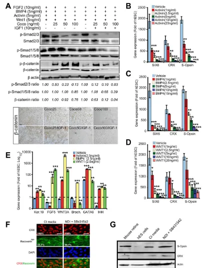
Antagonism between COCO and BMP/TGFβ/Wnt activities
To address the mechanism of COCO activity, hESCs cultured under a constant concentration of FGF2, BMP4, activin A and WNT1 recombinant proteins were exposed, or not, to increasing concentrations of COCO, or of COCO+10 ng/ml IGF1, for a period of 7 days. Because SMAD2/3 proteins are stabilized through
phosphorylation, we used β-actin to establish the phospho (
p)-SMAD2/3 and p-SMAD1/5/8 ratio (Funaba et al., 2002). We found
that phosphorylation of SMAD2/3 (a readout of BMP signaling)
and of SMAD1/5/8 (a readout of TGFβsignaling) was inhibited in a
dose-dependent manner by COCO, and that addition of IGF1 greatly increased COCO inhibitory activity (Fig. 7A).
p-β-catenin, which represents the β-catenin pool targeted for
proteosomal degradation, was also greatly increased by the addition of COCO, while addition of IGF1 ultimately resulted in
[image:8.612.88.518.56.565.2]a net reduction in p-β-catenin (at 50 and 100 ng/ml COCO), which
Fig. 6. Spontaneous development of a cellular sheet containing polarized cones.(A) Method for the derivation of a cellular sheet containing polarized cones. hESCs at confluence were exposed to CI medium (60 ng/ml COCO, daily media change) for 60 days and analyzed by confocal microscopy for 3D reconstruction, and by transmission electron microscopy. (B) Cellular sheet stained for CRX and analyzed by confocal microscopy. The boxed region is magnified to the right. (C) Cellular sheet stained for CRX, S-opsin and PNA. Scale bars: 40 µm. (D) Cellular sheet stained for connecting cilium markers (RPGR, acetylated
α-tubulin and RP2). (E) Cellular sheets analyzed by transmission electron microscopy. Numbers on the transverse section reveal cells organized in a rosette-like conformation. Arrows indicate the junction between the inner (is) and outer (os) segment. m, mitochondria; z, stratum limitans junction.
RESEARCH ARTICLE Development (2015) 142, 3294-3306 doi:10.1242/dev.125385
DEVEL
O
is likely to reflect depletion of the β-catenin pool. Accordingly,
the total level of β-catenin (a readout of Wnt signaling)
progressively decreased with the addition of COCO, while
β-catenin expression was nearly abolished with 50-100 ng/ml
[image:9.612.100.511.54.589.2]COCO+10 ng/ml IGF1 (Fig. 7A). Comparable results were also obtained by immunohistochemistry analysis (Fig. 7A, lower panel). In converse experiments, hESCs differentiated with CI were exposed to increasing concentrations of activin A, BMP4 or WNT1
Fig. 7. COCO cooperates with IGF1 to inhibit BMP/TGFβ/Wnt activities.(A) hESCs cultured as indicated for 7 days were subjected to western blot (top) or immunohistochemistry (bottom) analyses.β-actin was used to quantify protein loading. p-SMAD2/3, p-SMAD1/5/8 orβ-catenin levels are expressed as a ratio overβ-actin levels. (B-D) hESCs were cultured for 21 days in CI without (vehicle, PBS) or with increasing concentrations of activin A, BMP4 or WNT1 and analyzed by qPCR. Data are expressed as fold change over gene expression in undifferentiated hESCs. Results are mean±s.d. (n=3); *P<0.05, **P<0.01, ***P<0.001 as compared with gene expression levels in vehicle-treated cells. (E) hESCs were cultured in CI for 21 days with the addition of 2.5 ng/ml activin A, BMP4 or WNT1, and then analyzed by qPCR. Data are expressed as fold change over gene expression in undifferentiated hESCs. Results are mean±s.d. (n=3); *P<0.05, **P<0.01, ***P<0.001 as compared with gene expression levels in undifferentiated cells. (F,G) hESCs cultured in CI or NDI+SB431542 (TGFβinhibitor) for 21 days were analyzed by IF (F) or western blot (G). The percentage of positive cells is indicated (F). Scale bars: 40 µm.
DEVEL
O
for 3 weeks. We observed that even 1 ng/ml BMP4, activin A or WNT1 was sufficient to inhibit CI retinal-inducing activity and that this effect was dose dependent (Fig. 7B-D). Accordingly, adding a fixed concentration (2.5 ng/ml) of activin A, BMP4 or WNT1 to CI-treated cells induced differentiation of hESCs toward the epidermal (FGF5andKRT19) and mesendodermal [WNT3A, brachyury (T),
GATA6 and IHH] cell lineages (Fig. 7E). Conversely, hESCs
exposed to NDI+TGFβinhibitor (SB431542) could differentiate as
efficiently into S-cones as those exposed to CI when analyzed for CRX, recoverin and S-opsin expression (Fig. 7F,G).
Taken together, these results support the notion that COCO can promote neural and retinal cell fate from hESCs by simultaneous
inhibition of BMP/TGFβ/Wnt signaling, and are consistent with the
previously described inhibitory function of COCO on these pathways (Bell et al., 2003). They also suggest that IGF1 can
greatly enhance the inhibitory activity of COCO on BMP/TGFβ/
Wnt signaling.
DISCUSSION
We report on the rapid and efficient differentiation of hESCs into cone photoreceptors. ESCs were cultured under chemically defined, feeder-free conditions, and cell differentiation was induced under serum-free culture conditions without preselection procedures. Although COCO (+FGF2) displayed robust neuronal and retinal inducing activity, S-cone photoreceptor cell fate was induced to unprecedented levels when COCO was used in combination with IGF1. Differentiated cells expressed cone-specific genes and key proteins involved in phototransduction. After 35 days,
in vitro generated cells were similar in morphology to week 15 human embryonic cones and could degrade cGMP when exposed to light. Bypassing the EB induction step also allowed manipulation of
S- and M-cone cell fate by the addition of T3. After 60 days,in vitro
generated cells self-organized into a cellular sheet with polarized cone photoreceptors and showing evidence of connecting cilium and outer segment formation.
In the developing mouse retina, photoreceptor development apparently follows an S-cone default pathway that is determined by
Crx and Thrβ2; Crx induces the expression of S-opsin by default,
whereas Thrβ2 suppresses it and instead induces the expression of
M-opsin (Ng et al., 2001; Swaroop et al., 2010; Yanagi et al., 2002). Likewise, both Nrl and Notch1 inhibit cone formation to promote genesis of other retinal cell types (Jadhav et al., 2006; Mears et al., 2001; Yaron et al., 2006). We found here that most retinal progenitors derived from hESCs become S-cones upon exposure to COCO, FGF2 and IGF1. Notably, using a simplified neural induction protocol, M-cone genesis could also be induced by adding T3. During this process, 9-cis RA is, in principle, required
to activate RA receptorγ(Rarγ), which cooperates with Thrβ2 to
repress S-opsin expression (Roberts et al., 2006; Swaroop et al., 2010). Although we did not add RA to our cultures, the precursor of RA synthesis, retinyl acetate, is present in the B27 supplement.
We showed that antagonism between COCO and BMP4, Activin and WNT1 activities is required to allow hESC differentiation into retinal and photoreceptor cells. More surprisingly, we found that COCO (+FGF2) was sufficient to promote hESC differentiation into retinal and photoreceptor progenitors, with comparable efficiency to NCDI. This is notable because noggin and Dkk1 are unable to induce retinal cell fate from hESCs in the absence of IGF1 (Lamba et al., 2006). One explanation is that COCO, in contrast to
noggin and Dkk1, is also able to block TGFβsignaling. Hence,
pharmaceutical inhibition of TGFβ in combination with NDI
provided comparable efficiency to CI (Fig. 7). Likewise, the
relatively low efficiency of Dkk1 and LEFTY at inducing hESC differentiation into photoreceptors might be explained by the inability of LEFTY to block Activin signaling (Schier, 2009), combined with the absence of BMP inhibition (Osakada et al.,
2008). This raises the possibility that Wnt, BMP and TGFβ/Nodal/
Activin signaling also exert cone-inhibitory activities during normal
retinal development. Accordingly, in theNrl−/−mouse retina, which
is composed of an excessively large number of cones, theBmp4,
Smad4and Wnt/Ca2+signaling pathway genes are downregulated,
and Nrl was found to directly activate these genes in order to promote rods genesis at the expense of cones (Yu et al., 2004). Finally, both Wnt and Activin can promote rod genesis (Davis et al., 2000).
These findings suggest a working model whereby inhibition of
Wnt, BMP and TGFβ/Nodal/Activin is required to allow cone
genesis at the expense of rods (and possibly of other retinal cell types), and suggest that S-cones are generated by default if all inhibitory factors are blocked. Our data also support the possibility that retinal cell fate can be actively promoted, as evidenced by the improved differentiation of hESCs into S-cones when IGF1 was used in combination with COCO. Likewise, although early neural induction efficiency was similar when using either COCO+IGF1 or COCO+FGF2, anterior neural and retinal induction was more efficient with IGF1. However, the mechanism by which IGF1 operates in this context is still unclear. IGF1 retinal fate-promoting activity may in part be through inhibition of Smad signaling (Pera et al., 2001). Indeed, we observed robust cooperation between COCO and IGF1 in blocking SMAD2/3 and SMAD1/5/8
phosphorylation, as well as in reducingβ-catenin stabilization.
In eye diseases in which cones are severely affected, such as macular degenerations, cone dystrophies and cone-rod dystrophies, differentiation of hESCs into cones is of particular interest for cell therapy by transplantation (Michaelides et al., 2006). Although most forms of retinitis pigmentosa primarily affect rods, the disease is followed by loss of cones, possibly because of a reduction in the trophic support normally provided by rods to cones and/or increased metabolic stress (Mohand-Said et al., 2000; Punzo et al., 2009). Thus, the efficiency of cell replacement therapy to treat retinitis pigmentosa may also depend on our capacity to generate rods and cones. Using scanning electron microscopy, we observed that cells differentiated for 35 days developed a protruding inner segment and displayed morphological similarities to week 15 human embryonic cones. Prolonging the cell culture time to 60 days resulted in the formation of a cellular sheet composed of polarized cones with evidence of connecting cilium and outer segment formation. Self-organization of hESCs into a cone tissue sheet is reminiscent of the self-formation of hESCs into an optic cup (Nakano et al., 2012). Human cone tissue sheets may be used as a retinal patch for the treatment of macular degenerations, as shown for mouse retinal sheets transplanted into a retinitis pigmentosa mouse model (Assawachananont et al., 2014).
In conclusion, the availability of a highly enriched human cone photoreceptor population opens new avenues to study cone
biochemistry and developmental genetics. Using induced
pluripotent stem cell technology, it will also be possible to generate cones from cone-affected disease patients in order to study the disease mechanism and perform drug-screening assays (Jin et al., 2011).
MATERIALS AND METHODS Ethics statement
The Animal Care Committee of the Maisonneuve-Rosemont Hospital Research Centre approved the use of the animals in this study. Post-mortem
RESEARCH ARTICLE Development (2015) 142, 3294-3306 doi:10.1242/dev.125385
DEVEL
O
human eyes (see the supplementary Materials and Methods) were provided by the Banque d’yeux du Québec du Centre Michel-Mathieu and were used with approbation of the Comité d’Éthique à la Recherche de l’Hôpital Maisonneuve-Rosemont. hESCs were used in accordance with Canadian Institute Health Research (CIHR) guidelines and approved by the Comité de Surveillance de la Recherche sur les Cellules Souches (CSRCS) of the CIHR.
Cell cultures
The hESC line H9 (WiCell) was cultured on a Matrigel-coated plate (BD Biosciences) with a daily change of mTeSR medium according to the manufacturer’s instruction (Stemcell Technologies) (Thomson et al., 1998). The H9 hESC line was first established on mouse embryonic fibroblasts (MEFs) and then cultured on Matrigel in mTeSR medium. Undifferentiated hESC colonies were treated with dispase and induced to form EBs in ultra-low attachment plates (VWR) in neural induction medium, which consists of the NDI mix [DMEM-F12 medium (Invitrogen) containing 10% KnockOut serum, 2% B27, 1 ng/ml noggin, 1 ng/ml Dkk1 and 5 ng/ml IGF1], essentially as described (Lamba et al., 2006). Otherwise, cells were cultured in NCDI mix (NDI supplemented with 10 ng/ml COCO) using COCO alone or together with 10 ng/ml IGF1. EBs were plated 3 days later into Matrigel-or laminin-coated plates and cultured in DMEM-F12 medium supplemented with 2% B27, 1% N2, 10 ng/ml noggin, 10 ng/ml Dkk1, 10 ng/ml IGF1, 10 ng/ml COCO and 5 ng/ml FGF2 for an additional 4 weeks. The media were changed every 2 days. Unless otherwise stated, COCO was used at 30 ng/ml in CI media. Recombinant proteins were purchased from R&D Systems.
Western blot
Total protein extracts were prepared in the Complete Mini Protease Inhibitor Cocktail solution (Roche Diagnostics) and sonicated. Protein content was quantified using the Bradford reagent. Proteins in Laemmli buffer were resolved by SDS-PAGE and transferred to a nitrocellulose blotting membrane (Pall). Membranes were blocked for 1 h in 5% non-fat milk in 1×TBS containing 0.05% Tween 20 and incubated overnight with primary antibodies: mouse anti-CRX (1:1000; Genetex, GTX91782), rabbit anti-S-opsin (1:1000; Abcam, ab81017), mouse anti-rhodanti-S-opsin (4D2) (1:50, provided by the Robert S. Molday Laboratory, University of British Columbia, Canada), rabbit anti-M-opsin (1:500; Chemicon, AB5405), rabbit anti-Smad1/5/8 (1:250; Santa Cruz, sc-6031-R), rabbit anti-p-Smad1/ 5/8 (1:250; #9516), rabbit anti-Smad2/3 (1:300; #8685), rabbit anti-p-Smad2/3 (1:300; #8828), rabbit anti-β-catenin (1:300; #8480), rabbit
anti-p-β-catenin (1:300; #4176) (all from Cell Signaling), and mouse anti-β-actin (1:1000; Abcam, ab8226). Membranes were treated with the appropriate horseradish peroxidase-conjugated secondary antibodies (anti-rabbit, 1:15,000, A9169; anti-mouse, 1:10,000, A8924; Sigma) and developed using Immobilon western reagents (Millipore).
Immunohistochemistry
Fixed/permeabilized cells were incubated with primary antibodies overnight at 4°C, and analyzed using the Vectastain ABC Kit (Vector Laboratories); DAB (Sigma) was used as the peroxidase substrate. For grafting analyses, eye sections were incubated overnight with primary antibody solutions at 4°C, washed and incubated with Rhodamine-conjugated and FITC-conjugated secondary antibodies for 1 h at room temperature. Slides were mounted on coverslips in DAPI-containing mounting medium (Vector Laboratories). Observations were made under a Leica DMRE fluorescence microscope with a Retiga EX digital camera. For further details of sample preparation and the antibodies used, see the supplementary Materials and Methods.
In situhybridization
Forin situhybridizations on slices, tissues were dissected in PBS, embedded in CRYOMATRIX embedding medium (Thermo Shandon), snap frozen in liquid nitrogen and sections (8 µm) were cut and dried onto Superfrost glass slides (Fisher Scientific). For in situ hybridizations on whole embryos, embryos were dissected in PBS, then fixed overnight in 4% paraformaldehyde at 4°C. Whole embryos and slices were hybridized with
digoxigenin-labeled RNA probes [Dand5(Coco), accession #BC115659] and revealed with alkaline phosphatase-coupled anti-digoxigenin antibody (1:2000; Roche, 11093274910) and NBT/BCIP substrate (Boehringer) at pH 9.5.
RT-PCR
Total RNA (1 µg) was reverse transcribed with M-MLV reverse transcriptase (Invitrogen) and used in quantitative real-time PCR (qPCR) using Platinum SYBR Green SuperMix (Invitrogen) and an ABI Prism 7000 apparatus, with GAPDH as an internal standard. Experiments were performed at least in triplicate. For further details and primer sequences, see the supplementary Materials and Methods.
Phototransduction analysis
Phototransduction activity was assessed by measuring the light-induced hydrolysis of cyclic (c) GMP with an enzyme immunoassay kit (Biotrack EIA system) according to the manufacturer’s instructions (Amersham Bioscience GE Healthcare). Undifferentiated H9 hESCs and cells cultured in NDI, COCO and IGF1 were kept in the dark or exposed to ambient light. The PDE inhibitor IBMX (3-isobutyl-1-methylxanthine, Sigma) was added (1 mM) 72 h before determination of cGMP levels.
Fluorescence-activated cell sorting
For cell membrane epitope staining, dispase-dissociated cells were stained with Rhodamine-coupled PNA (1:500; Vector Laboratories, RL-1072). Cells were then fixed/permeabilized with the CytoFix/Cytoperm Kit according to the manufacturer’s instructions (BD Biosciences). Cells were incubated with rabbit anti-S-opsin antibody (1:2000; Chemicon, A5407) or Alexa-conjugated isotypic control goat anti-rabbit IgG antibody (1:500; Molecular Probes, A-11037). The primary antibody was visualized using FITC-conjugated goat anti-rabbit serum (1:500; Chemicon, AP124F). Sorting employed a FACSVantage flow cytometer (Becton-Dickinson) at a flow rate of less than 3000 cells/s. Dead cells and cell debris were excluded from acquisition by gating with FCS and SSC biparametric plot. After sorting, an aliquot of sorted cells was always reanalyzed to check for purity, which was usually greater than 95%.
Cell transplantation
Cell clamps (1 µl) containing∼10,000 cells were injected into the vitreous of anesthetized neonatal (P1) mice as described in the supplementary Materials and Methods.
Statistical analysis
Statistical differences were analyzed using Student’st-test for unpaired samples. An analysis of variance (ANOVA) followed by the Dunnett test was used for multiple comparisons with one control group.P-values are given in the figure legends.
Acknowledgements
We are grateful to D. Melton and the Harvard Stem Cell Institute for the HUES1, HUES8 and HUES9 cell lines, and to M. Nelea and S. Breault for technical assistance with electron microscopy.
Competing interests
The authors declare no competing or financial interests.
Author contributions
Conceived and designed the study and wrote the paper: G.B. Performed the experiments: S.Z., A.F., M.A., N.T., A.B., S.W. Analyzed the data: G.B., S.Z., A.F., M.A.
Funding
This work was supported by grants from the Foundation Fighting Blindness Canada, Turmel Family Foundation for Macular Degeneration Research, Canadian Stem Cell Network, C. Durand Foundation, the GO Foundation, and Natural Science and Engineering Research Council of Canada [grant #250970-2012]. A.F. was supported by fellowships from the Réseau Vision du Québec and from University of Montreal Molecular Biology Program. G.B. was supported by the Fonds de
Recherche en Santédu Québec.
DEVEL
O
Supplementary material
Supplementary material available online at
http://dev.biologists.org/lookup/suppl/doi:10.1242/dev.125385/-/DC1
References
Allikmets, R., Singh, N., Sun, H., Shroyer, N. F., Hutchinson, A., Chidambaram, A., Gerrard, B., Baird, L., Stauffer, D., Peiffer, A. et al.(1997). A photoreceptor cell-specific ATP-binding transporter gene (ABCR) is mutated in recessive Starqardt macular dystrophy.Nat. Genet.15, 236-246.
Assawachananont, J., Mandai, M., Okamoto, S., Yamada, C., Eiraku, M., Yonemura, S., Sasai, Y. and Takahashi, M. (2014). Transplantation of embryonic and induced pluripotent stem cell-derived 3D retinal sheets into retinal degenerative mice.Stem Cell Rep.2, 662-674.
Banin, E., Obolensky, A., Idelson, M., Hemo, I., Reinhardtz, E., Pikarsky, E., Ben-Hur, T. and Reubinoff, B.(2006). Retinal incorporation and differentiation of neural precursors derived from human embryonic stem cells. Stem Cells24, 246-257.
Bell, E., Muñoz-Sanjuán, I., Altmann, C. R., Vonica, A. and Brivanlou, A. H. (2003). Cell fate specification and competence by Coco, a maternal BMP, TGFbeta and Wnt inhibitor.Development130, 1381-1389.
Blanks, J. C. and Johnson, L. V.(1983). Selective lectin binding of the developing mouse retina.J. Comp. Neurol.221, 31-41.
Bouwmeester, T., Kim, S.-H., Sasai, Y., Lu, B. and De Robertis, E. M.(1996). Cerberus is a head-inducing secreted factor expressed in the anterior endoderm of Spemann’s organizer.Nature382, 595-601.
Burmeister, M., Novak, J., Liang, M.-Y., Basu, S., Ploder, L., Hawes, N. L., Vidgen, D., Hoover, F., Goldman, D., Kalnins, V. I. et al. (1996). Ocular retardation mouse caused by Chx10 homeobox null allele: impaired retinal progenitor proliferation and bipolar cell differentiation.Nat. Genet.12, 376-384. Chambers, S. M., Fasano, C. A., Papapetrou, E. P., Tomishima, M., Sadelain, M.
and Studer, L.(2009). Highly efficient neural conversion of human ES and iPS cells by dual inhibition of SMAD signaling.Nat. Biotechnol.27, 275-280. Chatoo, W., Abdouh, M., Duparc, R.-H. and Bernier, G. (2010). Bmi1
distinguishes immature retinal progenitor/stem cells from the main progenitor cell population and is required for normal retinal development.Stem Cells28, 1412-1423.
Chen, S., Wang, Q.-L., Nie, Z., Sun, H., Lennon, G., Copeland, N. G., Gilbert, D. J., Jenkins, N. A. and Zack, D. J. (1997). Crx, a novel Otx-like paired-homeodomain protein, binds to and transactivates photoreceptor cell-specific genes.Neuron19, 1017-1030.
Corbo, J. C., Myers, C. A., Lawrence, K. A., Jadhav, A. P. and Cepko, C. L. (2007). A typology of photoreceptor gene expression patterns in the mouse.Proc. Natl. Acad. Sci. USA104, 12069-12074.
Couly, G. and Le Douarin, N. M.(1988). The fate map of the cephalic neural primordium at the presomitic to the 3-somite stage in the avian embryo. Development103, 101-113.
Davis, A. A., Matzuk, M. M. and Reh, T. A.(2000). Activin A promotes progenitor differentiation into photoreceptors in rodent retina.Mol. Cell. Neurosci.15, 11-21. Ellis, P., Fagan, B. M., Magness, S. T., Hutton, S., Taranova, O., Hayashi, S., McMahon, A., Rao, M. and Pevny, L.(2004). SOX2, a persistent marker for multipotential neural stem cells derived from embryonic stem cells, the embryo or the adult.Dev. Neurosci.26, 148-165.
Ferda Percin, E., Ploder, L. A., Yu, J. J., Arici, K., Horsford, D. J., Rutherford, A., Bapat, B., Cox, D. W., Duncan, A. M. V., Kalnins, V. I. et al.(2000). Human microphthalmia associated with mutations in the retinal homeobox gene CHX10. Nat. Genet.25, 397-401.
Freund, C. L., Gregory-Evans, C. Y., Furukawa, T., Papaioannou, M., Looser, J., Ploder, L., Bellingham, J., Ng, D., Herbrick, J.-A. S., Duncan, A. et al.(1997). Cone-rod dystrophy due to mutations in a novel photoreceptor-specific homeobox gene (CRX) essential for maintenance of the photoreceptor.Cell91, 543-553. Funaba, M., Zimmerman, C. M. and Mathews, L. S.(2002). Modulation of
Smad2-mediated signaling by extracellular signal-regulated kinase.J. Biol. Chem.277, 41361-41368.
Furukawa, T., Morrow, E. M. and Cepko, C. L.(1997). Crx, a novel otx-like homeobox gene, shows photoreceptor-specific expression and regulates photoreceptor differentiation.Cell91, 531-541.
Gallardo, M. E., Lopez-Rios, J., Fernaud-Espinosa, I., Granadino, B., Sanz, R., Ramos, C., Ayuso, C., Seller, M. J., Brunner, H. G., Bovolenta, P. et al.(1999). Genomic cloning and characterization of the human homeobox gene SIX6 reveals a cluster of SIX genes in chromosome 14 and associates SIX6 hemizygosity with bilateral anophthalmia and pituitary anomalies.Genomics61, 82-91.
Ghosh, A. K., Murga-Zamalloa, C. A., Chan, L., Hitchcock, P. F., Swaroop, A. and Khanna, H.(2010). Human retinopathy-associated ciliary protein retinitis pigmentosa GTPase regulator mediates cilia-dependent vertebrate development. Hum. Mol. Genet.19, 90-98.
Graham, V., Khudyakov, J., Ellis, P. and Pevny, L.(2003). Sox2 functions to maintain neural progenitor identity.Neuron39, 749-765.
Hurd, T., Zhou, W., Jenkins, P., Liu, C.-J., Swaroop, A., Khanna, H., Martens, J., Hildebrandt, F. and Margolis, B.(2010). The retinitis pigmentosa protein RP2
interacts with polycystin 2 and regulates cilia-mediated vertebrate development. Hum. Mol. Genet.19, 4330-4344.
Idelson, M., Alper, R., Obolensky, A., Ben-Shushan, E., Hemo, I., Yachimovich-Cohen, N., Khaner, H., Smith, Y., Wiser, O., Gropp, M. et al.(2009). Directed differentiation of human embryonic stem cells into functional retinal pigment epithelium cells.Cell Stem Cell5, 396-408.
Jadhav, A. P., Mason, H. A. and Cepko, C. L. (2006). Notch 1 inhibits photoreceptor production in the developing mammalian retina.Development
133, 913-923.
Jean, D., Bernier, G. and Gruss, P.(1999). Six6 (Optx2) is a novel murine Six3-related homeobox gene that demarcates the presumptive pituitary/hypothalamic axis and the ventral optic stalk.Mech. Dev.84, 31-40.
Jin, Z.-B., Okamoto, S., Osakada, F., Homma, K., Assawachananont, J., Hirami, Y., Iwata, T. and Takahashi, M.(2011). Modeling retinal degeneration using patient-specific induced pluripotent stem cells.PLoS ONE6, e17084. Jomary, C. and Jones, S. E. (2008). Induction of functional photoreceptor
phenotype by exogenous Crx expression in mouse retinal stem cells.Invest. Ophthalmol. Vis. Sci.49, 429-437.
Lamba, D. A., Karl, M. O., Ware, C. B. and Reh, T. A.(2006). Efficient generation of retinal progenitor cells from human embryonic stem cells.Proc. Natl. Acad. Sci. USA103, 12769-12774.
Lamba, D. A., Gust, J. and Reh, T. A.(2009). Transplantation of human embryonic stem cell-derived photoreceptors restores some visual function in Crx-deficient mice.Cell Stem Cell4, 73-79.
Liu, W., Lagutin, O., Swindell, E., Jamrich, M. and Oliver, G.(2010). Neuroretina specification in mouse embryos requires Six3-mediated suppression of Wnt8b in the anterior neural plate.J. Clin. Invest.120, 3568-3577.
Livne-Bar, I., Pacal, M., Cheung, M. C., Hankin, M., Trogadis, J., Chen, D., Dorval, K. M. and Bremner, R.(2006). Chx10 is required to block photoreceptor differentiation but is dispensable for progenitor proliferation in the postnatal retina. Proc. Natl. Acad. Sci. USA103, 4988-4993.
Mathers, P. H., Grinberg, A., Mahon, K. A. and Jamrich, M.(1997). The Rx homeobox gene is essential for vertebrate eye development. Nature 387, 603-607.
Mears, A. J., Kondo, M., Swain, K. S., Takada, Y., Bush, R. A., Saunders, T. L., Sieving, P. A. and Swaroop, A.(2001). Nrl is required for rod photoreceptor development.Nat. Genet.29, 447-452.
Mellough, C. B., Collin, J., Khazim, M., White, K., Sernagor, E., Steel, D. H. and Lako, M.(2015). IGF-1 signaling plays an important role in the formation of three-dimensional laminated neural retina and other ocular structures from human embryonic stem cells.Stem Cells33, 2416-2430.
Michaelides, M., Hardcastle, A. J., Hunt, D. M. and Moore, A. T. (2006). Progressive cone and cone-rod dystrophies: phenotypes and underlying molecular genetic basis.Surv. Ophthalmol.51, 232-258.
Mohand-Said, S., Hicks, D., Dreyfus, H. and Sahel, J. A. (2000). Selective transplantation of rods delays cone loss in a retinitis pigmentosa model.Arch. Ophthalmol.118, 807-811.
Molday, L. L., Rabin, A. R. and Molday, R. S.(2000). ABCR expression in foveal cone photoreceptors and its role in stargardt macular dystrophy. Am. J. Ophthalmol.130, 689.
Muñoz-Sanjuán, I. and Brivanlou, A. H.(2002). Neural induction, the default model and embryonic stem cells.Nat. Rev. Neurosci.3, 271-280.
Nakano, T., Ando, S., Takata, N., Kawada, M., Muguruma, K., Sekiguchi, K., Saito, K., Yonemura, S., Eiraku, M. and Sasai, Y.(2012). Self-formation of optic cups and storable stratified neural retina from human ESCs.Cell Stem Cell10, 771-785.
Narayanan, K. and Wadhwa, S.(1998). Photoreceptor morphogenesis in the human retina: a scanning electron microscopic study.Anat. Rec.252, 133-139. Ng, L., Hurley, J. B., Dierks, B., Srinivas, M., Saltó, C., Vennström, B., Reh, T. A.
and Forrest, D.(2001). A thyroid hormone receptor that is required for the development of green cone photoreceptors.Nat. Genet.27, 94-98.
Ng, L., Lyubarsky, A., Nikonov, S. S., Ma, M., Srinivas, M., Kefas, B., St. Germain, D. L., Hernandez, A., Pugh, E. N., Jr and Forrest, D.(2010). Type 3 deiodinase, a thyroid-hormone-inactivating enzyme, controls survival and maturation of cone photoreceptors.J. Neurosci.30, 3347-3357.
Novarino, G., Akizu, N. and Gleeson, J. G.(2011). Modeling human disease in humans: the ciliopathies.Cell147, 70-79.
Osakada, F., Ikeda, H., Mandai, M., Wataya, T., Watanabe, K., Yoshimura, N., Akaike, A., Sasai, Y. and Takahashi, M.(2008). Toward the generation of rod and cone photoreceptors from mouse, monkey and human embryonic stem cells. Nat. Biotechnol.26, 215-224.
Pacione, L. R., Szego, M. J., Ikeda, S., Nishina, P. M. and McInnes, R. R.(2003). Progress toward understanding the genetic and biochemical mechanisms of inherited photoreceptor degenerations.Annu. Rev. Neurosci.26, 657-700. Pera, E. M., Wessely, O., Li, S.-Y. and De Robertis, E. M.(2001). Neural and head
induction by insulin-like growth factor signals.Dev. Cell1, 655-665.
Piccolo, S., Agius, E., Leyns, L., Bhattacharyya, S., Grunz, H., Bouwmeester, T. and De Robertis, E. M.(1999). The head inducer Cerberus is a multifunctional antagonist of Nodal, BMP and Wnt signals.Nature397, 707-710.
RESEARCH ARTICLE Development (2015) 142, 3294-3306 doi:10.1242/dev.125385
DEVEL
O
Punzo, C., Kornacker, K. and Cepko, C. L.(2009). Stimulation of the insulin/mTOR pathway delays cone death in a mouse model of retinitis pigmentosa. Nat. Neurosci.12, 44-52.
Rachel, R. A., Li, T. and Swaroop, A.(2012). Photoreceptor sensory cilia and ciliopathies: focus on CEP290, RPGR and their interacting proteins.Cilia1, 22. Reubinoff, B. E., Pera, M. F., Fong, C. Y., Trounson, A. and Bongso, A.(2000).
Embryonic stem cell lines from human blastocysts: somatic differentiation in vitro. Nat. Biotechnol.18, 399-404.
Roberts, M. R., Srinivas, M., Forrest, D., Morreale de Escobar, G. and Reh, T. A. (2006). Making the gradient: thyroid hormone regulates cone opsin expression in the developing mouse retina.Proc. Natl. Acad. Sci. USA103, 6218-6223. Rorick, A. M., Mei, W., Liette, N. L., Phiel, C., El-Hodiri, H. M. and Yang, J.(2006).
PP2A:B56epsilon is required for eye induction and eye field separation.Dev. Biol. 302, 477-493.
Schier, A. F.(2009). Nodal morphogens.Cold Spring Harb. Perspect. Biol.1, a003459.
Swaroop, A., Kim, D. and Forrest, D. (2010). Transcriptional regulation of photoreceptor development and homeostasis in the mammalian retina.Nat. Rev. Neurosci.11, 563-576.
Taranova, O. V., Magness, S. T., Fagan, B. M., Wu, Y., Surzenko, N., Hutton, S. R. and Pevny, L. H.(2006). SOX2 is a dose-dependent regulator of retinal neural progenitor competence.Genes Dev.20, 1187-1202.
Thomson, J. A., Itskovitz-Eldor, J., Shapiro, S. S., Waknitz, M. A., Swiergiel, J. J., Marshall, V. S. and Jones, J. M.(1998). Embryonic stem cell lines derived from human blastocysts.Science282, 1145-1147.
Tropepe, V., Hitoshi, S., Sirard, C., Mak, T. W., Rossant, J. and van der Kooy, D. (2001). Direct neural fate specification from embryonic stem cells: a primitive mammalian neural stem cell stage acquired through a default mechanism.Neuron
30, 65-78.
Tucker, B. A., Mullins, R. F., Streb, L. M., Anfinson, K., Eyestone, M. E., Kaalberg, E., Riker, M. J., Drack, A. V., Braun, T. A. and Stone, E. M.(2013). Patient-specific iPSC-derived photoreceptor precursor cells as a means to investigate retinitis pigmentosa.Elife2, e00824.
Walther, C. and Gruss, P.(1991). Pax-6, a murine paired box gene, is expressed in the developing CNS.Development113, 1435-1449.
Yanagi, Y., Takezawa, S. and Kato, S.(2002). Distinct functions of photoreceptor cell-specific nuclear receptor, thyroid hormone receptor beta2 and CRX in one photoreceptor development.Invest. Ophthalmol. Vis. Sci.43, 3489-3494. Yaron, O., Farhy, C., Marquardt, T., Applebury, M. and Ashery-Padan, R.(2006).
Notch1 functions to suppress cone-photoreceptor fate specification in the developing mouse retina.Development133, 1367-1378.
Yu, J., He, S., Friedman, J. S., Akimoto, M., Ghosh, D., Mears, A. J., Hicks, D. and Swaroop, A.(2004). Altered expression of genes of the Bmp/Smad and Wnt/ calcium signaling pathways in the cone-only Nrl-/- mouse retina, revealed by gene profiling using custom cDNA microarrays.J. Biol. Chem.279, 42211-42220.