Haemostasis treatment using dual red
imaging during endoscopic submucosal
dissection: a multicentre, open-label,
randomised controlled trial
Ai Fujimoto, 1,2 Yutaka Saito,3 Seiirhicro Abe,3 Shu Hoteya,4 Kosuke Nomura,4
Hiroshi Yasuda,5 Yasumasa Matsuo,5 Toshio Uraoka,6 Shiko Kuribayashi,6
Itaru Saito,7 Yosuke Tsuji,7 Tadateru Maehata,1 Yasutoshi Ochiai,1
Toshihiro Nishizawa,2 Naohisa Yahagi1
To cite: Fujimoto A, Saito Y, Abe S, et al. Haemostasis treatment using dual red imaging during endoscopic submucosal dissection: a multicentre, open-label, randomised controlled trial. BMJ Open Gastro 2019;6:e000275. doi:10.1136/ bmjgast-2019-000275 Received 12 January 2019 Revised 19 February 2019 Accepted 2 March 2019
For numbered affiliations see end of article.
Correspondence to
Dr Naohisa Yahagi; yahagi. keio@ gmail. com
Endoscopy
© Author(s) (or their employer(s)) 2019. Re-use permitted under CC BY-NC. No commercial re-use. See rights and permissions. Published by BMJ.
AbsTrACT
Introduction Patients scheduled to undergo oesophageal, gastric and colorectal endoscopic submucosal dissection (ESD) are to be investigated to verify the efficacy of dual red imaging (DRI) for establishing haemostasis during ESD. Methods and analysis The trial is designed as a multicentre, open-label randomised, parallel-group, controlled intervention study. Registered patients will be randomly assigned to DRI and white light imaging (WLI) groups. In the DRI group, the mucosal incision and submucosal dissection will be performed by WLI, and haemostasis will be managed by DRI when bleeding occurs. In the WLI group, the mucosal incision and submucosal dissection are to be performed by WLI and the haemostasis management is to be performed by WLI. The primary endpoint is the time from the recognition of bleeding up to the achievement of complete haemostasis (haemostasis time). The secondary endpoints are the operation time, the proportion of cases in which perforation occurs, and the psychological stress experienced by the endoscopist during haemostasis treatment.
Ethics and dissemination This trial was approved by the Keio University Review Board for Clinical Trials (5 December 2016).
Discussion This will be the first multicentre collaborative research using DRI for haemostasis treatment during ESD. When the safety and simplicity of DRI as a treatment for haemostasis during ESD can be proven, the ESD procedure can be simplified and disseminated more widely in clinical practice.
Trial registration number UMIN000025134
InTroDuCTIon
Endoscopic submucosal dissection (ESD) is now widely performed in some Asian countries, as a surgical procedure for the en bloc resec-tion of early-stage gastrointestinal cancer. So far, however, technical challenges in performing ESD have hindered the widespread adoption of the procedure in other parts of the world.1–3 Haemostasis procedure poses one of the greatest technical challenges in ESD, as the bleeding point can be difficult for the endoscopist to
visualise when encountering a haemorrhage. The time spent to achieve haemostasis and the poor visibility of the bleeding point during the process impose added psychological stress on the endoscopist and may also increase the risk of perforation.4–6 Recent developments and improvements in endoscopic devices and the ESD technique have shortened the operation time and improved the treatment outcomes and complication rates.7–9 Yet, few reports have offered endoscopists satisfactory methods for promptly managing bleeding events during the ESD procedure.10–12
Dual red imaging (DRI) is a novel endo-scopic technology using two long-wavelength narrow-band lights (600 nm and 630 nm), developed in our institute in cooperation with Olympus (Tokyo, Japan).13 Haemoglobin in blood more strongly absorbs orange light with a centre wavelength of 600 nm in compar-ison to light with a centre wavelength of 630 nm. Furthermore, light with a centre wave-length of 600 nm has relatively high sensi-tivity to changes in the density and thickness of blood in a bleeding site. When the density and thickness of the blood is different between the bleeding point and its peripheral area, it is considered that light with 600 nm provides more useful information regarding the differ-ences. Therefore, DRI which uses light with 600 nm makes it possible to show the different between bleeding point and its peripheral area with different colours, for example, bleeding point is depicted in orange colour, the periph-eral area in yellow colour (figures 1 and 2). When the endoscopist encounters an oozing haemorrhage from a vein, the colour contrast between the bleeding point and its peripheral area is dramatically enhanced by using DRI than white light imaging (WLI) as shown in
copyright.
on May 29, 2020 by guest. Protected by
http://bmjopengastro.bmj.com/
BMJ Open Gastroenterol: first published as 10.1136/bmjgast-2019-000275 on 30 March 2019. Downloaded from
copyright.
on May 29, 2020 by guest. Protected by
http://bmjopengastro.bmj.com/
BMJ Open Gastroenterol: first published as 10.1136/bmjgast-2019-000275 on 30 March 2019. Downloaded from
copyright.
on May 29, 2020 by guest. Protected by
http://bmjopengastro.bmj.com/
Figure 1 Mechanism for enhancing a bleeding by DRI. DRI which uses light with 600 nm makes it possible to express bleeding point and its peripheral area with different colours. When DRI is used, the following points are considered as the reason why the bleeding point appears clearly. (1) Haemoglobin in blood more strongly absorbs orange light with a centre wavelength of 600 nm. (2) Light with a centre wavelength of 600 nm has relatively high sensitivity to changes in the density and thickness of blood in a bleeding site. When the density and thickness of the blood is different between the bleeding point and its peripheral area, it is considered that light with 600 nm provides more useful information regarding the differences. DRI, dual red imaging.
[image:2.595.129.466.452.671.2]Figure 2 Mechanism for enhancing a bleeding by DRI. DRI which uses light with 600 nm makes it possible to express bleeding point and its peripheral area with different colours. When DRI is used, the following points are considered as the reason why the bleeding point appears clearly. (1) Haemoglobin in blood more strongly absorbs orange light with a centre wavelength of 600 nm. (2) Light with a centre wavelength of 600 nm has relatively high sensitivity to changes in the density and thickness of blood in a bleeding site. When the density and thickness of the blood is different between the bleeding point and its peripheral area, it is considered that light with 600 nm provides more useful information regarding the differences. DRI, dual red imaging.
figure 3. We speculate that DRI could facilitate haemo-stasis during the ESD procedure by depicting the bleeding points in a yellow colour that can be more easily seen by the endoscopist while inducing less psychological alarm than the red colour of actual bleeding.
METhoDs AnD AnAlysIs study objectives
This randomised controlled trial aims to verify the effi-cacy of DRI in establishing haemostasis during the ESD procedure.
copyright.
on May 29, 2020 by guest. Protected by
Open access
Figure 3 Difference in the visibility of the bleeding point using DRI and WLI. DRI facilitated the identification of the bleeding point due to the different rates of haemoglobin absorption in the artery and vein. (A, C). WLI. (B, D) DRI. DRI, dual red imaging; WLI, white light imaging. Arrow, bleeding point.
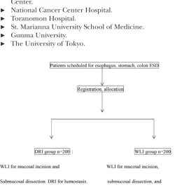
Figure 4 Overall design of this study. DRI, dual red imaging; ESD, endoscopic submucosal dissection; WLI, white light imaging.
study design
This trial is designed as a multicentre, open-label, randomised, parallel-group, controlled intervention study.
Participating centres
► National Hospital Organization Tokyo Medical
Center.
► National Cancer Center Hospital. ► Toranomon Hospital.
► St. Marianna University School of Medicine. ► Gunma University.
► The University of Tokyo.
study population
Hypotheses: Patients in the DRI group will achieve haemostasis in less time than the patients in the WLI group and endoscopists will experience less psycholog-ical stress when performing haemostasis on the DRI group than when performing haemostasis on the WLI group. The study population will consist of 400 patients evenly assigned to a standard treatment control group (WLI group, n=200) and experimental treatment group (DRI group, n=200) (figure 4).
The average time of one bleeding event requiring haemostasis per ESD procedure in a prior study was 50.8 s in a DRI group and 60.7 s in a WLI group. Haemo-stasis procedures had to be performed 1294 times in this study in order to determine the time reduction required to achieve haemostasis in the DRI group, as a result of calculation with a one-sided significance level of 0.05 and statistical power of 0.80. On average, 3.9 haemostasis procedures were required during each ESD intervention in the prior study, so 324 (1294/3.9) procedures were necessary in total. No bleeding occurred during the ESD in about 17% of the procedures in the prior study. In anticipation of drop-outs, the sample size for the current trial has been set at n=400 patients in total.
subjects
Patients who used DRI for haemostasis during ESD or and patients using WLI were enrolled.
Eligibility criteria Inclusion criteria
Patients who fulfil all of the following criteria will be included:
1. Scheduled to undergo ESD for an early-stage oesopha-geal, gastric or colorectal cancer (including colorectal adenoma).
2. Aged 20 years or older and 80 years or younger at the time of informed consent.
3. No pretreatment (ESD, surgery, radiation therapy, chemotherapy) has been performed in the organ to be examined.
4. Performance status of 0–1 on the Eastern Cooperative Oncology Group standard.
5. Written consent is provided at the patient’s own dis-cretion.
6. Antithrombotic drugs are on cessation according to the guideline.14
Exclusion criteria
Patients who fulfil any of the following criteria will be excluded:
1. Haemorrhagic diathesis (platelet count ≤100×109/L, haemoglobin count ≤80 g/L).
2. Scheduled to undergo ESD for multiple lesions. 3. Receiving maintenance dialysis.
4. Unable to stop antiplatelet regimen at the time of in-formed consent.
copyright.
on May 29, 2020 by guest. Protected by
http://bmjopengastro.bmj.com/
[image:3.595.42.293.432.707.2]5. Scheduled treatment for lesions with remnant recur-rence.
6. Deemed unsuitable for participation in this study by the principal investigator or other investigators.
study outline Treatment protocols
Patients found to have an early-stage oesophageal, gastric or colorectal cancer (including colorectal adenoma) for which ESD was indicated will be assigned to one of the two groups below. The endoscopist who carries out this study will be required to have performed more than 30 ESD procedures for each organ subject to the ESD proce-dure. A dual knife, hook knife and insulation-tipped diathermic (IT) knife will be used.
Standard treatment group (WLI group): After the periphery of the lesion is marked, Glyceol (10% glyceol and 5% fructose, Chugai Pharmaceutical, Tokyo, Japan) and MucoUp (hyaluronic acid, Boston Scientific Japan, Tokyo, Japan) will be injected into the submucosal layer and a mucosal incision will be made using WLI. When bleeding is occurred during ESD, the endoscopist will establish haemostasis using WLI with haemostat or knife. Once haemostasis is established, the submucosal dissection will be continued with WLI until the lesion is resected. The definition of bleeding in this study encom-passes bleeding of all types, from pulsatile bleeding to oozing. No bleeding will be assumed to occur, however, when the discharge of blood stops naturally or when only a very small amount of blood is discharged.
Experimental treatment group (DRI group): The initial marking, mucosal incision and submucosal dissec-tion in the DRI groups will be performed by the same methods used in the WLI group. If subsequent bleeding is occurred, the endoscopist will switch to DRI immedi-ately to confirm the bleeding point and establish haemo-stasis, then switch back to WLI once the haemostasis is achieved. As described above, the haemostasis treatment will be performed by switching to DRI only at times of bleeding during the procedure to fully resect the lesion.
Modified treatment schedule criteria
None specified.
Concurrent and supportive therapy
None specified.
Allocation
Patients are to be allocated to groups using an electronic data capture (EDC) system.
Method: Minimisation
Adjustment factors: Hospital, Organ.
Data collection and monitoring
Data management
An EDC system will be used to collect, monitor and manage the data from this trial. Study investigators or others designated as such will use strictly controlled personal electronic signatures (IDs and passwords) to
log into the EDC system. Collected patient information will be promptly entered into the EDC and sent to a data centre. Study investigators or others designated as such will enter and correct the data, and the former will confirm the accuracy of all data input. Case report forms will be saved separately from the data to be input to EDC, scanned and imported into charts.
Monitoring
This trial will employ a centralised monitoring system based on data collected via the EDC system. The centralised monitoring of the data will be itself monitored once a month. On-site monitoring will be conducted at each facility 1 month after the start of testing.
Endpoints
Primary endpoint
The time from the recognition of bleeding up to the achievement of complete haemostasis (haemostasis time).
Secondary endpoints
1. The psychological stress experienced by the endos-copist during haemostasis treatment, as assessed on a scale of 1–5 ((1) no stress; (2) minor stress; (3) moder-ate stress; (4) high stress; (5) very high stress).
2. The operation time defined as the time from the start of the mucosal incision to the complete excision of the lesion.
3. Proportion of cases in which perforation occurs.
Subanalysis
1. The haemostasis time and psychological stress due to the difference in the haemostasis device used (the knife tip or haemostatic forceps (Coagrasper)). 2. The haemostasis time in ESD for each organ
(oesoph-agus, stomach, colon, rectum).
statistical analysis Analysis sets
In accordance with the intention-to-treat (ITT) prin-ciple, the ITT set will consist of all cases registered and allocated to the groups in this study. The full analysis set (FAS) will be composed of all of the ITT cases except those in which treatment is cancelled for any reason after randomisation and those in which have no haemostasis treatment during ESD. Efficacy will be primarily analysed in the FAS set. The per-protocol set will be composed of all of the FAS cases except the following. With regard to the psychological stress experienced by the endosco-pists during haemostasis treatment and the haemostasis time: (1) Haemostasis treatment in cases which there is an omission in the data sheet; (2) haemostasis treatment by clipping; (3) if the endoscopist is switched midway through the treatment, all haemostasis procedures after the switch; (4) treatment with haemostasis in less than 5 s. With regard to the operation time: (1) cases in which treatment is discontinued due to adverse events, severe body movements while under procedure or poor
copyright.
on May 29, 2020 by guest. Protected by
Open access
circulation dynamics; (2) cases in which the endoscopist is switched during treatment; (3) cases with perforation and closure by clipping; (4) cases in which haemostasis is established not with the use of a knife or haemostat, but with clipping. The major axis of the resected specimen and the lesion site will be analysed as background data.
Primary endpoint analysis
Haemostasis time will be calculated for each group and compared between groups by the Mann-Whitney U test.
Secondary endpoint analysis
The operation time and psychological stress experienced by the endoscopist when working to achieve haemostasis are to be compared between the two groups by the Mann-Whitney U test. The proportion of cases in which perfo-ration occurs will be compared between the two groups by the χ2 test.
Subanalysis
The haemostasis time and psychological stress expe-rienced by the endoscopist will be calculated for each group and compared between groups by the Mann-Whitney U test.
Interim analysis
No interim analysis will be performed.
EThICs AnD DIssEMInATIon
In order to participate in the study, the patients must provide written consent at their own free will after receiving full explanations of the study by a study inves-tigator or research team member using an explana-tion-and-consent form. The explanaexplana-tion-and-consent form to be used meets the requirements of the Ethical Guidelines for Medical and Health Research Involving Human Subjects and has been approved for use by the Review Board for Clinical Trials.
Patient safety
Subjects will be monitored for adverse events by a study investigator or research team member. Any serious adverse event will be treated immediately and appro-priately: responding team members will make efforts to ensure the subject’s safety and investigate the cause of the event and will promptly report back to a study inves-tigator by telephone or facsimile. The responses to and results of any treatment-related death will be promptly reported to the Minister of Health, Labour and Welfare if the event could not have been predicted and the researchers are unable to rule out a causal relationship between the event and this study.
DIsCussIons
If the results go as planned, the study will demonstrate that DRI is a novel endoscope imaging technology that effectively simplifies and facilitates the achievement of haemostasis during ESD by shortening haemostasis
time and decreasing the psychological stress experi-enced by the endoscopist during haemostasis treatment. Naganuma et al reported that DRI was useful to assess the severity of inflammation in patients with ulcerative colitis using the deep vascular pattern.15 Ninomiya et al proposed that DRI may enhance both the safety and speed of the colorectal ESD procedure by improving the visibility of the arteries.16 Furuichi et al similarly found that DRI increased the visibility of the oesophageal varices and red colour sign.17 We also seek to verify the safety of haemostasis treatment with DRI, and the possi-bility of shortening the operation time by simplifying the haemostasis procedure.
Infrared endoscopy (IRE) has been found to facilitate the clear visualisation of bleeding sites during endoscopic treatment.12 The drawback of IRE is a required injection of indocyanine green, an agent that potentially induces toxic reactions such as urticaria, nausea, tachycardia or hypotension.18 In contrast, DRI requires no injection of medication, and the endoscopist can easily switch the endoscopic image from WLI to DRI with a single press of a button on the endoscope instrumentation.
It can sometimes be difficult to identify the bleeding point using WLI, because bleeding point and surrounding area are depicted in similar shades of red. Even when observing abundant bleeding with DRI, the dark yellow of the bleeding point appears in clear contrast against the surrounding ivory colour. Furthermore, the colour yellow is generally thought to uplift the emotions by elic-iting feelings of optimism and confidence.19 In contrast, the red colour of the bleeding imposes added psycholog-ical stress on the endoscopist, as it puts the endoscopist on alert much like the red signal of a traffic light. We speculate, therefore, that the endoscopist can manage haemostasis with less psychological stress using DRI. For the reasons above, we presume that DRI will shorten the haemostasis time in bleeding management during ESD.
One limitation of this study should be mentioned: the psychological stress is to be evaluated subjectively based on a scale the doctors declare themselves.
Author affiliations
1Division of Research and Development for Minimally Invasive Treatment, Cancer
Center, Keio University School of Medicine, Tokyo, Japan
2Department of Gastroenterology and Hepatology, National Hospital Organization
Tokyo Medical Center, Tokyo, Japan
3Endoscopy Division, National Cancer Center Hospital, Tokyo, Japan 4Department of Gastroenterology, Toranomon Hospital, Tokyo, Japan
5Division of Gastroenterology and Hepatology, St Marianna University School of
Medicine, Kawasaki, Japan
6Department of Gastroenterology and Hepatology, Gunma University, Maebashi,
Japan
7Department of Endoscopy and Endoscopic Surgery, The University of Tokyo, Tokyo,
Japan
Acknowledgements We appreciated support of Mitsuhiro Fujishiro, Masayoshi Ono, Osamu Goto, Motohiko Kato, Teppei Akimoto, Atsushi Nakayama, Yoshiyuki Kiguchi and Motoki Sasaki for this study.
Contributors AF participated in the design and statistical analysis plan of the study, and is responsible for managing data, handling monitoring. YS, SA, SH, HY, YM, TU and IS conceived of this study and participated in meetings to design the
copyright.
on May 29, 2020 by guest. Protected by
http://bmjopengastro.bmj.com/
study and coordinate details. SA, KN, YM, SK, IS and YT submitted the design to an ethics committee at each participating institution. NY oversaw the study as chief and provided personal research funding. AF, SA, KN, YM, SK, IS, YT, TN, TM and YO were responsible for registering the cases. All of the authors read and approved this manuscript.
Funding The authors have not declared a specific grant for this research from any funding agency in the public, commercial or not-for-profit sectors.
Competing interests None declared.
Patient consent for publication Not required.
Ethics approval This study complies with the World Medical Association’s Declaration of Helsinki, Ethical Guidelines for Medical and Health Research Involving Human Subjects, and Act on the Protection of Personal Information. It was approved by the Keio University Review Board for Clinical Trials (5 December 2016).
Provenance and peer review Not commissioned; externally peer reviewed.
open access This is an open access article distributed in accordance with the Creative Commons Attribution Non Commercial (CC BY-NC 4.0) license, which permits others to distribute, remix, adapt, build upon this work non-commercially, and license their derivative works on different terms, provided the original work is properly cited, appropriate credit is given, any changes made indicated, and the use is non-commercial. See: http:// creativecommons. org/ licenses/ by- nc/ 4. 0/.
rEFErEnCEs
1. Pimentel-Nunes P, Dinis-Ribeiro M, Ponchon T, et al. Endoscopic submucosal dissection: European Society of gastrointestinal endoscopy (ESGE) guideline. Endoscopy 2015;47:829–54. 2. Schlachterman A, Yang D, Goddard A, et al. Perspectives on
endoscopic submucosal dissection training in the United States: a survey analysis. Endosc Int Open 2018;6:E399–409.
3. Cao Y, Liao C, Tan A, et al. Meta-analysis of endoscopic submucosal dissection versus endoscopic mucosal resection for tumors of the gastrointestinal tract. Endoscopy 2009;41:751–7.
4. Tanaka S, Toyonaga T, Morita Y, et al. Efficacy of a new hemostatic forceps during gastric endoscopic submucosal dissection: a prospective randomized controlled trial. J Gastroenterol Hepatol
2017;32:846–51.
5. Toyonaga T, Nishino E, Man-I M, et al. Principles of quality controlled endoscopic submucosal dissection with appropriate dissection level and high quality resected specimen. Clin Endosc 2012;45:362–74.
6. Lee DW, Jeon SW. Management of complications during gastric endoscopic submucosal dissection. Diagn Ther Endosc
2012;2012:1–5.
7. Kikuchi D, Iizuka T, Yamada A, et al. Feasibility of a newly developed thumb control device for simultaneous manipulation of the endoscope and treatment devices in endoscopic submucosal dissection: a clinical feasibility study. Digestion 2016;94:123–8. 8. Huang R, Yan H, Ren G, et al. Comparison of o-type HybridKnife to
conventional knife in endoscopic submucosal dissection for gastric mucosal lesions. Medicine 2016;95:e3148.
9. Suzuki S, Gotoda T, Kobayashi Y, et al. Usefulness of a traction method using dental floss and a hemoclip for gastric endoscopic submucosal dissection: a propensity score matching analysis (with videos). Gastrointest Endosc 2016;83:337–46.
10. Libânio D, Pimentel-Nunes P, Dinis-Ribeiro M. Complications of endoscopic resection techniques for upper GI tract lesions. Best Pract Res Clin Gastroenterol 2016;30:735–48.
11. Fujishiro M, Yoshida S, Matsuda R, et al. Updated evidence on endoscopic resection of early gastric cancer from Japan. Gastric Cancer 2017;20:39–44.
12. Ishihara R. Infrared endoscopy in the diagnosis and treatment of early gastric cancer. Endoscopy 2010;42:672–6.
13. Yahagi N, Fujimoto A, Horii J, et al. Dual read imaging: a novel endoscopic imaging technology visualizing thick blood vessels of the
gastrointestinal wall. Endosc Int Open, 2018.
14. Kato M, Uedo N, Hokimoto S, et al. Guidelines for
gastroenterological endoscopy in patients undergoing antithrombotic treatment: 2017 appendix on anticoagulants including direct oral anticoagulants. Dig Endosc 2018;30:433–40.
15. Naganuma M, Yahagi N, Bessho R, et al. Evaluation of the severity of ulcerative colitis using endoscopic dual red imaging targeting deep vessels. Endosc Int Open 2017;5:E76–E82.
16. Ninomiya Y, Oka S, Tanaka S, et al. Clinical impact of dual red imaging in colorectal endoscopic submucosal dissection: a pilot study. Therap Adv Gastroenterol 2016;9:679–83.
17. Furuichi Y, Gotoda T, Moriyasu F, et al. Dual red imaging (novel advanced endoscopy) can increase visibility and can predict the depth in diagnosing esophageal varices. J Gastroenterol
2017;52:568–76.
18. Benya R, Quintana J, Brundage B. Adverse reactions to indocyanine green: a case report and a review of the literature. Cathet Cardiovasc Diagn 1989;17:231–3.
19. Okudaira Y, Sugiura T, Nakamura T, et al. "Physiological responses on color stimulation of video images", SCIS & ISIS 2008:1608–12.
copyright.
on May 29, 2020 by guest. Protected by
Correction:
Haemostasis treatment using dual red
imaging during endoscopic submucosal dissection: a
multicentre, open-label, randomised controlled trial
Fujimoto A, Saito Y, Abe S, et al. Haemostasis treatment using dual red imaging during endoscopic submucosal dissection: a multicentre, open-label, randomised controlled trial. BMJ Open Gastroenterology 2019;6:e000275. doi: 10.1136/ bmjgast-2019-000275
The third author’s name is misspelt. The correct spelling is ‘Seiichiro Abe’.
Open access This is an open access article distributed in accordance with the Creative Commons Attribution Non Commercial (CC BY-NC 4.0) license, which permits others to distribute, remix, adapt, build upon this work non-commercially, and license their derivative works on different terms, provided the original work is properly cited, appropriate credit is given, any changes made indicated, and the use is non-commercial. See:http:// creativecommons. org/ licenses/ by- nc/ 4. 0/.
© Author(s) (or their employer(s)) 2019. Re-use permitted under CC BY-NC. No commercial re-use. See rights and permissions. Published by BMJ.
BMJ Open Gastro 2019;6:e000275corr1. doi:10.1136/bmjgast-2019-000275corr1