Induction of Human Blood Group A Antigen Expression
on Mouse Cells, Using Lentiviral Gene Transduction
Xiaohu Fan,1 Haili Lang,6Xianpei Zhou,1Li Zhang,2Rong Yin,1Jessica Maciejko,1 Vasiliki Giannitsos,1 Bruce Motyka,1Jeffrey A. Medin,3 Jeffrey L. Platt,4and Lori J. West1,5
Abstract
The ABO histo-blood group system is the most important antigen system in transplantation medicine, yet no small animal model of the ABO system exists. To determine the feasibility of developing a murine model, we previ-ously subcloned the humana-1,2-fucosyltransferase (H-transferase, EC 2.4.1.69) cDNA and the human a-1,3-N-acetylgalactosaminyltransferase (A-transferase, EC 2.4.1.40) cDNA into lentiviral vectors to study their ability to induce human histo-blood group A antigen expression on mouse cells. Herein we investigated the optimal conditions for human A and H antigen expression in murine cells. We determined that transduction of a bicistronic lentiviral vector (LvEF1-AH-trs) resulted in the expression of A antigen in a mouse endothelial cell line. We also studied thein vivoutility of this vector to induce human A antigen expression in mouse liver. After intrahepatic injection of LvEF1-AH-trs, A antigen expression was observed on hepatocytes as detected by immunohisto-chemistry and real-time RT-PCR. In human group A erythrocyte-sensitized mice, A antigen expression in the liver was associated with tissue damage, and deposition of antibody and complement. These results suggest that this gene transfer strategy can be used to simulate the human ABO blood group system in a murine model. This model will facilitate progress in the development of interventions for ABO-incompatible transplantation and transfusion scenarios, which are difficult to develop in clinical or large animal settings.
Introduction
T
heABOblood group system, first described more than100 years ago (Landsteiner, 1900, 1901), is characterized by the expression of ABH antigens, which are expressed in humans not only on red blood cells but also on cells in a variety of organs, hence the term ‘‘histo-blood group anti-gens’’ (Clausen and Hakomori, 1989). As shown in Fig. 1, the ABH structures are terminal polysaccharides synthesized by sequential monosaccharide addition(s) to a common precur-sor (Yamamotoet al., 1990). With rare exceptions, all humans possess the gene encoding the a-1,2-fucosyltransferase en-zyme (H-transferase, EC 2.4.1.69) that catalyzes the addition ofl-fucose to ab-galactosyl residue, forming the H antigen
(Yamamoto et al., 1990). Blood group O individuals are characterized by expression of this antigen without further
modification. Blood group A and AB individuals also have the gene for thea-1,3-N-acetylgalactosaminyltransferase en-zyme (A-transferase, EC 2.4.1.40), which transfers a terminal
N-acetyl-d-galactosamine residue to the H chain, forming the
A antigen. Similarly, the B-transferase enzyme expressed by group B and AB individuals transfers a terminald-galactose
residue to the H antigen, forming the B antigen. These reac-tions are summarized in Fig. 1.
The ABO histo-blood group system has great clinical sig-nificance in transfusion and transplantation medicine. Acci-dental blood transfusion across the ABO barrier can have catastrophic consequences (Linden, 1999). ABO incompati-bility in stem cell transplantation is a source of clinical complications such as posttransplantation anemia and graft-versus-host disease (Stussiet al., 2002; Lee et al., 2003), and ABO-incompatible solid organ transplantation is usually
1Department of Pediatrics, University of Alberta, Edmonton, AB, T6G 2E1 Canada. 2
Department of Pediatrics, Hospital for Sick Children, University of Toronto, Toronto, ON, M5G 1X8 Canada.
3Division of Stem Cell and Developmental Biology, Ontario Cancer Institute, University Health Network, University of Toronto, Toronto,
ON, M5G 2M9 Canada.
4Department of Surgery, University of Michigan, Ann Arbor, MI 48109. 5
Department of Surgery and Department of Immunology, University of Alberta, Edmonton, AB, T6G 2E1 Canada.
6Department of Surgery, Section of Cardiothoracic Surgery, University of Nebraska Medical Center and Children’s Hospital, Omaha,
NE 68198.
DOI: 10.1089/hum.2008.089
followed by hyperacute antibody-mediated graft rejection (Paul and Baldwin, 1987; Stocket al., 1987). We have shown that the seemingly insurmountable ABO barrier to heart transplantation can be overcome in human infants (West
et al., 2001) and that ABO-incompatible heart transplantation during an ill-defined period of immunologic malleability during infancy leads to prolonged B cell tolerance to donor histo-blood group A/B antigens (Fan et al., 2004). Others have shown that it is possible to transplant kidneys from ABO-incompatible donors into group O individuals, using aggressive immunosuppressive strategies (Glooret al., 2003). This often results in a phenomenon known as graft accom-modation, which is characterized by resistance of the graft to injury despite antibody reaccumulation (Stevens and Platt, 1992; Kochet al., 2004). Despite these successes, the detailed mechanisms behind these clinical phenomena are poorly understood. Moreover, potential strategies need to be ex-plored to allow organ transplantation to be performed suc-cessfully with donors of any blood type in both infants and older recipients, thus expanding the potential donor pool and minimizing wastage of donor organs.
Limitations inherent to experimentation in humans neces-sitate the use of an animal model for the detailed investigation of ABO-incompatible organ transplantation; however, no small animal model of the human ABO histo-blood group system currently exists. Although a number of mammalian species, including chimpanzees and orangutans, are known to possess a homolog of the human A/B antigens (Saitou and Yamamoto, 1997), and ABO-incompatible heart transplanta-tion in baboons has been attempted in small numbers (Cooper
et al., 1987; Yeet al., 1994), there are disadvantages to studying ABO immunobiology in these nonhuman primates. In com-parison with models based on small animals, experiments involving nonhuman primates are substantially more ex-pensive to conduct and require more physical space. Further, experimental timelines are vastly shorter for murine than for nonhuman primate experiments, and as relatively little is known about the detailed immunobiology of the ABO blood group system, establishment of a murine model would greatly facilitate progress in this area.
We previously showed the feasibility of inducing human histo-blood group H and A antigen expression in mouse cells
in vitro by multiple gene transfer strategies using lentiviral vectors (Fanet al., 2005). The human H- and A-transferase cDNAs were subcloned and inserted into replication-deficient lentiviral vectors; these vectors were used in tandem to transduce human and mouse cell lines to generate human blood group A antigen. We demonstrated that mouse cells possess the appropriate precursor sugar chain required for synthesizing blood group H and A antigen. Cell surface H antigen expression was observed on mouse cells infected with LvCMV-H-trs particles (Fanet al., 2005). Unlike human cells, expression of the A antigen on mouse cells was com-pletely dependent on infection with both of the lentiviral particles (Fanet al., 2005). In the current study we sought to improve our original vector by constructing a bicistronic lentiviral vector with the A-transferase gene upstream of an encephalomyocarditis virus internal ribosome entry site (EMCV IRES) element and the H-transferase gene under the control of the human elongation factor-1a(EF-1a) promoter. In sensitized mice,in vivointrahepatic injection of LvEF1-AH-trs particles resulted in expression of A antigen in the liver
and simulated the process of hyperacute antibody-mediated rejection.
Materials and Methods
Animals
BALB/c female mice were obtained from Jackson Laboratory (Bar Harbor, ME) and maintained in a bio-containment facility at the University of Alberta (Edmonton, AB, Canada) under specific pathogen-free conditions. Sen-sitization was performed by intraperitoneal injection of 108 washed human blood group A erythrocytes given three times at 3-week intervals. Direct injection of lentiviral vector into the right inferior portal vein under surgical visualization was performed 1 week after the final erythrocyte injection. Injected liver lobes were harvested at various times under surgical visualization. Serum aspartate transaminase (AST) levels were assessed by Charles River Research Animal Diagnostic Services (Wilmington, MA). Animal protocols used for this study were approved by the Institutional Re-view Board of the University of Alberta in accordance with the Canadian Council on Animal Care guidelines.
Lentiviral vector production and titration
The full-length human H-transferase (a -1,2-fucosyltrans-ferase) cDNA and human A-transferase (a-1,3-N -acetylgal-actosaminyltransferase) cDNA were amplified from cDNA of MKN45 cells, using ThermAce proofreading polymerase (Invitrogen, Carlsbad, CA). The purified amplified DNA products of human H- and A-transferase genes were inserted into a lentiviral expression construct downstream of the human cytomegalovirus (CMV) promoter. The resulting plasmids were denoted pCMV-H-trs and pCMV-A-trs, re-spectively. The H- and A-transferase genes were also sub-cloned into thePmeI site of pWPI plasmid. The A-transferase gene was placed downstream of the human EF-1apromoter and upstream of the enhanced green fluorescent protein (eGFP) reporter gene, and the resulting plasmid was denoted pEF1-A-trs-GFP. pCMV-H-trs and pEF1-A-trs-GFP have been reported previously (Fanet al., 2005). A bicistronic len-tiviral plasmid was constructed by replacing eGFP with H-transferase, denoted pEF1-AH-trs. To package lentiviral vectors, 293T cells were cotransfected with one of the ex-pression plasmids and packaging plasmids, using the calcium phosphate method. Concentration by ultracentrifugation yielded titers of up to 109transducing units (TU)/ml.
Lenti-viral stock was either used immediately or stored at808C.
Cellular ELISA for quantifying cell surface H or A antigen expression
(TMB; Sigma-Aldrich) for color development. The reaction was stopped with sulfuric acid and absorbance was measured at 450 nm.
To assess epitope density of cell surface A antigen, human group A1 erythrocytes, A2 erythrocytes, MKN45 cells, and
fluorescence-activated cell-sorted (FACS) stable A antigen-expressing SVECs were counted and plated by serial dilution into 96-well V-bottom plates. Cells were stained with a high concentration (20 ng/ml) of murine anti-A IgG3 monoclonal antibody (mAb) (BD Biosciences, San Jose, CA) to saturate all antigen epitopes and subsequently labeled with horseradish peroxidase (HRP)-conjugated goat anti-mouse IgG3 (1:100 dilution; Southern Biotech, Birmingham, AL). After the stained cells were washed, TMB substrate was added and the reaction was stopped with sulfuric acid. The surface area of erythro-cytes was deduced according to the formulaS¼4pr2, as for other trypsinized cells. A microruler was used to determine the mean diameter of each cell type on the basis of 50 randomly chosen cells on photomicrographs. A relative surface area bearing a defined amount of blood group A antigen epitopes defined as an optical density (OD) reading of 1.0 in a cellular ELISA (CELISA) was determined for each cell type, based on these standard curves. The epitope density of each cell type was then deduced according to the known epitope density of A1erythrocytes (Gupteet al., 1985).
Flow cytometry
Washed SVECs were incubated for 30 min at 48C with anti-A IgM mAb (NOVACLONE, 1:100 dilution; Dominion Biologicals, Dartmouth, NS, Canada). Phycoerythrin (PE)-conjugated goat anti-mouse IgM antibody (1:1000 dilution; Southern Biotech) was used as a secondary detecting anti-body. Cell acquisition and analysis were performed with a FACScan flow cytometer (BD Biosciences) and WinMDI version 2.8 software. A antigen-positive SVECs were sorted with a FACSAria flow cytometer (BD Biosciences).
Immunofluorescence and immunohistochemistry staining
O.C.T. medium-embedded mouse tissues were cryosec-tioned at 4–6mm and processed according to standard im-munohistochemistry techniques. A antigen was detected with peroxidase-conjugated Helix pomatia lectin (Sigma-Aldrich). Labeling for IgM or IgG antibody deposition was performed with HRP-conjugated goat anti-mouse IgM or IgG mAb (Southern Biotech). Anti-CD3 (clone 17A2) and anti-CD19 (clone ID3) were obtained from BD Biosciences, and anti-CD11b (clone M1/70) and anti-F4/80 (clone BM8) were ob-tained from eBioscience (San Diego, CA). The cryosections were fixed in 4% paraformaldehyde (PFA) solution, washed with water, treated with 0.3% H2O2in methanol, and
incu-bated with 2% normal goat serum. The sections were then treated with primary antibody for 12 hr at 48C, washed, and incubated with HRP-conjugated goat anti-rat g-chain antibody (Southern Biotech). Bound antibody was visual-ized with 3,30-diaminobenzidine tetrahydrochloride and
counterstained with Mayer’s hematoxylin. Goat anti-mouse complement component C3d (R&D Systems, Minneapolis, MN) was applied at a 1:100 dilution for C3d staining by im-munofluorescence. Sections were washed and incubated with Alexa Fluor 546-labeled donkey anti-goat IgG(HþL)
anti-body (Invitrogen) and visualized with an inverted fluores-cence microscope (Leica Microsystems, Wetzlar, Germany).
Quantitative real-time RT-PCR
For kinetic quantification of human A-transferase expres-sion in injected mouse liver, total RNA was obtained from snap-frozen tissue specimens, using TRIzol (Invitrogen), and purified with RNeasy columns (Qiagen, Valencia, CA). For each specimen, 400 ng of RNA was separately reverse tran-scribed by using random hexamers and SuperScript II reverse transcriptase (Invitrogen). PCR amplification was done with each cDNA at empirically determined optimal concentrations of forward and reverse primers and a 6-carboxyfluorescein (FAM)-labeled TaqMan probe. Both b-actin and human A-transferase primers were used at 300 nM each and FAM/ TAMRA-labeled probe was used at 200 nM. Cycling condi-tions were as follows: initial denaturation at 958C for 10 min, followed by 40 cycles at 958C for 15 sec, then 608C for 1 min on an ABI Prism 7900HT fast real-time PCR system (Applied Biosystems, Foster City, CA). Theb-actin primers and human A-transferase primers and probe were designed as follows:
A-trs-FP (50-CTCGTTGCCAAGGATGGTCTA-30), A-trs-RP
(50-CCACGAGGACATCCTTCCTACA-30), and A-trs-Prob (50
-FAM-CCCCAGCCAAAGGTGCTGACACC-TAMRA-30); b
-actin-FP (50-ACGGCCAGGTCATCACTATTG-30), b-actin-RP
(50-CAAGAAGGAAGGCTGGAAAAG-30), and b-actin-Prob
(50-FAM-CAACGAGCGGTTCCGATGCCC-TAMRA-30).
Relative quantification of human A-transferase mRNA ex-pression levels was calculated by the comparativeCTmethod,
with theDCTvalue fromb-actin used as the calibrator.
To assess the transcriptional expression of endogenous murinea-1,2-fucosyltransferase and murinecis-AB-transferase genes in different tissues, 8- to 12-week-old BALB/c mice (n¼3) were killed and various tissues and organs were har-vested and homogenized in TRIzol. Total RNA (500 ng) from each tissue was used for reverse transcription with SuperScript II reverse transcriptase (Invitrogen). The resultant cDNA library was subjected to PCR with the following primers:
H-transferase: 50-CCACCTGAATGGAGGAAACT-30
(for-ward) and 50-TAGTCTCCACGACGCACATG-30(reverse);
A-transferase: 50-CATGGTGGGACACAAGGTCA-30(forward)
and 50-CTCCAGGGCTCAAAGTGTGT-30 (reverse); and b
-actin: 50-ATTGCCGACAGGATGCAGAA-30 (forward) and
50-GCTGATCCACATCTGCTGGAA-30(reverse).
Settings for PCR were as follows: 35 cycles, with each cycle consisting of 60 sec at 948C, 60 sec at 628C, and 60 sec at 728C. The first cycle was preceded by 5 min at 948C to ensure complete DNA denaturation. All assays were repeated three times to ensure the reproducibility of results.
Statistical analysis
The significance of differences in AST values was deter-mined by one-tailed t test.p<0.01 was considered statisti-cally significant between the experimental groups.
Results
Mice fail to exhibit native H-transferase or A-transferase expression in most tissues
weak A- and B-transferase activities. Although the cis-AB gene product is homologous to human A/B-transferase, its A- and B-transferase activities were much weaker than those encoded by the human genes. A or B antigens were not detected in mouse tissues, except the submaxillary glands, which exhibited weak expression (Yamamotoet al., 2001). The murine H-transferase (a-1,2-fucosyltransferase) homolog has also been identified, and its expression seems restricted to the gastrointestinal tract (Linet al., 2000). The expression of murine endogenous H-transferase or A-transferase has not been systematically studied and blood group A antigen expression in mouse tissues is still unclear; therefore we performed RT-PCR for these two genes and immunohistochemistry to detect potential A antigen expression in 13 tissues and organs of BALB/c mice. The RT-PCR experiments for all tissue types were repeated three times, using different mouse samples each time, and the results were consistently reproduced as shown in Table 1. Although H-transferase expression was detected in colon and relatively weak expression was also found in liver, spleen, thymus, and skin, only colonic epi-thelium and thymus expressed H antigen as detected by UEA1 lectin binding (Table 1). mRNA expression from the murine cis-AB-transferase gene was found in colon, stom-ach, and testes. Among these 13 organs, only colon was reproducibly found to coexpress both H-transferase andcis -AB-transferase, consistent with the fact that the colon was the only tissue showing Helix pomatia (A antigen-specific lectin) binding (Table 1). However, it must be stressed that the demonstration of A antigen expression on colonic epi-thelium may not be conclusive because expression may be attributed to cross-reactive detection of bacteria in gut flora, or A/B-like substances in the colonic contents. Bacteria in gut flora are known to contain A-like carbohydrate struc-tures believed to be the antigenic source for T-independent immune development of ‘‘natural antibodies’’ (Scheffel and Kim, 1979).
Construction of lentiviral expression vectors harboring human ABO-related glycosyltransferases
Human A-transferase and H-transferase genes were am-plified from an MKN45 cDNA library, using proofreading DNA polymerase. The full cDNA sequences of these genes were compared with available gene bank data before sub-cloning into lentiviral expression vectors. No nucleotide mutation was identified from PCR cloning. As we previously reported, pCMV-H-trs is a lentiviral expression vector with the H-transferase gene placed under the control of the CMV promoter (Fan et al., 2005). pEF1-A-trs-GFP is a bicistronic vector with the A-transferase gene placed upstream of the GFP gene. We constructed three new lentiviral expression vectors denoted as pCMV-A-trs, pEF1-H-trs, and pEF1-AH-trs, respectively, into which amplified full-length cDNA fragments of A-transferase and/or H-transferase genes were subcloned into lentiviral expression vectors with different promoters (Fig. 1). Thus, we obtained a series of different lentiviral vectors for further study of the synergistic inter-action between human A- and H-transferases on mouse cells. pEF1-AH-trs is a new bicistronic expression vector used to package bicistronic lentiviral vector LvEF1-AH-trs.
Dose-dependent A antigen production and H antigen consumption after various levels of A-transferase lentivirus exposure
Although we previously demonstrated that gene transfer-induced human A antigen expression on mouse cells requires transduction of both human H- and A-transferases (Fanet al., 2005), the extent of dependence on H-transferase by A-trans-ferase for optimal expression of A antigen was not determined. Understanding the dependence of the two transferases for A antigen expression was important in the design of a bicistronic vector for optimal coexpression of the transferase genes.
To address this question, serially diluted LvCMV-A-trs and LvCMV-H-trs lentiviral vectors at equal titers were
Table1. Tissue Expression of Murine Genes Homologous to Human ABO-Related Glycosyltransferase Genes and Expression of Related Carbohydrate Antigensa
RT-PCR IHC
Mouse tissue b-Actin mFUT mA-trs H-Antigen A-Antigen a-Gal
Bone marrow þ – – – – þ
Colon þ þ þ þ þ W
Heart þ – – – – W
Intestine þ – – – – þ
Kidney þ – – – – þ
Liver þ W – – – þ
Lung þ – – – – þ
Spleen þ W – – – þ
Stomach þ – W – – þ
Testes þ – þ – – þ
Thymus þ W – þ – W
Uterus þ – – – – þ
Tail skin þ W – – – þ
Abbreviations:mA-trs, murinea-1,3-N-acetylgalactosaminyltransferase; mFUT, murine gene homologous to a-1,2-fucosyltransferase; W, weak expression.
a
RT-PCR and immunohistochemistry (IHC) results from 13 different tissues and organs of naive adult BALB/c (n¼3) are listed.b-Actin was used as a housekeeping control for RT-PCR.
combined in a 96-well plate seeded with SVECs. Different amounts of each vector particle were added to each well (2-fold serial dilutions starting from 2106 TU/ml of either LvCMV-A-trs or LvCMV-H-trs added in the same volume to 1105preseeded SVECs). These two vectors synergisti-cally and dose-dependently induced A antigen expression on mouse SVECs as quantified by CELISA (Fig. 2A). When either LvCMV-A-trs or LvCMV-H-trs vectors were absent no A antigen was detected. With exposure to increasing quantities of LvCMV-H-trs lentiviral vector, H antigen ex-pression increased in a dose-dependent fashion from an undetectable level (Fig. 2B). With augmentation of LvCMV-A-trs vector, more A antigen was expressed on the cell surface. Interestingly, H antigen expression (Fig. 2B) decreased in a dose-dependent manner with increasing A-transferase transduction, suggesting that H antigen was consumed as the precursor for A antigen synthesis. These results confirmed the dependence of human A-transferase on
H-transferase to induce A antigen expression on mouse en-dothelial cells. In the CELISA wells with the highest amount of LvCMV-H-trs transduced, sufficient expression of H an-tigen was induced for LvCMV-A-trs to consume the appar-ently surplus H antigen when equal amounts of LvCMV-A-trs were used. Because usually the downstream gene will express less than the upstream gene in a bicistronic construct, this led to the design of a bicistronic vector (pEF1-AH-trs) with the H-transferase gene located downstream of the A-transferase gene.
EF-1ais a suitable promoter for long-term stable A antigen expression on mouse SVECs
[image:5.612.77.536.58.439.2]SVECs transduced with LvCMV-H-trs and LvCMV-A-trs, or with LvEF1-H-trs and LvEF1-A-trs-GFP, were sorted for high expression of A antigen by FACS. The purity of sorted SVECs was confirmed and the cells were cultured for
3 months, with passage every 2–3 days. Cells were cryo-preserved every third passage. FACS analysis of cryopre-served Aþ SVECs was performed to assess A antigen expression after culture for 30 passages (Fig. 3). Of cells transduced with vectors containing either the CMV promoter or the EF-1apromoter, 100% exhibited A antigen expression immediately after sorting. However, with long-term culture A antigen expression gradually decreased in CMV-trans-duced cells over time, and by 30 cell passages A antigen was detectable on only 23% of CMV-transduced SVECs. In con-trast, EF-1a-transduced cells exhibited remarkably stable A antigen expression, with the percentage of cells expressing A antigen remaining relatively constant (more than 90%) from 1 to 90 days posttransduction (Fig. 3).
Lentiviral vector-transduced stable
A antigen-expressing mouse SVECs express an epitope density comparable to that of human MKN45 cells
To clarify whether lentiviral vector (a bicistronic vector with the EF-1apromoter) transduction was sufficient to in-duce stable A antigen expression at a level comparable to human cells, we estimated the density of A antigen ex-pressed on our stable AþSVEC line after 30 passages. We performed CELISA with serially diluted A1erythrocytes, A2
erythrocytes, the human MKN45 cell line, and Aþ SVECs. For all four cell types assessed, a linear relationship was achieved between total cell surface area and A antigen level (Fig. 4A). On the basis of pooled A1 erythrocytes bearing
about 106blood group A epitopes per cell (Economidouet al., 1967; Gupteet al., 1985), the epitope density on A2
erythro-cytes, MKN45 cells, and AþSVECs was estimated (Fig. 4B). Not surprisingly, blood type A1erythrocytes had the highest
A antigen density, whereas A2erythrocytes expressed less A
antigen, as previously noted using other methods (Econo-midouet al., 1967). Although not as high as erythrocytes, the MKN45 human gastric cancer cell line is known to express a high level of A antigen (Yamamotoet al., 2001). The stable transgenic SVEC line transduced with the lentiviral vector showed comparable antigen density to human MKN45 cells.
A antigen expressionin vivoand immune response in mouse liver after lentiviral vector injection
To explore the usefulness of our lentiviral vectors toward modeling ABO histoincompatibilityin vivo, we performed a pilot study in which lentiviral vectors were injected directly into mice. Anti-A antibodies of both IgM and IgG isotypes were measured by ELISA in the serum of mice sensitized by injection of human A erythrocytes (n¼20), and in control mice (n¼5). Nonsensitized mice produced relatively low levels of ‘‘natural’’ antibody specific to A antigen (Fig. 5A), consistent with previously reported data (Ne´ron and Lemieux, 1994). The isotype of the natural anti-A antibodies was almost exclusively IgM (Fig. 5A and B). After sensitiza-tion, strong anti-A antibody responses of both IgM and IgG isotypes were detected (Fig. 5A and B).
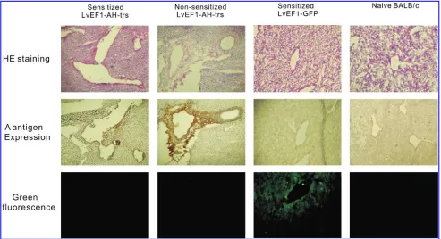
[image:6.612.63.553.58.286.2]Under direct visualization, 5107TU of LvEF1-AH-trs was injected into the right inferior portal vein of sensitized mice (n¼20) and nonsensitized mice (n¼5). As the control experi-ment, LvEF1-GFP vector was injected into the portal vein of five sensitized mice. The sensitized animals injected with LvEF1-AH-trs vector were killed at multiple time points (2, 3, 5,
FIG. 2. Dose-dependent A antigen production and H antigen consumption after combined exposure to serially diluted LvCMV-A-trs and LvCMV-H-trs lentiviral vectors. Serially diluted LvCMV-A-trs and LvCMV-H-trs lentiviral vectors were combined on a 96-well plate seeded with SVECs. A different amount of each vector particle was added to each well (2-fold serial dilution starting from 2106TU/ml of either LvCMV-A-trs or LvCMV-H-trs) to preseeded SVECs. Blood group A antigen (A)
and 10 days) and nonsensitized mice and LvEF1-GFP vector-injected sensitized mice were killed on day 3 after vector in-jection, after which half of the right inferior liver lobe was examined by immunohistochemistry for A antigen expression, antibody deposition and C3d, and phenotype of infiltrating cells. The other half of the liver lobe was subjected to RT-PCR to detect human A-transferase RNA expression. The hepatic re-gions supplied by the injected portal vein strongly expressed blood group A antigen as early as 48 hr after vector injection into sensitized mice (Fig. 5C and D). Hematoxylin and eosin (H&E) staining of this area revealed tissue injury of the A antigen-expressing tissues (Fig. 5E and F). A large mass of necrosis and degeneration of liver parenchymal cells was observed inside the lentiviral vector-transduced region, accompanied by diffuse hemorrhage and microvascular destruction. In addition, mononuclear cell infiltration was seen surrounding the area of A antigen expression (Fig. 5D and F). To clarify whether the detected blood group A antigen could be due to antigen incorporated into vector particles from the transfected producer cell line, we performed real-time RT-PCR to quantify human A-transferase RNA in in-jected liver lobes. RNA expression was measured at five time points (days 0, 2, 3, 5, and 10) in five animals per time point. After lentiviral vector injection, human A-transferase RNA
was highest on day 2 and decreased thereafter (Fig. 5G). It is possible that on day 3, expression levels were decreased as a result of tissue destruction and loss in the injection area as the result of an anti-A antigen-mediated immune response. In a separate experiment, animals injected with a lower titer of vector (1107TU) showed a low level of A antigen expression
with only mild changes in histology (data not shown). Human A-transferase RNA in five LvEF1-GFP vector-injected mice (72 hr) was undetectable (Fig. 5G). Aspartate transaminase (AST) levels for each animal group were assessed as an indi-cator of acute liver damage. The normal range of AST for female BALB/c mice is 8041 IU/liter (Frith et al., 1980). Sensitized mice injected with LvEF1-AH-trs vector showed significantly elevated AST serum levels at 72 hr that recovered to baseline levels by 10 days postinjection. Nonsensitized mice (n¼5) also showed a moderately elevated AST level com-pared with mice injected with control GFP vector (LvEF1-GFP) (n¼5), which did not show AST levels above baseline at 72 hr (Fig. 5H).
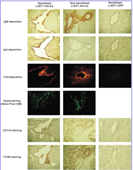
[image:7.612.64.545.58.380.2]Obvious tissue loss was apparent in A antigen-expressing areas 72 hr after vector injection. This led mainly to staining for A antigen in the margin of the lesion area, and to depo-sition of IgM and IgG (Figs. 6 and 7). This suggests that the expression of foreign A antigen in the liver led to a humoral
response in the systemically sensitized mice. Notably, as shown in Fig. 7, IgG deposition was observed in sensitized mice and not in nonsensitized mice. This is consistent with the observation that nonsensitized mice did not possess the IgG isotype of anti-A antibody in sera as measured by ELISA (Fig. 5B). A milder histological lesion in terms of tissue loss was observed in the liver of nonsensitized mice 72 hr after lentiviral vector injection (Figs. 6 and 7). For mice injected with LvEF1-GFP, GFP was expressed (Fig. 6). Importantly, no obvious histological lesions were observed (Figs. 6 and 7). In addition, no A antigen expression, antibody, or complement fragment C3d deposition was observed in mice injected with control GFP lentiviral vector (n¼5) (Figs. 6 and 7).
C3d is a product of complement activation that covalently binds to tissue near sites of complement activation and has been considered to be a marker of the presence of antibody-mediated rejection (AMR), particular in ABO-incompatible transplantation (Haas et al., 2006). Immunofluorescence
staining was performed and C3d deposition was found in the lesion area that also showed A antigen expression and de-position of IgM and IgG (Fig. 7).
To determine the specificity of deposited mouse antibodies, tissue was stained with biotin-conjugated tetrasaccharide blood group A antigen (A4–polyacrylamide [PAA]–biotin)
and B antigen (B4–PAA–biotin). A4–PAA–biotin staining was
positive in areas of IgG and IgM deposition in sensitized mice, and in areas of IgM deposition in nonsensitized mice (Fig. 7). Conversely, these areas were negative for B4–PAA–biotin
staining (data not shown).
[image:8.612.79.534.59.292.2]To characterize the phenotype of infiltrating mononuclear cells surrounding the region of A antigen expression, im-munostaining was performed for murine CD3, CD19, CD11b, and F4/80. No CD3þor CD19þcells were found in the lesion area (data not shown). However, many infiltrating cells stained for expression of CD11b and F4/80 (Fig. 7), which suggests that many of these cells were macrophages.
FIG. 4. Lentiviral vector-transduced stable A antigen-positive SVECs express a high A epitope density similar to that of human MKN45 cells. Human group A1erythrocytes, group A2erythrocytes, MKN45 cells, and FACS-sorted stable A
antigen-expressing SVECs (AþSVECs) were counted and plated into 96-well V-bottom plates in a serial dilution manner. CELISA was performed and the relation of total cell surface to A antigen expression was plotted (A). The mean value of triplicate assay wells was taken for all results. Using data from the most linear region of all four standard curves, cell surface A antigen density (B) for the four cell types was estimated on the basis of the known antigen density of A1erythrocytes.
FIG. 5. Injection of A-lentiviral vector into the liver of sensitized mice leads to local A antigen expression and a humoral immune response. Sera collected from nonsensitized BALB/c mice (n¼5) and human A erythrocyte-sensitized BALB/c mice (n¼20) were evaluated by ELISA for the presence of anti-A IgM (A) or IgG (B). LvEF1-AH-trs vector (5107TU) was injected into the right branch of the portal vein supply to the right inferior lobe of the liver of sensitized BALB/c mice. Forty-eight hours after injection of LvEF1-AH-trs vector into sensitized mice, A antigen expression was detected withHelix pomatialectin, as shown at original magnifications of100 (C) and400 (D). On a contiguous section, H&E staining shows the region into which vector was administered (E). One marginal area close to the supply portal vein branch (F) is shown at higher power (original magnification,400). Real-time RT-PCR for human A-transferase RNA expression is shown in injected liver lobe at time points 2, 3, 5, and 10 days after injection (n¼5 for each time point). A-transferase RNA expression in livers from the day 3 LvEF1-GFP-injected group is also shown (G). AST levels in mouse sera were assessed from serum samples (n¼5 mice per group) collected at 72 hr for all treatment groups and 10 days after injection for the sensitized mouse group. The normal high and normal low levels of AST for female BALB/c mice are indicated by horizontal lines. (H). Error bars indicate the SD of data collected from all five animals in each group. Color images available online at www.liebertonline.com/hum.
Among all these pathological observations, vector-injected nonsensitized mice did not possess significant differences in comparison with vector-injected sensitized mice except for the degree of tissue loss and elevated AST levels 72 hr after gene therapy, as summarized in Table 2. Overall, these find-ings suggest that the expression of foreign A antigen in the liver of mice sensitized to A antigen resulted in a hyperacute humoral reaction similar to that observed during AMR in ABO-incompatible grafts.
Discussion
It has been suggested that one or more genetic manipu-lations may be necessary in order to establish a mouse model of the ABO histo-blood group system (Yamamoto et al., 2001). Candidate manipulations include increased murine
cis-AB gene expression, modification of the murine cis-AB gene to retain only A- or B-transferase activity, introduction of a nonmurine A- or B-transferase gene, introduction of the H-transferase gene under control of a strong promoter, and knockout of thea-1,3-galactosyltransferase gene. In the cur-rent study, we have determined that lentiviral delivery of the human H- and A-transferase genes in a bicistronic lentiviral vector under strong promoter control is sufficient for trans-duction of A antigen expression on the surface of mouse cells bothin vitroandin vivo.
Although mice possess a gene homologous to the human A/B-transferase gene and another homolog to the human H-transferase gene, the enzymatic activity of the mousecis
-AB-transferase gene is reduced compared with the respective human genes (Yamamotoet al., 2001). Furthermore, as we have shown by RT-PCR and immunohistochemical analysis (Table 1), the cis-AB-transferase gene is expressed only in limited tissues in the mouse and likely does not lead to widespread A antigen expression because of the deficiency in H antigen expression. Lack of H antigen expression may also be due to the presence of strong expression ofa -l,3-galacto-syltransferase in most mouse tissues, because this competes with H-transferase for terminalN-acetyllactosamine (Sharma
et al., 1996). This study confirmed that in mice, A antigen is expressed only on epithelial cells in the colon. This finding is consistent with the RT-PCR analysis that showed coexpres-sion of both mouse cis-AB-transferase and H-transferase genes only in the colon. Furthermore, A antigen is unlikely to be expressed as a self-antigen on other organs and tissues that we have not studied because BALB/c mice produce abundant amounts of natural anti-A antibodies without developing autoimmune disease (Ne´ron and Lemieux, 1994).
[image:10.612.61.554.57.325.2]Our gene transfer vector also proved to induce human blood group A antigen expressionin vivo. Importantly, the induced liver expression of A antigen in mice previously sensitized to A antigen led to a humoral response character-ized by deposition of IgM, IgG antibody, complement C3d, infiltration of macrophages, and tissue damage. Macrophages are frequently present in areas of inflammation and are often demonstrated to be present in transplanted organs during acute AMR. The International Society for Heart and Lung Transplantation (ISHLT) listed the presence of macrophages
FIG. 7. Characterization of immune molecule deposition and phenotype of infiltrating cells by immunofluorescence and immunohistochemical staining. Deposition of both mouse IgM and IgG was detected in livers of sensitized mice as shown in representative images 72 hr after AH-trs injection. Simultaneous staining of nonsensitized mouse liver 72 hr after LvEF1-AH-trs portal vein injection showed the absence of IgG deposition. Images of liver sections from control LvEF1-GFP vector-injected mice (72 hr after injection) are also shown. H&E staining of tissue in the same vicinity as GFP expression on step sections revealed neither tissue destruction nor antibody deposition. C3d deposition was found in injected tissues in the same area as IgM and IgG deposition. Representative images of C3d deposition are shown from the liver of a mouse 72 hr after injection. Staining for CD11b and the pan-macrophage marker F4/80 was found in areas of infiltrating mononuclear cells in injected tissues. To confirm the specificity of deposited mouse antibodies, biotin-conjugated synthesized tetrasaccharide blood type A (A4–PAA–biotin) was used to stain tissues, followed by Alexa Fluor 488 labeling. A4–PAA–biotin staining was found in the
as a criterion for diagnosis of AMR in cardiac allografts (Stewart et al., 2005). Because the A IgM and IgG anti-bodies are preformed antibody in this model, an instant antigen–antibody response characterized by antibody depo-sition and complement activation was observed in vector-injected liver once human blood type A antigen expression evolved in the injected tissue. This antibody-mediated im-mune response was associated with tissue injury and de-struction, accompanied by acute inflammation. Most likely, macrophages were recruited to inflammatory tissue as a natural physiological response, rather than acting as a key player in this immune response. These results suggest that this gene transfer strategy can simulate the hyperacute rejec-tion pathophysiology of human ABO-incompatible organ transplantation in a murine model. The ability to replicate ABO-incompatible related humoral rejection in a small animal model will greatly facilitate more extensive study of immune interference in hyperacute rejection, accommodation induc-tion, and B cell tolerance strategies, which are difficult to perform in clinical or large animal settings.
Because the EF-1apromoter is not liver specific it is possi-ble that confounding factors played a role in modulating the humoral response observed in our experiments. It has been reported previously that lentiviral vectors expressing GFP in the liver of mice resulted in an immune response to the transgene (Brown et al., 2006). This immune response was thought to have been elicited by transgene expression in nonhepatic cells, perhaps as a result of the systemic delivery of the vector via the tail vein (Brownet al., 2006). Thus, it is possible that antibodies could be directed against the ex-pressed glycosyltransferase transgenes. However, in our study, vector administration via the portal vein directly into the liver would be highly likely to result in primary localiza-tion to the liver and limit the possibility of transgene expres-sion in nonhepatic tissues. Numerous lines of evidence suggest that the induced immune response after LvEF1-AH-trs vector injection was directed mainly toward the A antigen rather than the vector or expressed transgene (glycosyl-transferase). First, IgM and IgG antibody deposition within the liver was found to correspond to areas of A antigen
spe-cifically, as determined by A–PAA–biotin labeling. Second, IgG deposition in the liver was likely to be specific to the A antigen, as IgG deposition was observed only in mice that were sensitized to the A antigen. Third, there was no overt immune damage to the liver after the administration of the control LvEF1-GFP vector. Although beyond the scope of the present study, future investigations should include a detailed study of LvEF1-AH-trs vector biodistribution after portal vein administration.
The bicistronic lentiviral vector gene transfer approach provides great flexibility to ABO-related research in ma-nipulation of the murine model, from the location and ex-tent of expression in organs, to introduction of A antigen during various states of immunological competence. This lentiviral system of gene transfer may also allow the con-struction of a transgenic mouse expressing the human H-and A-transferase genes by transducing mouse embryos with lentiviral vectors (Pfeifer, 2004). The use of lentiviral vectors to generate a transgenic mouse with both genes would be considerably less costly and more efficient than other methods presently available, such as DNA microin-jection and oncoretroviral gene transfer. Furthermore, be-cause the lentiviral vectors can transduce nondividing cells, it has been considered an ideal vector for therapeutic gene transduction of hematopoietic stem cells with extensive applications (Guenecheaet al., 2000; Scherr and Eder, 2002). Our vector may also be used to transduce donor bone marrow cells in a murine bone marrow transplantation model. Thus, similar to transgenic mice, this approach may produce A antigen-positive ‘‘blood type A erythrocytes’’ and other lineage blood cells in recipient mice. Induction of A antigen expression in blood cells in a small animal model could prove to be useful in the study of hemolytic diseases or rescue therapy for accidental ABO-mismatched transfu-sion (Honig and Bove, 1980; Sazama, 1990). In conclutransfu-sion, we have developed a lentiviral gene therapy system that may provide a useful platform to investigate ABO-related transplantation immunology and microbiology issues that would be difficult to investigate in detail in the human setting.
Table2. Summary of Pathological Changes afterin VivoGene Therapya
Observation
A antigen sensitized/ LvEF1-AH-trs
Nonsensitized/ LvEF1-AH-trs
A antigen sensitized/ LvEF1-GFP
Preformed anti-A IgM (OD) 1.710.74 0.560.13 1.750.76 Preformed anti-A IgG (OD) 1.430.61 0.110.03 1.400.57
A antigen expression 5/5b 5/5 0/5
A-trs expression (RQ log10) 1.180.76 2.100.85 0.020.04
Tissue destruction at 72 hr 5/5 4/5 0/5 AST elevation (IU/liter) 17516.7 10318.2 83.69.7
IgM deposition 5/5 5/5 0/5
IgG deposition 5/5 0/5 0/5
C3d deposition 5/5 4/5 0/5
T cell infiltration (CD3þ) 0/5 0/5 0/5 B cell infiltration (CD19þ) 0/5 0/5 0/5 Macrophage infiltration (CD11bþF4/80þ) 5/5 4/5 0/5
Abbreviations:AST, aspartate transaminase; OD, optical density; RQ, relative quantification units.
aResults shown are from animals that were injected with the indicated vectors (n¼5 animals for each group). Quantitative data
are expressed as meansSD.
Acknowledgments
The authors are grateful to Dr. D. Trono for providing the pWPI vector, and to Ms. Heather Wilson for assistance in manuscript editing. Research in the West Laboratory is supported by grants from the Heart and Stroke Foundation of Canada, the Canadian Institutes of Health Research, the U.S. National Institutes of Health (HL79067), the Alberta Heritage Foundation for Medical Research, and the Cana-dian Foundation for Innovation/Canada Research Chairs program. X. Fan was also supported by a fellowship grant from the Heart and Stroke Foundation of Canada (Ontario).
Author Disclosure Statement
No competing financial interests exist for all authors.
References
Brown, B.D., Venneri, M.A., Zingale, A., Sergi, L., and Naldini, L. (2006). Endogenous microRNA regulation suppresses trans-gene expression in hematopoietic lineages and enables stable gene transfer. Nat. Med. 12, 585–591.
Clausen, H., and Hakomori, S. (1989). ABH and related histo-blood group antigens: Immunochemical differences in carrier isotypes and their distribution. Vox Sang. 56, 1–20.
Cooper, D.K., Lexer, G., Rose, A.G., Rees, J., Keraan, M., Du Toit, E., and Oriol, R. (1987). Cardiac allograft survival in ABO blood group incompatible baboons. Transplant. Proc. 19, 1036–1038. Economidou, J., Hughes-Jones, N.C., and Gardner, B. (1967). Quantitative measurements concerning A and B antigen sites. Vox Sang. 12, 321–328.
Fan, X., Ang, A., Pollock-Barziv, S.M., Dipchand, A.I., Ruiz, P., Wilson, G., Platt, J.L., and West, L.J. (2004). Donor-specific B-cell tolerance after ABO-incompatible infant heart transplan-tation. Nat. Med. 10, 1227–1233.
Fan, X., Ang, A., Tao, K., and West, L.J. (2005). Induction of human histo-blood group A antigen expression in mouse cells by gene therapy using lentiviral vectors harboring human ABH-related glycosyltransferase genes. Transplant. Proc. 37, 265–267.
Frith, C.H., Suber, R.L., and Umholtz, R. (1980). Hematologic and clinical chemistry findings in control BALB/c and C57BL/6 mice. Lab. Anim. Sci. 30, 835–840.
Gloor, J.M., Lager, D.J., Moore, S.B., Pineda, A.A., Fidler, M.E., Larson, T.S., Grande, J.P., Schwab, T.R., Griffin, M.D., Prieto, M., Nyberg, S.L., Velosa, J.A., Textor, S.C., Platt, J.L., and Stegall, M.D. (2003). ABO-incompatible kidney transplantation using both A2 and non-A2 living donors. Transplantation 75, 971–977.
Guenechea, G., Gan, O.I., Inamitsu, T., Dorrell, C., Pereira, D.S., Kelly, M., Naldini, L., and Dick, J.E. (2000). Transduction of human CD34þCD38bone marrow and cord blood-derived SCID-repopulating cells with third-generation lentiviral vec-tors. Mol. Ther. 1, 566–573.
Gupte, S.C., Thiel, E., Majdic, O., Thierfelder, S., and Bhatia, H.M. (1985). Quantitation of antibody uptake on A group erythrocytes using immunoautoradiography and monoclonal IgM anti-A. Blut 51, 251–258.
Haas, M., Rahman, M.H., Racusen, L.C., Kraus, E.S., Bagnasco, S.M., Segev, D.L., Simpkins, C.E., Warren, D.S., King, K.E., Zachary, A.A., and Montogmery, R.A. (2006). C4d and C3d staining in biopsies of ABO- and HLA-incompatible renal al-lografts: Correlation with histologic findings. Am. J. Trans-plant. 6, 1829–1840.
Honig, C.L., and Bove, J.R. (1980). Transfusion-associated fatal-ities: Review of Bureau of Biologics reports 1976–1978. Transfusion 20, 653–661.
Koch, C.A., Khalpey, Z.I., and Platt, J.L. (2004). Accommodation, preventing injury in transplantation and disease. J. Immunol. 172, 5143–5148.
Landsteiner, K. (1900). Zur kenntnis fer antifermentativen, lytischen und agglutinierenden Wirkungen des Blut serums und der Lymphe. Zentralbl. Bakteriol. 27, 357–362.
Landsteiner, K. (1901). Uber agglutinationserscheinungen nor-malen menschlichen Blutes. Wien Klin. Wochenschr. 14, 1132– 1134.
Lee, J.H., Lee, J.H., Choi, S.J., Kim, S., Seol, M., Lee, J.S., Kim, W.K., and Lee, K.H. (2003). Changes of isoagglutinin titres after ABO-incompatible allogeneic stem cell transplantation. Br. J. Haematol. 120, 702–710.
Lin, B., Hayashi, Y., Saito, M., Sakakibara, Y., Yanagisawa, M., and Iwamori, M. (2000). GDP-fucose: b-Galactoside a 1,2-fucosyltransferase, MFUT-II, and not MFUT-I or -III, is in-duced in a restricted region of the digestive tract of germ-free mice by host–microbe interactions and cycloheximide. Bio-chim. Biophys. Acta. 27, 275–285.
Linden, J.V. (1999). Errors in transfusion medicine: Scope of the problem. Arch. Pathol. Lab. Med. 123, 563–565.
Ne´ron, S., and Lemieux, R. (1994). Type 2 T-cell-independent murine immune response to the human ABO blood group antigens. Vox Sang. 67, 68–74.
Paul, L.C., and Baldwin, W.M., III (1987). Humoral rejection mechanisms and ABO incompatibility in renal transplanta-tion. Transplant. Proc. 19, 4463–4467.
Pfeifer, A. (2004). Lentiviral transgenesis. Transgenic Res. 13, 513–522.
Saitou, N., and Yamamoto, F. (1997). Evolution of primate ABO blood group genes and their homologous genes. Mol. Biol. Evol. 14, 399–411.
Sazama, K. (1990). Reports of 355 transfusion-associated deaths, 1976 through 1985. Transfusion 30, 583–590.
Scheffel, J.W., and Kim, Y.B. (1979). Role of environment in the development of ‘‘natural’’ hemagglutinins in Minnesota min-iature swine. Infect. Immun. 26, 202–210.
Scherr, M., and Eder, M. (2002). Gene transfer into hematopoietic stem cells using lentiviral vectors. Curr. Gene Ther. 2, 45–55. Sharma, A., Okabe, J., Birch, P., McClellan, S.B., Martin, M.J., Platt, J.L., and Logan, J.S. (1996). Reduction in the level of Gal(a1,3)Gal in transgenic mice and pigs by the expression of ana (1,2)fucosyltransferase. Proc. Natl. Acad. Sci. U.S.A. 93, 7190–7195.
Stevens, R.B., and Platt, J.L. (1992). The pathogenesis of hyper-acute xenograft rejection. Am. J. Kidney Dis. 20, 414–421. Stewart, S., Winters, G.L., Fishbein, M.C., Tazelaar, H.D.,
Kobashigawa, J., Abrams, J., Andersen, C.B., Angelini, A., Berry, G.J., Burke, M.M., Demetris, A.J., Hammond, E., Itescu, S., Marboe, C.C., McManus, B., Reed, E.F., Reinsmoen, N.L., Rodriguez, E.R., Rose, A.G., Rose, M., Suciu-Focia, N., Zeevi, A., and Billingham, M.E. (2005). Revision of the 1990 working formulation for the standardization of nomenclature in the diagnosis of heart rejection. J. Heart Lung Transplant. 24, 1710–1720.
Stock, P., Sutherland, D.E., Fryd, D.S., Ascher, N.L., Payne, W.D., Simmons, R.L., and Najarian, J.S. (1987). Detrimental effect of ABO mismatching in renal transplantation. Trans-plant. Proc. 19, 711–712.
of ABO incompatibility in allogeneic hematopoietic stem cell transplantation. Bone Marrow Transplant. 30, 87–93.
West, L.J., Pollock-Barziv, S.M., Dipchand, A.I., Lee, K.J., Cardella, C.J., Benson, L.N., Rebeyka, I.M., and Coles, J.G. (2001). ABO-incompatible heart transplantation in infants. N. Engl. J. Med. 344, 793–800.
Yamamoto, F., Clausen, H., White, T., Marken, J., and Hakomori, S. (1990). Molecular genetic basis of the histo-blood group ABO system. Nature 345, 229–233.
Yamamoto, M., Lin, X.H., Kominato, Y., Hata, Y., Noda, R., Saitou, N., and Yamamoto, F. (2001). Murine equivalent of the human histo-blood group ABO gene is a cis-AB gene and encodes a glycosyltransferase with both A and B transferase activity. J. Biol. Chem. 276, 13701–13708.
Ye, Y., Niekrasz, M., Kehoe, M., Rolf, L.L., Jr., Martin, M., Baker, J., Kosanke, S., Romano, E., Zuhdi, N., and Cooper, D.K. (1994).
Cardiac allotransplantation across the ABO-blood group bar-rier by the neutralization of preformed antibodies: The baboon as a model for the human. Lab. Anim. Sci. 44, 121–124.
Address correspondence to:
Dr. Lori J. West Departments of Pediatrics, Surgery, and Immunology 6-002 Health Research Innovation Facility East University of Alberta Edmonton, AB, Canada T6G 2E1
E-mail:ljwest@ualberta.ca Received for publication June 27, 2008;