R E S E A R C H A R T I C L E
Open Access
Clinical and radiological outcome following
treatment of displaced lateral clavicle fractures
using a locking compression plate with lateral
extension: a prospective study
Marc Beirer
*, Sebastian Siebenlist, Moritz Crönlein, Lukas Postl, Stefan Huber-Wagner, Peter Biberthaler
and Chlodwig Kirchhoff
Abstract
Background:Treatment of lateral fractures of the clavicle is challenging and has been controversially discussed for a long time due to high non-union rates in non-operative treatment and high complication rates in surgical treatment. Acromioclavicular joint instability due to the injury of the closely neighbored coraco-clavicular ligaments can result in a cranialization of the medial clavicle shaft. A recently developed implant showed a promising functional outcome in a small collective of patients.
Methods:In this prospective study, 20 patients with a mean age of 40.7 ± 11.3 years with a dislocated fracture of the lateral clavicle (Jäger&Breitner I-III, Neer I-III) were enrolled. All patients were surgically treated using the locking compression plate (LCP) for the superior anterior clavicle (Synthes®). Functional outcome was recorded using the Munich Shoulder Questionnaire (MSQ) allowing for qualitative self-assessment of the Shoulder Pain and Disability Index (SPADI), of the Disability of the Arm, Shoulder and Hand (DASH) score and of the Constant Score.
Acromioclavicular joint stability was evaluated using the Taft-Score.
Results:The mean follow-up was 14.2 ± 4.0 months. The mean MSQ was 87.0 ± 7.4 points, the mean SPADI 91.1 ± 11.3 points, the mean DASH score 7.6 ± 7.3 points and the mean normative age- and sex-specific Constant Score 85.6 ± 8.0 points. The mean Taft Score resulted in 10.7 ± 1.0 points. The mean Taft Score in lateral clavicular fractures with fracture gap between the coracoclavicular ligaments in combination with a rupture of the conoid ligament (J&B II a, Neer II B; n =11) was with 10.3 ± 0.9 points significantly lower than the mean Taft Score of all other types of lateral clavicle fractures (J&B I, II b, III; n =9) which resulted in 11.3 ± 0.9 points (p < 0.05).
Conclusions:The Synthes® LCP superior anterior clavicle plate allows for a safe stabilization and good functional outcome with high patient satisfaction in fractures of the lateral clavicle. However, in fractures type Jäger&Breitner II a, Neer II B a significant acromioclavicular joint instability was observed and additional reconstruction of the coracoclavicular ligaments should be performed.
Trial registration:ClinicalTrials.gov NCT02256059. Registered 02 October 2014.
Keywords:Lateral clavicle fracture, Distal clavicle fracture, Locking compression plate, Superior anterior clavicle plate with lateral extension, Instability
* Correspondence:marc.beirer@gmx.de
Department of Trauma Surgery, Klinikum rechts der Isar, Technical University of Munich, Ismaningerstrasse 22, Munich 81675, Germany
Background
Fractures of the lateral end of the clavicle account for approximately 18% of all clavicular fractures [1] and occur most commonly in young adults following sports injury and less frequently following simple falls [2]. Non-operative treatment of lateral clavicle fractures (Neer type II) results in a non-union rate of 33% [3], whereas surgical treatment leads to a union rate of up to 95% [4]. Previous operative approaches such as osteo-synthesis using the hook plate or k-wires led to an in-creased complication rate of up to 22% [3], most likely due to biomechanically insufficient stabilization of the short, metaphyseal fracture fragment. Therefore treat-ment of lateral clavicle fractures was controversially dis-cussed for a long time. Recently several authors reported promising clinical and radiological results using a new, precontoured, locking compression plate with lateral ex-tension, the so-called LCP superior anterior clavicle plate (Depuy-Synthes®, 4528 Zuchwil, Switzerland), us-able for both internal fixation of lateral clavicle fractures [5,6] and operative management of clavicular non-union [7]. However, up to now, there are no studies focusing on long term follow-up.
Another crucial issue concerning bony union and functional results of surgically treated distal clavicle frac-tures is the potential correlation to the integrity of the acromio-clavicular (AC) complex. Besides integrity of the coraco-clavicular (CC) and AC ligaments, the weight of the arm, scapular rotation and muscle forces (latissi-mus dorsi (latissi-muscle, pectoralis major and minor (latissi-muscle, trapezius muscle) have been reported to impair bony union [8]. Several authors reported good functional and radiological results after plate fixation of distal clavicle fractures J&B II a in combination with reconstruction of the CC ligaments [6,9] and therefore it must be consid-ered whether specific fracture types require more than single plate fixation.
In summary the aim of this prospective study was to evaluate the clinical and radiological outcome after a mean follow-up of one year and especially to assess the correlation between fracture type and postoperative AC instability using the Taft Score [10].
Methods Patients
The study protocol was approved by the local ethics com-mittee (Ethics Comcom-mittee of the medical faculty, Klinikum rechts der Isar, Technical University of Munich, Germany; study number 5536/12). Patients suffering from a dislocated fracture of the lateral clavicle presenting at our emergency department were identified and prospectively enrolled in the study after informed consent. All patients gave their written consent to publish their data in Table 1. All frac-tures were classified according to the Jäger&Breitner (J&B)
classification [11] as an extension of the Neer classifica-tion [8]. Preoperative standard radiographs of the clav-icle (anterior-posterior perpendicular to cassette and anterior-posterior 30 degree angle cephalad) were per-formed. Patients with a history of any other pathology such as preexisting rotator cuff tear, gleno-humeral in-stability, glenohumeral osteoarthritis (> Samilson I), AC joint instability, AC osteoarthritis, calcifying tendonitis, biceps pathology or signs of cervical root symptoms were excluded from the study. Written informed con-sent was obtained from each patient.
The implant
The LCP superior anterior clavicle plate with lateral ex-tension (Depuy-Synthes®, 4528 Zuchwil, Switzerland) is an anatomically precontoured fixation system with three to eight medial shaft holes for 3.5 mm locking or 3.5 mm cortex screws and six lateral 2.7 mm divergent locking or 2.4 mm cortex screws.
Surgical technique and rehabilitation
All patients underwent surgical intervention with open re-duction and internal fixation (ORIF) in beachchair pos-ition with the affected arm in a mobile pospos-ition. A transverse skin incision was made upon the clavicle with lateral extension to the lateral edge of the acromion. The AC joint capsule was not incised. After sharp dissection of the periosteum and debridement of fracture hematoma, the fracture was sparingly exposed. To gain anatomical reduction the fracture was temporarily reduced using two k-wires as temporary arthrodesis of the AC joint or using a reduction forceps. The position was checked using fluoroscopy. The AC joint was located by temporary in-sertion of a needle. The plate was centered onto the cla-vicular shaft and after confirmation of correct plate positioning in fluoroscopy, screw holes were consecu-tively drilled. If necessary inferior butterfly fragments of the CC-insertion were fixed using a cerclage (FiberWire 2, Arthrex, Naples, USA).
Post surgery the arm was immobilized in a sling (Medi Sling, Medi SAK, Bayreuth, Germany) and patients started physiotherapy on the first postoperative day fol-lowing a standard rehabilitation protocol: abduction and flexion were restricted to 90° for the first six weeks. With decreasing pain, this training was progressed with strengthening exercises of the rotator cuff and shoulder muscles. Return to sportive activity of the upper extrem-ities was allowed after another 6 weeks.
Follow-up
Clinical and radiological outcome was assessed during routine follow-up examinations 6, 12, 36 weeks and one year after surgery in our outpatient clinic. The Munich Shoulder Questionnaire (MSQ) presents an universally
Beireret al. BMC Musculoskeletal Disorders2014,15:380 Page 2 of 9
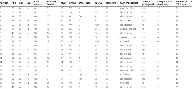
Table 1 Patient demographics and outcomes
Number Age Sex J&B Plate removed
Follow up
(months) MSQ SPADI DASH score Rel. CS Taft score Injury mechanism
Dominant side injured
Delay trauma-repair (days)
Time length for FFR (days)
1 39 M IIa Yes 13 91 97 7 93 11 vehicle accident Yes 10 35
2 26 M I Yes 16 91 98 4 90 12 bike accident Yes 3 26
3 33 W I Yes 12 72 68 26 84 10 bike accident No 5 44
4 35 M IIa Yes 16 86 92 9 87 11 ski accident Yes 4 31
5 59 M IIa Yes 14 98 99 0 100 11 bike accident No 2 37
6 29 M IIa No 12 86 86 7 81 10 jogging accident No 2 23
7 45 M IIa No 12 89 97 2 84 10 bike accident No 7 31
8 28 M III No 12 93 98 2 87 12 jogging accident Yes 6 37
9 47 W III No 13 96 100 0 100 12 simple fall No 6 46
10 31 W I Yes 12 86 95 8 84 11 bike accident Yes 3 42
11 37 M IIa No 13 84 89 13 85 11 ski accident Yes 2 48
12 46 M I Yes 15 90 95 1 85 12 ski accident No 6 39
13 53 M IIa No 12 87 95 6 85 9 bike accident Yes 1 38
14 36 M IIa Yes 19 94 98 0 90 11 bike accident Yes 5 34
15 39 M IIa Yes 21 92 100 1 83 11 bike accident No 4 41
16 21 W IIb No 14 83 88 14 87 12 vehicle accident Yes 4 28
17 52 W IIa No 12 70 58 20 65 9 simple fall Yes 8 44
18 54 M IIb No 7 77 77 18 75 10 bike accident Yes 9 39
19 61 W IIa No 26 82 92 8 76 9 simple fall No 1 43
20 43 M I No 12 92 99 6 91 11 bike accident Yes 2 26
Jäger&Breitner (J&B); Munich Shoulder Questionnaire (MSQ); Shoulder Pain and Disability Index (SPADI); Disability of the Arm, Shoulder and Hand (DASH) score; relative Constant Score (Rel. CS); full functional recovery (FFR).
BMC
Musculoskel
etal
Disorders
2014,
15
:380
Page
3
o
f
9
ntral.com/1
applicable instrument for the self-assessment of the shoul-der function. It was developed for an effective follow-up of shoulder patients allowing for a quantitative assessment of the Shoulder Pain and Disability Index (SPADI), the Dis-ability of the Arm, Shoulder and Hand (DASH) score and the Constant Score. The MSQ has been validated previ-ously and its accuracy and effectiveness for follow-up as-sessment was demonstrated [12,13]. Original Constant Score values were used to calculate a normative age- and sex-specific Constant Score (relative Constant Score) ac-cording to Gerber et al. [14]. AC joint stability was assessed by determining the Taft score which includes subjective, objective and radiologic criteria [10].
Statistics
Data is given in terms of the arithmetic mean ± standard deviation and the range in brackets. The results were compared by calculating the Wilcoxon rank-sum test. A p-value <0.05 determined significance. Statistics were calculated using commercially available programs (Sigma-Stat 3.1, SigmaPlot 8.02, Systat Software Inc., Chicago, USA). A p-value less than 0.05 was considered as statisti-cally significant.
Results
Demographics and fracture morphology
Between June 2011 and September 2013, 20 dislocated fractures of the lateral clavicle in 20 patients (14 men, 6 women) with a mean age of 40.7 ± 11.3 years (21–61 years) were enrolled in the study and surgically treated using the Synthes® LCP superior anterior clavicle plate in a prospective clinical trial (see Table 1). The mean inter-val between surgery and follow-up was 14.2 ± 4.0 months (7–26 months). According to the Jäger&Breitner (J&B) classification [11] 5 patients had a type I, 11 a type II a, 2 a type II b and 2 a type III fracture.
Surgery characteristics
The skin incisions had an average length of 5.1 ± 0.8 cm (4–7 cm). A 3-hole plate was implanted in 6, a 4-hole plate in 8, a 5-hole plate in 5 cases and a 6-hole plate in 1 case. The shaft holes were placed with at least one 3.5 mm locking screw. The plate size and the number of implanted screws depended on the fracture pattern espe-cially on the medial extent of the fracture. On average 5 ± 1 holes (4–6 holes) of the lateral extension were placed with 2.7 mm locking screws. All four surgeons (CK, PB, SH, SS) who performed the procedure were experienced upper extremity surgeons. CK performed surgery in 8, PB in 5, SH in 3 and SS in 4 cases. Surgery
had an average duration of 89.2 ± 26.7 min (46–
127 min), mean dose area product as degree of intraop-erative fluoroscopy was 30 ± 26 cGycm2 (6,69-113,31 cGycm2).
Complications
There were no major complications such as wound-healing problems, infections, implant failures or revision surgeries to be reported. In 9 of 20 patients the implant was removed on average 17.6 ± 2.7 months (range 13–22 months) after surgery. Main reasons for hardware re-moval were either the patients’ explicit request (n = 4), implant-associated pain while carrying a heavy backpack (n = 3) or sensitivity to weather changes (n = 2).
Patient reported and radiological outcomes
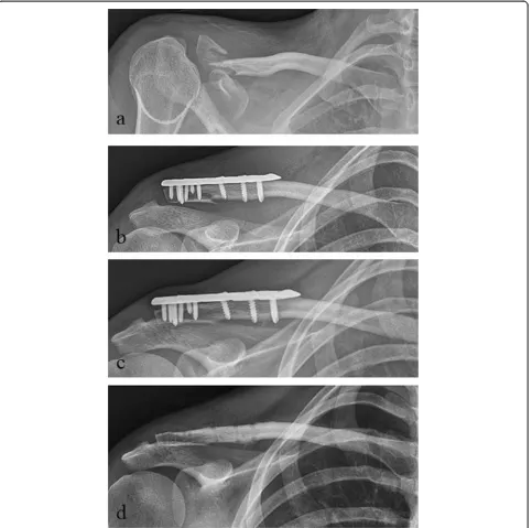
The mean MSQ was 87.0 ± 7.4 points (70–98), the mean SPADI 91.1 ± 11.3 points (58–100), the mean DASH score 7.6 ± 7.3 points (0–26) and the mean normative age- and sex-specific Constant Score 85.6 ± 8.0 points (65–100) (Table 1, Figure 1). The mean Taft Score was 10.7 ± 1.0 points (9–12 points). The mean Taft Score in lateral clavicular fractures with fracture gap between the coracoclavicular ligaments in combination with a rup-ture of the conoid ligament (J&B II a, Neer II B; n =11) was with 10.3 ± 0.9 points significantly lower than the mean Taft Score of all other types of lateral clavicle frac-tures (J&B I, II b, III; n =9) which resulted in 11.3 ± 0.9 points (p < 0.05 according to the Wilcoxon rank-sum test; Figure 2). Radiologic bony union occurred in all 20 patients after a mean interval of 6–10 weeks postopera-tive. Figures 3 and 4 show the radiological outcome of two lateral clavicle fractures (J&B II a and II b).
Discussion
Treatment of dislocated lateral clavicle fractures, especially of type IIb lesions is controversially discussed [15,16]. This is due to high non-union rates in conservative and high complication rates in surgical treatment using conven-tional implants such as the hook plate or k-wire fixation [3]. However, recently developed locking compression plates (LCP) showed promising functional results in small case series [5,6,17]. In this prospective clinical trial the re-sults of treating dislocated fractures of the lateral clavicle in 20 patients are reported using a new angular stable im-plant, the Synthes® LCP superior anterior clavicle plate.
Demographics
The presented study collective consists of 20 consecutive patients with a mean age of 40.7 ± 11.3 years and a male–female ratio of 14:6 comparable to other outcome studies concerning age and gender [6,18]. Interpretation of previous case series describing follow-up of locking compression plates for treating lateral clavicle fractures is mostly limited due to small patient collectives (7–14 patients) [5,6,19]. Hence, the strength of the obtained re-sults of follow-up examinations in low-incident diseases, such as lateral clavicle fractures, can only provide the fundament and a good starting point for further analysis.
Beireret al. BMC Musculoskeletal Disorders2014,15:380 Page 4 of 9
Complications
No major complications had to be reported in our study. However, 9 of 20 enrolled patients had elective removal of the plate in terms of implant removal. In the current literature, routine removal of metal implants re-mains a controversial issue [20,21] with a lack of evident guidelines [22]. Due to the typical surgical risks and
[image:5.595.57.539.89.338.2]complications, implant removal should only be performed in symptomatic patients after a detailed discussion of available treatment options. In our collective, removal of implants was performed in 9 patients due to subjective complaints such as pain if carrying a heavy backpack or sensitivity to weather changes. Compared to other bones of the upper extremity, removal of metalwork is
[image:5.595.58.539.487.695.2]Figure 2Mean Taft Score of lateral clavicle fractures Jäger&Breitner (J&B) II a (Neer II B) versus J&B I, II b and III at a mean follow-up of 14 months.Data are given as vertical boxplots (median: horizontal boxline; 25–75% interquartile ranges). *p < 0.05 group J&B II a (Neer II B) vs. group J&B I, II b, III; Wilcoxon rank-sum test.
most commonly performed in the clavicle [22], most likely due to the prominent subcutaneous position of the im-plant especially in athletes with poor soft tissue coverage.
Functional and radiological outcomes
Since clinical examination by surgeons rating their own patients is characterized by several bias, follow-up as-sessment was based on a patient-reported outcome questionnaire. The MSQ is a self-administrated and valid questionnaire to assess different aspects of the shoulder function. It allows for a qualitative self-assessment of the SPADI, the DASH score and the Constant Score [12,13]. In the presented study good to excellent functional re-sults at a mean follow-up of one year after surgery were
found. The presented results are comparable to other authors who used a locking compression plate for treat-ing fractures of the lateral clavicle [5,6,17]. Advantages of angular stable plate systems in comparison to previ-ous conventional osteosynthetic procedures (such as maintaining sufficient blood perfusion of the periosteum reducing the risk of bony non-union [23]) is one of the reasons for reduced complication rates. The presented results also seem to confirm the promising clinical and radiological outcome of previous smaller case series using the same implant (Synthes® LCP superior anterior clavicle plate) [5,6].
[image:6.595.61.537.88.518.2]Fractures of the lateral clavicle are closely anatomically neighbored to the CC ligaments, which are essentially Figure 3Radiological outcome of a lateral clavicle fracture J&B II a (Neer II B). apreoperative;bpostoperative;c1-year follow-up;dafter plate removal.
Beireret al. BMC Musculoskeletal Disorders2014,15:380 Page 6 of 9
important for the AC stability. Especially a disruption of the more vulnerable, medial conoid ligament can lead to vertical AC instability resulting in a cranialization of the medial clavicle shaft [24]. In our study collective, pa-tients with a lateral clavicular fracture type J&B II a (Neer II B) with disruption of the conoid ligament pre-sented a significant lower postoperative Taft Score com-pared to all other fracture types according to J&B with intact conoid ligament (I, II b, III; p < 0.05). However the number of patients in the compared groups is relatively
[image:7.595.59.542.89.568.2]low and therefore the reliability of significance is limited. Several authors showed good clinical and radiological re-sults after reconstruction of the CC ligaments in lateral clavicular fractures type J&B II a (Neer II B) which was additionally performed to the actual treatment of the fracture using a locking plate osteosynthesis [6,17,25]. Loriaut et al. [26] and Takase et al. [27] reported a satis-factory outcome after single arthroscopic reconstruction of the CC ligaments in unstable lateral clavicle fractures type J&B II a (Neer II B) without plate osteosynthesis. Figure 4Radiological outcome of a lateral clavicle fracture J&B II b. apreoperative;bpostoperative;c1-year follow-up;dafter
However, biomechanical evaluation showed increased stiffness, higher resistance to compression and decreased displacement of locking plate osteosynthesis combined with CC reconstruction compared to either technique alone [28]. Hence initial evidence is provided that single reconstruction of the CC ligaments in unstable lateral clavicle fractures type J&B II a (Neer II B) could be an insufficient approach due to the remaining instability allowing for fragment movement with an increased risk of pain and non-union.
Limitations
Despite of the prospective nature of our study it has sev-eral limitations to be mentioned. First of all the small number of included patients is considered as limitation. The number of patients in the compared groups is rela-tively low and therefore the reliability of significance is limited. However the literature does only provide evalua-tions of the same implant with even lower numbers of patients and therefore we contribute to the poor data situation in literature of this low-incidence-disease with a study of a comperatively high number of patients. A second limitation constitutes the uneven age distribution in both groups regarding AC instability. Although the disruption of the conoid ligament in lateral clavicle frac-tures J&B IIa presents a relevant reason for postopera-tive AC instability, we cannot exclude the higher patient age in this group as a substantial confounder. A third drawback of our study is of course that the postoperative rehabilitation was performed on an outpatient basis and thus was not performed in a standardized way. Although physiotherapy should be done according to a standard protocol we cannot guarantee the patient’s compliance.
Conclusions
By the development of locking compression plates for the treatment of lateral clavicle fractures new perspectives for the so far frustrating surgical therapy have arised. The pre-contoured Synthes® LCP superior anterior clavicle plate leads to sufficient stabilization and good functional out-come with high patient satisfaction when used for dis-placed lateral clavicle fractures. In fractures between the coracoclavicular ligaments in combination with a rupture of the conoid ligament (J&B II a, Neer II B) the recon-struction of the CC ligaments additionally to locking plate osteosynthesis showed superior biomechanical stability re-sults. Hence we favor an arthroscopic CC reconstruction additional to open LCP osteosynthesis in lateral clavicle fractures type J&B II a (Neer II B). However, our results still need to be substantiated by analyzing greater patient cohorts which is the focus of our study group.
Abbreviations
AC:Acromio-clavicular; CC: Coraco-clavicular; J&B: Jäger and Breitner;
questionnaire; SPADI: Shoulder pain and disability index; DASH: Disability of the arm, shoulder and hand; LCP: Locking compression plate.
Competing interests
The authors declare that they have no competing interests.
Authors’contributions
MB and CK were substantially involved in conception and design of the study, coordination and supervision of data collection, statistics, drafting the initial version of the manuscript and final approval of the version to be published. They are responsible for the overall content as guarantors. MC, LP and SS were involved in conception and design of the study, data collection and drafting the initial manuscript. They approved the final manuscript as submitted. SH and PB carried out the initial analyses, reviewed and revised the manuscript and approved the final manuscript as submitted. All authors read and approved the final manuscript.
Acknowledgements
We would like to explicitly thank Fritz Seidl, MA Interpreting and Translating and expert for statistics, for his excellent language copyediting and for his statistical analysis. The authors declare that they have no competing interests and that there is no source of funding.
Received: 23 September 2014 Accepted: 5 November 2014 Published: 19 November 2014
References
1. Nowak J, Mallmin H, Larsson S:The aetiology and epidemiology of clavicular fractures. A prospective study during a two-year period in Uppsala, Sweden.Injury2000,31(5):353–358.
2. Donnelly TD, Macfarlane RJ, Nagy MT, Ralte P, Waseem M:Fractures of the clavicle: an overview.Open Orthop J2013,7:329–333.
3. Oh JH, Kim SH, Lee JH, Shin SH, Gong HS:Treatment of distal clavicle fracture: a systematic review of treatment modalities in 425 fractures.
Arch Orthop Trauma Surg2011,131(4):525–533.
4. Good DW, Lui DF, Leonard M, Morris S, McElwain JP:Clavicle hook plate fixation for displaced lateral-third clavicle fractures (Neer type II): a functional outcome study.J Shoulder Elbow Surg2012,21(8):1045–1048. 5. Tiren D, Vroemen JP:Superior clavicle plate with lateral extension for
displaced lateral clavicle fractures: a prospective study.J Orthop Traumatol2013,14(2):115–120.
6. Schliemann B, Rosslenbroich SB, Schneider KN, Petersen W, Raschke MJ, Weimann A:Surgical treatment of vertically unstable lateral clavicle fractures (Neer 2b) with locked plate fixation and coracoclavicular ligament reconstruction.Arch Orthop Trauma Surg2013,133(7):935–939. 7. Kirchhoff C, Banke IJ, Beirer M, Imhoff AB, Biberthaler P:Operative
management of clavicular non-union: Iliac crest bone graft and anatomic locking compression plate.Oper Orthop Traumatol2013,25(5):483–498. 8. Neer CS 2nd:Fractures of the distal third of the clavicle.Clin Orthop Relat
Res1968,58:43–50.
9. Seppel G, Lenich A, Imhoff AB:Distal clavicle fracture.Oper Orthop Traumatol2014,26(3):254–262.
10. Taft TN, Wilson FC, Oglesby JW:Dislocation of the acromioclavicular joint. An end-result study.J Bone J Surg Am Vol1987,6(7):1045–1051. 11. Jager M, Breitner S:Therapy related classification of lateral clavicular
fracture.Unfallheilkunde1984,87(11):467–473.
12. Schmidutz F, Beirer M, Braunstein V, Bogner V, Wiedemann E, Biberthaler P: The Munich Shoulder Questionnaire (MSQ): development and validation of an effective patient-reported tool for outcome measurement and patient safety in shoulder surgery.Patient Saf Surg2012,6(1):9. 13. Biberthaler P, Beirer M, Kirchhoff S, Braunstein V, Wiedemann E, Kirchhoff C:
Significant benefit for older patients after arthroscopic subacromial decompression: a long-term follow-up study.Int Orthop2013,37(3):457–462. 14. Yian EH, Ramappa AJ, Arneberg O, Gerber C:The Constant score in normal
shoulders.J Shoulder Elbow Surg2005,14(2):128–133.
15. Khan LA, Bradnock TJ, Scott C, Robinson CM:Fractures of the clavicle.J Bone Joint Surg Am2009,91(2):447–460.
16. Robinson CM, Cairns DA:Primary nonoperative treatment of displaced lateral fractures of the clavicle.J Bone Joint Surg Am2004,86-A(4):778–782. 17. Hohmann E, Hansen T, Tetsworth K:Treatment of Neer type II fractures of
Beireret al. BMC Musculoskeletal Disorders2014,15:380 Page 8 of 9
TightRope augmentation of the coraco-clavicular ligaments.Arch Orthop Trauma Surg2012,132(10):1415–1421.
18. Bhatia DN, Page RS:Surgical treatment of lateral clavicle fractures associated with complete coracoclavicular ligament disruption: clinico-radiological outcomes of acromioclavicular joint sparing and spanning implants.
Int J Shoulder Surg2012,6(4):116–120.
19. Herrmann S, Schmidmaier G, Greiner S:Stabilisation of vertical unstable distal clavicular fractures (Neer 2b) using locking T-plates and suture anchors.Injury2009,40(3):236–239.
20. Richards RH, Palmer JD, Clarke NM:Observations on removal of metal implants.Injury1992,23(1):25–28.
21. Sanderson PL, Ryan W, Turner PG:Complications of metalwork removal.
Injury1992,23(1):29–30.
22. Jamil W, Allami M, Choudhury MZ, Mann C, Bagga T, Roberts A:Do orthopaedic surgeons need a policy on the removal of metalwork? A descriptive national survey of practicing surgeons in the United Kingdom.Injury2008,39(3):362–367.
23. Hanschen M, Biberthaler P:Mono- versus polyaxial locking plates.
Unfallchirurg2013,116(8):733–741. quiz 742–733.
24. Trompetter R, Seekamp A:Clavicle fractures.Unfallchirurg2008,111(1):27–38. quiz 39. 25. Martetschlager F, Kraus TM, Schiele CS, Sandmann G, Siebenlist S, Braun KF,
Stockle U, Freude T, Neumaier M:Treatment for unstable distal clavicle fractures (Neer 2) with locking T-plate and additional PDS cerclage.Knee Surg Sports Traumatol Arthrosc2013,21(5):1189–1194.
26. Loriaut P, Moreau PE, Dallaudiere B, Pelissier A, Vu HD, Massin P, Boyer P: Outcome of arthroscopic treatment for displaced lateral clavicle fractures using a double button device.Knee Surg Sports Traumatol Arthrosc2013.
27. Takase K, Kono R, Yamamoto K:Arthroscopic stabilization for Neer type 2 fracture of the distal clavicle fracture.Arch Orthop Trauma Surg2012, 132(3):399–403.
28. Rieser GR, Edwards K, Gould GC, Markert RJ, Goswami T, Rubino LJ: Distal-third clavicle fracture fixation: a biomechanical evaluation of fixation.J Shoulder Elbow Surg2013,22(6):848–855.
doi:10.1186/1471-2474-15-380
Cite this article as:Beireret al.:Clinical and radiological outcome following treatment of displaced lateral clavicle fractures using a locking compression plate with lateral extension: a prospective study. BMC Musculoskeletal Disorders201415:380.
Submit your next manuscript to BioMed Central and take full advantage of:
• Convenient online submission
• Thorough peer review
• No space constraints or color figure charges
• Immediate publication on acceptance
• Inclusion in PubMed, CAS, Scopus and Google Scholar
• Research which is freely available for redistribution