Open Access
Research article
Evaluation of a novel nanocrystalline hydroxyapatite paste Ostim
®
in comparison to Alpha-BSM
®
- more bone ingrowth inside the
implanted material with Ostim
®
compared to Alpha BSM
®
Franz-Xaver Huber*
1, Nicholas McArthur
2, Lydia Heimann
3,
Elvira Dingeldein
3, Héloïse Cavey
4, Xavier Palazzi
4, Gaëlle Clermont
4and
Jean-Pierre Boutrand
4Address: 1Klinik für Unfallchirurgie, Orthopädie und Wiederherstellungschirurgie, Klinikum Ansbach, Escherichstraße 1 91522 Ansbach, Germany, 2Medway Maritime Hospital, Gillingham, Kent, ME7 5 NY, UK, 3aap bio implants group, Industrie Center Obernburg, D - 63784 Obernburg, Germany and 4BIOMATECH-NAMSA, Zone Industrielle de I'Islon, 115 Rue Pasteur, F - 38670 Chasse-sur-Rhone, France
Email: Franz-Xaver Huber* - [email protected]; Nicholas McArthur - [email protected]; Lydia Heimann - [email protected]; Elvira Dingeldein - [email protected]; Héloïse Cavey - [email protected];
Xavier Palazzi - [email protected]; Gaëlle Clermont - [email protected]; Jean-Pierre Boutrand - [email protected] * Corresponding author
Abstract
Background: The purpose of this study was to evaluate the performance a newly developed nanocrystalline hydroxyapatite, OSTIM® following functional implantation in femoral sites in
thirty-eight sheep for 1, 2 or 3 months. Ostim® 35 was compared to an established calcium phosphate,
Alpha BSM®.
Methods: Biomechanical testing, μ-CT analysis, histological and histomorphological analyses were
conducted to compare the treatments including evaluation of bone regeneration level, material degradation, implant biomechanical characteristics.
Results: The micro-computed tomography (μCT) analysis and macroscopic observations showed
that Ostim® seemed to diffuse easily particularly when the defects were created in a cancellous
bone area. Alpha BSM® remained in the defect.
The performance of Ostim was good in terms of mechanical properties that were similar to Alpha BSM® and the histological analysis showed that the bone regeneration was better with Ostim® than
with Alpha BSM®. The histomorphometric analysis confirmed the qualitative analysis and showed
more bone ingrowth inside the implanted material with Ostim® when compared to Alpha BSM ®
at all time points.
Conclusions: The successful bone healing with osseous consolidation verifies the importance of the nanocrystalline hydroxyapatite in the treatment of metaphyseal osseous volume defects in the metaphyseal spongiosa.
Published: 22 December 2009
BMC Musculoskeletal Disorders 2009, 10:164 doi:10.1186/1471-2474-10-164
Received: 10 May 2009 Accepted: 22 December 2009
This article is available from: http://www.biomedcentral.com/1471-2474/10/164
© 2009 Huber et al; licensee BioMed Central Ltd.
Background
Operative reconstruction of bone defects beyond a certain size still remains a challenge to trauma and orthopedic surgeons. Every year, millions of people worldwide are suffering from bone defects arising from trauma, tumor or bone diseases. In approximately 10% of all traumatically related loss of bone structure or even tumor related bone defects, spontaneous bone healing is not able to restore the required physiological stability. In such cases bone replacement materials are often necessary to reconstruct the anatomical morphology and restore stability of the bone[1].
The use of autologous pelvic bone is still considered as the gold standard in the reconstruction of bone defects because of its unsurpassed biological activity even in implant sites with low osteogenic potential. Pelvic bone harvesting from the iliac crest does, however, presents unacceptable rates of morbidity at the grafting site and at the same time may only provide a limited amount of can-cellous bone[2-8]. Chronic pain can be present in up to 39% of patients at the donor site after iliac crest harvest-ing[2]. Other published complications include: fractures, infection, nerve and arterial injury[7].
Other bone sources include bone allografts which carry the potential of disease transmission, immunogenicity and possibly lower union rates[4,9]. Furthermore, the structural, mechanical, and resorption properties of allo-grafts are usually much altered by processing, preserva-tion, and sterilization techniques[4,10,11]. The relative concerns over the use of either autograft or allograft have led to the development of numerous bone graft substi-tutes[12-20].
In the ideal case artificial bone replacement materials should present a similar structure and composition to human bone and thus be able to present bone function. The materials should be osteoconductive and osteoinduc-tive by allowing osteoblast and osteoclast activity. At present there are over 100 approved bone replacement materials in Germany alone. The spectrum encompasses mainly hydroxyapatite ceramics, absorbable calcium phosphate cements, various metals, plastics and a variety of composites. The most commonly used synthetic min-eral substitutes for bone defect and trauma applications as implant coatings and defect fillers are hydroxyapatite cements, which have already undergone comprehensive animal testing and have also established themselves in many surgical procedures on human patients[18-39].
Ostim® represents a brand new development among the
purely synthetically produced and rapidly absorbable Hydroxyapatite compounds. It has been widely and
suc-cessfully used in the fields of oral and maxillofacial sur-gery and orthopedic and trauma sursur-gery[40-46].
The aim of the following study was to compare the newly
developed Ostim® with tricalcium phosphate cement
Alpha-BSM®, an already established bone replacement
material, in relation to their biocompatibility and bone ingrowth in a bone defect.
Methods
Material properties of the hydroxyapatite compounds used
Ostim®
Ostim®, (aap biomaterials GmbH, Dieburg, Germany) is a
newly developed, fully synthetic and fully resorbable
injectable nanocrystalline paste [Ca10(PO4)6(OH)2] and
consists of a suspension of pure HA in water prepared by a wet chemical reaction. The needle shaped HA crystals form agglomerates as shown by transmission electron microscopy (see fig. 1). XRD Analysis reveals an average
crystallite size of 19 nm. Ostim® paste does not harden
after application into the bone and is free of endothermal heating. It is characterized by a large bioactive specific
sur-face area of 106 m2 g-1[47]. The atomic ratio of
calcium-phosphorus is 1.67.
The product is supplied in a ready-to-use syringe to which a needle or a flexible 5 cm nozzle can be attached in order to inject the paste into deeper voids.
Alpha BSM®
Alpha BSM® (ETEX Corp, CAMBRIDGE, MA 02139, USA),
an established calcium phosphate cement, is an endother-mically setting apatitic calcium phosphate bone substi-tute. The final product (means after the conversion reaction is complete) consists of purely crystalline HA
with an average crystallite size of 12-14 nm. Alpha BSM®
has a paste like consistency until it is injected and hardens at body temperature. In 20 min it achieves complete con-version and mechanical strength. The reported mechani-cal strength is 15 MPa[34]. Specific surface area of the
dried product is 78 m²/g.
None of two materials has macropores and both are applied as wet pastes. The water content of Ostim is 65%, freshly prepared alpha-BSM paste has a water content of 55%.
Animal experiment protocol
reserve (38 + 3) female sheep (Ovisaries), between 53 and 73 kg and between 2 and 5 years old were used for the ani-mal experiments.
The evaluation of the performance and the biocompatibil-ity of a bone substitute was tested following functional implantation in femoral sites in thirty-eight sheep for 1, 2
or 3 months (total of 76 sites). The Ostim® material (n =
38 sites) was compared to Alpha-BSM® (n = 38 sites)
fol-lowing implantation in a cancellous bone defect (see additional file 1).
Each animal received an implantation of each bone replacement material in the cancellous bone of the right and left medial distal femoral epiphysis respectively. The treatment allocation was randomly assigned between the right and left leg. Pre-medication and anesthesia were per-formed by intravenous injection of
thiopental-pentobar-bital mixture (NESDONAL®, MERIAL, France;
pentobarbital sodique, CEVA Santé animale, France) and atropine (Atropinum sulfuricum, AGUETTANT, France) followed by inhalation of a O2 - isoflurane (1-4%) mix-ture. Each animal received an analgesic (flunixine,
FINA-DYNE® Injectable, Schering-Plough, France)
pre-operatively. As a prophylactic measure, perioperative anti-biotics penicillin-procaine and penicillin benzathine
(DUPHAPEN® LA, FORT DODGE, France) was given.
Using standard aseptic techniques, a cutaneous incision was made on the medial face of the knee. The medial con-dyle of the stifle joint of each animal was exposed by blunt dissection of the subcutaneous tissue and the fascia. The muscle and joint capsule were gently dissected and the supracondyle bone area was approached. The periosteum was carefully removed from the femoral epiphysis and metaphysis to expose the implant site. A core drill was used to drill a 10 mm diameter × 15 mm deep hole in the distal femoral epiphysis. After drilling the defect to the required depth, an extractor was inserted and driven down to the bottom of the defect. The extractor was turned to break the bone trabecula at the bottom of the defect. Depth of the defect was measured. Each defect was filled with either the test or the control article. The incision was then closed by closing the subcutaneous layers one by one with absorbable sutures (Vicryl 2-0 and Vicryl 1, ETHICON, France), and the skin layer was closed using surgical staples. The animal was repositioned and the other bone replacement material implanted in the same fashion. At the appropriate termination interval, the des-ignated sheep were terminated by lethal injection of bar-biturate (DOLETHALND, VETOQUINOL, France). A macroscopic examination of the implanted femoral epi-physes was performed. The epiepi-physes were then harvested and identified. The epiphyses from the 2 and 3 months animals were placed in 10% buffered formalin. After
fixa-tion, these samples were submitted for μ-CT analysis. The
epiphyses from the 3 months interval were arbitrarily divided so that sixteen sites sampled from eight animals (eight test and eight control) were selected for biome-chanical testing and the remaining sites were fixed in 10% buffered formalin.
Preparation of the samples
The epiphysis from the eight designated sheep from the 3 months termination interval was subjected to biomechan-ical testing. The soft tissues were removed from these sam-ples. The defect was located using X-rays. A diamond saw was used to obtain two parallel and flat surfaces. The approximate final dimensions of the sample were 4 × 4 × 1.8 cm (see fig 2). Care was taken not to disrupt the implant from the defect site at this stage.
The test specimens were prepared so that the implantation site were in the centre of the sample. The tissue samples
Sample dimensions
Figure 1
were provided in physiological serum and the trials were performed within 48 h after removal. Their thickness has been measured using a slide caliper at the implantation point.
Biomechanical testing
First, the thickness of the samples was measured with a calliper. Following thickness measurements, the samples were immersed in room temperature saline solution. The samples were submitted to an indentation test. Load was applied axially to the specimens by a pin with a diameter of 4 mm and with a displacement speed of 0.4 mm/min until failure. To determine the mechanical characteristics of the samples, the data collected were stiffness and Young's modulus, yield point, maximum load and corre-sponding stress and strain. This biomechanical testing was also performed on four non implanted test articles.
Non implanted samples were prepared by placing Ostim samples in a cylindrical container of a diameter of 12 mm and a height of 20 mm. They were tested immediately thereafter.
-CT analysis
Following fixation, the epiphyses sites for the 2 months animals and the ten animals designated from the 3 months interval were used to quantify the mean BV/TV (Bone + substitute volume/Total bone defect volume).
The μCT measurements were conducted at ETH Zürich by
means of a μCT 80, Scanco Medical AG, Switzerland. The
measured data was filtered using a constrained Gaissian
filter with finite filter support (1 Voxel) and filter width (σ
= 1.2). The images were then binarized to separate the object from the background using a global thresholding procedure. For density analysis a cylinder of 11.7 mm in diameter and 17 mm length was analyzed. The cylinder
dimension was obtained from the μCT images and was
chosen to represent the original defect as good as possible.
For this the cylinder was morphed along the axis of the defect zone in the following way: A circle with 11.7 mm was positioned over the defect zone on the first and the
last μCT slice of the given range of the substitute (17 mm)
and the cylinder was morphed in-between. Within this cylinder the apparent density was computed as the ratio of substitute versus background.
Histopathological technique
Each implant site and surrounding tissue was isolated using a band saw. After labelling and fixing in 10% buff-ered formalin solution, the sites were longitudinally divided in two blocks. One half of each site was dehy-drated in alcohol solutions of increasing concentrations, cleared in xylene, decalcified and embedded in paraffin.
Embedded sites were then longitudinally cut at 5 to 7 μm
using a microtome (MICROM®, France). One paraffin
sec-tion per site was prepared and stained with Masson's tri-chrome. The other half of each site was dehydrated in alcohol solutions at increasing concentrations, cleared in xylene and embedded in polymethylmethacrylate (PMMA). One longitudinal section was prepared using the Exakt system [25,50]. The resin sections were stained with a modified Paragon.
Qualitative evaluation of implanted sites
Histological sections were observed using a Nikon micro-scope (Eclipse E600) fitted with ×4, ×10, ×25 and ×40 objectives. Semi-quantitative analysis was performed in accordance with ISO 10993-6 standard: local tolerance criteria and performance were analyzed and graded. (necrosis, fibrinous exudate, tissue degeneration, inflam-matory cells, fibrous tissue, osteolysis, newly formed bone, osteointegration, osteoconduction, and material degradation.)
Particular attention was devoted to:
- the neoformed bone, the direct bone/implant interface and the osteoconduction process
- inflammatory reaction,
- presence of fibrosis
- material degradation.
The semi-quantitative scoring system is shown in addi-tional file 2
Quantitative histomorphometry of implanted sites
The histological slides obtained from resin blocks were evaluated under a Zeiss Axioscope microscope fitted with ×5, ×10, ×20 and ×40 objectives and equipped with a
color image analyzing system SAMBA® (SAMBA
TECH-Ostim® agglomerates in transmission electron microscopy
Figure 2
Ostim® agglomerates in transmission electron
microscopy.
4 cm
15 mm
4 cm
NOLOGIES, France). Two standard areas of investigation were located peripherally to the Bone/Implant junction, and two standard areas of investigation were located inter-nally to the Bone/Implant junction.
In these areas of investigation, whenever possible, the fol-lowing parameters were measured in the control resin slides and in the test resin slides at one, two and three months:
- bone to implant contact, BIC (expressed in percentage)
- bone density around the material (expressed in percent-age)
- bone density inside the material (expressed in percent-age)
At each time point a statistical analysis (ANOVA method) was performed for these 3 parameters, considering control versus test article.
Statistical analysis
The statistical tests used for a comparison between groups for the main parameters of biomechanical testing and quantitative histological analysis were Mann and Whitney
or ANOVA tests performed with a software (SPSS® version
14.0, edited by SPSS Inc, Chicago).
Results
Biomechanical testing
Biomechanical testing was performed on 16 samples from the 3 month time-period (see additional file 3). At yield
point, the stress was higher in the Alpha-BSM® Group and
the strain was higher in the Ostim® group. Resistance,
stress and strain were higher in the Alpha-BSM® group.
Elasticity, stiffness and Young modulus were higher in the
Ostim® group. However, no statistically significant
differ-ences were observed for any parameters. The biomechan-ical testing performed on four non implanted test articles showed a maximum load for resistance and yield point at least ten times lower than the values observed after implantation, suggesting a significant hardening and/or bone ingrowth during the 3 month implantation period.
CT analysis
All samples were successfully scanned with μCT. From
these measurements it can be seen that the Alpha BSM material was very dense and had a very well confined shape. For this reason, the median for these groups was higher than 95%, which means that there was almost no porosity. The mean BV/TV (Bone + substitute volume/ Total volume) values were found to be 95.2% for the 2 month Alpha BSM sites and 90.4% for the 3 month Alpha BSM sites. The Ostim material was in most samples very
granular and showed some tendency to penetrate into the surrounding area. However, bone ingrowth into the sub-stitute could not be determined as it was not possible to automatically differentiate the bone from the substitute due to similar contrast. In samples where the substitute disappeared from the original defect, there was only little bone formation that could be seen in the defect. The apparent density in the Ostim sites was lower than in Alpha BSM sites. The mean BV/TV values were found to be 68.3% for the 2 month Ostim sites and 79.9% for the 3 month Ostim sites. The relatively large range indicates that these samples were much more inhomogeneous as
the control samples. The μCT measurement were limited
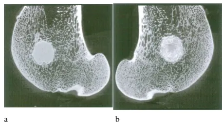
by the fact that the bone replacement materials had a very similar attenuation for X-rays as bone and that thus two phases could not be separated automatically. It was observed that the Ostim article seemed to diffuse easily in particular when the defects were created in a spongious bone area, close to the diaphysis or the joint-space nar-rowing whereas the more compact Alpha BSM article used in similar conditions stayed in the defect. This led to a lower apparent density within the defects treated with the Ostim when compared to the Alpha BSM (see fig. 3a - b).
Microscopic analysis of femoral sites at 1 month (see additional file 4)
Alpha BSM
All ten implanted sites were identified on the histological sections. These sites were filled with a dense green-red stained biomaterial (on paraffin sections) with diffuse optically empty round cavities. On resin sections the material was very dense, dark pink colored and it showed no signs of open porosity. Limited signs of inflammatory reaction were visible in the area surrounding the product and were accompanied by the presence of macrophages and giant multinucleated cells. The Alpha-BSM showed
[image:5.612.318.546.538.668.2]Defects centralized, confined (a) Alpha-BSM® and (b) Ostim®
Figure 3
Defects centralized, confined (a) Alpha-BSM® and (b)
Ostim®. The μCT images after three months of the bone replacement material implantation.
good signs of osseointegration. A slight grade of fibrocon-nective tissue was often observed at the bone-implant interface. Almost no signs of implant degradation were visible. Few osteoblasts were observed directly on the sur-face of the implant. The overall osteoconduction process and newly formed bone were of limited grade and local-ized onto the interface between the edge of the original bone defect and the implant surface. No or very limited signs of bone ingrowth within the implant were observed, likely due to the absence of open porosity of the implant. As a consequence the bone density within the implant was of very limited grade (see fig. 4)
Ostim®
Ten implant sites were identified. The implant was recog-nized as green stained particles (on paraffin sections) of
various sizes ranging from a few μm to 100-200 μm. In
resin sections, the implant particles often formed central dense and peripheral loose aggregates, thus delimiting widely open interconnected peripheral porosities. No continuity with the neighboring articular cartilage could be observed microscopically on both paraffin and resin sections. A slight to moderate grade of inflammatory reac-tion was observed in the area surrounding the implant particles and was accompanied by the presence of a vascu-larized fibroconnective inflammatory tissue,
macro-phages and multinucleated giant cells. The Ostim® 35
bone substitute showed good signs of osseointegration (moderate grade), osteoconduction (moderate grade) and bone density within the implant (slight to moderate grade). Notable was a degree of heterogeneity was observed amongst the animals regarding new bone
forma-tion and bone density. Limited signs of material degrada-tion were observed (see fig. 5).
Microscopic analysis of femoral sites at 2 months (see additional file 4)
Alpha BSM
The ten sites implanted with Alpha BSM showed a similar appearance compared to those observed at 1 month, with only slight signs of peripheral degradation after 2 months. Almost no residual signs of inflammatory reaction were visible: no biologically significant amounts of macro-phages, giant multinucleated cells and fibroconnective tis-sue were observed. The osseointegration of the
Alpha-BSM® slightly progressed with few signs of penetration of
thin bone trabecules within the outer portion of the bone substitute. Few signs of bone remodeling were observed (see fig. 6)
Ostim®
The overall aspects of Ostim® after 2 months were
compa-rable to those observed at 1 month, with some sites show-ing a heterogeneous structure of the material. The degradation process of the test article progressed signifi-cantly between 1 and 2 months with an overall reduced size of the particles in many animals. No continuity with the neighboring articular cartilage could be observed microscopically on both paraffin and resin sections.
[image:6.612.317.548.432.616.2]Alpha-BSM® at 1 month
Figure 4
Alpha-BSM® at 1 month. Masson's tricrome staining and 2× magnification of decalcified bone with calcium phosphate appearing as a still relatively compact matter. The osteocon-duction process and newly formed bone were of limited grade and localized onto the interface (black arrows) between the edge of the original bone defect and the implant surface. (New bone = blue, Alpha-BSM = red)
Ostim® at 1 month
Figure 5
[image:6.612.60.292.482.621.2]Ostim® particles were associated with few signs of
inflam-matory reaction consisting of a limited grade of macro-phages and giant multinucleated cells. A variable grade of fibrocytic tissue was visible within the implanted bone defect. Mean osteoconduction and bone density parame-ters progressed very significantly. Few signs of bone remodeling were observed (see fig. 7).
Microscopic analysis of femoral sites at 3 months (see additional file 4)
Alpha-BSM®
Ten implanted sites were identified on the histological sections. In paraffin sections the sites were filled with a
dense red stained biomaterial with diffuse optically empty round cavities. In resin sections, the material was still quite dense, but some signs of open porosity were observed. Limited signs of inflammatory reaction were visible in the area surrounding the product and were accompanied by the presence of macrophages. Moderate evidences of bone ingrowth within the implant were observed. As a consequence the bone density within and around the implant was of slight to moderate grade, and a steady and homogeneous progression of new bone for-mation was observed amongst the animals in resin sec-tions. Some signs of bone remodeling were observed in one animal only on resin sections (see fig. 8)
Ostim®
The overall histopathological picture was very similar to the one observed at 2 months. The mean amount of inflammatory reaction surrounding the implant particles was slight and tended to decrease over time. Still there were great individual variations regarding the amount of fibrous tissue ranging from absent to marked grade,
resulting in respectively high or low bone density. Ostim®
showed good signs of mean osseointegration, osteocon-duction and bone density within the implant that improved over time. Moderate signs of material degrada-tion were constantly observed, which increased over time. Some signs of bone remodeling were observed in resin sections (see fig. 9).
Quantitative histolomorphometrical analysis
At early time points, Ostim® showed much better
micro-scopic signs of osteointegration, osteoconduction and overall bone ingrowth when compared to the
Alpha-BSM®. After 2 and 3 months, the bone regeneration level
within the Ostim® treated sites was better than the
Alpha-BSM® treated sites, but new bone formation tended to
[image:7.612.63.291.89.235.2]Alpha-BSM at 2 months
Figure 6
Alpha-BSM at 2 months. Masson's tricrome staining and 20× magnification of decalcified bone 2 months after Alpha-BSM® implantation.
Ostim® at 2 months
Figure 7
Ostim® at 2 months. Modified paragon staining and 20× magnification of Ostim® core surrounded by new bone
for-mation.
[image:7.612.59.294.500.668.2]Alpha-BSM® at 3 months
Figure 8
Alpha-BSM® at 3 months. Modified paragon staining and 10× magnification of Alpha BSM® cement on the left upper
slow down when compared to the steadier progression and fewer inter-individual variations observed with
Alpha-BSM® (see additional file 5 and 6).
Bone to Implant Contact (BIC)
After 1 month, bone to implant contact was significantly different between alpha BSM and Ostim groups (p = 0.004). Bone to implant contact around the material was found higher in the alpha BSM than in the Ostim group. This difference was transient and not observed at later time-points (see additional file 7).
Bone density inside the material
After 1 and 2 months, bone density inside the material
was significantly different between Alpha-BSM® and
Ostim® groups. Bone density was higher in Ostim® group
when compared to Alpha-BSM® as no bone was found
inside the Alpha-BSM® material. After 3 months, this same
parameter was again significantly higher in the Ostim®
group as compared to the Alpha-BSM® group (p = 0.012)
although some bone was found inside the Alpha-BSM®
material (see fig. 10).
Bone density around the material did not show statisti-cally significant variations at all considered time-points.
Discussion and Conclusions
There were a number of attempts in the past to replace bone loss with bone replacement materials. Hamilton in 1881 used disinfected sponges and Gluck in 1891 implanted ivory into bone defects. Dreesmann even tried gypsum in 1892. In the 20th century Maatz experimented with bovine bone and Tarsoly filled the defects with egg
shells[51]. Modern bone replacement materials com-posed of calcium phosphate are either extracted from bovine cancellous bone or are produced syntheti-cally[14,18,52,53]. The requirements of an ideal bone replacement material have not changed over the past 30 years: simple application, osteoconductive properties, and complete resobarbility during bone ingrowth[53,54].
Alpha-BSM® fulfils these requirements very well as a
syn-thetic calcium phosphate bone replacement material. Compared to other calcium phosphates which require five years or more to be resorbed by the host bone[55,56],
Alpha BSM® appears to undergo more rapid
resorp-tion[22,52]. It does, however, have less compressive strength than other types of cements [34]. Compared to autologous cancellous bone, calcium phosphate cements have shown superior compressive strength. Welch et al reported a significantly lower secondary loss of correction
with the use of Alpha-BSM®, in the treatment of tibia head
fractures in goats compared to autologous spongiosa[34]. Russell also revealed a significantly lower subsidence in
tibia head fractures with the use of Alpha-BSM® compared
to autologous bone grafts in the scope of a multicentre study with 119 Patients[39].
The cement microstructure of Alpha-BSM®, which results
from a reaction of precipitation, is not equivalent to the crystalline structure of human bone[18,57,58]. The
nanocrystalline structure of Ostim® on the other hand
resembles human bone much closer (Calcium/Posphate
ratio of 1,675 and specific surface area of 106 m2g-1 is
syn-thesized by a wet chemical reaction of precipitation under
[image:8.612.320.548.90.272.2]Ostim® at 3 months
Figure 9
Ostim® at 3 months. Modified paragon staining and 5× magnification of non-decalcified bone 3 months after Ostim®
implantation. Ostim® bone showed good osseointegration
and bone remodeling.
[image:8.612.61.289.90.257.2]Bone density inside the material in percent
Figure 10
permanent pH control using CaO dispersed in water under constant stirring to maintain a suspension state and
H3PO4 as starting material [47,58]. Ostim® has been
observed as needle shaped HA crystals forming agglomer-ates in transmission electron microscopy [1,47]. Accord-ing to the hypotheses put forward by Constanz and Knaack such HA-compounds accelerate the bony ingrowth into the CSD as they closely mimic the required resorptive and osseointegrative properties of poor crystal-line apatitic structure of natural bone[52,57,58]. Moreo-ver Kilian et al also presented evidence of angiogenesis within bone defects filled with nanocrystalline hydroxya-patite [59,1,60]. These properties appear to explain the
significantly increased ingrowth of bone of the Ostim®
group compared to the Alpha-BSM® group. There was no
significant difference between the two materials with regard to their biomechanical stability.
Ostim® 35 showed limited signs of degradation after 1
month but degradation increased over time and was of moderate grade after 3 months in our experimental series. This confirms the results obtained in a number of differ-ent experimdiffer-ental animal models of other studies. Grigor-yan et al described the rapid bone ingrowth at a low complication rate following the treatment of jaw defects
with Ostim® in dogs in 2000 [61]. Our own animal
exper-iments dealing with the filling of critical size defects with Ostim in New Zealand white rabbits also resulted in swift and uniform bone ingrowth[62]. The results were further enhanced by the additional use of a sintered
hydroxyapa-tite ceramic[62]. Ostim® was always present in the bone
after 60 days following implantation, as in the present study[62]. Laschke reported a guided neovascularization
directed toward areas of Ostim® degradation in Syrian
golden hamsters [63]. Spiess further confirmed the good biocompatibility, osteoconductivity and bone ingrowth
in New Zealand whites, but reported a halt in the Ostim®
degradation process 6 weeks following implantation[64].
Comprehensive clinical experience of using the hydrated HA-paste as a void filling exists in the field of maxillofa-cial surgery. Various stomatology publications describe an accelerated fracture healing and bone density increase at a high degree of tolerance. In 1996 Zuev treated 395 patients with jaw defects and peridontal abscesses. The complication rate of the 200 patients in the Ostim group was at 1.5% compared to 3.6% in the group with 195 patients treated with allografts[44]. Bezrukow achieved excellent results in 1998 after treating 49 patients with
Ostim® following cystectomy of benign cyst-tumours of
the jaw. The defects in all 49 patients were replaced with fresh bone three months following the defect filling [43].
Gerlachfilled 44 mandibular cysts with Ostim® and
reported complete material resorption with an extemely low complication rate[65]. Strietzel et al performed a
lat-eral alveolar ridge augmentation with Ostim® in 14
patients. Six months later histological results showed good bone ingrowth of the defect and small amounts of
Ostim® remnants [46].
Ostim® in combination with conventional or angularly
stable osteosynthesis has also been successfully used in the field of traumatology. Particularly good results were achieved in prospective cohort studies with patients for the treatment of radial fractures, with closed intraarticular calcaneus fractures, as well as tibia compression fractures [40-42]. Histological examinations of bone harvested from patients during elective metal removal, have shown excellent bone ingrowth into the past bone defect with well structured cortical and cancellous bone tissue and without any inflammatory reaction orosteofibrosis. The implanted material was mainly resorbed[62].
Our data verifies the importance of the nanocrystalline hydroxyapatite in the treatment of metaphyseal osseous volume defects in the metaphyseal spongiosa, even though this study was done on an experimental animal model. Further clinical studies need to be conducted to validate the significance of Ostim in human bone defects. Due to its good biocompatibility and high rates of resorp-tion we can see a number of indicaresorp-tions for the use of Ostim in the field of traumatology. In our opinion the
application of Ostim® should always be combined with
some form of stable osteosynthesis, preferably with an angularly stable plate, due to its lack of dimensional sta-bility.
Competing interests
Aap biomaterials is manufacturing and selling Ostim. Aap did finance the animal study which was performed by Bio-matech. E. Dingeldein and L. Heinmann receive their sal-ary and are employed by aap Biomaterials. E. Dingeldein holds shares in aap.
H. Cavey, J-P. Boutrand, X. Palazzi, and G. Clermont receive salary from Biomatech.
F-X. Huber and N. McArthur have no competing interests.
None of the authors holds or is applying for patents related to the content of the manuscript.
Authors' contributions
Additional material
References
1. Schnettler R, Stahl JP, Alt V, Pavlidis T, Dingeldein E, Wenisch S: Cal-cium phosphat-based bone substitutes. European Journal of Trauma 2004, 4:219-229.
2. Arrington ED, Smith WJ, Chambers HG, Bucknell AL, Davino NA: Complications of iliac crest bone graft harvesting. Clin Orthop Relat Res 1996:300-309.
3. Blakemore ME: Fractures at cancellous bone graft donor sites.
Injury 1983, 14:519-522.
4. Boyce T, Edwards J, Scarborough N: Allograft bone. The influ-ence of processing on safety and performance. Orthop Clin North Am 1999, 30:571-581.
5. Goulet JA, Senunas LE, DeSilva GL, Greenfield ML: Autogenous iliac crest bone graft. Complications and functional assess-ment. Clin Orthop Relat Res 1997:76-81.
6. Ubhi CS, Morris DL: Fracture and herniation of bowel at bone graft donor site in the iliac crest. Injury 1984, 16:202-203. 7. Younger EM, Chapman MW: Morbidity at bone graft donor
sites. J Orthop Trauma 1989, 3:192-195.
8. Wippermann BW, Schratt HE, Steeg S, Tscherne H: [Complica-tions of spongiosa harvesting of the ilial crest. A retrospec-tive analysis of 1, 191 cases]. Chirurg 1997, 68(12):1286-1291. 9. Stevenson S: Biology of bone grafts. Orthop Clin North Am 1999,
30(4):543-552.
10. Goldberg VM, Stevenson S: Natural history of autografts and allografts. Clin Orthop Relat Res 1987:7-16.
11. Mankin HJ, Doppelt S, Tomford W: Clinical experience with allo-graft implantation. The first ten years. Clin Orthop Relat Res
1983:69-86.
12. Bauer TW, Muschler GF: Bone graft materials. An overview of the basic science. Clin Orthop Relat Res 2000:10-27.
13. Burstein FD: Bone substitutes. Cleft Palate Craniofac J 2000, 37:1-4. 14. Delloye C, Cnockaert N, Cornu O: Bone substitutes in 2003: an
overview. Acta Orthop Belg 2003, 69:1-8.
15. Hollinger JO, Brekke J, Gruskin E, Lee D: Role of bone substitutes.
Clin Orthop Relat Res 1996:55-65.
16. Van Heest A, Swiontkowski M: Bone-graft substitutes. Lancet
1999, 353(Suppl 1):SI28-SI29.
17. Pietrzak WS, Ronk R: Calcium sulfate bone void filler: a review and a look ahead. J Craniofac Surg 2000, 11:327-333.
18. Schnurer SM, Gopp U, Kuhn KD, Breusch SJ: [Bone substitutes].
Orthopade 2003, 32:2-10.
19. Briem D, Linhart W, Lehmann W, Meenen NM, Rueger JM: [Long-term outcomes after using porous hydroxyapatite ceramics (Endobon) for surgical management of fractures of the head of the tibia]. Unfallchirurg 2002, 105:128-133.
20. Mahan KT, Carey MJ: Hydroxyapatite as a bone substitute. J Am Podiatr Med Assoc 1999, 89:392-397.
21. Cassidy C, Jupiter JB, Cohen M, li-Santi M, Fennell C, Leinberry C, et al.: Norian SRS cement compared with conventional fixation in distal radial fractures. A randomized study. J Bone Joint Surg Am 2003, 85-A:2127-2137.
22. Sarkar MR, Wachter N, Patka P, Kinzl L: First histological obser-vations on the incorporation of a novel calcium phosphate bone substitute material in human cancellous bone. J Biomed Mater Res 2001, 58(3):329-334.
23. Trenholm A, Landry S, McLaughlin K, Deluzio KJ, Leighton J, Trask K,
et al.: Comparative fixation of tibial plateau fractures using alpha-BSM, a calcium phosphate cement, versus cancellous bone graft. J Orthop Trauma 2005, 19(10):698-702.
24. Simpson D, Keating JF: Outcome of tibial plateau fractures managed with calcium phosphate cement. Injury 2004, 35:913-918.
25. Gierse H, Donath K: Reactions and complications after the implantation of Endobon including morphological examina-tion of explants. Arch Orthop Trauma Surg 1999, 119:349-355. 26. Hing KA, Best SM, Tanner KE, Bonfield W, Revell PA: Mediation of
bone ingrowth in porous hydroxyapatite bone graft substi-tutes. J Biomed Mater Res A 2004, 68:187-200.
27. Jarcho M: Calcium phosphate ceramics as hard tissue pros-thetics. Clin Orthop Relat Res 1981:259-278.
28. Fernandez E, Gil FJ, Ginebra MP, Driessens FC, Planell JA, Best SM: Calcium phosphate bone cements for clinical applications. Part II: precipitate formation during setting reactions. J Mater Sci Mater Med 1999, 10:177-183.
Additional file 1
Study design. Number of sheep, number of investigation sites and analy-ses performed.
Click here for file
[http://www.biomedcentral.com/content/supplementary/1471-2474-10-164-S1.DOCX]
Additional file 2
Semi-quantitative scoring system. The table shows how each score was correlated to various degrees of reaction.
Click here for file
[http://www.biomedcentral.com/content/supplementary/1471-2474-10-164-S2.DOCX]
Additional file 3
Results of biomechanical testing. Geometry, Elasticity Yield point and Resistance were tested for Ostim and Alpha-BSM.
Click here for file
[http://www.biomedcentral.com/content/supplementary/1471-2474-10-164-S3.DOCX]
Additional file 4
Mean values of the semi-quantitative analysis. A number of histological parameters have been compared between Ostim and Alpha-BSM at 1, 2 and 3 months.
Click here for file
[http://www.biomedcentral.com/content/supplementary/1471-2474-10-164-S4.DOCX]
Additional file 5
Mean histomorphometric values around the material. Values in % are shown for both Ostim and Alpha-BSM concerning fibrous to implant con-tact, bone to implant concon-tact, implant density, fibrous density and bone density around the implant.
Click here for file
[http://www.biomedcentral.com/content/supplementary/1471-2474-10-164-S5.DOCX]
Additional file 6
Mean histomorphometric values inside the material. Values in % are shown for both Ostim and Alpha-BSM concerning fibrous to implant con-tact, bone to implant concon-tact, implant density, fibrous density and bone density inside the implant.
Click here for file
[http://www.biomedcentral.com/content/supplementary/1471-2474-10-164-S6.DOCX]
Additional file 7
Significant differences between Ostim and Alpha-BSM. The table shows that only bone density inside material remained significantly differ-ent between Ostim and Alpha-BSM throughout the 3 month study period. Click here for file
29. Fernandez E, Gil FJ, Ginebra MP, Driessens FC, Planell JA, Best SM: Calcium phosphate bone cements for clinical applications. Part I: solution chemistry. J Mater Sci Mater Med 1999, 10:169-176.
30. Lobenhoffer P, Gerich T, Witte F, Tscherne H: Use of an injectable calcium phosphate bone cement in the treatment of tibial plateau fractures: a prospective study of twenty-six cases with twenty-month mean follow-up. J Orthop Trauma 2002, 16:143-149.
31. Keating JF, Hajducka CL, Harper J: Minimal internal fixation and calcium-phosphate cement in the treatment of fractures of the tibial plateau. A pilot study. J Bone Joint Surg Br 2003, 85:68-73.
32. Gangopadhyay S, Ravi K, Packer G: Dorsal plating of unstable dis-tal radius fractures using a bio-absorbable plating system and bone substitute. J Hand Surg [Br] 2006, 31(1):93-100. 33. Bajammal SS, Zlowodzki M, Lelwica A, Tornetta P, Einhorn TA,
Buck-ley R, et al.: The use of calcium phosphate bone cement in frac-ture treatment. A meta-analysis of randomized trials. J Bone Joint Surg Am 2008, 90(6):1186-1196.
34. Welch RD, Zhang H, Bronson DG: Experimental tibial plateau fractures augmented with calcium phosphate cement or autologous bone graft. J Bone Joint Surg Am 2003, 85-A(2):222-231.
35. Kopylov P, Adalberth K, Jonsson K, Aspenberg P: Norian SRS ver-sus functional treatment in redisplaced distal radial frac-tures: a randomized study in 20 patients. J Hand Surg [Br] 2002, 27:538-541.
36. Sanchez-Sotelo J, Munuera L, Madero R: Treatment of fractures of the distal radius with a remodellable bone cement: a pro-spective, randomised study using Norian SRS. J Bone Joint Surg Br 2000, 82:856-863.
37. Dickson KF, Friedman J, Buchholz JG, Flandry FD: The use of Bone-Source hydroxyapatite cement for traumatic metaphyseal bone void filling. J Trauma 2002, 53:1103-1108.
38. Zimmermann R, Gabl M, Lutz M, Angermann P, Gschwentner M, Pechlaner S: Injectable calcium phosphate bone cement Norian SRS for the treatment of intra-articular compression fractures of the distal radius in osteoporotic women. Arch Orthop Trauma Surg 2003, 123:22-27.
39. Russell TA, Leighton RK: Comparison of autogenous bone graft and endothermic calcium phosphate cement for defect aug-mentation in tibial plateau fractures. A multicenter, pro-spective, randomized study. J Bone Joint Surg Am 2008, 90(10):2057-2061.
40. Huber FX, Hillmeier J, Herzog L, McAthur N, Kock HJ, Meeder PJ: Open reduction and palmar plate-osteosynthesis in combi-nation with a nanocrystalline hydroxyapatite spacer in the treatment of comminuted fractures of the distal radius. J Hand Surg [Br] 2006, 31(3):298-303.
41. Huber FX, McArthur N, Hillmeier J, Kock HJ, Baier M, Diwo M, et al.: Void filling of tibia compression fracture zones using a novel resorbable nanocrystalline hydroxyapatite paste in combina-tion with a hydroxyapatite ceramic core: first clinical results.
Arch Orthop Trauma Surg 2006, 126:533-540.
42. Huber FX, Hillmeier J, McArthur N, Kock HJ, Meeder PJ: The use of nanocrystalline hydroxyapatite for the reconstruction of cal-caneal fractures: Preliminary results. J Foot Ankle Surg 2006, 45:322-328.
43. Bezrukov VM, Grigor'iants LA, Zuev VP, Pankratov AS: [The surgi-cal treatment of jaw cysts using hydroxyapatite with an ult-rahigh degree of dispersity]. Stomatologiia (Mosk) 1998, 77:31-35. 44. Zuev VP, Dmitrieva LA, Pankratov AS, Filatova NA: [The compar-ative characteristics of stimulators of reparcompar-ative osteogene-sis in the treatment of periodontal diseases]. Stomatologiia (Mosk) 1996, 75:31-34.
45. Schwarz F, Bieling K, Latz T, Nuesry E, Becker J: Healing of intrab-ony peri-implantitis defects following application of a nanoc-rystalline hydroxyapatite (Ostim) or a bovine-derived xenograft (Bio-Oss) in combination with a collagen mem-brane (Bio-Gide). A case series. J Clin Periodontol 2006, 33:491-499.
46. Strietzel FP, Reichart PA, Graf HL: Lateral alveolar ridge aug-mentation using a synthetic nano-crystalline hydroxyapatite bone substitution material (Ostim). Preliminary clinical and histological results. Clin Oral Implants Res 2007, 18:743-751.
47. Rauschmann MA, Wichelhaus TA, Stirnal V, Dingeldein E, Zichner L, Schnettler R, et al.: Nanocrystalline hydroxyapatite and cal-cium sulphate as biodegradable composite carrier material for local delivery of antibiotics in bone infections. Biomaterials
2005, 26:2677-2684.
48. Aebli N, Stich H, Schawalder P, Theis JC, Krebs J: Effects of bone morphogenetic protein-2 and hyaluronic acid on the osseointegration of hydroxyapatite-coated implants: an experimental study in sheep. J Biomed Mater Res A 2005, 73(3):295-302.
49. Cimerman M, Cor A, Ceh M, Kristan A, Pizem J, Tonin M: Micro-structural analysis of implant-bone interface of hydroxyapa-tite-coated and uncoated Schanz screws. J Mater Sci Mater Med
2005, 16(7):627-634.
50. Donath K, Breuner G: A method for the study of undecalcified bones and teeth with attached soft tissues. The Sage-Schliff (sawing and grinding) technique. J Oral Pathol 1982, 11:318-326. 51. Draenert K, Wiese FG, Garde U, Draenert Y, Helber U, Börner M: Syn-thetic bone substitutes based on hydroxyapatites and trical-cium phosphates. Trauma und Berufskrankheiten 2001, 3:293-300. 52. Knaack D, Goad ME, Aiolova M, Rey C, Tofighi A, Chakravarthy P, et
al.: Resorbable calcium phosphate bone substitute. J Biomed Mater Res 1998, 43:399-409.
53. Noetzel J, Kielbassa AM: [Calcium phosphate cements in medi-cine and dentistry--a review of literature]. Schweiz Monatsschr Zahnmed 2005, 115(12):1148-1156.
54. Giannoudis PV, Dinopoulos H, Tsiridis E: Bone substitutes: an update. Injury 2005, 36(Suppl 3):S20-S27.
55. Welch RD, Berry BH, Crawford K, Zhang H, Zobitz M, Bronson D,
et al.: Subchondral defects in caprine femora augmented with in situ setting hydroxyapatite cement, polymethylmethacr-ylate, or autogenous bone graft: biomechanical and histo-morphological analysis after two-years. J Orthop Res 2002, 20(3):464-472.
56. Frankenburg EP, Goldstein SA, Bauer TW, Harris SA, Poser RD: Bio-mechanical and histological evaluation of a calcium phos-phate cement. J Bone Joint Surg Am 1998, 80(8):1112-1124. 57. Constantz BR, Ison IC, Fulmer MT, Poser RD, Smith ST, VanWagoner
M, et al.: Skeletal repair by in situ formation of the mineral phase of bone. Science 1995, 267:1796-1799.
58. Posner AS: The mineral of bone. Clin Orthop Relat Res 1985:87-99. 59. Kilian O, Alt V, Heiss C, Jonuleit T, Dingeldein E, Flesch I, et al.: New blood vessel formation and expression of VEGF receptors after implantation of platelet growth factor-enriched biodegradable nanocrystalline hydroxyapatite. Growth Factors 2005, 23:125-133. 60. Blokhuis TJ, Termaat MF, den Boer FC, Patka P, Bakker FC, Haarman
HJ: Properties of calcium phosphate ceramics in relation to their in vivo behavior. J Trauma 2000, 48:179-186.
61. Grigor'ian AS, Grigor'iants LA, Podoinikova MN: [A comparative analysis of the efficacy of different types of filling materials in the surgical elimination of tooth perforations (experimental morphological research)]. Stomatologiia (Mosk) 2000, 79:9-12. 62. Huber FX, Berger I, McArthur N, Huber C, Kock HP, Hillmeier J, et
al.: Evaluation of a novel nanocrystalline hydroxyapatite paste and a solid hydroxyapatite ceramic for the treatment of critical size bone defects (CSD) in rabbits. J Mater Sci Mater Med 2008, 19(1):33-38.
63. Laschke MW, Witt K, Pohlemann T, Menger MD: Injectable nanoc-rystalline hydroxyapatite paste for bone substitution: in vivo analysis of biocompatibility and vascularization. J Biomed Mater Res B Appl Biomater 2007, 82:494-505.
64. Spies C, Schnurer S, Gotterbarm T, Breusch S: [Animal study of the bone substitute material Ostim within osseous defects in Gottinger minipigs]. Z Orthop Unfall 2008, 146(1):64-69. 65. Gerlach KL, Niehues D: Treatment of jaw cysts with a new kind
of nanoparticular hydroxylapatite. Mund Kiefer Gesichtschir
2007, 11:131-137.
Pre-publication history
The pre-publication history for this paper can be accessed here: