Bone Regeneration Enhanced by Antigen-Extracted
Xenogeneic Cancellous Bone Graft with rhBMP-2 in
Rabbits Mandibular Defect Repair
*
Renfa Lai#†, Zejian Li†, Ye Zhang, Zhiying Zhou
The Medical Centre of Stomatology, The 1st Affiliated Hospital of Jinan University, Guangzhou, China Email: #tlrf@jnu.edu.cn
Received December 2012
ABSTRACT
The effects of large piece xenogeneic bone which was separated from healthy pigs as a scaffold on repair of mandibular defect was investigated and the applicability of antigen-extracted xenogeneic cancellous bone (AXCB) soaked with rhBMP-2 in bone defect repair was assessed. Mandibular defects were created in 48 New Zealand Rabbits, and then randomly divided into 4 groups, which was grafted in the mandibular defects with AXCB, AXCB soaked with rhBMP-2, autograft bone, or blank. Equal number of animals from each group was classified into three time points (4, 8, and 12 weeks) after operation for gross pathological observation, hematoxylin and eosin (H & E) staining, radiographic examination, and bone density measurement. H & E staining revealed that the area percentage of bone regeneration in the group of AXCB/rhBMP-2 graft was 27.72 ± 4.68, 53.90 ± 21.92, and 77.35 ± 9.83 when at 4, 8, and 12 weeks, which was better than that of auto bone graft, prompting that the group of AXCB/rhBMP-2 graft had commendable osteogenic effect. And comparing with the AXCB without rhBMP-2, of which the area percentage of bone regeneration was only 14.03 ± 5.02, 28.49 ± 11.35, and 53.90 ± 21.92, the osteogenic effect of AXCB/rhBMP-2 graft was demon- strated to be much better. In the group of AXCB/rhBMP-2 graft, the area percentage of bone regeneration increased, and the implanted materials were gradually degraded and replaced by autogenous bone regeneration over time. We con- cluded that antigen-extracted xenogeneic cancellous bone (AXCB) graft soaked with rhBMP-2 had shown excellent osteogenic effect in repair of bone defects, with good biocompability.
Keywords: Recombinant Human Bone Morphogenetic Protein-2(rhBMP-2); Antigen-Extracted Xenogeneic
Cancellous Bone (AXCB); Defect Repair; Bone Regeneration; Mandible Defect
1. Introduction
Large bone defect in oral and maxillofacial region is frequently seen in human patients, and its proper repair is a big challenge due to the anatomical complexity of this region and the cosmetic issue. The main method to repair the bone defect so far is bone transplantation, which in- cludes autologous bone graft, bone allograft and xeno- graft. Autologous bone graft provides not only a scaffold but also a certain number of osteoblasts, and it has the best osteogenic effect. Therefore, it is considered the gold standard for bone defect repair [1]. The autologous cancellous bone are usually taken from the iliac cancell- ous bone, the distal femur, greater trochanter or proximal tibia [2]. However, autologous bone graft has limitated bone sources, and needs a second operation area, which
will increase extra trauma to the patients and increase the duration of operation. Bone allograft is another way of providing a scaffold for bone regeneration, but it may have a high risk of disease transmission. In addition, some medical ethics issues may also limit the clinical application of allogeneic bone graft [3]. Xenograft is a good source of scaffold for bone regeneration, but it also has a potential risk of disease transmission, for example, the bovine spongiform encephalopathy (BSE) from bo- vine bone xenograft, which is currently the most com-monly used one across the world. Nevertheless, as the development of the advanced specific antigen extraction technology, the risk of disease transmission from xeno- graft is no longer a health concern [4]. Recently, the source of heterogeneous bone from pigs, cattle, sheep, and dogs has become the focus of study for development of biomaterials for bone regeneration. In this study, we investigate the biocompatibility of antigen-extracted xe- nogeneic cancellous bone (AXCB) as a scaffold, and its
*This work was financially supported by a grant from Guangdong
Science and Technology Foundation (No:2011B080701053) #Corresponding author.
osteogenic efficiency, in combination with bone morpho- genetic protein (AXCB-BMP), in repairing defects of the mandibular bone in rabbits, aiming to identify a new and better approach for bone defect repair in the oral and maxillofacial region using allogeneic bone as a scaffold.
2. Materials and Methods
2.1. Materials
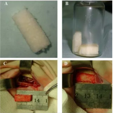
The iliac bones are separated from healthy pigs, and cut into 15 mm × 6 mm × 4 mm pieces for preparation of xenogeneic antigen-extracted cancellous bone (AXCB). The bone pieces were soaked in acetone for 48 hours to remove the fatty composition, demineralized in 0.6 M HCl, completely washed with water, treated with enzyme, washed with water again according to patented technol- ogy by the Guangdong Guan-Hao Technology Co. ( Fig-ure 1A), and then freeze-dried for preparation of rhBMP- 2 incorporation. The rhBMP-2 was produced by recom- binant expression in Escherichia coli at the Institute of Biomedical Engineering, Jinan University (Guangzhou, China), and purified to more than 98% purity, which was then dissolved in gelatin solution with 0.1% acetic acid (10 mg/ml). Each piece of AXCB was soaked with 1 ml of gelatin solution containing rhBMP-2 (2.0 mg/ml) for 24 hours, sterilized by γ-ray irradiation with a radiation do- sage of 25 k Gy, then freeze-dried, and stored frozen until use (Figure 1B).
Figure 1. Implantation of AXCB incorporated with BMP-4 (AXCB/ rhBMP-2) in mandibular defect in rabbits. (A) The prepared AXCB; (B) AXCB scaffold incorporated with rhBMP-2; (C) Creation of a 15mm × 6mm × 6mm mandi- bular defect in rabbit. (D) Implantation of AXCB/rhBMP-2 into the created mandibular defect.
2.2. Animal Experiment
Forty-eight adult New Zealand White rabbits weighing 3.0 - 3.5 kg (Experimental Animal Center of Guangdong Province) were used for the experiment, and the protocol was approved by the Institutional Animal Care and Use Committee (IACUC) at Jinan University Health Science Center. The animals were randomly divided into 4 groups (AXCB graft with rhBMP-2, AXCB graft only, autolog- ous bone graft, and non-graft control), with 3 subgroups (4, 8 and 12 weeks) for each group (4 animals for each con- dition). Prior to operation, the animals were anesthetized by intravenous injection of Nembutol (pentobarbital so- dium) (30 mg/kg). In the autologous bone graft group, the animals were first subjected to bilateral abdominal inci- sions parallel to the iliac crest; a 15 mm × 6 mm × 4 mm bone piece was excised from the iliac bone on each side, and then placed in saline solution until use. Preparation of the AXCB, and those soaking with rhBMP-2 were as described above. Then a bone defect with a size of 15 mm × 6 mm × 4 mm was created on both mandibles in each animal for all 4 groups (Figure 1C). In the graft groups, the created bone defect was implanted with the prepared AXCB soaked with rhBMP-2, AXCB only, or autologous bone (Figure 1D); while in the control (non-graft) group, the skin incision was directly closed with sutures after creation of the bone defect. All animals were then housed in the same condition and monitored for postoperative activities, emotional response, and wound healing. The animals were sacrificed at 4, 8, or 12 weeks after opera- tion, and the whole mandible was harvested from each side for investigation.
2.3. X-Ray Examination
X-ray examination of the harvested mandibles was per- formed (DR3000, Kodak, USA), and the image data were scored by three technicians blindly based on Lane- Sandhu scoring method [5], and analyzed using the the Leica Image Analysis System for assessment of bone regeneration following the mandibular defect.
2.4. Bone Mineral Density Measurement
The obtained bone samples were fixed with formalin in posphate buffer, and bone mineral density was measured for the repaired area using the bone density meter platform Lunar Prodigy (GE, USA). The bone mineral content (BMC) was presented as g/cm2.
2.5. Preparation of Bone Samples for Pathological Staining
[image:2.595.58.289.437.667.2]stained slides were observed under optical microscope at 50× magnification for evaluation of new bone formation and calcification, new blood vessel and fibrous tissue generation, inflammatory cell infiltration, and implanted scaffold degradation.
2.6. Quantitative Analysis of the New Bone
The bone samples were fixed in 10% neutral buffered formalin, immersed in Technovit 7200 VLC (Heraeus- Kulzer, Germany) after dehydration, and sectioned into 5 thin slices of approximately 40 - 80 μm and mounted onto slides after 24 hours’ solidification. The slides were H & E-stained, and histomorphometry was performed using Leica Image Analysis System.
2.7. Statistical Analysis
One-way ANOVA was used for the statistical analysis, and the data were presented as means ± standard devia- tion.
3. Results
3.1. Gross Observation
All animals from all groups were alive after surgery, and the wound healed well, though temporary postoperative swelling was noted in all animals. Neither signs of loss, displacement, and discharge of the implants, nor fracture and wound infection was observed during the whole ob- servation period.
3.2. Histological Observation
[image:3.595.310.539.84.190.2]In the group with implantation of xenogeneic antigen- extracted pig massive cancellous bone, at 4 weeks after operation, there found some fibrous tissue, capillary pro- liferation, trace of trabecular bone degradation, a small number of osteoblasts, and a small amount of new bone formation around the edge of the implant; at 8 weeks after operation, there were partial degradation of trabecular bone, and a large number of osteoblasts around the edge of the implant; at 12 weeks after operation, there was little mature trabecular bone tissue (Figure 2A). In the group with implantation of xenogeneic antigen-extracted pig massive cancellous bone soaked with rhBMP-2, at 4 weeks after operation, the trabecular bone of the implant was partially degraded, a large number of new bone for- mation was observed, and around the new bone, there were a large number of osteoblasts and mesenchymal cells, capillary ingrowth, and osteoid formation; at 8 weeks after operation, there were a small area of unabsorbed implant, a large amount of trabecular bone tissue and new bone formation, and capillary ingrowth, with a lot of osteob- lasts and mesenchymal cells around; at 12 weeks after operation, there were almost complete degradation of the
Figure 2. Histological images of the rabbit mandible defect (×100). (A) A representative result at 12 weeks after surgery in AXCB group; (B) A representative result at 12 weeks after surgery in AXCB-rhBMP-2 group.
implant, a large amount of new bone formation with ma- ture trabecular bone and some bone marrow. Histological examination showed rigorous bone regeneration around the implant. In the non-graft control group, the mandible showed only a small amount of new bone formation, and the created bone defect was mainly occupied by fibrous tissue at all time points. In the autograft group, there showed a large amount of new mature trabecular bone, and the mandibular defect was mostly occupied by the newly formed bone (Figure 2B).
3.3. The Radiographic Evaluation
Lateral and vertical radiography was used to evaluate bone regeneration and healing of the mandible defect during follow-ups. New bone formation was assessed by Lane-Sandhu scoring method. Score 0 indicated “no new bone formation”, 1, “new bone occupied 25% of the de- fect”, 2, “new bone occupied 50% of the defect”, and 3, “new bone occupied 75% of the defect”. The average scores were 1.00, 7.50, and 11.00 in the autograft group, 1.00, 5.25, and 7.50 in the AXCB/ rhBMP-2 group, and 0.20, 2.75 and 3.75 in the AXCB alone group at 4, 8, and 12 weeks after operation, respectively, indicating that scaffold graft alone had limited effect on bone regenera- tion and addition of rhBMP-2 greatly enhanced bone regeneration, which is comparable to auto bone graft (Figure 3). New bone generation increased over time.
3.4. Bone Mineral Density
Bone mineral density test revealed significant difference in bone mineral density across groups (autogenous bone group > AXCB/ rhBMP-2 group > AXCB only group > control group) (P < 0.05). And the bone mineral density was significantly increased over time (at 4, 8, and 12 weeks) within each individual group (P < 0.05).
3.5. Quantitative Assessment of Bone Regeneration
Figure 3. X-ray photos of the rabbit mandible defect. (a) A representative result at 12 weeks after surgery in AXCB group; (b) A representative result at 12 weeks after surgery in AXCB- rhBMP-2 group; (c) A representative result at 12 weeks after surgery in Control group; (d) A representative result at 12 weeks after surgery in Autograft group.
lated under microscopic view of the H & E stained slides. The area percentage of bone regeneration in the group of AXCB/rhBMP-2 graft was 27.72 ± 4.68, 53.90 ± 21.92, and 77.35 ± 9.83, that in the group of AXCBgraft was 14.03 ± 5.02, 28.49 ± 11.35, and 55.87 ± 10.20, and that in the group of autograft bone graft was 30.19 ± 1.46, 49.73 ± 2.68, 68.18 ± 3.92 at 4, 8, and 12 weeks, respec- tively. Statistical analysis result suggested that the area of bone regeneration of the mandibular defect was signifi- cantly greater in the group of AXCB/ rhBMP-2 (scaffold with morphogen) than in the group of xenogeneic anti- gen-extracted pig massive cancellous bone (scaffold only) (P < 0.05), and there was a significant increase of bone regeneration over time (at 4, 8, and 12 weeks after opera- tion) within each group (P < 0.05). There was no signifi- cant difference in the area of new bone formation between the group grafted with autogenous bone and the group grafted with AXCB/ rhBMP-2
4. Discussion
Tumors, especially malignant tumors, severe trauma, and congenital malformation in the oral and maxillofacial region often lead to a large area of bone defect. Because of the anatomical particularity and the three-dimensional structure complexity, the restoration of bone defects in oral and maxillofacial region remains a challenge for surgeons. The restoration of the original shape of the facial skull is a prerequisite for the restitution of facial
appearance. Scientists are trying to develop new ap- proaches aiming at the enhancement of bone regeneration instead of using autogenous bone grafts. Autologous bone can provide the transplant scaffolds while providing a certain number of osteoblasts, and it has the best os- teogenic effect and has been widely used as the gold standard method for repair of bone defects. However, autologous bone usually doesn’t provide an anatomically preformed shape and meet the requirement for mechani- cal properties, and its source and volume are very limited. Autogenous bone graft requires a second operation area and causes new damage for the bone-donated area, which greatly increases the duration of operation and may result in more complications [6]. Recent progress in regenera- tive medicine and bone tissue engineering raises the hope of repairing bone defects with a combination of biomate- rials and growth factors. Application of the large can- cellous bone (ilium) as a morphogen carrier for rhBMP-2 in skeletal repair has been extensively researched during the past decade [7]. Bone morphogenetic proteins (BMPs) have been successfully applied in the reconstruction of long bones, spine and the facial skeleton in preclinical studies [8].
and subsequently assessed the bone regenerative effect [16]. The results revealed that the group grafted with AXCB soaked with BMP-4 (AXCB/ rhBMP-2) had much better and more extensive bone regeneration than the group grafted with AXCB only, and the bone rege- neration increased over time from 4 weeks to 12 weeks after operation, which indicated that rhBMP-2 has sig- nificant bone regenerative effect over time with AXCB as a scaffold, and AXCB is probably a good carrier for BMP-4, which can help rhBMP-2 release slowly and work effectively. On the other hand, the AXCB was found to be gradually degraded over time, and at 12 weeks after operation, the implanted bone was almost completely replaced by newly regenerated bone tissue, which showed apparent mature trabecular structure. There were no appreciable histological signs of inflam- mation or immune rejection of the graft.
In conclusion, the osteogenic effect of AXCB graft soaked with rhBMP-2 is proved much better than AXCB graft alone (without rhBMP-2, which shows no signifi- cant difference with the autologous bone graft). Xeno- geneic antigen-extracted pig massive cancellous bone has shown good biocompatibility and it may potentially re- place autologous bone graft in repair of large bone defects. This study has provided a new reference for bone rege- neration in the oral and maxillofacial region.
5. Acknowledgements
This work was supported by a grant from the Guangdong Science and Technology Foundation (No: 2011B0807- 01053). We thank the Department of Nuclear Medicine, the Animal Center, and the Institute of Biomedical En-gineering at Jinan University, and Guangdong Guan-Hao Science and Technology Development Co. Ltd for their generous support.
REFERENCES
[1] C. Madrigal, R. Ortega, C. Meniz, et al., “Study of Avai- lable Bone Forinterforaminal Implant Treatment Using Cone-Beam Computed Tomography,” Medicina Oral Pa- tologia Oral y Cirugia Bucal, Vol. 1, No. 13, 2008, pp. E307-312.
[2] W. G. De Long Jr., T. A. Einhorn, K. Koval, et al., “Bone Grafts and Bone Graft Substitutes in Orthopaedic Trauma Surgery,” The Journal of Bone & Joint Surgery, Vol. 89, 2007, pp. 649-658.
[3] E. N. Ebbesen, J. S. Thomsen and L. Mosekilde, “Nonde- structive Determination of Iliac Crest Cancellous Bone Strength by pQCT,” Bone, Vol. 21, 1997, pp. 535-540.
[4] C. G. Finkemeier, “Bone-Grafting and Bone-Graft Subs- titutes,” The Journal of Bone & Joint Surgery, Vol. 84A, 2002, pp. 454-464.
[5] A. S. Herford and P. J. Boyne, “Reconstruction of Man- dibular Continuity Defects with Bone Morphogenetic Protein-2 (rhBMP-2),” Journal of Oral and Maxillofacial Surgery, Vol. 66, 2008, pp. 616-624.
[6] S. A. Jovanovic, D. R. Hunt, et al., “Bone reconstruction Following Implantation of rhBMP-2 and Guided Bone Regeneration in Canine Alveolar Ridge Defects,” Clinical Oral Implants Research, Vol. 18, 2007, pp. 224-230. [7] Z. Luo, Y. Hu and Q. Wang, “The Experimental Studies
of Immune Response of Antigen-Extracted Bovine Can- cellous Bone Grafting,” Zhonghua Wai Ke Za Zhi, Vol. 35, 1997, pp. 690-693.
[8] S. Oeberg, C. Johansson and J. B. Rosenquist, “Bone For- mation after Implantation of Autolysed Antigen Extracted Allogeneic Bone in Ovariectomized Rabbits,” Interna- tional Journal of Oral and Maxillofacial Surgery, Vol. 32, 2003, pp. 628-632.
[9] H. Schliephake, “Application of Bone Growth Factors: The Potential of Different Carrier Systems,” Oral and Maxillofacial Surgery, Vol. 14, 2010, pp. 17-22.
[10] K. H. Schuckert, S. Jopp and S. H. Teoh, “Mandibular Defect Reconstruction Using Three-Dimensional Polyca- prolactone Scaffold in Combination with Platelet-Rich Plasma and Recombinant Human Bone Morphogenetic Protein-2: De Novo Synthesis of Bone in a Single Case,” Tissue Engineering Part A, Vol. 15, 2009, pp. 493-499.
[11] C. Shi, W. Chen, Y. Zhao, et al., “Regeneration of Full- Thickness Abdominal Wall Defects in Rats Using Colla- gen Scaffolds Loaded with Collagen-Binding Basic Fi- broblast Growth Factor,” Biomaterials, Vol. 32, 2011, pp. 753-759.
[12] D. I. Spector, J. H. Keating and R. J. Boudrieau, “Imme-
diate Mandibular Reconstruction of a 5 cm Defect Using rhBMP-2 after Partial Mandibulectomy in a Dog,” Vete- rinary Surgery, Vol. 36, 2007, pp. 752-759.
[13] R. Visser, P. M. Arrabal, J. Becerra, et al., “The Effect of
an rhBMP-2 Absorbable Collagen Sponge-Targeted Sys- tem on Bone Formation in Vivo,” Biomaterials, Vol. 30, 2009, pp. 2032-2037.
[14] B. Wenz, B. Oesch and M. Horst, “Analysis of the Risk
of Transmitting Bovine Spongiform Encephalopathy through Bone Grafts Derived from Bovine Bone,” Biomaterials, Vol. 22, 2001, pp. 1599-1606.
[15] J.-C. Xu, G.-H. Wu, H.-L. Liu, et al., “The Effect of Lep-
tin on the Osteoinductive Activity of Recombinant Hu- man Bone Morphogenetic Protein-2 in Nude Mice,” Sau- di Medical Journal, Vol. 31, 2010, pp. 615-621.
Morphogenetic Protein-2 Implanted in Situ,” Biomate- rials, Vol. 31, 2010, pp. 4935-4943.