type Ca
2+
channels in vivo by Ca
2+
/calmodulin-dependent protein kinase II
Junlan Yao, … , Roger J. Colbran, Paula Q. Barrett
J Clin Invest.
2006;
116(9)
:2403-2412.
https://doi.org/10.1172/JCI27918
.
Ang II receptor activation increases cytosolic Ca
2+levels to enhance the synthesis and
secretion of aldosterone, a recently identified early pathogenic stimulus that adversely
influences cardiovascular homeostasis. Ca
2+/calmodulin-dependent protein kinase II
(CaMKII) is a downstream effector of the Ang II–elicited signaling cascade that serves as a
key intracellular Ca
2+sensor to feedback-regulate Ca
2+entry through voltage-gated Ca
2+channels. However, the molecular mechanism(s) by which CaMKII regulates these
important physiological targets to increase Ca
2+entry remain unresolved. We show here
that CaMKII forms a signaling complex with
a
1HT-type Ca
2+channels, directly interacting
with the intracellular loop connecting domains II and III of the channel pore (II-III loop).
Activation of the kinase mediated the phosphorylation of Ser1198 in the II-III loop and the
positive feedback regulation of channel gating both in intact cells in situ and in cells of the
native adrenal zona glomerulosa stimulated by Ang II in vivo. These data define the
molecular basis for the in vivo modulation of native T-type Ca
2+channels by CaMKII and
suggest that the disruption of this signaling complex in the zona glomerulosa may provide a
new therapeutic approach to limit aldosterone production and cardiovascular disease
progression.
Research Article
Cardiology
Find the latest version:
Research article
Molecular basis for the modulation
of native T-type Ca
2+
channels in vivo by
Ca
2+
/calmodulin-dependent protein kinase II
Junlan Yao,1 Lucinda A. Davies,1 Jason D. Howard,1 Scott K. Adney,1 Philip J. Welsby,1
Nancy Howell,2 Robert M. Carey,2 Roger J. Colbran,3 and Paula Q. Barrett1
1Department of Pharmacology and 2Department of Medicine, University of Virginia School of Medicine, Charlottesville, Virginia, USA. 3Department of Molecular Physiology and Biophysics, Vanderbilt University School of Medicine, Nashville, Tennessee, USA.
Ang II receptor activation increases cytosolic Ca
2+levels to enhance the synthesis and secretion of
aldoste-rone, a recently identified early pathogenic stimulus that adversely influences cardiovascular homeostasis.
Ca
2+/calmodulin-dependent protein kinase II (CaMKII) is a downstream effector of the Ang II–elicited
sig-naling cascade that serves as a key intracellular Ca
2+sensor to feedback-regulate Ca
2+entry through
volt-age-gated Ca
2+channels. However, the molecular mechanism(s) by which CaMKII regulates these important
physiological targets to increase Ca
2+entry remain unresolved. We show here that CaMKII forms a signaling
complex with
α
1HT-type Ca
2+channels, directly interacting with the intracellular loop connecting domains II
and III of the channel pore (II-III loop). Activation of the kinase mediated the phosphorylation of Ser1198 in
the II-III loop and the positive feedback regulation of channel gating both in intact cells in situ and in cells
of the native adrenal zona glomerulosa stimulated by Ang II in vivo. These data define the molecular basis
for the in vivo
modulation of native T-type Ca
2+channels by CaMKII and suggest that the disruption of this
signaling complex in the zona glomerulosa may provide a new therapeutic approach to limit aldosterone
production and cardiovascular disease progression.
Introduction
Abnormal intracellular Ca2+ homeostasis is a common feature
of cardiovascular disease (1, 2). In the human failing heart, arrhythmogenic remodeling is characterized by dysregulated sar-coplasmic reticulum Ca2+ uptake and release, enhanced Na-Ca2+
exchange activity, and increased activity of L-type high voltage– activated (HVA) Ca2+ channels (α1C
) (3–5) in cardiomyocytes. Cen-tral to these pathologies is an increase in adrenergic tone and a marked activation of the renin-angiotensin aldosterone axis. The importance of aldosterone as an early-onset pathogenic stimulus has been highlighted by recent findings. Following myocardial infarction, inappropriate mineralocorticoid receptor activation increases L-type Ca2+
channel density before the onset of structur-al hypertrophy (6), and chronic levels of circulating aldosterone correlate directly with the density of L-type HVA Ca2+ channels in
ventricular myocytes that are devoid of morphological change (7). In addition, in the setting of high Na+ intake, the progression to
left-ventricular hypertrophy and failure is advanced by an eleva-tion in aldosterone levels that stimulates perivascular fibrosis (8–10). Thus, long recognized to regulate Na+ and K+ balance,
blood volume, and blood pressure, aldosterone has a newly appre-ciated adverse influence on cardiovascular homeostasis.
Aldosterone is synthesized and secreted from the adrenal zona glomerulosa (ZG) cell principally in response to Ang II, ACTH, and physiological increases in extracellular potassium (11). These
secretagogues depolarize the ZG cell and/or potentiate Ca2+
chan-nel activity (12). Aldosterone production is Ca2+ dependent, and
low voltage–activated (LVA), T-type Ca2+ channels provide the Ca2+
that is necessary to sustain its stimulated production (13–15). Voltage-dependent Ca2+ channels change electrical signals into
biochemical events. But, because this conversion is adjustable, via changes in channel activity and density, Ca2+ channels also can
exert additional controls on Ca2+-dependent cellular processes.
In the heart, L-type Ca2+ channels use Ca2+
/calmodulin-depen-dent protein kinase II (CaMKII) to adjust signal transmission by inducing a gating change that is characterized by frequent channel openings (16). CaMKII expression is increased in structural heart disease (17), and inhibition of its activity reduces Ca2+ current and
ameliorates dysfunction (18). In the ZG cell, CaMKII induces a gat-ing shift of α1H T-type Ca2+ channels that enables more channels
to open at hyperpolarized potentials, enhancing Ca2+ entry and
thus stimulating aldosterone production (19). Although CaMKII regulates both α1C HVA (16, 20, 21) and α1H LVA channels (19,
22, 23), the molecular basis for these effects in vitroand in vivo remains unresolved. Here we analyzed the interaction of CaMKII with α1H T-type Ca2+ channels. Our data show that α1H channels
use tethered CaMKII as a Ca2+ sensor to dynamically regulate
channel activity. Moreover, our data indicate that α1H channels
are regulated robustly by protein phosphorylation in situ and in vivo, identifying these channels for the first time to our knowledge as authentic physiological targets of kinse activity.
Results
LVA channels are encoded by 3 separate genes: α1G, α1H, and α1I,
belonging to the Cav3.0 family of voltage-gated Ca2+ channels (24).
CaMKII activation increases whole-cell and single-channel currents mediated by α1H channels by inducing an approximately 12-mV
Nonstandard abbreviations used: [Ca2+]i, intracellular [Ca2+]; CaM, calmodulin;
CaMKII, Ca2+/calmodulin-dependent protein kinase II; D5W, 5% dextrose; GST,
glutathione-S-transferase; HVA, high voltage–activated; LVA, low voltage–activated; PFA, paraformaldehyde; ROI, region of interest; ZG, zona glomerulosa.
Conflict of interest: The authors have declared that no conflict of interest exists.
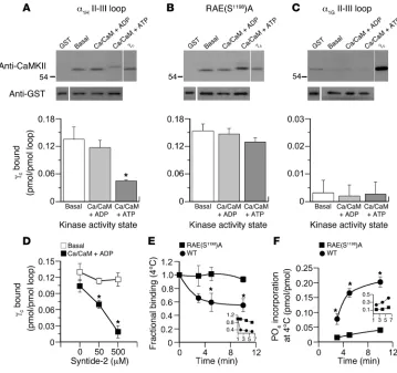
hyperpolarizing shift in the half-activation potential for channel opening. This change in activation gating mediated by CaMKII depends on the intracellular loop (residues 1019–1293) connecting channel transmembrane domains II and III and can be abrogated by a Ser1198-to-Ala point mutation (23). Therefore, to characterize the interaction of CaMKII with α1H
channels, we began by investi-gating the binding of purified recombinant CaMKII to the II-III loop, which was expressed as a bacterial fusion protein (see Meth-ods). After the proteins were mixed (8 hours, 4°C), bound kinase was separated using glutathione-sepharose. Binding was quantified against purified recombinant CaMKII protein standards (0.25–1 pmol) in immunoblots using a pan-CaMKII–specific antibody.
Because CaMKII displays differ-ing activity states, we evaluated binding under conditions that promoted each configuration. We activated CaMKII with Ca2+/
calmodulin (Ca2+/CaM) to release
the substrate-binding domain of the kinase from autoinhibitory contacts made in the basal state (25–27) or included Ca2+/CaM
and ATP in the binding buffer to allow the transphosphoryla-tion of Thr287 between adjacent kinase monomers within the dodecameric holoenzyme (28). Autophosphorylation sustains kinase activity in the absence of Ca2+ elevation and also exposes
additional sites for protein-pro- tein interactions (29, 30). Nota- bly, inactive kinase, in its auto-inhibited conformation, bound robustly to the channel loop (0.14 ± 0.03 pmol/pmol loopat 20 nM CaMKIIγC; n = 5) (Figure
1A). This binding was specific to the loop and could not be attrib-uted to the agarose bead or to the N terminus of the glutathione-S-transferase (GST) fusion protein. Moreover, syntide-2, a CaMKII substrate modeled after the CaMKII phosphorylation site of glycogen synthase, could not compete for binding of the inac-tive kinase at either 50 or 500
μM (Figure 1D), suggesting that the substrate-binding domain of CaMKII is not mediating effector contact. Surprisingly, activation of CaMKII by Ca2+ (0.5 mM), CaM
(5mM), and ADP (0.5 mM) had no significant effect on the level of binding (0.12 ± 0.02 pmol/ pmol loop; n = 5; NS). Nonethe-less, the binding of active kinase was effectively competed for by syntide-2. Syntide-2 reduced the binding of active kinase by 38% ± 4% (n = 4; P < 0.05) and 89% ± 9% (n = 3; P < 0.05) at 50 μM and 500 μM, respectively (Figure 1D). Taken together these data suggest that the inactive kinase interacts with the α1H II-III loop at a primary contact site that lies outside of
its classical substrate-binding domain.
[image:3.585.46.405.82.419.2]We also examined the binding of catalytically active kinase by replacing ADP with ATP in the activation buffer. ATP (0.5 mM) reduced CaMKII binding by more than 60% to 0.05 ± 0.001 pmol/ pmol (n = 5), indicating that either CaMKII autophosphorylation or CaMKII phosphorylation of the II-III loop disrupts binding (Figure 1A). Therefore, we examined the binding of CaMKII to a mutated II-III loop in which the major CaMKII phosphorylation
Figure 1
CaMKIIγC binds to the α1H II-III loop and is released by loop phosphorylation. (A–C) Binding of purified
recombinant CaMKIIγC (5 pmol/250 μl)to bead-bound GST-(II-III) loop fusion proteins (2 pmol). Wild-type
α1H II-III loop (A), RAE(S1198)A–α1H II-III loop (B), or α1G II-III loop (C) under autoinhibited (basal), activated
(Ca2+/CaM + ADP), or autophosphorylated (Ca2+/CaM + ATP) conditions. Upper panels: immunoblots of
bound kinase detected by anti-CaMKII antibody; 0.25 pmol standard for comparison. Equal fusion load-ing was verified independently in parallel runs (see Methods), and anti-GST detection is shown below.
Histograms: quantified mean binding data ± SEM for 5 studies. *P < 0.05, Ca2+/CaM + ATP versus basal
and Ca2+/CaM + ADP (Kruskal-Wallis ANOVA on Ranks, Dunn’s method). (D)Competition for
autoin-hibited or Ca2+/CaM + ADP–activated CaMKII/α1H II-III loop by syntide-2. *P < 0.05 (ANOVA,
Student-Newman-Keuls method). (E and F)Time course of 4°C CaMKIIγC dissociation and loop phosphorylation
following activation of autoinhibited CaMKIIγC prebound to GST–α1H II-III loop fusion proteins [wild type
vs. RAE(S1198)A]. Activators, Ca2+/CaM + ATP (0.5 mM/5 μM + 0.5 mM), added at t = 0. (E)
Activated-kinase binding determined from quantified immunoblots expressed relative to preactivation values. (F)
Phosphorylation stoichiometry of GST–α1H II-III loop fusions determined from 32P incorporation. *P < 0.05,
wild type binding at 5 or 10 minutes versus 0 minutes (ANOVA, Student-Newman-Keuls method). Inset:
research article
site, Ser1198 (23), was mutated to Ala (Figure 1B). The mutated loop bound similar amounts of inactive CaMKII compared with the wild-type loop (0.15 ± 0.01 pmol/pmol; n = 5). Notably, CaMKII activation (Ca2+/CaM + ADP) or autophosphorylation (Ca2+/CaM
+ ATP) had no significant effect on this interaction (0.15 ± 0.01 pmol/pmol [ADP], 0.13 ± 0.01 pmol/pmol [ATP]; n = 5; NS). Thus, Ser1198 substrate phosphorylation appears to initiate the release of the kinase from the channel loop. As expected, we observed no evidence for the binding of inactive, active, or autonomously active CaMKII to the α1G II-III loop fusion protein despite overexposure
of our immunoblots (Figure 1C), in agreement with the lack of regulation of α1G channels by CaMKII (22).
To corroborate the above-described findings, we preassembled complexes of inactive CaMKII with wild-type or S1198A-mutated II-III loops and followed the time course of reduction in binding upon activation of the kinase with Ca2+/CaM and ATP/[γ-32P]ATP
at 4°C to slow enzymatic activity. Kinase preassociated with the wild-type loop (Figure 1E) was released with a time course (T1/2 = 2.1
minutes, for 0–5 minutes, where T1/2
represents half-time of dissoci- ation) that approximately paralleled the time course of phosphory-lation (Figure 1F) of the loop (T1/2 = 2.7 minutes, for 0–5 minutes).
At 30°C the rate of debinding was dramatically increased (Figure 1E, inset), consistent with the increased rate of loop phosphorylation (Figure 1F, inset). Mutation of Ser1198 dramatically reduced both phosphate incorporation (73% ± 10%; n = 3; Figure 1F) and the dissociation of the kinase (94% ± 4% still bound at 10 minutes; n = 3; Figure 1E). Thus, our data show that the interaction of CaMKII with the II-III loop depends on its activity state: inactive kinase forms a stable interaction, whereas the interaction of active kinase is transient. These features are quite distinct from the long-lasting interactions of CaMKII withN-methyl-d
-aspartic acid (NMDA) receptors (31–35) or ether-a-go-go channels (36, 37) that can keep the active kinase tethered in a catalytically active conformation even in the absence of Ca2+ elevation or autophosphorylation.
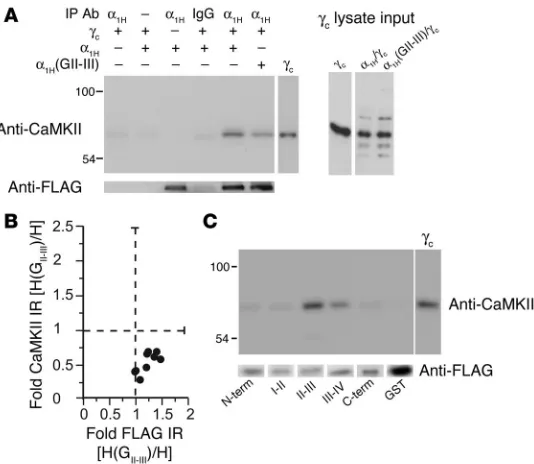
To determine whether CaMKII interacts with α1H
chan-nels in situ, we coexpressed CaMKIIγC and FLAG-tagged
α1H
channels in HEK 293 cells and tested for coimmuno- precipitation. Immunoblotting with a pan-CaMKII–spe-cific antibody often revealed 2 bands of immunoreactivity that coimmunoprecipitated with CaMKIIγC-transfected
α1H channels: an upper band corresponding to CaMKIIγC
and a lower band of weaker immunoreactivity, which was less consistently observed and likely to be a trunca-tion product of the expressed kinase construct (Figure 2A). Moreover, CaMKII was not detected in the absence of an immunoprecipitating antibody, in the absence of channel or CaMKII expression, or in the immunopre-cipitate of a control immunoglobulin, validating the specificity of the observed coassociation. Because the II-III loop of α1H channels confers regulation to α1G
channels that are not modulated by CaMKII, we hypoth-esized that substitution of the α1H II-III loop with the
II-III loop from α1G channels would reduce the amount
of CaMKII coimmunoprecipitated. As illustrated in Fig-ure 2A in a representative immunoblot, the chimeric channel immunoprecipitated a fraction of the CaMKII immunoprecipitated by the wild-type α1H channel,
despite equivalent levels of CaMKII expression detected in the lysate input. In 3 of 9 experiments where the efficiency of channel immunoprecipitation was equivalent, the α1H(GII-III)
chimera immunoprecipitated 33.7% ± 0.5% of the CaMKII immu-noreactivity that was immunoprecipitated with the wild-type channel (Figure 2B). Thus, contact sites in the II-III loop play an important role in CaMKII binding to α1H
channels in situ. None-theless, because one-third of the immunoreactivity was retained by the chimeric channel, we tested for the coassociation of CaMKII with other intracellular channel domains that were expressed as GST fusion proteins. These GST fusion proteins were loaded onto glutathione-sepharose and used in in vitro binding reactions with purified CaMKII. No evidence was obtained for the interaction of CaMKII with the N terminus, the I-II loop, or the C terminus (Figure 2C). However, significant binding to the III-IV loop, representing 10–16% of the binding to the α1H II-III
loop, was observed. In principle, CaMKII binding to the III-IV loop can account in part for the residual binding of CaMKII to the α1H(GII-III) chimeric channel.
The identification of α1H
[image:4.585.45.312.82.314.2]channels as a CaMKII-binding part- ner and the importance of Ser1198 for the modulation of chan-nel gating classify α1H channels as candidate CaMKII substrates. Figure 2
CaMKIIγC forms a signaling complex with α1H channels dependent on the
II-III loop. (A) Immunoblot of channel immunoprecipitates from HEK 293 cells
transiently expressing FLAG-tagged α1H wild-type or α1H(GII-III) chimeric
channels, most cotransfected with CaMKIIγC, using: goat IgG or anti-α1H for the
immunoprecipitation, anti-FLAG for verification of channel immunoprecipitation, and anti-CaMKII for detection of CaMKII coassociation. Input lysates show
expression level of CaMKIIγC with 0.25 pmol purified CaMKIIγC standard for
comparison. (B) An analysis of 9 experiments. Integrated optical band
den-sities of FLAG and CaMKII immunoreactivities (IR) in α1H(GII-III) channel
immunoprecipitates expressed relative to immunoreactivities in wild type.(C)
In vitrobinding of purified CaMKIIγC (5 pmol/250 μl)to bead-bound channel
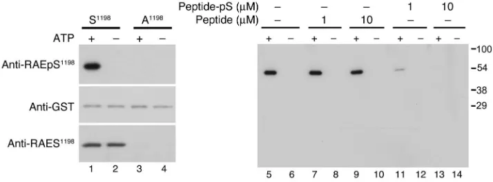
However, previously there was no direct evidence that Ser1198 was phosphorylated by CaMKII in intact cells. To evaluate Ser1198 phosphorylation in intact cells and tissue, we developed a novel phosphomotif-specific antibody (anti-RAEpS1198
) and as a byprod-uct a motif-specific antibody (anti-RAES1198
), enabling us to deter-mine whether α1H channels are authentic kinase substrates. The
specificity of the affinity-purified antibody anti-RAEpS1198 was
evaluated initially using recombinant II-III loop proteins that con-tained the wild-type sequence or an Ala point mutation at Ser1198. Anti-RAEpS1198 recognized only the wild-type II-III loop protein
following CaMKII phosphorylation. This phosphorylation- dependent interaction was competed with excess phosphopep-tide antigen (pS1198
peptide) but not with the non-phosphopep-tide (S1198 peptide) (Figure 3) and contrasted with anti-RAES1198
that recognized the wild-type fusion protein without regard for its phosphorylation state. Moreover, mutation of Ser1198 to Ala precluded recognition by anti-RAEpS1198 and anti-RAES1198. Thus,
anti-RAEpS1198 exhibits appropriate phosphorylation-dependent
specificity in vitro.
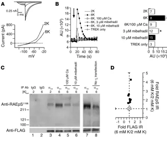
We generated a HEK 293 cell line stably expressing a mouse leak K+ channel (mTREK) and FLAG-tagged α
1H Ca2+ channel (α1H/
mTREK) for use in intact cell phosphorylation studies. Ca2+
chan-nel currents are robust in these cells, and 6 mM K+ depolarizes
the cell an approximate 20 mV, as indicated in Figure 4A by the rightward shift in the zero current potential of the macroscopic currents, which were measured using a voltage-ramp protocol. Six millimolar K+ depolarized the membrane potential from –83 ± 4
mV at 2 mM K+ to –65 ± 3 mV (n = 5). Six millimolar K+
depolariza-tion also elicited a transient rise in intracellular Ca2+
concentra-tion [Ca2+]i, as measured by fluo-4 fluorescence (Figure 4B). This
increase in [Ca2+]
i was fully dependent on extracellular Ca2+
, dose-dependently attenuated by cellular preincubation with a blocker of LVA Ca2+ channel entry (mibefradil at 3 μM and 10 μM), and
precluded in cells stably expressing mTREK alone, despite similar changes in membrane potential elicited upon cell depolarization with 6 mM K+ (–76 ± 3 mV at 2 mM K+, –52 mV ± 6 mV at 6 mM K+; n = 6). Taken together these data argue strongly that the measured
rise in intracellular Ca2+
is medi-ated by α1H channels.
To i n v e s t i g a t e w h e t h e r Ser1198 was phosphorylated in situ, we compared the state of phosphorylation of α1H
chan-nels in CaMKIIγC-transfected
α1H/mTREK cells that had been
incubated with 2 mM or 6 mM K+. To isolate the channel
pro-tein, we immunoprecipitated
α1H channels from whole-cell
lysates using a goat α1H-specific
antibody directed against the C-terminal tail of the channel (anti-α1H). Cell stimulation with 6
mM K+ increased [Ca2+]i and also
increased the phosphorylation state of the α1H channel RAES1198
motif in cells expressing CaMKIIγC
(Figure 4C). The detected increase in RAEpS site immunoreactiv-ity was dependent on Ca2+ entry
through α1H channels, as it was appropriately attenuated by low
calcium (100 μM) or mibefradil (10 μM) preincubation and, as expected, was not observed in the immunoprecipitate of goat IgG. To quantify the increase in phosphorylation state, we evaluated the fold increase in anti-RAEpS immunoreactivity (6 mM K+/
2 mM K+) and compared it to anti-FLAG immunoreactivity in the
immunoprecipitate using a mouse monoclonal FLAG antibody to detect the FLAG-tagged channels. In 8 experiments, K+
-medi-ated depolarization increased α1H
channel RAEpS site immuno-reactivity 2- to 4-fold in cells expressing CaMKIIγC (230% ± 40%; n = 8; P < 0.001) compared with FLAG immunoreactivity (Fig-ure 4D), which remained unchanged (103% ± 0.03%; n = 8; NS). By contrast, α1H channel RAEpS site immunoreactivity was not
increased in the absence of CaMKII transfection (100% ± 0.04%;
n = 5; NS). Thus, we conclude that Ser1198 on α1H channels is
a bone fide CaMKII phosphorylation site in situ and that this phosphorylation is controlled by membrane depolarization that activates the LVA channel.
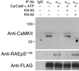
Based on our in vitro CaMKII-binding experiments, we hypothe-sized that the phosphorylation of the RAES motif by CaMKII would disrupt the coassociation of CaMKII and the channel protein. To maximize the degree of kinase autoinhibition and promote docking on the channel, we immunoprecipitated the channel-kinase com-plex from α1H/mTREK cells that were maintained at 2 mM K+ and
transfected with CaMKIIγC. CaMKII in the immunoprecipitated
complex was maintained in an inactive conformation or was acti-vated with Ca2+
/CaM plus ATP, either alone or following preincu-bation and maintained incubation with an active (10 μM KN-93) or inactive (10 μM KN-92) CaMKII inhibitor (Figure 5). In the absence of cellular activation, α1H channels formed a complex with
[image:5.585.45.402.83.215.2]the kinase and showed no evidence of RAES site phosphorylation. Activation of the associated kinase in vitro promoted the phos-phorylation of the RAES motif and the complete dissociation of the channel-kinase complex, which was prevented by preincubation with the active but not the inactive CaMKII inhibitor. Thus, autoin-hibited CaMKII forms a stable complex with the channel that can be fully disrupted by CaMKII activity.
Figure 3
Characterization of phosphomotif-specific antibody targeting RAEpS1198. Immunoblots of in vitro
phos-phorylation reactions of GST–α1H II-III loop fusion proteins (1 μM) — wild-type (S1198) and mutant
(A1198) — catalyzed by 10 nM CaMKIIγC (Ca2+ [0.5 mM], CaM [2 μM] ± ATP [100 μM]) showing selective
recognition of pS1198 by anti-RAEpS1198 (top row, lanes 1, 5, 7, 9). Below is an anti-GST immunoblot for
each fusion, indicating equal loading (middle row, lanes 1–4). Notably, anti-RAES1198 recognizes S1198
without regard for phosphorylation state (bottom row, lanes 1 and 2). Anti-RAEpS1198 preadsorption
with phosphopeptide (Peptide-pS, lanes 11–14) but not non-phosphopeptide (Peptide, lanes 7–10)
suppressed anti-RAEpS1198 immunoreactivity, showing phosphorylation-dependent interaction,
research article
To confirm the specificity of the anti-RAEpS1198 antibody in the
context of complex interacting cellular proteins, we evaluated the state of phosphorylation of the RAES motif in our α1H/mTREK
test system. As illustrated in representative immunoblots (Figure 6A), we detected strong anti-RAEpS1198 diaminobenzidine (DAB)
immunostaining in response to 6 mM K+ depolarization. Notably,
this staining was competed with the antigenic peptide (pS1198 peptide)
but was not competed with the non-phosphopeptide (S1198 peptide).
By contrast anti-RAEpS1198 DAB immunostaining of cells maintained
at 2 mM K+ was weak. Thus, we conclude that anti-RAEpS1198
immu-noreactivity reports the phosphorylation state of the RAES motif and immunohistochemistry can be used to determine whether Ser1198 phosphorylation of native LVA channels is also modulated in vivo.
Ang II is a major physiological regulator of aldosterone produc-tion from cells of the ZG that robustly express α1H channels (38,
39). Moreover, Ang II activates CaMKII (40), and CaMKII activity is important for sustaining Ang II–stimulated aldo-sterone secretion (41, 42). Therefore, we harvested adrenal glands from rats fol-lowing a 30-minute infusion of Ang II (50–200 ng/kg/min) or saline vehicle and compared the phosphorylation of Ser1198 by immunohistochemistry (Fig-ure 6B) to localize the α1H antigen within
the ZG and to avoid its dilution with adre-nal proteins from other regions. To blunt the fright response and the attendant surge in release of ACTH, itself an aldo-sterone secretagogue, we administered dexamethasone to the animals 24 hours before study (see Methods). Ang II infu- sion raised plasma aldosterone concentra-tion 4-fold, from 247 ± 105 to 978 ± 92 pg/ml (mean ± SEM; n = 4 and 7 animals, respectively; P < 0.004). Following Ang II stimulation, we detected strong anti-RAEpS1198 DAB immunostaining among
cells of the ZG that reside directly under-neath the capsular envelope (Figure 6B, Ang II/primary Ab). Significantly, this tissue staining was competed with the antigenic pS1198 peptide (Figure 6B,
Ang II + pS1198 peptide) but not with the
S1198 peptide (Figure 6B, Ang II + S1198
peptide) and replicated the zonal distri-bution of α1H mRNA detected by in situ
hybridization (39). Sections from control tissue (Figure 6B, Control) exhibited con-sistently weaker DAB immunostaining than those from stimulated tissue (Fig- ure 6B, Ang II). Quantification of anti-RAEpS1198 DAB immunostaining of the
ZG (Figure 6C) revealed a 6-fold increase in the staining of Ang II–stimulated tis-sue (3.9% ± 0.7% to 24.7% ± 3.9% region of interest [ROI]; P < 0.006) that was reduced by approximately 60% by preadsorption with an 80-fold molar excess of antigenic phosphopeptide, establishing the α1H
channel as a bona fide kinase substrate in vivo.
To determine whether CaMKII was indeed mediating the Ang II– elicited change in S1198 phosphorylation in vivo, we endeavored to
[image:6.585.42.369.81.357.2]blunt CaMKII activation by direct delivery of the water-soluble, cell-permeant CaMKII inhibitor (KN-93) to the subcapsular inter-stitium of the adrenal cortex. Following uniadrenalectomy and a short surgical recovery period, the remnant adrenal gland was infused in vivo (1 μl/min, 30 minutes) with 5% dextrose (D5W) or D5W containing 100 μM KN-93, prior to and during a systemic infusion of Ang II (50 ng/kg/min, 30 minutes). Blood samples were taken for aldosterone measurement after uniadrenalectomy before the onset and upon the termination of subcapsular infusion to determine the secretory response of the remnant adrenal to Ang II infusion. The remnant gland was harvested, and the phosphory-lation state of Ser1198 was compared with that of its preinfused
Figure 4
CaMKIIγC-induced phosphorylation of α1H channels in situ. (A) Sample currents from α1H/mTREK,
transiently expressing CaMKIIγC.Top: α1H tail currents elicited upon repolarization to –60 mV
(Vtest = –50, –30, –10, 0, +10 mV). Bottom: Whole-cell currents elicited by a voltage ramp (–120
to –20 mV). Zero-current potential defines membrane potential (Vm); dashed line highlights Vm
change. (B) [Ca2+]i plotted in arbitrary units of fluo-4-fluorescence with time in α1H/mTREK cells
(filled circles, open squares, open triangles, filled triangles) or mTREK-only cells (open circles).
Six millimolar K+ (6K) added at 16 seconds. The plain line indicates timed response to 2 mM K+
challenge in α1H/mTREK cells. Traces are mean response of cells in 3 plate wells. Histograms
plot peak (20 seconds) rise in [Ca2+]i (mean ± SEM; n as indicated). Cells were preincubated
with 100 μM Ca2+ or 10 μM mibefradil 30 minutes prior to 6 mM K+challengeand during
mea-surement. *P < 0.05, 2 mM K+ versus treatment (ANOVA, Student-Newman-Keuls method).
(C) Immunoblot of channel-enriched immunoprecipitates (as obtained in Figure 2) from cells
challenged with 2 (lanes 1 and 3) or 6 (lanes 2 and 4–8) mM K+ with (lanes 1–7) or without (lane
8) CaMKIIγC transfection. Channels were immunoprecipitated with a control IgG or anti-α1H.
Anti-FLAG verified immunoprecipitation, and anti-RAEpS1198 evaluated phosphorylation. Cells
were preincubated with 100 μM Ca2+ or 10 μM mibefradil as in B (lanes 5–6). (D) Analysis of
8 immunoprecipitation experiments. Integrated optical band densities of FLAG and RAEpS1198
immunoreactivities in 6 mM K+–stimulated samples expressed relative to immunoreactivities in 2
paired control (Figure 6C). As illustrated in Figure 6C and quanti-fied in the top panel, the direct cortical delivery of KN-93 abrogated the 2-fold increase in RAEpS1198 immunostaining of the Ang II–
stimulated tissue (20% ± 5.7% ROI [Ang II] versus 3.3% ± 2.7% ROI [Ang II + KN-93]); n = 3 each; P < 0.05) and concomitantly blunted the aldosterone secretory response of the remnant adrenal to Ang II (1,004 ± 179 pg/ml [Ang II]; 569 ± 40 pg/ml [Ang II + KN-93]);
n = 3 each; P < 0.05). Taken together, our data indicate the partici-pation of a Ca2+/CaM-dependent kinase in the dynamic regulation
of Ser1198 by physiological stimuli that activate aldosterone secre-tion and suggest an important role for channel regulation in the control of aldosterone secretion.
Discussion
Despite decades of research on the regulation of voltage-gated ion channels by protein phosphorylation, few phosphorylation events that alter ion channel activity have been shown to be physiologi-cally modulated in vivo (43–45). This is especially true of the posi-tive feedback regulation of L-type and T-type channels by CaMKII. CaMKII-induced facilitation of L-type Ca2+ channels requires active
kinase (16, 46, 47), yet the phosphorylation site(s) of functional importance have eluded identification (21, 48).
Previous studies suggested that Ser1198 in α1H channels is
required for the potentiation of T-type Ca2+ channel current by
CaMKII, implicating protein phosphorylation as the mechanism by which CaMKII regulates α1H
channels (23). In addition, single-channel experiments using membrane patches excised from native bovine ZG cells demonstrated that CaMKII is contained within the domain of the excised patch, suggesting that channel regulatory elements are locally contained (19). The present study significantly enhances our understanding of the nature of CaMKII–α1H channel
interactions and the existence of a CaMKII-channel signaling com- plex. Our findings provide a biochemical and molecular explana-tion for how CaMKII modulates LVA Ca2+ channels and show that
a physiological agonist activates this mechanism to enhance the phosphorylation of α1H channels at Ser1198 in vivo.
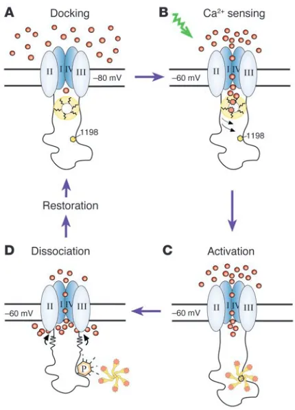
We have uncovered what we believe to be a novel mechanism for Ca2+-dependent autoregulation of α
1H channels that exploits the
changing conformational states of CaMKII. A cycle of autoactiva-tion begins with the docking of inactive CaMKII on the II-III loop of α1H channels under basal conditions (Figure 7A). As a binding
partner of the II-III loop, CaMKII is well positioned to sense local Ca2+
concentrations that arise from the opening of the LVA chan-nel itself (Figure 7B). The reception of this signal by the kinase induces its activation as well as the transfer of signaling informa-tion to Ser1198 as CaMKII shifts its primary interaction with the II-III loop to its catalytic domain as it initiates phosphotransfer (Figure 7C). As is characteristic of interactions between a protein kinase and its substrates (49), phosphorylation of Ser1198 dis-rupts this association (Figure 7D), dissociating the kinase either to begin a new cycle of autoregulation at another channel or to execute other functions.
These interaction features describe a new paradigm for modula-tion of ion channel substrates by CaMKII. Inactive kinase does not significantly interact with the cytoplasmic regions of NMDA recep-tors (31–35), ether-a-go-go channels (36, 37), ryanodine receptors (50), or α1C L-type Ca2+ channels (21). Moreover, the recruitment
of active kinase to these channels forms long-lasting associations when the kinase is autophosphorylated and new sites of interac- tion are exposed (T sites). In most instances T site binding pro-duces constitutive kinase activity, keeping the kinase tethered in a catalytically active conformation in the absence of Ca2+ elevation
or autophosphorylation (31–37).
Notably, our data show that the proposed mechanism for α1H
channel modulation by CaMKII is engaged in a model cell by changes in membrane potential that are induced by extracellular K+
at concentrations that physiologically stimulate ZG cells. Eleva-tion in extracellular K+
from 2 to 5 mM potentiates the aldoste-rone secretory response to physiological doses of Ang II (51, 52). Thus, although extracellular K+
and Ang II can be considered inde-pendent regulators of aldosterone production, the physiological potency of these agonists depends on their combined activities and their synergy. Our data provide strong evidence for the opera- tion of this mechanism in the adrenal ZG in response to Ang II sig-naling in vivo. Thus, Ser1198 in α1H channels is, to our knowledge,
the first residue in voltage-gated T-type Ca2+ channels shown to be
dynamically phosphorylated in vivo. The targeted disruption of docking and/or CaMKII-mediated phosphorylation of α1H
chan- nels may offer an alternative therapeutic strategy to that of min-eralocorticoid receptor antagonism for limiting the development and progression of cardiovascular damage in heart failure.
Methods
Molecular cloning. A [His]6-FLAG tag was introduced into the wild-type
construct GST-HII-III using the mega-primer 5′
-AGAAGGTCATCACA- CACAAGTCTAGAGAGAATCTGTATTTCCAAGGCCACCATCACCAT- CACCATGACTACAAGGACGACGATGACAAGAATTCATCGTGACT-GACTGACG-3′ and its reverse compliment in a QuikChange reaction (Stratagene). The GST-HII-III S1198A mutant construct was generated by
PCR. All constructs were confirmed by DNA sequencing.
Fluorescence measurement of [Ca2+]i.Cells grown on 96-well plates pretreated
(1 hour) with Krebs-HEPES buffer containing 2 mM potassium loaded with fluo-4 AM (30 minutes, 37°C; Invitrogen) were washed prior to experimenta-tion. Fluorescence was monitored with a FlexStation scanning fluorometer (excitation 485 nm, emission 538 nm; Molecular Devices). After 16 seconds
baseline scan, signal intensity was monitored (2 minutes, 1.5 Hz) after chal-Figure 5
CaMKIIγC forms a signaling complex with α1H channels that is
dis-rupted by S1198 phosphorylation. Typical immunoblot of channel
immunoprecipitates from FLAG- α1H/mTREK double-stable cells
incu-bated at 2 mM K+ expressing transfected CaMKIIγC, using a α1H C
termi-nus–specific antibody for immunoprecipitation and anti-FLAG for verifica-tion of channel immunoprecipitaverifica-tion. CaMKII in the immunoprecipitated complex was kept inactive or was activated with Ca2+/CaM + ATP, either
alone or following preincubation and maintained incubation with 10 μM
of a CaMKII inhibitor, KN-93, or its inactive analog, KN-92. Anti-CaMKII
detected CaMKII retained in the immunoprecipitate, and anti-RAEpS1198
[image:7.585.95.229.84.205.2]research article
lenge with 2 or 6 mM K+. When used, the indicated concentration of mibefradil
or 100 μM calcium was present at baseline and during potassium challenge.
Expression and purification of recombinant fusion proteins. Full-length α1H
and α1G II-III loop proteins included an N-terminal GST and a C-terminal
[His]6
-FLAG tag, enabling tandem affinity purification of full-length pro-tein.Transformedbacteria were grown (37°C, OD600 0.4) before induction
with isopropyl-β-D-thiogalactopyranoside (IPTG; 0.5 mM, 16 hours, 20°C) and mechanically lysed in PBS. Cleared lysate was affinity purified on Ni-NTA Agarose (QIAGEN) with 6 M urea, refolded overnight using a urea gradient (6 to 0 M), and eluted with 300 mM imidazole.
CaMKII binding and phosphorylation of HII-III.
Ni-NTA–purified fusions (GST-HII-III-His-FLAG, GST-GII-III-His-FLAG, or GST-HII-III-RAEA-His-FLAG) were
bound to Glutathione Sepharose 4B (Amersham Biosciences; GE Health-care) beads for 2 hours at 4°C to achieve a final bead loading of 2 pmol of channel loop per sample. Prebound loop was incubated with 5 pmol purified CaMKIIγC at pH 7.5 under basal (100 mM NaCl, 10 mM MgCl2,
0.1% Triton, 50 mM HEPES, 1 mM DTT) or kinase-activating conditions (with 0.5 mM Ca2+, 5 μM CaM, 100 μM ATP, or 100 μM ADP) at 4°C for
[image:8.585.55.534.80.477.2]8 hours, at which steady-state binding was achieved. Beads equivalently loaded with GST alone served as negative controls. Protein-bound beads were transferred to a Wizard PCR mini-column to facilitate rapid washing with minimal bead loss, washed with PBS, eluted with hot 2× SDS-PAGE loading buffer following 60 seconds high-heat microwave treatment, and recovered by centrifugation (53). Coassociating kinase was immunodetected
Figure 6
Phosphorylation-state of Ser1198 in α1H channels in situ and in vivo. Immunohistochemical detection of pS1198 in: CaMKIIγC-transfected
α1H/mTREK double-stable cells following 6 mM K+ depolarization (1 minute) (A); thin sections of rat adrenal glands harvested 30 minutes after
vehicle or Ang II infusion, 50–200 ng/kg/min (n = 4 and 7 animals, respectively) (B); thin sections of rat adrenal glands subcapsularly perfused
(1 μl/min) with D5W with or without 100 μM KN-93 30 minutes before and during a 30-minute systemic infusion of Ang II at 50 ng/kg/min in
uni-adrenalectomized rats (n = 3 animals each) (C). Anti-RAEpS1198 immunohistochemistry revealed DAB immunostaining in the subcapsular ZG (B
and C). Note the increase in signal strength after stimulation (A–C). Signal competed with an 80-fold molar excess of antigenic phosphopeptide, pS1198 peptide, but not non-phosphopeptide, S1198 peptide, evaluated at either ×40 (B,bottom row; and C) or ×100 (B, top row; and A). All samples
were counterstained with hematoxylin. Quantification of ZG DAB immunostaining expressed as percent of ROI. #P < 0.05, control S1198 peptide
versus antibody alone. ##P < 0.05, stimulated pS1198 peptide versus antibody alone (ANOVA on ranks, Student-Newman-Keuls method). †P < 0.05,
with CaMKII antibody (RU16; a gift of P. Greengard, Rockefeller Univer-sity, New York, New York, USA) and quantified with recombinant CaMKII standards (0.125–1 pmol) from scanned immunoblots. Equal loading of GST fusion proteins was evaluated on duplicate gels using BSA standards (25–200 ng) stained with SYPRO Ruby (Bio-Rad) or SimplyBlue SafeStain (Invitrogen) (sensitivities of detection >10 ng). Variation in GST loading was tolerated only if values fell within the standard curve and within an experiment did not differ by more than 40%. Binding values were corrected for GST loading differences within the tolerated range. Nonspecific GST- loaded bead binding was typically 0.017 ± 0.012 pmol/pmol loop indepen-dent of CaMKII activity state and was subtracted from the totals. In each experiment, samples were analyzed in duplicate.
Phosphorylation-dissociation assays. Inactive CaMKII was prebound (8 hours, 4°C) to channel loop and then activated with Ca2+, CaM, and ATP
(4°C). Residual binding was quantified as described above. When concomi-tantly assessing α1HII-III loop phosphorylation, 32P-ATP was added and
SDS-PAGE–separated radiolabeled loop was excised for liquid scintillation counting to determine direct phosphate incorporation.
CaMKII binding to GST-channel loops and termini . PCR fragments corre-sponding to human α1H (NM_021098) NH2 terminus (aa 1–99), I-II loop
(aa 419–788), II-III loop (aa 1019–1293), III-IV loop (aa 1556–1616), and COOH terminus (aa 1862–2353) were cloned into pGEX-2T and fusions expressed as described above. Glutathione-sepharose beads were pre-blocked with untransfected HEK cell lysates (1 hour, 4°C) and washed with
PBS. Fusion proteins were loaded onto preblocked beads from clarified bacterial lysates containing PBS, 1% Triton X-100, 1 mM PMSF (2 hours, 4°C), achieving a 2-pmol final loading to be used in the CaMKIIγC binding
assay described above.
Generation and characterization of RAEpS antibody.A phosphomotif-specific antibody recognizing Ser1198 phosphorylation on α1H
II-III loop was gen-erated by BioSource, Invitrogen. Rabbits were immunized with acetylated phosphopeptide Ac-LRRAE(pS)LDPC-amide, corresponding to residues 1193–1201 (human α1H
). Relevant antisera were purified on non–phos- phopeptide–conjugated sepharose to remove non-phosphomotif–spe-cific antibodies, generating anti-RAES1198, and the flow-through further
purified on a phosphopeptide-affinity resin to select for anti-RAEpS1198.
Phosphorylation reactions (in mM: 0.5 Ca2+, 0.002 CaM, 10 MgCl, 1 DTT,
45 HEPES, pH 7.5, with or without 0.04 ATP; 37°C, 30 minutes) using purified CaMKIIγC (10 nM) and GST−HII-III-His-FLAG and GST-HII-III
-RAEA-His-FLAG loop fusions (1 μM) were used to evaluate phosphomotif specificity (32). Products separated on 10% SDS-PAGE and transferred to PVDF were immunoblotted with anti-RAEpS1198 (0.04 μ
g/ml) or anti-RAES1198 (0.1 μg/ml). Specificity was assessed by preadsorbing antibody
with 1 or 10 μM phospho or non-phosphopeptide for 1 hour.
Immunoprecipitation. HEK293 or α1H/mTREK double-stables were
transfected (48 hours) (Lipofectamine 2000; Invitrogen) with CaMKIIγC,
and α1H or α1H(GII-III) where appropriate. When stimulated (Figure 4),
cells were pretreated (1 hour) with Krebs-HEPES buffer (2 mM K), before 6 mM K+ (final) or 2 mM K+ (final, control) challenge (1 minute, 37°C).
Termination was initiated by snap-freezing on dry-ice/methanol. Frozen cells were solubilized (in mM: 300 NaCl, 50 Tris HCl, pH 7.5, protease and phosphatase inhibitors [0.025 leupeptin, 0.025 aprotinin, 20 β -glycerol-phosphate, 0.0005 microcystin, 10 pyrophosphate, 0.0001 vanadate], and 0.5% Triton X-100 [1 hour, 4°C]) and centrifuged, and supernatant was precleared with protein G–sepharose. Four micrograms each of anti-α1H
antibody or goat IgG (Santa Cruz Biotechnology Inc.) was incubated with 2 mg precleared lysate (2 hours, 4°C), before incubation with protein G (1 hour, 4°C). After sequential bead washing (low-salt buffer: 10 mM Tris-HCl, pH 7.5, 0.2% NP-40, 10 mM EDTA, 150 mM NaCl; high-salt buffer: as above with 500 mM NaCl; and final buffer: 5 mM Tris-HCl, pH 7.5), immunoprecipitated proteins were eluted at 80°C with 2× SDS sample buffer, resolved by 4–15% SDS-PAGE, and analyzed by immunoblot using anti-FLAG (Sigma-Aldrich) and anti-CaMKII (RU-16; P. Greengard).
Electrophysiology. Voltage commands were applied via an EPC7 patch-clamp amplifier and digitized with Digidata 1322A analog-to-digital converter (Axon Instruments). To record macroscopic currents, cells were held at –50 mV, and membrane currents in response to a hyperpolariz-ing ramp (–20 to –120 mV) were recorded at a constant interval (0.1 Hz) as described previously (54). The bath solution was (in mM): 140 NaCl, 3 KCl, 2 MgCl2, 2 CaCl2
, 10 HEPES, 10 glucose, pH 7.4; the pipette solu-tion: 120 KMeSO4, 4 NaCl, 1 MgCl2, 0.5 CaCl2, 10 HEPES, 10 EGTA,
3 MgATP, 0.3 GTP-Tris, pH 7.2. Ca2+ channel tail currents were elicited in
response to 15 test depolarizations in 5-mV increments (–60 to +10 mV; 10.4 ms) from a holding potential of –90 mV during repolarization to –60 mV (45 ms), as previously described (23). The bath solution was (in mM): 127 TEA-Cl, 10 CaCl2, 0.5 MgCl2, 10 HEPES, 5 dextrose, 32 sucrose,
pH 7.4 (CsOH adjusted); the pipette solution: 115 CsCl, 1 tetrabutylam-monium chloride, 1 MgCl2, 5 Mg ATP, 1 LiGTP, 20 HEPES, 0.9 mM CaCl2,
11 BAPTA, pH 7.2 (CsOH adjusted).
[image:9.585.58.273.80.379.2]Animal surgeries. Animals were pretreated with dexamethasone (i.p.: 0.3–1 mg/kg) 16 and 4 hours before surgery, which inhibited any ACTH-medi- ated fright response induced by animal handling and anesthesia adminis-tration. Female rats (~220 g) were treated (30 minutes) by jugular infusion of either an isotonic D5W solution or one containing 50–200 ng/kg/min
Figure 7
Four-stagemechanistic model of CaMKII and α1H channel interactions.
(A) Inactive CaMKII docks on the II-III loop of α1H Ca2+ channels poised
to sense Ca2+ influx. (B) Depolarization-mediated Ca2+ influx elicits a
local rise in Ca2+ and activates CaMKII. (C) Activated CaMKII migrates
to S1198 for phosphotransfer. (D) CaMKII phosphorylates S1198,
pro-voking further Ca2+ influx, and then dissociates. Membrane potential is
research article
Ang II. Plasma aldosterone levels were assayed before and after infusion (Aldosterone Coat-a-Count; Diagnostic Products Corp.). Following infu-sion, rats were perfused transcardially with fresh 4% paraformaldehyde (PFA) and the adrenals harvested.
Uniadrenalectomy studies. Following anesthesia, the left adrenal vein and artery were ligated for gland removal into 4% PFA (control), the right femo-ral vein cannulated and prepared for infusion, and the right femoral artery cannulated to facilitate mean arterial blood pressure recording. In addi-tion, a PE-10 catheter was placed in the subcapsular area of the remnant adrenal gland, secured using Vetbond (3M Tissue Adhesive; 3M Animal Care Products), and infused in vivo (1 μl/min, 30 minutes) with D5W alone or containing 100 μM KN-93, prior to and during a systemic infusion of Ang II (50 ng/kg/min, 30 minutes). To harvest adrenals, rats were perfused transcardially with fresh 4% PFA. All animal experiments were performed in accordance with NIH policies and approved by the University of Virginia Medical Center Institutional Animal Care and Use Committee.
Immunohistochemistry.Harvested adrenals were dehydrated and paraffin embedded before sectioning (5 μ m) onto slides. Sections were deparaf-finized, rehydrated, and quenched with H2O2. The tissue antigenic sites
were unmasked (unmasking reagent; Vector Laboratories) and slides washed extensively in PBS with porcine gelatin (PBS/PG). Sections pre-blocked with 10% goat serum in PBS/PG were incubated sequentially with: anti-RAEpS1198
(4°C, 16 hours; 1:800) alone or preadsorbed with phospho-peptide (pS1198 peptide) or non-phosphopeptide (S1198 peptide) at 80-fold
excess, biotinylated goat anti-rabbit secondary antibody (room tempera-ture, 1 hour; Vector Laboratories), and avidin-biotin-HRP complex (room temperature, 0.5 hours; VECTASTAIN Elite ABC kit; Vector Laboratories). The HRP complex was detected using DAB (Dako), slides counterstained with hematoxylin (Richard-Allan Scientific), and dehydrated in ethanol then xylene before coverslipping in Krystalon (EM Science Harleco) (55). Images were obtained using QImaging Retiga 1300C digital camera fitted to a Zeiss microscope and processed using IPLab (Scanalytics). Four quadrants of each section were photographed and the ZG area selected using IPLab’s polygon ROI for analysis of percent DAB staining. IPLab software version 3.65 (BD) was scripted to identify pixels with Hue, Saturation, and Value (HSV) parameters appropriate to define brown hues of varying intensity.
Immunocytochemistry. α1H/mTREK cells grown on poly-lysine–coated
12-well HTC super-cured slides (Cel-Line) were pretreated (1 hour) with
standard Krebs-HEPES buffer (2 mM K+) stimulated (1 minute, 37°C) with
6 mM K+ final or 2 mM K+ (control), immediately fixed with 4% PFA, and
permeabilized with 1% Triton X-100.
Statistics . Multiple comparisons were evaluated using ANOVA with Stu-dent-Newman-Keuls method for post-hoc testing or an unpaired 2-tailed Student’s t test when appropriate (SigmaStat; Jandel Scientific, SPSS).
P values less than 0.05 were considered statistically significant.
Specialized reagents. A cDNA encoding porcine CaMKIIγC, a generous gift
from C.M. Schworer and H.A. Singer (Albany Medical College, Albany, New York, USA), was cloned into PVL1392 to generate recombinant baculovi- rus. Sf9 insect cells infected with plaque-purified recombinant baculovi-rus (MOI = 10) were harvested after 72 hours. The protein was purified by ammonium sulfate precipitation followed by affinity chromatography on CaM-agarose. CaMKIIγC was dialyzed against 50 mM HEPES pH 7.5
containing 50% (vol/vol) glycerol, 10% (vol/vol) ethylene glycol, 2.5 mM EDTA, 1 mM DTT and stored (–20°C). Protein purity (>95%) was assessed by Coomassie blue–stained SDS-polyacrylamide gels and expressed a spe-cific activity of 10–20 μmol/min/mg (syntide-2 substrate, 20 μM) (56).
Acknowledgments We acknowledge support from NIH grants (HL36977 to P.Q. Barrett and MH63232 to R.J. Colbran) and American Heart Association MidAtlantic-Affiliate postdoctoral fellowships to J. Yao and L.A. Davies. Received for publication January 13, 2006, and accepted in revised form June 20, 2006. Address correspondence to: Paula Q. Barrett, Department of Phar-macology, University of Virginia School of Medicine, 1300 Jefferson Park Avenue, Charlottesville, Virginia 22908, USA. Phone: (434) 924-5454; Fax: (434) 982-3878; E-mail: [email protected]. J.D. Howard’s present address is: Department of Pharmacology and Molecular Sciences, Johns Hopkins University School of Med-icine, Baltimore, Maryland, USA. P.J. Welsby’s present address is: Department of Physiology, Trinity College, Dublin, Ireland. 1. Pogwizd, S.M., and Bers, D.M. 2004. Cellular basis of triggered arrhythmias in heart failure. Trends Cardiovasc. Med. 14:61–66.
2. Anderson, M.E. 2005. The fire from within: the biggest Ca channel erupts and dribbles. Circ. Res.
97:1213–1215.
3. Bers, D.M., Eisner, D.A., and Valdivia, H.H. 2003. Sarcoplasmic reticulum Ca2+ and heart failure: roles of diastolic leak and Ca2+ transport. Circ. Res.
93:487–490. 4. Pogwizd, S.M., Schlotthauer, K., Li, L., Yuan, W., and Bers, D.M. 2001. Arrhythmogenesis and con- tractile dysfunction in heart failure: roles of sodi-um-calcium exchange, inward rectifier potassium current, and residual beta-adrenergic responsive-ness. Circ. Res. 88:1159–1167.
5. Schroder, F., et al. 1998. Increased availability and open probability of single L-type calcium channels from failing compared with nonfailing human ven-tricle. Circulation. 98:969–976.
6. Perrier, E., et al. 2004. Mineralocorticoid receptor antagonism prevents the electrical remodeling that precedes cellular hypertrophy after myocardial infarction. Circulation. 110:776–783.
7. Perrier, R., et al. 2005. A direct relationship between
plasma aldosterone and cardiac L-type Ca2+ cur-rent in mice. J. Physiol. (Lond.). 569:153–162.
8. Brilla, C.G., and Weber, K.T. 1992. Mineralocorti- coid excess, dietary sodium, and myocardial fibro-sis. J. Lab. Clin. Med. 120:893–901.
9. Funder, J.W. 1995. Steroids, hypertension and car-diac fibrosis. Blood Press. Suppl. 2:39–42.
10. Pitt, B., Stier, C.T., Jr., and Rajagopalan, S. 2003. Mineralocorticoid receptor blockade: new insights into the mechanism of action in patients with car-diovascular disease. J. Renin Angiotensin Aldosterone Syst. 4:164–168.
11. Quinn, S.J., and Williams, G.H. 1988. Regula-tion of aldosterone secretion. Annu. Rev. Physiol.
50:409–426.
12. Spat, A., and Hunyady, L. 2004. Control of aldoste-rone secretion: a model for convergence in cellular signaling pathways. Physiol. Rev. 84:489–539.
13. Barrett, P.Q., Isales, C.M., Bollag, W.B., and McCar-thy, R.T. 1991. Ca2+ channels and aldosterone secretion: modulation by K+ and atrial natriuretic peptide. Am. J. Physiol. 261:F706–F719.
14. Rossier, M.F., Burnay, M.M., Vallotton, M.B., and Capponi, A.M. 1996. Distinct functions of T- and L-type calcium channels during activation of bovine adrenal glomerulosa cells. Endocrinology.
137:4817–4826.
15. Rossier, M.F., et al. 1996. Sources and sites of action of calcium in the regulation of aldosterone biosyn-thesis. Endocr. Res. 22:579–588.
16. Dzhura, I., Wu, Y., Colbran, R.J., Balser, J.R., and Anderson, M.E. 2000. Calmodulin kinase deter-mines calcium-dependent facilitation of L-type calcium channels. Nat. Cell Biol. 2:173–177. 17. Hoch, B., Meyer, R., Hetzer, R., Krause, E.G., and
Karczewski, P. 1999. Identification and expression of delta-isoforms of the multifunctional Ca2+/ calmodulin-dependent protein kinase in failing and nonfailing human myocardium. Circ. Res.
84:713–721.
18. Zhang, R., et al. 2005. Calmodulin kinase II inhibi-tion protects against structural heart disease. Nat. Med. 11:409–417.
19. Lu, H.K., Fern, R.J., Nee, J.J., and Barrett, P.Q. 1994. Ca(2+)-dependent activation of T-type Ca2+ chan-nels by calmodulin-dependent protein kinase II.
Am. J. Physiol. 267:F183–F189.
20. Dzhura, I., et al. 2002. Cytoskeletal disrupting agents prevent calmodulin kinase, IQ domain and voltage-dependent facilitation of L-type Ca2+ channels [erratum 2003, 546:955]. J. Physiol.
545:399–406.