Differential sympathetic neural control of
oxygenation in resting and exercising human
skeletal muscle.
J Hansen, … , W J Parsons, R G Victor
J Clin Invest.
1996;
98(2)
:584-596.
https://doi.org/10.1172/JCI118826
.
Metabolic products of skeletal muscle contraction activate metaboreceptor muscle afferents
that reflexively increase sympathetic nerve activity (SNA) targeted to both resting and
exercising skeletal muscle. To determine effects of the increased sympathetic
vasoconstrictor drive on muscle oxygenation, we measured changes in tissue oxygen
stores and mitochondrial cytochrome a,a3 redox state in rhythmically contracting human
forearm muscles with near infrared spectroscopy while simultaneously measuring muscle
SNA with microelectrodes. The major new finding is that the ability of reflex-sympathetic
activation to decrease muscle oxygenation is abolished when the muscle is exercised at an
intensity > 10% of maximal voluntary contraction (MVC). During high intensity handgrip,
(45% MVC), contraction-induced decreases in muscle oxygenation remained stable despite
progressive metaboreceptor-mediated reflex increases in SNA. During mild to moderate
handgrips (20-33% MVC) that do not evoke reflex-sympathetic activation, experimentally
induced increases in muscle SNA had no effect on oxygenation in exercising muscles but
produced robust decreases in oxygenation in resting muscles. The latter decreases were
evident even during maximal metabolic vasodilation accompanying reactive hyperemia. We
conclude that in humans sympathetic neural control of skeletal muscle oxygenation is
sensitive to modulation by metabolic events in the contracting muscles. These events are
different from those involved in either metaboreceptor muscle afferent activation or reactive
hyperemia.
Research Article
Find the latest version:
J. Clin. Invest.
© The American Society for Clinical Investigation, Inc. 0021-9738/96/07/0584/13 $2.00
Volume 98, Number 2, July 1996, 584–596
Differential Sympathetic Neural Control of Oxygenation in Resting and Exercising
Human Skeletal Muscle
Jim Hansen, Gail D. Thomas, Shannon A. Harris, William J. Parsons, and Ronald G. Victor
Department of Internal Medicine, Cardiology Division, Molecular Cardiology Laboratories, University of Texas Southwestern Medical Center, Dallas, Texas 75235-8573
Abstract
Metabolic products of skeletal muscle contraction activate
metaboreceptor muscle afferents that reflexively increase
sympathetic nerve activity (SNA) targeted to both resting
and exercising skeletal muscle. To determine effects of the
increased sympathetic vasoconstrictor drive on muscle
oxy-genation, we measured changes in tissue oxygen stores and
mitochondrial cytochrome
a,a
3redox state in rhythmically
contracting human forearm muscles with near infrared
spectroscopy while simultaneously measuring muscle SNA
with microelectrodes. The major new finding is that the
ability of reflex-sympathetic activation to decrease muscle
oxygenation is abolished when the muscle is exercised at an
intensity
.
10% of maximal voluntary contraction (MVC).
During high intensity handgrip (45% MVC),
contraction-induced decreases in muscle oxygenation remained stable
despite progressive metaboreceptor-mediated reflex increases
in SNA. During mild to moderate handgrips (20–33%
MVC) that do not evoke reflsympathetic activation,
ex-perimentally induced increases in muscle SNA had no effect
on oxygenation in exercising muscles but produced robust
decreases in oxygenation in resting muscles. The latter
de-creases were evident even during maximal metabolic
va-sodilation accompanying reactive hyperemia.
We conclude that in humans sympathetic neural control
of skeletal muscle oxygenation is sensitive to modulation by
metabolic events in the contracting muscles. These events
are different from those involved in either metaboreceptor
muscle afferent activation or reactive hyperemia. (
J. Clin.
Invest.
1996. 98:584–596.) Key words: sympathetic nervous
system
•microelectrodes
•muscle contraction
•vasocon-striction
•tissue oxygenation
Introduction
The sympathetic nervous system produces many of the cardio-vascular adjustments during exercise, including reflex in-creases in blood pressure, heart rate, and regional vascular
re-sistance. Although these adjustments match oxygen delivery to the metabolic demands of exercising skeletal muscle, the un-derlying mechanisms by which this matching occurs remain poorly understood.
As early as 1841, A.W. Volkmann (1) proposed that the ac-cumulation of chemical products of contraction in skeletal muscle somehow signals the brain of a mismatch between mus-cle blood flow and metabolism and evokes compensatory neu-rocirculatory responses to minimize this perfusion mismatch. There now is unequivocal evidence that metabolic products of contraction (such as H1) activate chemically sensitive skeletal
muscle afferents that reflexively increase efferent-sympathetic vasoconstrictor discharge (2–15). This reflex mechanism (termed the “muscle metaboreflex”) has been shown to trigger parallel sympathetic activation in resting and exercising human skele-tal muscle (16), but the resultant effect of this reflex-sympa-thetic activation on muscle blood flow and oxygenation re-mains poorly understood.
In quiescent skeletal muscle, reflex-sympathetic activation produces vasoconstriction (10), which is thought to optimize blood flow to metabolically active muscles. However, the func-tional consequence of reflex-sympathetic activation in con-tracting skeletal muscle has been the subject of considerable debate. Previous studies in intact animals and humans have suggested that sympathetic vasoconstriction in contracting muscle is both (a) well preserved (17–28), thereby partially off-setting metabolic vasodilation to maintain blood pressure (29); and (b) largely negated by metabolic vasodilation (30–38), thereby optimizing muscle perfusion. The latter concept, ini-tially termed “functional sympatholysis” (30) recently has been extended by reductionist microcirculatory preparations (39–45) demonstrating that certain local metabolic conse-quences of contraction (e.g., intramuscular acidosis, hypoxia) interfere with specific signal transduction pathways mediating alpha-adrenergic vasoconstriction. Furthermore, the alpha adrenergic receptors that are most susceptible to such meta-bolic inhibition are those located on the distal nutrient arteri-oles, which are most accessible to metabolic products of con-traction; in contrast, adrenergic receptors located on more proximal resistance arteries are not so accessible to these met-abolic products (42). Our recent studies (33) further extend this concept by implicating a key role for intramuscular acido-sis, since sympathetically mediated vasoconstriction in rat hindlimb muscle was greatly attenuated during intense con-traction of highly glycolytic, but not of oxidative, muscles.
Taken together, these recent animal studies led us to hy-pothesize that contraction-induced metabolic inhibition of sympathetic vasoconstriction in mainly nutrient arterioles ne-gates an otherwise deleterious effect of reflex-sympathetic ac-tivation on muscle oxygenation. The ability to continuously mea-sure changes in tissue oxygen stores and mitochondrial oxygen availability in contracting skeletal muscle with near infrared spectroscopy, while simultaneously measuring muscle
sympa-Address correspondence to Ronald G. Victor, Department of Inter-nal Medicine Molecular Cardiology Laboratories, UT Southwestern Medical Center, 5323 Harry Hines Blvd., NB 11.200, Dallas, TX 75235-8573. Phone: 214-648-1400; FAX: 214-648-1450; E-mail: vic-tor@ryburn.swmed.edu
thetic nerve activity (SNA)1 with intraneural microelectrodes,
provided a new opportunity to test this hypothesis in humans. Specifically, we asked: (a) do reflex increases in muscle SNA lead to decreased oxygenation in resting, but not in contracting skeletal muscle; and (b) is such functional sympatholysis evi-dent mainly during intense rather than mild muscle contrac-tion? We postulated that the minimum level of contraction that elicits functional sympatholysis is the same high intensity that is needed to elicit metaboreflex-mediated increases in muscle SNA. This would implicate a common metabolic mech-anism, such as glycolytic production of H1, in the coordinate
regulation of reflex-sympathetic activation and mitochondrial oxygenation in human skeletal muscle.
Methods
The study protocols were approved by the Institutional Review Board at University of Texas Southwestern Medical Center, and writ-ten informed consent was obtained from each subject prior to the study. We studied a total of 29 healthy volunteer subjects, 21 male and 8 fe-male, 19 to 47 years of age (mean age of 25). Several of the subjects participated in more than one protocol on separate occasions.
General methods
Subjects were studied in the supine position. Heart rate was mea-sured continuously by electrocardiography and arterial pressure by finger photoplethysmography (Finapres; Ohmeda, Englewood, CO). Respiratory excursions were monitored with a strain-gauge pneumo-graph; during the experimental protocols, subjects were instructed to avoid performance of a Valsalva maneuver or prolonged expiration because these respiratory maneuvers can stimulate sympathetic nerve activity (46). All measurements were recorded continuously on an FM tape recorder (R-71; TEAC, Japan) and on an electrostatic re-corder (ES 1000; Gould Inc., Cleveland, OH).
Recording of sympathetic nerve discharge
Multiunit recordings of postganglionic SNA targeted to the skeletal muscle bed were obtained with unipolar tungsten microelectrodes in-serted selectively into muscle nerve fascicles of the peroneal nerve posterior to the fibular head by microneurography (47). Briefly, the neural signals were amplified (by 20–50 3 103), filtered (bandwidth of
700 to 2,000 Hz), rectified, and integrated (time constant of 0.1 s) to obtain a mean voltage display of sympathetic activity. A recording of muscle SNA was considered acceptable when the neurograms re-vealed spontaneous, pulse-synchronous bursts of neural activity, with a minimum signal-to-noise ratio of 3:1, that increased during phases II and III of the Valsalva maneuver but not during arousal stimuli (loud noise, skin pinch).
Sympathetic bursts were detected by inspection of the filtered and mean voltage neurograms; the interobserver and intraobserver variability in identifying bursts is , 10 and , 5%, respectively (6). In-advertent contraction of the leg muscles adjacent to the recording electrode produces electromyographic artifacts that are easily distin-guished from sympathetic bursts; neurograms that revealed such arti-facts were excluded from analysis. SNA was expressed as (a) the number of bursts of sympathetic activity per minute, and (b) the num-ber of bursts per minute multiplied by the mean burst amplitude in that minute (total activity).
In vivo near infrared multiwavelength spectroscopy
Near infrared spectroscopy was performed according to the method
of Piantadosi and co-workers (48–50). Detailed descriptions of near infrared spectroscopy applied to skeletal muscle, cardiac muscle, and brain have been published previously (e.g., references 51–56) and the theoretical background and in vivo validation studies have been re-viewed (48–50, 57–61).
Briefly, the near infrared method exploits the principle that laser light with wavelengths in the 700–900-nm range penetrates tissues with relative ease, and is absorbed by the copper moiety (CuA) in
cy-tochrome a,a3 and by the iron-porphyrin moieties in oxygenated and
deoxygenated hemoglobin and myoglobin. Changes in absorption are proportional to changes in the relative concentrations of oxidized cy-tochrome a,a3, oxygenated hemoglobin and myoglobin, and
deoxy-genated hemoglobin and myoglobin. Thus, the technique provides continuous measurement of several indices of the adequacy of muscle oxygen delivery relative to use at the intracellular and microcircula-tory level. These include:
Redox state of cytochrome a,a3. Cytochrome a,a3 is the terminal
member of the electron transport chain and is located on the inner mitochondrial membrane where it catalyzes the transfer of electrons to molecular oxygen, thereby making free energy available for the synthesis of ATP via oxidative phosphorylation. The final step in mi-tochondrial respiration is a four-electron transfer between cyto-chrome a,a3 and oxygen. In view of the high affinity constant, rapid
rate, and compartmental properties of the reaction between cyto-chrome a,a3 and molecular O2, the redox state of cytochrome a,a3 is a
sensitive indicator of relative changes in mitochondrial oxygen avail-ability. Thus, a decrease in cytochrome a,a3 (cyt a,a3) redox state
sug-gests diminished mitochondrial O2 availability relative to the rate of
electron transport, which is determined by metabolic demand. Tissue oxygen stores. The near infrared method also permits measurement of changes in the absorption by iron–porphyrin moi-eties in oxygenated hemoglobin plus myoglobin (tHbO21MbO2, or
“tissue oxygen stores”), and deoxygenated hemoglobin plus myoglo-bin (tHb1Mb). It is not possible to estimate the fraction of O2 in
tis-sue hemoglobin vs myoglobin, since the absorption spectra of oxyhe-moglobin and oxymyoglobin (and the spectra of deoxyheoxyhe-moglobin and deoxymyoglobin) are nearly identical. Importantly, the oxygen-dependent changes in the hemoglobin absorption reflect changes oc-curring mainly in the microvessels, rather than in the resistance ves-sels. Due to the high extinction coefficients of whole blood, vessels greater than 1 mm in diameter must be considered maximal absorb-ers of photons. Thus, photons that successfully migrate through mus-cle will primarily proceed through minimal absorbers, such as small arterioles, capillaries, and venules (61). Furthermore, since most of the hemoglobin is located in capacitance vessels, the hemoglobin component of the optical signals is most sensitive to changes in capil-lary and venous O2 content. In the present study, similar directional
changes in the cytochrome a,a3 redox state and tHbO21MbO2, and
reciprocal changes in tHb1Mb reflect changes in muscle oxygen-ation.
In the present experiments, near infrared signals were obtained by means of two fiber optical bundles (optrodes), which were placed over the region of the flexor digitorum profundus muscle of the left arm, which is the main active muscle mass recruited during the type of handgrip performed (62). The optrodes were applied to the skin oriented parallel to each other z 2.3 cm apart for optimal reflectance measurements. The average depth of penetration of the near infrared light is a function of the distance between the optrodes (57). The pho-tons traverse a banana-shaped region underlying the two optrodes with the average depth of penetration equaling half the distance be-tween the optrodes (z 1.2 cm). In preliminary experiments, we found that rhythmic handgrip can cause two types of motion artifacts, which are readily detectable and preventable: (a) complete loss of contact between optrodes and skin, which leads to an obvious loss of all opti-cal signals, and (b) inadvertent movement of the optrodes from their optimal placement, but without complete displacement from the skin. This latter motion artifact is readily detectable by (a) abrupt changes in all optical signals and (b) failure of the signals to return to the pre-1. Abbreviations used in this paper: cyt a,a3, cytochrome a,a3; LBNP,
exercise baseline values during recovery. Such motion artifacts were avoided with the use of a spring loaded stereotactic instrument that effectively maintained the stable position of the optrodes during rhyth-mic handgrip. In each experiment, we documented absence of a base-line shift by ensuring that during recovery all optical signals returned to within 5% of the total labile signal (TLS) (54). All near infrared measurements (i.e., cyt a,a3, tHbO21MbO2 and tHb1Mb) were
ob-tained at a sampling rate of 1 Hz, and each data point displayed as the mathematical average of 10 consecutive measurements. The maximal change in the near infrared signals was determined in each experi-ment as the difference between baseline and complete deoxygenation accomplished with circulatory arrest (pneumatic cuff at 280 mmHg at the level of the brachial artery for 8 min in resting muscle or for 2 min after handgrip exercise). The magnitude of this difference defines the TLS for each parameter, and all changes are expressed as a percent-age of the total labile signal (52). The reproducibility of changes in optical density to a given intervention in a given subject was deter-mined in preliminary studies where handgrip at 45% maximal volun-tary contraction (MVC), lower body negative pressure (LBNP) at
220 mmHg, and forearm circulatory arrest (to obtain the TLS) was performed twice on four subjects. The differences (mean6SD) in the change in optical density between the two tests were (cyt a,a3,
tHbO21MbO2, tHb1Mb): HG: 20.0260.04, 0.0160.06, and 0.056
0.02. TLS: 20.0160.04, 20.0160.04, and 0.0160.03. LBNP: 20.026
0.02, 20.0160.01, and 20.0160.02. This corresponds to coefficients of variability (SD of the difference/mean change with intervention) of optical density changes between 4 and 16%.
The near infrared spectrophotometer, which was constructed in Dr. Claude Piantadosi’s laboratory (53–55), consists of four GaAIAs laser diodes and circuitry for photodetection, demodulation, and log-arithmic amplification of the optical signals. The four lasers in se-quence provide pulses of monochromatic light (1.5-nm bandwidth) at 775, 810, 870, and 904 nm. One fiber-optic bundle was used for laser pulse transmission and monitoring of incident light by a reference photomultiplier tube, and a second fiber-optic bundle was used to re-ceive photons from the tissue region of illumination, for detection by a second photomultiplier tube. Photocurrents were integrated, de-multiplexed, and fed through a log-ratio amplifier. These voltages were then input to a microprocessor and algorithms for the contribu-tion of each absorber at each wavelength were used to solve for three unknowns: changes in cyt a,a3, tHbO21MbO2, and tHb1Mb. These
metabolic signals were displayed on a computer screen in real time as changes in optical density (DOD).
Near infrared spectroscopy has been validated extensively (48–50, 59–61) and the algorithms used to separate cyt a,a3, tHbO21MbO2,
and tHb1Mb signals have been published (54, 55, 60). The validity of these algorithms has been established in experimental animal models using exchange transfusions with perfluorocarbon blood substitutes (60). The seminal observation is that after five exchanges, cyt a,a3
from rat brain remains stable (decreases by , 4%), despite a 75% re-duction in tHbO21MbO2 (60). Furthermore, supporting evidence has
been obtained using venous occlusion in the human forearm, a tech-nique that does not alter arterial inflow of oxygenated blood such that the tHbO21MbO2 signal remains unchanged, but which
redis-tributes the oxygenated blood away from the nutrient circulation, re-sulting in a 58% decrease in cyt a,a3 (59). The main limitations of near
infrared spectroscopy currently are (a) the difficulty in assessing the precise degree of separation between the cyt a,a3 and the tHbO21
MbO2 and tHb1Mb signals; and (b) the optical responses reflect
rela-tive rather than absolute changes in the concentrations of the absorb-ers, because the exact pathlength of transmitted photons is unknown (50). These limitations, however, are not relevant to the interpreta-tion of the present experiments, where all the optical indices of mus-cle oxygenation were used to indicate changes in oxygen delivery rel-ative to metabolic demand.
Venous occlusion plethysmography
Forearm blood flow during LBNP was measured with venous
occlu-sion plethysmography according to the technique of Siggaard-Ander-sen (63). An air-filled latex cuff was placed z 5 cm distal to the an-tecubital fossa and the arm was elevated to produce venous drainage. Circulation to the hand was arrested by inflating a cuff on the wrist to 250 mmHg during determinations of forearm flow. The pressure of the venous congesting cuff on the upper arm was 40 mmHg. Forearm blood flow was measured at 15-s intervals and expressed in milliliters per minute per 100 ml of forearm volume. Forearm vascular resis-tance was calculated as mean arterial pressure/forearm blood flow. Mean arterial pressure was calculated as diastolic pressure 1 (pulse pressure/3).
Handgrip exercise
Handgrip exercise was performed with a custom-made handgrip dy-namometer connected to a force transducer (Interface; MFG, Scotts-dale, AZ). Force output from the handgrip device was recorded on paper and displayed on an oscilloscope to provide the subject with vi-sual feedback. Before the experiment, each subject’s MVC was deter-mined as the best of four to five trials with verbal encouragement to improve at each trial. The exercise protocols consisted of 5 min of in-termittent isometric exercise where subjects matched force produc-tion to a visual target, to the rhythm of a metronome (40 beats/min) with a 50% duty cycle.
Rhythmic handgrip at 45% MVC is accompanied by increases in muscle SNA due to activation of the muscle metaboreflex (7–9). In contrast, rhythmic handgrip at 5, 10, 20, or 33% MVC does not en-gage the muscle metaboreflex and does not increase muscle SNA (7–9).
Lower body negative pressure
The subject’s lower body was enclosed in a negative pressure cham-ber to the level of the iliac crest. An opening was created on one side of the chamber to allow performance of the microneurographic tech-nique for recording SNA from the peroneal nerve in the right leg. Once a stable recording of SNA was obtained, the opening was closed and sealed during the protocol. The pressure inside of the LBNP chamber was measured by a Statham transducer (Gould Inc., Oxnard, CA). LBNP at 220 mmHg was used to selectively unload the cardiopulmonary baroreceptors, thereby producing highly repro-ducible increases in muscle SNA, without concomitant changes in sys-temic arterial pressure (64, 65).
Administration of pharmacologic agents
In some studies, bretylium tosylate (Astra Scientific Intl., Westbor-ough, MA) was administered to one forearm by a regional intrave-nous technique (“Bier-block”) to selectively inhibit sympathetic neu-rotransmission in the forearm (66). An intravenous catheter was inserted into a forearm vein, the forearm was elevated above heart level and exsanguinated with a compressing bandage. A tourniquet cuff was inflated on the upper arm and kept at a constant pressure of 280 mmHg. Bretylium tosylate (1 mg/kg body wt) was dissolved in 40 ml saline and instilled into the forearm, which was kept ischemic for 20 min after the bretylium administration. We determined in pilot-studies that this technique selectively abolishes sympathetic vasocon-striction in the experimental forearm, as evidenced by absence of the forearm vasoconstrictor response to LBNP, for at least 4 h after the procedure, but it has no effect on systemic blood pressure or on sym-pathetic vasoconstriction in the contralateral forearm.
Specific protocols
Blood pressure, heart rate, respiration, force output, muscle SNA (recorded from the peroneal nerve), and near infrared signals (from the left flexor digitorum profundus muscle) were recorded at baseline and during 5 min of rhythmic handgrip at 45% MVC performed with the left forearm. Immediately before the end of the exercise, a tourni-quet cuff was inflated around the upper arm and kept at 280 mmHg for 2 min to produce postexercise forearm ischemia. This maneuver rapidly elicits the maximal response in the near infrared signals and it sustains activation of the muscle metaboreflex by trapping the meta-bolic products of contraction in the forearm (6).
The near infrared signals were recorded from the exercising fore-arm muscles. However, because of the technical difficulty in recording SNA to exercising forearm muscles, SNA to resting leg muscle was used as an index of SNA to exercising forearm muscle. The validity of this approach is provided by our previous experiments in which we docu-mented that metaboreflex activation triggers parallel increases in SNA to resting and exercising human skeletal muscle in the lower leg
(16), and the documentation that, during handgrip exercise, the mus-cle metaboreflex has been demonstrated to trigger parallel increases in SNA targeted to the skeletal muscle of both the arm and leg (67).
Protocol 2. Effects of reflex-sympathetic activation on muscle oxygenation in resting human skeletal muscle (4 experiments per-formed on 4 subjects). The aim of this protocol was to validate the assumption that, during nonhypotensive LBNP, decreases in the oxy-genation of resting forearm muscle are mediated by reflex-sympa-thetic vasoconstriction.
[image:5.612.58.554.255.638.2]Blood pressure, heart rate, near infrared signals, and forearm blood flow (plethysmography) were measured for 2 min each before, during, and after application of LBNP at 220 mmHg. This level of LBNP reproducibly increases muscle SNA by selectively unloading mainly cardiopulmonary baroreceptors, without changing arterial blood pressure (i.e., muscle perfusion pressure) (65). These experi-ments were performed both before and 1 h after regional forearm sympathetic blockade with bretylium.
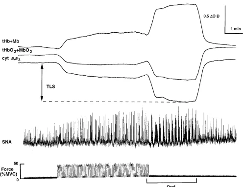
Figure 1. Segments of an original record showing simultaneous recordings of near infrared optical signals from forearm muscle (top), mean volt-age neurogram of muscle-sympathetic nerve activity (center), and handgrip force (bottom). Continuous tracings are shown at rest, during 5 min of rhythmic handgrip at 45% MVC, 2 min of post-exercise forearm vascular occlusion, and recovery. In this subject, handgrip produced rapid de-creases in cyt a,a3 and in tissue oxygen stores (tHbO21MbO2), and a corresponding increase in deoxyhemoglobin and deoxymyoglobin
Protocol 3. Effects of forearm sympathetic blockade on the changes in muscle oxygenation during high intensity rhythmic hand-grip (4 experiments performed on 4 subjects). The aim of this proto-col was to determine if pharmaproto-cologic blockade of sympathetic neu-rotransmission in the exercising arm would alter the magnitude or time course of muscle deoxygenation during handgrip. If the re-sponses to handgrip were similar after sympathetic blockade, it would support our hypothesis that the effect of functional sympatholysis normally produced during handgrip equals the effect of chemical sympatholysis in these experiments.
Blood pressure, heart rate, force output, and near infrared signals (from the left flexor digitorum profundus muscle) were recorded at baseline and during 5 min of rhythmic handgrip at 45% MVC per-formed with the left forearm before and 1 h after forearm sympa-thetic blockade with bretylium. Each exercise bout was followed by 2 min of forearm circulatory arrest to establish the maximal decrease in the near infrared signals. The efficacy of sympathetic blockade was documented by loss of reflex deoxygenation in the forearm in re-sponse to 2 min of LBNP at 220 and 240 mmHg.
Protocol 4. Effects of reflex-sympathetic activation on muscle oxygenation in exercising human skeletal muscle (16 experiments per-formed on 10 subjects). The aim of this protocol was to test our main hypo-thesis that the ability of reflex-sympathetic activation to elicit decreases in muscle oxygenation is attenuated when the muscle is exercised. To test this, reflex increases in muscle SNA evoked by a given level of LBNP (220 mmHg) were superimposed on levels of rhythmic handgrip ex-ercise (20 or 33% MVC) that by themselves do not alter muscle SNA. Blood pressure, heart rate, respiration, handgrip force, muscle SNA, and near infrared signals were recorded in response to LBNP (220 mmHg for 2 min) and rhythmic handgrip (20 or 33% MVC for 5 min each) performed alone or in combination. The LBNP was ap-plied during the 3rd and 4th min of each 5-min exercise period. Each exercise bout was followed by two min of forearm circulatory arrest to establish the maximal decrease in the near infrared signals. All 10 subjects performed handgrip at 20% MVC and in 6 of these subjects the experiments were repeated using a handgrip at 33% MVC, a level that causes greater muscle deoxygenation than handgrip at 20% MVC, but without producing reflex increases in muscle SNA.
Protocol 4a. Effects of mechanical vascular occlusion on muscle oxygenation in exercising human skeletal muscle (5 experiments
per-formed on 5 subjects). The aim of this subprotocol was to compare the effects of reduced oxygen delivery on the time-dependent de-creases in cyt a,a3 and tHbO21MbO2 at rest and during rhythmic
handgrip. In preliminary experiments, we determined that 1 min of complete circulatory arrest (pneumatic cuff inflated to 280 mmHg) of a resting forearm produced decreases in muscle oxygenation equiva-lent to those produced by 2 min of LBNP at 220 mmHg. The brief circulatory arrest was used as an internal control for decreases in oxy-gen delivery resulting from reflex-sympathetic vasoconstriction.
Blood pressure, heart rate, handgrip force, and near infrared sig-nals were recorded in response to 1 min of complete forearm circula-tory arrest at rest and during rhythmic handgrip (33% MVC for 5 min) performed alone or in combination. The forearm circulatory arrest was applied during the 3rd min of each 5-min exercise period. Each exercise bout was followed by 2 min of forearm circulatory arrest to establish the maximal decrease in the near infrared optical signals.
Protocol 4b. Effects of reflex-sympathetic activation on muscle oxygenation in resting forearm muscle during handgrip exercise with the contralateral arm (4 experiments performed on 4 subjects). In the above subprotocol, near infrared signals were recorded from the ex-ercising forearm muscles. The aim of this subprotocol was to test the hypothesis that functional sympatholysis is localized to the exercising muscle and is not caused by some systemic effect of exercise. To ac-complish this aim, near infrared signals were recorded from the rest-ing left forearm durrest-ing exercise of the contralateral right forearm.
Blood pressure, heart rate, force output, and near infrared signals from the left forearm were measured during LBNP at 220 mmHg and rhythmic handgrip at 33% MVC with the right arm performed alone and in combination.
Protocol 5. Effects of reflex-sympathetic activation on muscle oxygenation in exercising human forearm skeletal muscle during graded low intensity handgrip (12 experiments performed on 4 sub-jects). The aim of this protocol was to determine the minimum inten-sity of rhythmic handgrip at which functional sympatholysis could be detected, as evidenced by attenuation in the decrease in muscle oxy-genation evoked by LBNP.
[image:6.612.57.558.86.288.2]Blood pressure, heart rate, force output, and near infrared signals were measured in response to LBNP alone and superimposed during rhythmic handgrip performed as separate 5-min bouts of 5, 10, or 20% MVC. The order was random with at least 15 min of recovery
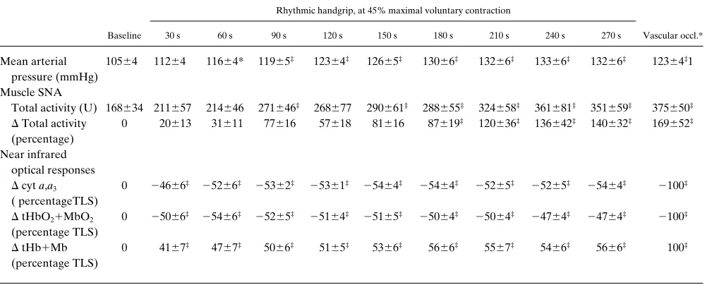
Table I. Responses during Rhythmic Handgrip at 45% Maximal Voluntary Contraction and Posthandgrip Forearm Vascular Occlusion
Rhythmic handgrip, at 45% maximal voluntary contraction
Baseline 30 s 60 s 90 s 120 s 150 s 180 s 210 s 240 s 270 s Vascular occl.*
Mean arterial pressure (mmHg)
10564 11264 11664* 11965‡ 12364‡ 12665‡ 13066‡ 13266‡ 13366‡ 13266‡ 12364‡1
Muscle SNA
Total activity (U) 168634 211657 214646 271646‡ 268677 290661‡ 288655‡ 324658‡ 361681‡ 351659‡ 375650‡
D Total activity (percentage)
0 20613 31611 77616 57618 81616 87619‡ 120636‡ 136642‡ 140632‡ 169652‡
Near infrared optical responses
D cyt a,a3
( percentageTLS)
0 24666‡ 25266‡ 25362‡ 25361‡ 25464‡ 25464‡ 25265‡ 25265‡ 25464‡ 2100‡
D tHbO21MbO2
(percentage TLS)
0 25066‡ 25466‡ 25265‡ 25164‡ 25165‡ 25064‡ 25064‡ 24764‡ 24764‡ 2100‡
D tHb1Mb
(percentage TLS)
0 4167‡ 4767‡ 5066‡ 5165‡ 5366‡ 5666‡ 5567‡ 5466‡ 5666‡ 100‡
between exercise bouts. 8 min of complete circulatory arrest was per-formed at the end of the protocol.
Protocol 6. Effects of reflex-sympathetic activation on muscle oxygenation during reactive (as opposed to exercise-induced)
[image:7.612.60.430.64.682.2]hyper-emia (4 experiments performed on 4 subjects). Reactive hyperhyper-emia was used as an internal control for any nonspecific effects of exercise-induced hyperemia on the changes in muscle oxygenation exercise-induced by a given reflex increase in muscle SNA (evoked by LBNP).
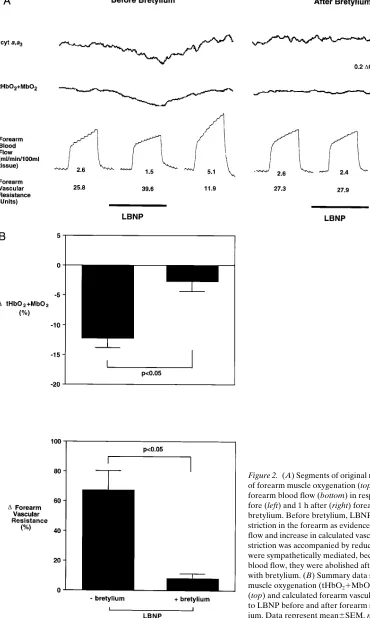
Figure 2. (A) Segments of original near infrared optical measurements of forearm muscle oxygenation (top) and plethysmographic tracings of forearm blood flow (bottom) in response to nonhypotensive LBNP be-fore (left) and 1 h after (right) forearm-sympathetic blockade with bretylium. Before bretylium, LBNP produced marked reflex vasocon-striction in the forearm as evidenced by the decrease in forearm blood flow and increase in calculated vascular resistance. This reflex vasocon-striction was accompanied by reductions in muscle oxygenation that were sympathetically mediated, because, like the decreases in forearm blood flow, they were abolished after forearm-sympathetic blockade with bretylium. (B) Summary data showing the changes in forearm muscle oxygenation (tHbO21MbO2, percentage of total labile signal)
Blood pressure, heart rate, and near infrared signals were mea-sured in response to LBNP (2 min at 220 mmHg applied at baseline, and 30 sec after release of a complete ischemia (pneumatic cuff at 280 mmHg for 8 min) of the resting forearm.
Data analysis
Statistical analysis was performed using repeated measures analysis of variance with Dunnett’s post hoc test to detect values that were different from baseline values. Responses to handgrip before and af-ter bretylium were compared using two-factor analysis of variance for repeated measures. Single comparisons were performed using a t test for paired or unpaired comparisons. P, 0.05 was considered signifi-cant. Data are expressed as mean6SEM.
Results
During high intensity rhythmic handgrip, decreases in muscle oxygenation are temporally dissociated from metaboreflex-medi-ated increases in muscle sympathetic nerve activity (Protocol 1, Fig. 1, and Table I). During the 1st min of rhythmic handgrip at 45% MVC, both cyt a,a3 and tHbO21MbO2 decreased
rap-idly to new steady state levels, but muscle SNA remained un-changed from baseline. During the subsequent 4 min of the ex-ercise, no additional changes in muscle oxygenation were observed even though muscle SNA increased progressively.
Reflex sympathetic vasoconstriction evokes a decrease in the oxygenation of resting forearm muscle (Protocol 2, Fig. 2). Nonhypotensive LBNP evoked reflex decreases in forearm blood flow and increases in forearm vascular resistance that were ac-companied by decreases in muscle oxygenation. These decreases in muscle oxygenation indeed were sympathetically mediated because, like the decreases in forearm blood flow, they were abolished by regional sympathetic blockade with bretylium.
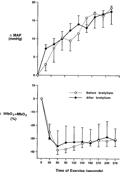
During high intensity rhythmic handgrip, contraction-induced decreases in muscle oxygenation are unaffected by local block-ade of sympathetic neurotransmission in the forearm (Protocol 3, Fig. 3). Decreases in muscle oxygenation and increases in systemic blood pressure (muscle perfusion pressure) were in-distinguishable during high intensity handgrip before and after regional sympathetic blockade of the exercising arm, demon-strating that forearm-sympathetic neural activation was not an important determinant of oxygenation in the contracting mus-cle. Sympathetic blockade was highly effective since it abol-ished optical responses at rest to LBNP at 220 mmHg (DtHbO21MbO2: 21363 vs 0.161.2% total labile signal
be-fore vs after bretylium, P, 0.05) and 240 mmHg (DtHbO21
MbO2: 21662 vs 2162% of the total labile signal before vs.
after bretylium, P, 0.05). Corresponding changes were ob-served in the cyt a,a3 and tHb1Mb signals.
Sympathetically mediated decreases in muscle oxygenation are eliminated when the forearm muscle is exercised, providing direct evidence for functional sympatholysis in human skeletal muscle. (Protocol 4, Figs. 4 and 5, and Table II). The reflex in-crease in muscle SNA induced by LBNP was identical when LBNP was applied before, during, or after performance of rhythmic handgrip at 20 or 33% MVC. The magnitude of this response, a fourfold increase in SNA over baseline, was at least as great as (and actually greater than) that produced by metaboreflex activation during handgrip at 45% MVC. This reflex increase in muscle SNA, which produced reproducible decreases in muscle oxygenation both before and after hand-grip, had no detectable effect on muscle oxygenation when the same muscle was exercised during rhythmic handgrip at either 20 or 33% MVC. Thus, handgrip at 20 and 33% MVC pro-duced steady state decreases in cyt a,a3 (23264 and 24565%
of the total labile signal, respectively) and tHbO21MbO2
(23365 and 24567% of the total labile signal), but when the LBNP was superimposed during rhythmic handgrip, no addi-tional decrement in these signals was observed.
In contrast, the decrease in muscle oxygenation elicited by brief mechanical occlusion of the forearm circulation was pre-served and even augmented during handgrip. When the vascular occlusion was applied to the resting forearm, tHbO21 MbO2
decreased by 1362% of the total labile signal (P, 0.05). Dur-ing rhythmic handgrip at 33% MVC alone, tHbO21MbO2
de-creased by 3266% of the total labile signal (P, 0.05 vs base-line), to a new steady state level. Most importantly, when the vascular occlusion was applied to the exercising forearm, tHbO21MbO2 decreased by an additional 3966% of the total
labile signal (P , 0.05 vs response at rest). Corresponding changes were observed in the cyt a,a3 and tHb1Mb signals.
The observed exercise-induced impairment in sympatheti-cally mediated decreases in muscle oxygenation (i.e.,
func-Figure 3. Summary data showing the decreases in muscle oxygen-ation (tHbO21MbO2) and increases in mean arterial pressure (MAP)
[image:8.612.60.297.59.398.2]Figure 4. (A) Segments of an original record showing simultaneous re-cordings of near infrared optical signals from forearm muscle (top), muscle SNA (center), and handgrip force (bottom). Measurements are shown in response to lower body negative pressure (LBNP) at rest, during handgrip at 20% MVC, and 10 min after the exercise. LBNP produced marked and virtually identical reflex increases in muscle SNA before, during, and after exercise. This reflex increase in muscle SNA reproducibly decreased muscle oxygenation before and after ex-ercise, but had no effect on oxygenation when the muscle was exercis-ing, indicating functional sympatholysis. (B) Summary data showing the changes in forearm muscle oxygenation (tHbO21MbO2,
tional sympatholysis) is localized to muscle that is being exer-cised. Thus, the same LBNP-induced increase in muscle SNA evoked equivalent decreases in the oxygenation of a resting forearm muscle regardless of whether the contralateral fore-arm was resting or exercising (rhythmic handgrip at 33% MVC): DtHbO21MbO2: 21262% vs 21161% of the total
la-bile signal (P5 NS, rest vs contralateral forearm exercise). 10–20% MVC is the minimum level of rhythmic handgrip needed to detect functional sympatholysis by near infrared spec-troscopy (Protocol 5, Fig. 6). Handgrip alone at 5, 10, and 20% MVC produced graded decreases in muscle oxygenation that
reached steady state within the first 2 min of exercise:
DtHbO21MbO2: 2862, 21365, and 24064% of the total labile
signal, respectively, with corresponding changes in the cyt a,a3
and tHb1Mb signals. Reflex increases in muscle SNA induced by LBNP produced similar decreases in the oxygenation of forearm muscle when the muscle was at rest or when it was ex-ercised during rhythmic handgrip at 5 or 10% MVC. In contrast, this sympathetically mediated decrease in muscle oxygenation was abolished during rhythmic handgrip at 20% MVC.
Sympathetically mediated decreases in forearm muscle oxy-genation, although abolished during the hyperemia that
accom-Figure 5. (A) Original near infrared optical re-cordings of forearm muscle tHbO21MbO2 in
response to brief (1 min) periods of forearm vascular occlusion (Occl.) performed at rest and during rhythmic handgrip at 33% MVC. After handgrip, forearm vascular occlusion was maintained for 2 min. At rest, 1 min of vascu-lar occlusion produced a decrease in muscle oxygenation comparable to that normally pro-duced by 2 min of LBNP. When the muscle was exercised, vascular occlusion produced a much greater decrease in muscle oxygenation, suggesting that the sensitivity of the method to detect decreases in oxygenation (e.g., from re-flex-sympathetic vasoconstriction), if present, would be at least as great in exercising as in resting muscle. (B) Summary data showing changes in muscle oxygenation (tHbO21MbO2,
[image:10.612.56.395.56.386.2]percentage of total labile signal) in response to lower body negative pressure and brief vascu-lar occlusion at rest and during rhythmic hand-grip at 33% MVC. Responses to LBNP at rest and during rhythmic handgrip at 33% MVC are reproduced from protocol 3 for compari-son. Data represents mean6SEM, n5 5 for vascular occlusion data, n5 6 for LBNP data.
Table II. Responses to LBNP before, during, and after Rhythmic Handgrip at 20 and 33% MVC
Rhythmic handgrip at 20% MVC Rhythmic handgrip at 33% MVC
Before During After Before During After
D Mean arterial
pressure (mmHg) 461 461 161 162 762* 2162
D Muscle SNA
(percentage total activity) 292672* 305659* 300678* 3336119* 266670* 321627*
D cyt a,a3
(percentage TLS) 21262* 2264 22062* 21762* 2164 21764*
D tHbO21MbO2
(percentage TLS) 21261* 2164 21762* 21662* 464 21863*
D tHb1Mb
(percentage TLS) 761* 2164 1062* 1162* 266 1363*
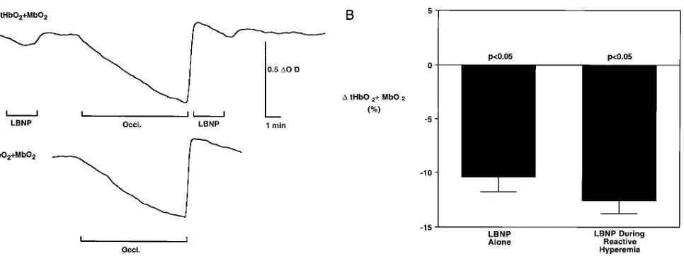
[image:10.612.59.555.562.723.2]panies mild forearm exercise, are well preserved during maxi-mal reactive hyperemia after ischemia of the resting forearm (Protocol 6, Fig. 7). Sympathetically mediated decreases in forearm muscle oxygenation elicited by LBNP were compara-ble at baseline and during peak reactive hyperemia after 8 min of forearm ischemia.
Discussion
Contraction-induced metabolic inhibition of sympathetic vaso-constriction (i.e., functional sympatholysis) has been demon-strated recently in anesthetized animal preparations (33, 44).
We now have provided new evidence for functional sym-patholysis in conscious humans using near infrared spectros-copy. Reflex sympathetic activation, which consistently de-creased oxygenation in resting forearm muscles, had no effect on oxygenation when the muscles were exercised. We also found that the threshold intensity of contraction required to evoke functional sympatholysis is unexpectedly mild, 10–20% MVC. Because this threshold intensity is far below that re-quired for muscle metaboreflex activation (i.e., 33–45% MVC), the data suggest that the two processes are governed by differ-ent mechanisms.
By using several different experimental approaches, we re-peatedly dissociated decreased oxygenation from increased SNA in contracting skeletal muscle. During high intensity rhythmic handgrip (45% MVC), the contraction-induced de-crease in muscle oxygenation reached a nadir before the onset of reflex-sympathetic activation and it did not decrease further despite the subsequent slow and progressive increase in mus-cle SNA. This temporal dissociation provided the first clue to the existence of functional sympatholysis, since the decreases in muscle oxygenation were far from maximal and therefore would not have precluded detection of an additional decre-ment in muscle oxygenation when muscle SNA was increasing during the latter part of the exercise. We further dissociated muscle oxygenation from muscle SNA during high intensity handgrip by showing that the contraction-induced deoxygen-ation was unaffected by eliminating sympathetic neurotrans-mission in the exercising muscles. During milder levels of rhythmic handgrip (20–33% MVC), experimentally induced (i.e., baroreflex-mediated) increases in muscle SNA had no ef-fect on oxygenation in the exercising muscles, but they de-creased oxygenation in the resting muscles. From these obser-vations, we conclude that reflex-sympathetic activation in contracting skeletal muscle is not an important determinant of muscle oxygenation.
[image:11.612.55.299.55.240.2]That these observations demonstrate functional sym-patholysis is predicated on the assumption that the sensitivity
Figure 6. Summary data showing the effect of LBNP-induced re-flex-sympathetic activation on forearm muscle oxygenation (tHbO21MbO2) at baseline and during graded rhythmic handgrip
at 5, 10, and 20% MVC. Data represent mean6SEM, n5 4. From these data, the threshold intensity of handgrip required to produce functional sympatholysis is between 10 and 20% MVC.
Figure 7. (A) Original near infrared optical recordings of forearm muscle tHbO21MbO2 in response to LBNP (top) at baseline and during peak
reactive hyperemia after 8 min of forearm ischemia (Occl.). The response to forearm ischemia alone (bottom) is shown for comparison. Reflex sympathetic activation with LBNP evoked a comparable decrease in muscle oxygenation at rest and during reactive hyperemia after forearm vascular occlusion. (B) Summary data showing forearm muscle oxygenation (tHbO21MbO2, percentage of total labile signal) in response to
[image:11.612.66.549.501.682.2]of near infrared spectroscopy to detect decreased oxygen de-livery is preserved when muscle blood flow and oxygen con-sumption are increased as during exercise. Because near infra-red indices of muscle oxygenation reflect the balance between oxygen delivery and demand, a given decrease in oxygen deliv-ery would be expected to produce, if anything, a larger crease in muscle oxygenation during contraction (high de-mand) than at rest (low dede-mand). Indeed, Mancini et al. (61) demonstrated that brachial artery infusion of vasoconstrictor agents produced decreases in forearm muscle oxygenation that were greater during mild rhythmic forearm exercise than at rest. We demonstrated that elimination of oxygen delivery with forearm circulatory arrest produced a larger decrease in muscle oxygenation when applied during handgrip than at rest. We also demonstrated that sympathetically mediated reflex decreases in muscle oxygenation were well preserved during reactive hyperemia; i.e., during increases in blood flow and he-moglobin in the muscle vessels of similar magnitude to those occurring during exercise.
The present experiments demonstrate that functional sym-patholysis is localized to the exercising muscles, which is con-sistent with our previous findings in rats (33). Because sympa-thetically mediated decreases in muscle oxygenation were preserved in resting forearm muscles during contralateral fore-arm muscle contraction, we conclude that functional sym-patholysis is mediated by some local metabolic event(s) and not by some blood-borne factor.
The demonstration of functional sympatholysis by near in-frared spectroscopy by no means refutes previous hemody-namic data indicating sympathetic vasoconstriction in exercis-ing human skeletal muscle (20–28). In this regard, near infrared spectroscopy provides several important advantages over traditional hemodynamic approaches to study metabolic modulation of sympathetic vasoconstriction in exercising hu-mans. First, the technique provides continuous measurement of oxygen availability at the level of microcirculation, the part of the vascular tree most accessible to metabolic products of contraction (48–50, 61). In contrast, measurements of whole-limb blood flow with techniques such as venous occlusion plethysmography (21–24, 26, 28) or indicator dilution (20, 27) reflect the vasomotor tone of mainly resistance vessels, which are not so accessible to metabolites produced in the muscle in-terstitium. We speculate that during exercise sympathetic vaso-constriction in upstream resistance vessels contributes to blood pressure regulation while sympatholysis in mainly the down-stream nutrient vessels optimizes muscle perfusion (33, 34, 42, 68). Second, the optrode placement is sufficiently stable to ac-quire measurements even during high intensity rhythmic con-traction. This is a major advantage over techniques (e.g., venous occlusion plethysmography) that permit blood flow measurements only after the cessation of exercise, since many of the neurocirculatory adjustments during exercise return to baseline within seconds after the cessation of exercise (6, 69).
Third, and most importantly, the spatial resolution of near infrared spectroscopy is sufficient to reflect changes in oxygen-ation in the truly active (rather than adjacent inactive) small muscle groups of the forearm. This is a major advantage for studying sympatholysis, a phenomenon localized to only active muscle. In contrast, venous effluent blood is an admixture of blood draining active and adjacent nonactive muscle groups, thereby reducing the sensitivity of venous oxygen saturation to detect changes in oxygenation in the active muscle (25, 26). We
suggest that sympatholysis in the active muscle coupled with sympathetic vasoconstriction in adjacent inactive muscle opti-mally redistributes intramuscular blood flow to the most active fibers (33, 34, 42, 70).
The present study does not determine whether functional sympatholysis is mediated primarily via a pre- or postjunc-tional site of action. In ex vivo vascular preparations, prejunc-tional inhibition of norepinephrine release from peripheral sympathetic nerve terminals has been demonstrated by cotrans-mitters such as neuropeptide Y (71) or substances such as ni-tric oxide, adenosine, or K1 (72–75), whose intramuscular
con-centration may increase during exercise. In humans, however, norepinephrine spillover from exercising skeletal muscle clearly is not inhibited during contraction; during unilateral quadriceps exercise, norepinephrine spillover is even greater from the exercising than from the nonexercising muscle (76). In previous animal studies, functional sympatholysis was evi-dent with either sympathetic nerve stimulation or direct appli-cation of exogenous alpha-adrenergic agonists, indicating a postjunctional site of action (33).
Although the local metabolic events mediating functional sympatholysis remain to be defined, we performed several ad-ditional sets of experiments that provide some important clues regarding potential underlying mechanisms. Sympathetically mediated decreases in muscle oxygenation, while abolished during contraction-induced hyperemia, were well preserved during reactive hyperemia. This observation demonstrates that functional sympatholysis is specific for exercise and is not merely a nonspecific effect of muscle hyperemia overwhelm-ing sympathetic vasoconstrictor drive. This observation further suggests that the various local vasodilator metabolites that have been implicated in mediating reactive hyperemia (ade-nosine, inorganic phosphate, histamine, and prostaglandins, reference 73) all are unlikely candidates to be primary media-tors of functional sympatholysis.
Hydrogen ion (H1) initially was an attractive candidate to
mediate functional sympatholysis in humans for several reasons. Our previous animal experiments (33) advanced the hypothe-sis that glycolytic production of H1 mediates functional
sym-patholysis in rat hindlimb muscle. This is consistent with micro-circulatory studies demonstrating that adrenergic vasoconstriction is impaired by experimental acidosis (39, 42). Previous human microneurographic studies (8, 11, 13) advanced the hypothesis that glycolytic production of H1 in exercising skeletal muscle
mediates activation of the muscle metaboreflex. This is consis-tent with single-fiber recordings of muscle metaboreceptor af-ferent discharge in cats, demonstrating that lactic acid is one of the most potent stimuli to these afferents (5). Taken together, this previous work led us to hypothesize that glycolytic produc-tion of H1 is involved in the coordinate regulation of
reflex-sym-pathetic activation and mitochondrial oxygenation in contract-ing skeletal muscle. Based on the present experiments however, H1 is unlikely to be the primary mediator of functional
but may be related to several factors including species, general anesthesia, mode of contraction (electrically evoked vs volun-tary), or sensitivity for the detection of functional sympatholy-sis using Doppler velocimetry vs near infrared spectroscopy.
A salient finding of the present study is that even mild lev-els of exercise (10–20% MVC) are accompanied by sizable de-creases in muscle oxygenation, raising the possibility that skel-etal muscle hypoxia per se is a primary determinant of functional sympatholysis. This is an attractive hypothesis be-cause tissue hypoxia has been shown to impair sympathetic vasoconstriction in rodent and feline skeletal muscle microves-sels both (a) directly, by interfering with oxygen-sensitive adren-ergic signal transduction pathways (such as those involving ATP-sensitive K1 channels) in vascular smooth muscle, and
(b) indirectly, by causing release of vasodilator metabolites from the surrounding skeletal muscle tissue in which the ves-sels are embedded (41, 45). The relative importance of such cellular mechanisms in mediating functional sympatholysis is beyond the scope of the present study.
Regardless of the precise underlying mechanisms involved, the present study demonstrates in humans that sympathetic neural control of skeletal muscle oxygenation normally is very sensitive to modulation by local metabolic events in the con-tracting muscles. Based on our experimental findings in healthy humans, we speculate that, in the clinical setting, im-paired functional sympatholysis may contribute to decreased exercise capacity in pathophysiologic conditions such as heart failure or renal failure, which are characterized by profound alterations in both skeletal muscle metabolism and sympa-thetic nerve activity.
Acknowledgments
We thank Dr. Claude A. Piantadosi for generously providing the near infrared technology, and Mark Hall for superb technical assistance. We thank Dr. Jere H. Mitchell for his critical review of our work.
Dr. Hansen is the recipient of the National Institutes of Health’s (NIH’s) Fogarty International Research Fellowship (NIH-1-F05-TW04949-01) and was supported by grants from the Danish Heart Foundation, the Simonsen & Weel Foundation, and the Danish Re-search Academy. Dr. Thomas was supported by an NIH training grant (T32-HL-07360). Dr. Parsons is the recipient of a Clinician-Sci-entist Award from the American Heart Association and Genentech. Dr. Victor is an Established Investigator of the American Heart As-sociation. This research was supported by a grant to Dr. Victor from the NIH (PO1-HL-06296) and by American Heart Association Texas Affiliate grant-in-aid (96G-064) to Dr. Thomas.
References
1. Volkmann, A.W. 1841. Bewegungen des atmens und schluckens mit be-sonderer berucksichtigung neurologischer streitfragen. Arch. Anat. Physiol. 1: 332–360.
2. Mitchell, J.H., M.P. Kaufman, and G.A. Iwamoto. 1983. The exercise pressor reflex: its cardiovascular effects, afferent mechanisms, and central path-ways. Annu. Rev. Physiol. 45:229–242.
3. Mense, S., and M. Stahnke. 1983. Responses in muscle afferent fibres of slow conduction velocity to contractions and ischemia in the cat. J. Physiol. 342: 383–397.
4. Kaufman, M.P., J.C. Longhurst, K.J. Rybicki, J.H. Wallach, and J.H. Mitchell. 1983. Effects of static muscular contraction on impulse activity of groups III and IV afferents in cats. J. Appl. Physiol. 55:105–112.
5. Rotto, D.M., C.L. Stebbins, and M.P. Kaufman. 1989. Reflex cardiovas-cular and ventilatory responses to increasing H1 activity in cat hindlimb muscle. J. Appl. Physiol. 67:256–263.
6. Mark, A.L., R.G. Victor, C. Nerhed, and B.G. Wallin. 1985. Microneuro-graphic studies of the mechanisms of sympathetic nerve responses to static
ex-ercise in humans. Circ. Res. 57:461–469.
7. Victor, R.G., D.R. Seals, and A.L. Mark. 1987. Differential control of heart rate and sympathetic nerve activity during dynamic exercise. Insight from intraneural recordings in humans. J. Clin. Invest. 79:508–516.
8. Victor, R.G., L.A. Bertocci, S.L. Pryor, and R.L. Nunnally. 1988. Sympa-thetic nerve discharge is coupled to muscle cell pH during exercise in humans. J. Clin. Invest. 82:1301–1305.
9. Victor, R.G., and D.R. Seals. 1989. Reflex stimulation of sympathetic outflow during rhythmic exercise in humans. Am. J. Physiol. 257:2017–2024.
10. Seals, D. 1989. Sympathetic neural discharge and vascular resistance during exercise in humans. J. Appl. Physiol. 66:2472–2478.
11. Pryor, S.L., S.F. Lewis, R.G. Haller, L.A. Bertocci, and R.G. Victor. 1990. Impairment of sympathetic activation during static exercise in patients with muscle phosphorylase deficiency (McArdle’s disease). J. Clin. Invest. 85: 1444–1449.
12. Scherrer, U., S.L. Pryor, L.A. Bertocci, and R.G. Victor. 1990. Arterial baroreflex buffering of sympathetic activation during exercise-induced eleva-tions in arterial pressure. J. Clin. Invest. 86:1855–1861.
13. Ettinger, S., K. Gray, S. Whisler, and L. Sinoway. 1991. Dichloroacetate reduces sympathetic nerve responses to static exercise. Am. J. Physiol. 261: 1653–1658.
14. Sinoway, L.I., R.F. Rea, T.J. Mosher, M.B. Smith, and A.L. Mark. 1992. Hydrogen ion concentration is not the sole determinant of muscle metabore-ceptor responses in humans. J. Clin. Invest. 89:1875–1884.
15. Costa, F., and I. Biaggioni. 1994. Role of adenosine in the sympathetic activation produced by isometric exercise in humans. J. Clin. Invest. 93:1654– 1660.
16. Hansen, J., G.D. Thomas, T.N. Jacobsen, and R.G. Victor. 1994. Muscle metaboreflex triggers parallel sympathetic activation in exercising and resting human skeletal muscle. Am. J. Physiol. 266:2508–2514.
17. Thompson, L.P., and D.E. Mohrman. 1983. Blood flow and oxygen con-sumption in skeletal muscle during sympathetic stimulation. Am. J. Physiol. 245:66–71.
18. Peterson, D.F., R.B. Armstrong, and H.M. Laughlin. 1988. Sympathetic neural influences on muscle blood flow in rats during submaximal exercise. J. Appl. Physiol. 65:434–440.
19. O’Leary, D.S., L.B. Rowell, and A.M. Scher. 1991. Baroreflex-induced vasoconstriction in active skeletal muscle of conscious dogs. Am. J. Physiol. 260:37–41.
20. Secher, N.H., J.P. Clausen, K. Klausen, I. Noer, and J. Trap-Jensen. 1977. Central and regional circulatory effects of adding arm exercise to leg ex-ercise. Acta Physiol. Scand. 100:288–297.
21. Williams, C.A., J.G. Mudd, and A. R. Lind. 1985. Sympathetic control of the forearm blood flow in man during brief isometric contractions. Eur. J. Appl. Physiol. 54:156–162.
22. Sinoway, L.I., J.S. Wilson, R. Zelis, J. Shenberger, D.P. McLaughlin, D.L. Morris, and F.P. Day. 1988. Sympathetic tone affects human limb vascular resistance during a maximal metabolic stimulus. Am. J. Physiol. 255(4 Pt 2): H937–946.
23. Sinoway, L., and S. Prophet. 1990. Skeletal muscle metaboreceptor stimulation opposes peak metabolic vasodilation in humans. Circ. Res. 66:1576– 1584.
24. Joyner, M.J., R.L. Lennon, D.J. Wedel, S.H. Rose, and J.T. Shepherd. 1990. Blood flow to contracting human muscles: influence of increased sympa-thetic activity. J. Appl. Physiol. 68:1453–1457.
25. Joyner, M.J. 1991. Does the pressor response to ischemic exercise im-prove blood flow to contracting muscles in humans? J. Appl. Physiol. 71:1496– 1501.
26. Joyner, M.J., L.A. Nauss, M.A. Warner, and D.O. Warner. 1992. Sym-pathetic modulation of blood flow and O2 uptake in rhythmically contracting human forearm muscles. Am. J. Physiol. 263:1078–1083.
27. Richter, E.A., B. Kiens, M. Hargreaves, and M. Kjær. 1992. Effect of arm-cranking on leg blood flow and noradrenaline spillover during leg exercise in man. Acta Physiol. Scand. 144:9–14.
28. Saito, M., A. Kagaya, F. Ogita, and M. Shinohara. 1992. Changes in muscle sympathetic nerve activity and calf blood flow during combined leg and forearm exercise. Acta Physiol. Scand. 146:449–456.
29. Rowell, L.B. 1993. Human Cardiovascular Control. Oxford University Press, New York. 471–475.
30. Remensnyder, J.P., J.H. Mitchell, and S.J. Sarnoff. 1962. Functional sympatholysis during muscular activity. Circ. Res. 11:370–380.
31. Kjellmer, I. 1965. On the competition between metabolic vasodilatation and neurogenic vasoconstriction in skeletal muscle. Acta Physiol. Scand. 63: 450–459.
32. Donald, D.E., D.J. Rowlands, and D.A. Ferguson. 1970. Similarity of blood flow in the normal and the sympathectomized dog hind limb during graded exercise. Circ. Res. 26:185–199.
33. Thomas, G.D., J. Hansen, and R.G. Victor. 1994. Inhibition of alpha 2-adrenergic vasoconstriction during contraction of glycolytic, not oxidative, rat hindlimb muscle. Am. J. Physiol. 266:920–929.
Suppl. 472:146–167.
35. Hartling, O.J., and J. Trap-Jensen. 1983. Hemodynamic and metabolic effects of alpha-adrenoceptor blockade with phentolamine at rest and during forearm exercise. Clin. Sci. (Lond.). 65:247–253.
36. Savard, G.K., E.A. Richter, S. Strange, B. Kiens, N.J. Christensen, and B. Saltin. 1989. Norepinephrine spillover from skeletal muscle during exercise in humans: role of muscle mass. Am. J. Physiol. 257:1812–1818.
37. Rowell, L.B., M.V. Savage, J. Chambers, and J.R. Blackmon. 1991. Car-diovascular responses to graded reductions in leg perfusion in exercising hu-mans. Am. J. Physiol. 261:1545–1553.
38. Richardson, R.S., B. Kennedy, D.R. Knight, and P.D. Wagner. 1995. High muscle blood flows are not attenuated by recruitment of additional mus-cle mass. Am. J. Physiol. 269:1545–1552.
39. Medgett, I.C., P.E. Hicks, and S.Z. Langer. 1987. Effect of acidosis on alpha1 and alpha2 adrenoreceptor mediated vasoconstrictor responses in iso-lated arteries. Eur. J. Pharmacol. 135:443–447.
40. Boegehold, M.A., and P.C. Johnson. 1988. Response of arteriolar net-work of skeletal muscle to sympathetic nerve stimulation. Am. J. Physiol. 254: 919–928.
41. Boegehold, M.A., and P.C. Johnson. 1988. Periarteriolar and tissue PO2 during sympathetic escape in skeletal muscle. Am. J. Physiol. 254:929–936.
42. McGillivray-Anderson, K.M., and J.E. Faber. 1990. Effect of acidosis on contraction of microvascular smooth muscle by alpha1- and alpha2-adrenocep-tors. Implications for neural and metabolic regulation. Circ. Res. 66:1643–1657.
43. McGillivray-Anderson, K.M., and J.E. Faber. 1991. Effect of reduced blood flow on alpha1- and alpha2-adrenoceptor constriction of rat skeletal mus-cle microvessels. Circ. Res. 69:165–173.
44. Anderson, K.M., and J.E. Faber. 1991. Differential sensitivity of arteri-olar alpha 1- and alpha 2-adrenoceptor constriction to metabolic inhibition dur-ing rat skeletal muscle contraction. Circ. Res. 69:174–184.
45. Tateishi, J., and J.E. Faber. 1995. ATP-sensitive K1 channels mediate alpha 2D-adrenergic receptor contraction of arteriolar smooth muscle and re-versal of contraction by hypoxia. Circ. Res. 76:53–63.
46. Delius, W., K.-E. Hagbarth, A. Hongell, and B. Wallin. 1972. Manoeu-vres affecting sympathetic outflow in human nerves. Acta Physiol. Scand. 84: 82–94.
47. Vallbo, Å., K.-E. Hagbarth, H. Torebjork, and B. Wallin. 1979. Soma-tosensory, proprioceptive, and sympathetic activity in human peripheral nerves. Physiol. Rev. 59:919–957.
48. Piantadosi, C.A. 1989. Near infrared spectroscopy: principles and appli-cation to noninvasive assessment of tissue oxygenation. J. Crit. Care. 4:308–318. 49. Piantadosi, C.A., and B.J. Comfort. 1989. Development and validation of multiwavelength algorithms for in vivo near infrared spectroscopy. In Pho-ton Migration in Tissue. B. Chance, editor. Plenum Publishing Corp., New York. 69–82.
50. Piantadosi, C.A. 1993. Absorption spectroscopy for assessment of mito-chondrial function in vivo. In Mitochondrial Dysfunction. Vol. 2. L.H. Lash and D.P. Jones, editors. Academic Press Inc., San Diego, CA. 107–126.
51. Jobsis, F.F. 1977. Noninvasive infrared monitoring of cerebral and myo-cardial and circulatory parameters. Science (Wash. DC). 198:1264–1267.
52. Hampson, N.B., and C.A. Piantadosi. 1988. Near infrared monitoring of human skeletal muscle oxygenation during forearm ischemia. J. Appl. Physiol. 64:2449–2457.
53. Parsons, W.J., J.C. Rembert, R.P. Bauman, J.C. Greenfield, Jr., and C.A. Piantadosi. 1990. Dynamic mechanisms of cardiac oxygenation during brief ischemia and reperfusion. Am. J. Physiol. 259:1477–1485.
54. Duhaylongsod, F.G., J.A. Griebel, D.S. Bacon, W.G. Wolfe, and C.A. Piantadosi. 1993. Effects of muscle contraction on cytochrome a,a3 redox state. J. Appl. Physiol. 75:790–797.
55. Parsons, W.J., J.C. Rembert, R.P. Bauman, F.G. Duhaylongsod, J.C. Greenfield, Jr., and C.A. Piantadosi. 1993. Myocardial oxygenation in dogs dur-ing partial and complete coronary artery occlusion. Circ. Res.. 73:458–464.
56. Piantadosi, C.A., and F.G. Duhaylongsod. 1994. Near infrared
spectros-copy: in situ studies of skeletal and cardiac muscle. Adv. Exp. Med. Biol. 361: 157–161.
57. Chance, B., and W. Bank. 1995. Genetic disease of mitochondrial func-tion evaluated by NMR and NIR spectroscopy of skeletal tissue. Biochim. Bio-phys. Acta. 1271:7–14.
58. Tamura, M., O. Hazeki, S. Nioka, and B. Chance. 1989. In vivo study of tissue oxygen metabolism using optical and nuclear magnetic resonance spec-troscopies. Annu. Rev. Physiol. 51:813–834.
59. Piantadosi, C.A., J.A. Griebel, and N.B. Hampson. 1988. Intramito-chondrial oxygen decreases in forearm muscle during venous congestion. Clin. Res. 36:37. (Abstr.)
60. Piantadosi, C.A. 1989. Behavior of the copper band of cytochrome c ox-idase in rat brain during FC-43-for-blood substitution. Adv. Exp. Med. Biol. 248:81–90.
61. Mancini, D.M., L. Bolinger, H. Li, B. Kendrick, B. Chance, and J.R. Wilson. 1994. Validation of near infrared spectroscopy in humans. J. Appl. Physiol. 77:2740–2747.
62. Fleckenstein, J.L., D. Watumull, L.A. Bertocci, R.W. Parkey, and R.M. Peshock. 1992. Finger-specific flexor recruitment in humans: depiction by exer-cise-enhanced MRI. J. Appl. Physiol. 72:1974–1977.
63. Siggaard-Andersen, O. 1970. Venous occlusion plethysmography on the calf. Dan. Med. Bull. 17:1–68.
64. Victor, R.G., and W.N. Leimbach, Jr. 1987. Effects of lower body nega-tive pressure on sympathetic discharge to leg muscles in humans. J. Appl. Phys-iol. 63:2558–2562.
65. Jacobsen, T.N., B.J. Morgan, U. Scherrer, S.F. Vissing, R.A. Lange, N. Johnson, W.S. Ring, P.S. Rahko, P. Hanson, and R.G. Victor. 1993. Relative contributions of cardiopulmonary and sinoaortic baroreflexes in causing sym-pathetic activation in the human skeletal muscle circulation during orthostatic stress. Circ. Res. 73:367–378.
66. Lefkowitz, R.J., B.B. Hoffman, and P. Taylor. 1990. Neurohumoral Transmission. In Goodman and Gilman’s The Pharmacologic Basis of Thera-peutics. A. Goodman, A. Gilman, T. W. Rall, A. S. Nies, and P. Taylor, editors. Pergamon Press Inc., Tarrytown, NY. 84–122.
67. Wallin, B.G., R.G. Victor, and A.L. Mark. 1989. Sympathetic outflow to resting muscles during static handgrip and postcontraction muscle ischemia. Am. J. Physiol. 256:105–110.
68. Granger, H.J., A.H. Goodman, and D.N. Granger. 1976. Role of resis-tance and exchange vessels in local microvascular control of skeletal muscle ox-ygenation in the dog. Circ. Res. 38:379–385.
69. Victor, R.G., N.H. Secher, T. Lyson, and J.H. Mitchell. 1995. Central command increases muscle sympathetic nerve activity during intense intermit-tent isometric exercise in humans. Circ. Res. 76:127–131.
70. Nellis, S.H., S.F. Flaim, K.M. McCauley, and R. Zelis. 1980. alpha-Stim-ulation protects exercise increment in skeletal muscle oxygen consumption. Am. J. Physiol. 238:331–339.
71. Lundberg, J.M., A. Franco-Cereceda, J.S. Lacroix, and J. Pernow. 1990. Neuropeptide Y and sympathetic neurotransmission. Ann. N.Y. Acad. Sci. 611: 166–174.
72. Vanhoutte, P.M., T.J. Verbueren, and R.C. Webb. 1981. Local modula-tion of adrenergic neuroeffector interacmodula-tion in the blood vessel wall. Physiol. Rev. 61:151–247.
73. Shepherd, J.T. 1983. Circulation to skeletal muscle. In Peripheral Circu-lation and Organ Blood Flow. Vol. 3. J.T. Shepherd and F.M. Abboud, editors. American Physiological Society, Bethesda, MD. 319–370.
74. Cohen, R.A., and R.M. Weisbrod. 1988. Endothelium inhibits norepi-nephrine release from adrenergic nerves of rabbit carotid artery. Am. J. Phys-iol. 254:871–878.
75. Burnstock, G. 1990. Local mechanisms of blood flow control by perivas-cular nerves and endothelium. J. Hypertens. 8(Suppl. 7):95–106.