Type 3 deiodinase is critical for the maturation
and function of the thyroid axis
Arturo Hernandez, … , Valerie Anne Galton, Donald St.
Germain
J Clin Invest. 2006;
116(2)
:476-484.
https://doi.org/10.1172/JCI26240
.
Developmental exposure to appropriate levels of thyroid hormones (THs) in a timely manner
is critical to normal development in vertebrates. Among the factors potentially affecting
perinatal exposure of tissues to THs is type 3 deiodinase (D3). This enzyme degrades THs
and is highly expressed in the pregnant uterus, placenta, and fetal and neonatal tissues. To
determine the physiological role of D3, we have generated a mouse D3 knockout model
(D3KO) by a targeted inactivating mutation of the Dio3 gene in mouse ES cells. Early in life,
D3KO mice exhibit delayed 3,5,3
¢
-triiodothyronine (T3) clearance, a markedly elevated
serum T3 level, and overexpression of T3-inducible genes in the brain. From postnatal day
15 to adulthood, D3KO mice demonstrate central hypothyroidism, with low serum levels of
3,5,3
¢
,5
¢
-tetraiodothyronine (T4) and T3, and modest or no increase in thyroid-stimulating
hormone (TSH) concentration. Peripheral tissues are also hypothyroid. Hypothalamic T3
content is decreased while thyrotropin-releasing hormone (TRH) expression is elevated.
Our results demonstrate that the lack of D3 function results in neonatal thyrotoxicosis
followed later by central hypothyroidism that persists throughout life. These mice provide a
new model of central hypothyroidism and reveal a critical role for D3 in the maturation and
function of the thyroid axis.
Research Article
Endocrinology
Find the latest version:
Type 3 deiodinase is critical for the maturation
and function of the thyroid axis
Arturo Hernandez,1 M. Elena Martinez,1 Steven Fiering,2 Valerie Anne Galton,3
and Donald St. Germain1,3
1Department of Medicine, 2Department of Microbiology and Immunology, and 3Department of Physiology,
Dartmouth Medical School, Lebanon, New Hampshire, USA.
Developmental exposure to appropriate levels of thyroid hormones (THs) in a timely manner is critical to
normal development in vertebrates. Among the factors potentially affecting perinatal exposure of tissues to
THs is type 3 deiodinase (D3). This enzyme degrades THs and is highly expressed in the pregnant uterus,
pla-centa, and fetal and neonatal tissues. To determine the physiological role of D3, we have generated a mouse
D3 knockout model (D3KO) by a targeted inactivating mutation of the
Dio3
gene in mouse ES cells. Early in
life, D3KO mice exhibit delayed 3,5,3
′
-triiodothyronine (T3) clearance, a markedly elevated serum T3 level, and
overexpression of T3-inducible genes in the brain. From postnatal day 15 to adulthood, D3KO mice
demon-strate central hypothyroidism, with low serum levels of 3,5,3
′
,5
′
-tetraiodothyronine (T4) and T3, and modest
or no increase in thyroid-stimulating hormone (TSH) concentration. Peripheral tissues are also hypothyroid.
Hypothalamic T3 content is decreased while thyrotropin-releasing hormone (TRH) expression is elevated. Our
results demonstrate that the lack of D3 function results in neonatal thyrotoxicosis followed later by central
hypothyroidism that persists throughout life. These mice provide a new model of central hypothyroidism and
reveal a critical role for D3 in the maturation and function of the thyroid axis.
Introduction
Thyroid hormones (THs) are essential for normal vertebrate devel-opment. In mammals, including humans, precise levels of THs during fetal and neonatal life are critical for appropriate cell pro-liferation and differentiation (1). Reduced or excessive THs during these developmental stages can result in severe abnormalities (2, 3). Particularly dramatic is the observation of important altera-tions in the maturation and function of the CNS in mammals with congenital hypothyroidism (2, 3). Some of the important actions of THs during development occur at a time when TH levels are much lower than those in the mother (4, 5) and the hypothalamic-pituitary-thyroid (HPT) axis is not fully functional.
Type 3 deiodinase (D3), a conserved selenoprotein coded by the
Dio3 gene, is responsible in part for the low TH levels in the fetus.
Dio3 is subject to genomic imprinting and is preferentially expressed from the paternal allele in the mouse fetus (6, 7). D3 catalyzes the conversion of the hormones secreted by the thyroid, the active hormone 3,5,3′-triiodothyronine (T3) and prohormone 3,5,3′,5′ -tetraiodothyronine (T4), into biologically inactive metabolites by removing an iodine atom from the tyrosyl ring of both compounds to form 3,3′-diiodothyronine (3,3′-T2) and 3,3′,5′-triiodothyronine (reverse T3) (8, 9). Thus, D3 is an inactivator of THs and serves as a modulator of intracellular TH levels and TH action.
In rodents, D3 activity is abundant in the pregnant uterus and placenta (10–13) and most fetal tissues (14), including the CNS (15). In contrast, during late neonatal and adult life, D3 activity is more limited, being significant in the skin (16) and central
ner-vous system (15, 17, 18), detectable in certain endocrine organs (18), and very low or absent in other tissues.
Though the physiological significance of D3 is unknown, its biochemical function and tissue expression pattern suggest that it plays a role in protecting tissues, particularly those in the develop-ing fetus, from inappropriate levels of THs. This is of particular importance as fetal serum levels of THs in the rat are only 5% of those present in the mother (19). As the HPT axis matures dur-ing late gestation and the neonatal period, serum TH levels rise steadily and reach values comparable to those in the adult in the second week of life (5, 19).
In order to characterize the physiological relevance of D3, we have disrupted the gene coding for D3 in mouse embryonic stem cells and generated a D3 KO mouse (D3KO) with no detectable D3 activity. The D3KO mouse manifests marked abnormalities in thyroid status and physiology, underscoring the critical role of this enzyme in the development and function of the HPT axis.
Results
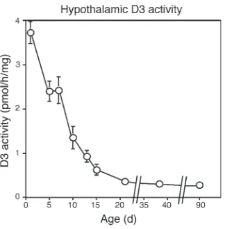
Neonatal hypothalamic D3 expression. To understand the potential role of D3 in the developing HPT axis, we first determined D3 activity in the mouse hypothalamus during neonatal life. We observed a high level of expression during the first week of life, with a marked dimi-nution immediately thereafter (Figure 1). This pattern is similar to that described in the rat (20) and suggests a role for D3 in limiting hypothalamic exposure to THs during early development.
Generation of D3-deficient mice. We have recently described a suc-cessful strategy utilizing homologous recombination in mouse ES cells for disruption of the Dio3 gene (6). The neomycin cassette used for positive selection of recombinant ES cells was excised with appropriate breeding with a Cre-expressing mouse as described in Methods. After this excision, the final structure of the mutated
Dio3 locus is illustrated in Figure 2A. The mutant mice carry a triple point mutation affecting 2 codons, one of them coding for Nonstandard abbreviations used: D3, type 3 deiodinase; HPT,
hypothalamic-pitu-itary-thyroid; neo-T4, neonatal T4; reverse T3, 3,3′,5′-triiodothyronine; S14, spot 14; T3, 3,5,3′-triiodothyronine; T4, 3,5,3′,5′-tetraiodothyronine; TH, thyroid hormone; TRH, thyrotropin-releasing hormone; TSH, thyroid-stimulating hormone.
Conflict of interest: The authors have declared that no conflict of interest exists.
research article
The Journal of Clinical Investigation http://www.jci.org Volume 116 Number 2 February 2006 477
selenocysteine, an active site residue that is critical for enzyme function (8, 21). That this construct codes for an inactive enzyme was shown by transfection experiments in COS-7 cells (data not shown), indicating that the mutation completely abolished enzyme activity. In addition to the triple mutation, mutant mice carry a residual insertion of a loxP site, 34 bp in length, located in the 3′-untranslated region. The presence of this insertion was used for routine genotyping as illustrated in Figure 2B.
This targeting strategy was designed to fully inactivate D3 while at the same time producing a minimal disruption in the Dio3 locus. This is critically important for 2 reasons. First, this locus is imprint-ed and belongs to a larger imprintimprint-ed domain in which long-range mechanisms may control gene expression (22, 23). Second, an addi-tional gene, termed Dio3os, is expressed from the opposite DNA strand and features multiple transcripts that result from alternative splicing (6, 24). The full structure and function of the Dio3os gene have not yet been determined, but partial exonic sequences from a specific Dio3os transcript lie within the Dio3 exon and promoter, 5′ to the point mutations and the residual loxP site (24).
Thus, a large deletion in the Dio3 locus might result in unwanted effects in gene expression within this imprinted region or in the disruption of Dio3os gene expression. To confirm that this was not the case, we performed Northern blot analysis to determine
Dio3os mRNA expression in fetuses homozygous for the Dio3
mutation. As shown in Figure 2C, there is no noticeable change in the pattern of Dio3os transcripts, suggesting that the small modi-fications introduced in the Dio3 locus do not disrupt the expres-sion of Dio3os transcripts in these mice. This is consistent with our previous observations that no Dio3os transcripts are detected by Northern blot analysis when using as a probe a genomic frag-ment comprising the Dio3 3′-untranslated region (24), where the residual loxP site is located. Although it is uncertain whether the triple point mutation introduced in the Dio3 coding region lies within exonic sequence of the Dio3os gene, this mutation would not disrupt any of the potential open reading frames coding for a hypothetical Dio3os protein.
D3 activity is undetectable in D3KO mice. Unless stated otherwise, all the WT and D3KO animals (homozygous for the mutated allele) used in the present work were born to heterozygous mothers of the 129/Sv strain. D3 activity in WT and D3KO mice was determined
in various tissues known to express D3, such as the pregnant uter-us, placenta, adult midbrain, cerebral cortex and ovary and in E14.5 whole fetuses (Table 1). As expected, no D3 activity was detected in tissues from D3KOmice except in the placenta. Low levels of D3 activity (2.5% of that in the WTs) were measured in the placentas of D3KO fetuses that were conceived by heterozygous mothers. This can be attributable to the presence in the tissue of a residual cell population of maternal origin that expresses D3. Indeed, no placental D3 activity was found in D3KO fetuses that were carried by D3KOmothers (Table 1). These results demonstrate that the introduced mutation completely inactivates the D3.
General phenotype of the D3KO mouse. The proportion of D3KO pups obtained from heterozygous matings was lower than the 25% expected from Mendelian laws. Out of 349 newborns produced from heterozygous parents, only 61 (17.5%) were D3KO (P =0.05). This observation suggests partial lethality of D3KO mice that occurs at or before the time of birth. D3-deficient mice also exhib-ited impaired reproductive function. Fertility rates were very low in D3KO mice of both sexes. In addition, both male and female D3KO mice were markedly growth retarded (Figure 3). This retar-dation was already apparent at weaning, when their weight was only 65% of that of the WT mice. This reduction in size persists into adulthood and is still observed in 1-year-old animals (data not shown). Body length is approximately proportional to weight, as shown in the picture included in Figure 3. This general phenotype, with slight variations, is observed in 129/Sv, C57BL/6, and mixed 129/Sv/C57BL/6 genetic backgrounds.
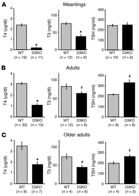
[image:3.585.80.242.81.246.2]Weanling and adult D3KO mice manifest central hypothyroidism. In the late postnatal period, D3KO mice were hypothyroid. Compared with WT mice, the serum T4 level in D3KO weanlings was reduced Figure 1
[image:3.585.315.518.431.650.2]Neonatal hypothalamic D3 activity. Each point represents the mean ± SEM of determinations in 4 animals.
Figure 2
by more than 95%, and the serum T3 concentration was reduced by 50% of normal. However, the serum thyroid-stimulating hor-mone (TSH) level was unaffected (Figure 4A). In adult D3KO mice, serum T4 and T3 levels were also low (27% and 80% of those in WT animals, respectively) while the serum TSH level was elevated 50% (Figure 4B). A very similar pattern of thyroid parameters was observed in older adults (Figure 4C), indicating that the central hypothyroidism persists through adult life. The increase in the TSH level in adults was much lower than what would be antici-pated, given the low circulating levels of THs. As a comparison, a 90-fold increase in TSH concentrations has been observed in mice in which comparably low TH levels were induced by feeding a low-iodine diet containing propylthiouracil (25). This failure of the serum TSH level to be elevated appropriately in the face of low circulating T4 and T3 levels points to a central etiology of the hypothyroidism. Values for T3 uptake were comparable in WT and D3KO mice, 60.1 ± 1.2 and 60.7 ± 1.2, respectively, suggesting that free fractions of T4 and T3 are the same in the 2 strains.
These low serum TH levels observed in adult D3KOmice resulted in tissue hypothyroidism. Thus, hepatic expression of TH-inducible genes such as spot 14 (S14) (26) and type 1 deio-dinase (D1) (27) was significantly decreased. Liver S14 mRNA expression was reduced by more than 80% in D3KO mice, both in adults and weanlings (Figure 5A). D1 activity and mRNA were also diminished in D3KO weanlings (Figure 5B).
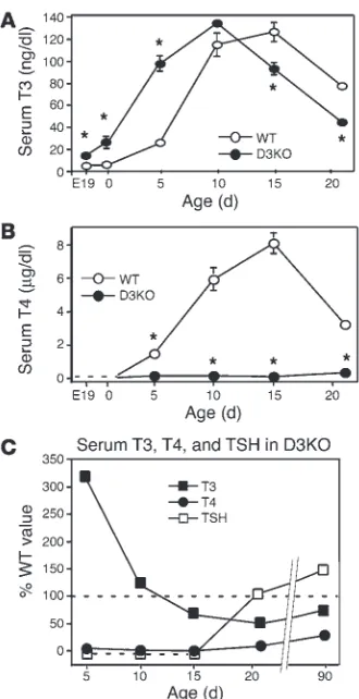
Perinatal D3KO mice are thyrotoxic. In contrast to the hypothy-roidism present in adult mice, D3KO neonates were thyrotoxic, based on a markedly elevated serum T3 level. At P5, the serum T3 level in D3KO mice was elevated 4-fold compared with that of WT animals (Figure 6A). By P10, the serum T3 level in WT and D3KO mice was comparable, and then, during the next 11 days of life, D3KO mice transitioned to the hypothyroid state observed in weanlings and adults. Thus, at P15, the serum T3 level in D3KO mice was significantly lower (Figure 6A) and by day 21 was only 50% of that observed in WT mice. It is notable that through-out the same period, serum T4 levels in D3KO mice were barely detectable and much lower than in WT mice (Figure 6B). In WT mice, the serum T4 level increased steadily during neonatal life to reach a peak around P15. No such neonatal T4 surge occurred in D3-deficient animals.
D3KOmice were also thyrotoxic in utero and at the time of birth; compared with WT mice, E19.5 D3KO fetuses and 1-day-old neonates showed a several-f1-day-old elevation in the serum T3 level
(Figure 6A). At these stages, serum T4 levels were undetectable (<0.15 µg/dl) in both mutant and WT mice (Figure 6B).
Neonatal serum TSH was markedly suppressed in D3KO mice. In WT animals, serum TSH values (in mU/l) were 76.8 ± 13.5 (n = 8), 47.3 ± 7.7 (n = 7), and 30.5 ± 8.0 (n = 5) at P5, P10, and P15, respec-tively. In contrast, serum TSH in D3KO mice at the same ages was undetectable (<10 mU/l in the assay, n = 5, 3, and 3 animals in each age group, respectively). Figure 6C summarizes the changes in thy-roid parameters that occurred in D3KO mice through 90 days of age, expressed as a percentage of the values determined in WT ani-mals. This panel shows 3 phases in the abnormalities of the D3KO thyroid axis. D3KO mice were thyrotoxic early in life with markedly elevated serum T3 and suppressed serum T4 and TSH. At day P15, a pattern of central hypothyroidism is apparent with low T4, T3, and TSH. After weaning and into adulthood, TSH became mildly elevated though the central hypothyroidism persisted.
To gain further insight into the cause of the high T3 levels observed during the perinatal period in D3KO mice, we evaluated the rate of T3 clearance from the serum. WT and D3KO 2-day-old pups were injected with a tracer amount of [125I]-T3, and the [125I]-T3 present in the blood was measured at different times. Two hours after injection, the level of [125I]-T3 in the serum of D3KO pups was double that in the WT mice (Figure 7A). Seven hours after injection, [125I]-T3 had decreased significantly in WT pups but very little in the D3KO pups. From the slopes of the curves, we estimated that D3KO newborns exhibited a significant reduc-tion in the serum T3 clearance rate. Serum [125I]-T3 in WT mice decreased approximately 5 times faster than in D3KO animals. Small amounts (<5% of the total) of other radioactive metabolites were noted in the serum of injected animals. In addition, values for serum T3 uptake were comparable in WT and D3KO mice at P2 (Figure 7B), suggesting no difference in serum binding of T3 between the 2 strains. Thus D3KO neonates demonstrate a dimin-ished T3 clearance, and this likely plays an important role in the high serum level of T3 observed at this age.
Brain thyrotoxicosis in D3KO neonates. To determine the effects on the brain of the elevated serum T3 level during the neonatal peri-Table 1
D3 activities in different tissues of WT and D3KO mice
Tissue WT D3KO
Pregnant uterus 12402 ± 552 Undetectable E14.5 fetus 988 ± 92 Undetectable Adult cortex 1024 ± 42 Undetectable Rest of adult cerebrum 1066 ± 102 Undetectable Adult ovary 73 ± 3 Undetectable Placenta (heterozygous mother) 3070 ± 267 77 ± 6 Placenta (D3KO mother) Undetectable
[image:4.585.53.272.111.204.2]D3 activities (fmol/h/mg protein) represent the mean ± SEM. Uterus and placenta samples correspond to E17.5 gestational age. Adult tissues were obtained from 3-month-old mice. From 4 to 6 samples were used for each determination. Undetectable activities in the assay conditions were lower than 2 fmol/h/mg protein.
Figure 3
[image:4.585.323.504.508.647.2]research article
The Journal of Clinical Investigation http://www.jci.org Volume 116 Number 2 February 2006 479
od, we analyzed the mRNA expression of RC3 and hairless, 2 genes that are upregulated by THs in the CNS during the neonatal peri-od (28, 29). In 1- to 3-day-old total brains, hairless expression was significantly elevated in D3KO animals (Figure 8A). The hairless
mRNA level in the hypothalamus of D3KO mice was also mark-edly elevated at P5 (Figure 8A). These observations indicate that the elevated serum T3 level is accompanied by enhanced T3 action in the neonatal brain. Indeed, the T3 concentration in the brains of D3KO neonates at P2 was more than double that in WT mice (Figure 8B). At P3, RC3 mRNA expression was also stimulated in the brain of D3KO mice as compared with that of WT mice (Figure 8C), but its expression was lower than normal by P15 (Figure 8C), following the onset of central hypothyroidism. These results indi-cate that the brain evolves from a thyrotoxic state in the newborn to a hypothyroid state in late neonatal life, undergoing a transition in thyroid status analogous to that observed in serum.
An important factor regulating TH action in the brain is type 2 deiodinase (D2), an enzyme that converts the prohormone T4
into the active hormone T3 (8, 9). D2 protects the brain from low levels of THs, and its activity is markedly increased when T4 lev-els are low (8, 30). In the present study, the profile of D2 activity in the brain of D3KOmice was consistent with the observed low serum T4 level. Thus, D2 activity was significantly elevated in the cerebral cortex of D3KO mice on P5 (Figure 8D) and was dramati-cally elevated (10- to 15-fold higher than in WT mice) from P10 to P21. A significant increase in D2 activity was also found in the adult cortex at P90.
Thyrotropin-releasing hormone expression and hypothalamic T3 content. To investigate the nature of the central hypothyroidism found in weanlings and adult D3KO mice, we measured hypothalamic T3 content and thyrotropin-releasing hormone (TRH) mRNA expres-sion. At 15 days of age, no difference was observed in the T3 con-tent of the hypothalamus between WT and D3KO mice (Figure 9A). However, a moderate but significant decrease in the T3 con-tent was observed in the hypothalamus of adult D3KO mice. No difference in TRH mRNA was observed at P15 (Figure 9B), but TRH expression was elevated in adult D3KO mice, consistent with the diminished hypothalamic T3 content observed at this age.
Discussion
[image:5.585.51.277.81.398.2]We have found that mice lacking an active D3 exhibit a number of abnormalities, including partial perinatal mortality, growth retar-dation, and impaired fertility. D3-deficient neonatal mice also manifest decreased T3 clearance and striking alterations in serum and tissue TH levels during perinatal life and adulthood. Thus, Figure 4
[image:5.585.340.492.474.741.2]Thyroid status of D3KO mice. Serum T4, T3, and TSH levels in (A) weanlings (21 days old), (B) adults (90 days old), and (C) older adults (8 to 13 months old). Number of animals tested are indicated in parentheses. Bars represent the mean ± SEM. †P < 0.05; #P < 0.01; *P < 0.0001, WT versus D3KO.
Figure 5
our findings in this new mouse model indicate that D3 is critical for both normal development and function of the thyroid axis.
We have shown that the D3KO mouse has no detectable D3 enzymatic activity and that the pattern of transcripts for the
Dio3os gene that is transcribed from the opposite strand at the
Dio3 locus does not appear to be disrupted. Thus, the abnormali-ties observed in the D3KO mice likely are due solely to the absence of an active D3 enzyme.
Our observations indicate that the lack of D3 results in a mark-edly elevated level of T3 in the serum of fetuses and early neonates. As it is during this developmental stage that D3 expression is at its highest in the hypothalamus and in most tissues, our results demonstrate that D3 plays a critical role in maintaining low levels of THs during fetal and early neonatal life. In the absence of D3, the clearance of T3 is diminished, and this likely contributes to the perinatal thyrotoxicosis observed in D3KO mice. In addition to an increased serum T3 level, increased T3 action in the brain and the hypothalamus is also observed. In the normal brain, the expression of various T3-inducible genes increases steadily at a well-defined rate during neonatal life (31). However, in the D3-deficient new-born, this developmental pattern of expression is disrupted; the expression of T3-responsive genes is higher than normal at early neonatal ages and then lower than normal later in development. The functional consequences of this altered gene expression pat-tern are as yet undefined but likely to be significant.
The thyrotoxic status in the D3KOnewborn evolves to hypo-thyroidism after 2 weeks of life, and this persists in adulthood. This is accompanied by tissue hypothyroidism, as demonstrated by a marked decrease in liver expression of T3-inducible genes. The
nature of this hypothyroidism is clearly central, as serum TSH is suppressed, unchanged, or only slightly elevated in D3KO at dif-ferent stages despite the low serum concentrations of both T4 and T3. In the adult, a 50% increase in TSH is observed in the presence of low T4 and T3 levels. However, this TSH elevation is very mod-est compared with a model of primary hypothyroidism that shows similar serum TH levels (25) and where TSH levels are increased 90-fold. This comparison indicates that the hypothyroidism in D3KO mice is due to central abnormalities of the HPT axis. Indeed, the TH parameters in the D3KO mouse are remarkably similar to those observed in the TRH and TRH-receptor KO mouse models (32, 33), where serum T4 and T3 are decreased and serum TSH is modestly elevated or unchanged, respectively.
In the adult D3KO mouse, the inability of increased TSH levels to normalize TH concentrations may reflect a defect in the thyroid gland. Although this possibility cannot be discarded, preliminary observations indicate that the size and appearance of the thyroid gland is normal. A more plausible explanation for the lack of TSH effect is that TSH bioactivity is diminished. TRH signaling is criti-cal for the proper maturation and glycosylation of TSH (34, 35), and these posttranslational modifications have been shown to be necessary for full TSH bioactivity in human cases of central hypo-thyroidism of hypothalamic origin (36). This has also been shown in the TRH KO mouse (37).
[image:6.585.82.247.80.401.2]In considering the etiology of the central hypothyroidism in the adult D3KO mouse, the rodent model referred to as the neonatal T4 (neo-T4) syndrome may be of relevance (38, 39). In this model, the injection of pharmacological doses of T4 (or T3) to rats for 3 to 5 days immediately after birth results in the development of central hypothyroidism in adults. These animals also show an
Figure 7
Neonatal serum T3 clearance and uptake. (A) [125I]-T3 levels in 2-day-old newborn mice after a single intraperitoneal injection of [125I]-T3 (see Methods). (B) Percentage of T3 uptake by serum from 2-day-old neo-nates. Each point represents the mean ± SEM of determinations in 4 (A) and 7 (B) animals in each group. *P < 0.0001, WT versus D3KO.
Figure 6
[image:6.585.312.531.554.679.2]research article
The Journal of Clinical Investigation http://www.jci.org Volume 116 Number 2 February 2006 481
impaired pituitary response to TRH (40, 41). Thus, the neo-T4 rat model demonstrates that exposure to inappropriately high levels of THs before the HPT axis is functionally mature results in pitu-itary and/or hypothalamic abnormalities that affect its regulatory mechanisms and set point.
Similar observations have been made in humans. Several articles have described infants who experienced thyrotoxicosis in utero as a result of poorly controlled maternal hyperthyroidism and who subsequently developed transient neonatal central hypothyroid-ism (42, 43). In view of these observations, we suggest that the main cause of central hypothyroidism in D3KO mice is their over-exposure to T3 during a critical period of thyroid axis develop-ment. The molecular parameters mediating this occurrence have not been identified in any of these models.
Notably, the central hypothyroidism observed in D3KO mice is more severe than that in neo-T4 rats whereas in the human cases mentioned above, the central hypothyroidism appears to be tran-sient. Although common mechanisms may cause the abnormali-ties of the HPT axis in the 3 models, the increased severity of the D3KO phenotype may be due to a higher degree and/or longer
duration of tissue T3 overexposure. In this regard, it is worth noting that both the neo-T4 rats and human infants born to hyperthyroid mothers have functional D3 that may be upregulated in the face of hyperthy-roidism (44) and thus partially protect the brain from exposure to high TH levels. D3KO mice have no such protective mechanism to ameliorate overexposure to T3. Hence, a more severe phenotype is not unexpected.
D3KO mice possess intact TRH and TRH-receptor genes and, indeed, TRH expression is increased in the adult D3KO hypothalamus, presumably in response to the demonstrated decrease in T3 content. Thus, D3 deficiency does not lead to excessive T3 effects (e.g., suppression of TRH) in the hypothalamus of the adult D3KO animal. This observation implies that the mechanism or mechanisms responsible for TSH dys-regulation in the D3KO animal differ fundamentally from that of the TRH KO animal and involve other molecular changes that alter the set point of TSH secretion in response to TH feedback. Of interest in this regard is that THs are known to regulate TRH-receptor mRNA levels in the pituitary gland, and conceivably this could be part of a
develop-Figure 8
Brain expression of T3-regulated genes. (A) Expression of
[image:7.585.42.319.80.413.2]hairless in the newborn brain and neonatal hypothalamus. Representative Northern blots are shown, and quantification of expression was performed in the number of animals indi-cated in parentheses. Each bar represents the mean ± SEM. Ribosomal staining or cyclophilin expression was used as a control to correct for the amount of RNA loaded per lane. (B) Brain T3 content of P2 newborns. (C) Northern blot analy-sis of RC3 expression in the brain at P3 and P15. The most abundant RC3 transcript is shown. (D) Neonatal and adult brain D2 activity. Each point represents the mean ± SEM of determinations in 6 animals. *P < 0.0001, WT versus D3KO.
Figure 9
[image:7.585.339.492.495.741.2]mental program to determine the set point of the thyroid axis control mechanisms (45).
The thyroid status of the D3KO hypothalamus at P15 does not reflect the circulating low TH levels; the T3 content of the hypo-thalamus is not different from that observed in WT mice despite significant decreases in serum T3 and T4 levels. This is possibly due to the marked increase in D2 activity or the fact that the brain is still evolving from a thyrotoxic state and has yet to adjust to lower serum TH levels. In the adult, however, the thyroid status of the hypothalamus in the D3KO mouse reflects the hypothy-roid serum levels and results in a modest increase in TRH mRNA expression that qualitatively mimics the situation in WT animals (46). This finding of a relatively small increase in TRH expression might be due to the fact that only a fraction of hypothalamic neu-rons display T3-sensitive TRH expression (45).
Other phenotypic abnormalities observed in the D3KO mouse include impaired growth, low fertility, and partial perinatal lethal-ity. It is well established that THs exert profound effects on growth hormone expression (47). Thus, alterations in the growth hormone axis may play a role in the growth retardation observed in D3KO mice. Impaired viability in D3KO mice may be due to perinatal thyrotoxicosis, as a similar observation has been made in rats and humans (48, 49). Concerning the reproductive function of the D3KO mouse, severe hypothyroidism may affect fertility in both sexes, as demonstrated by hyt/hyt mice (50, 51). Of note, fertility in the TRH KO mouse, which manifests a milder degree of hypo-thyroidism that is similar to that in the D3KO mouse, is normal (32). Thus, factors in addition to alterations in adult thyroid sta-tus likely play a role in the impaired fertility of the D3KO mouse. For instance, in utero thyrotoxicosis may also contribute to the perceived lower fertility of D3KO female mice, as the absence of a functional maternal D3 in tissues involved in implantation and placentation may be detrimental to early embryonic viability.
In summary, our results demonstrate that D3 is critical for the normal development and function of the thyroid axis and plays a role in maintaining appropriate TH levels in the fetus and neonate. D3 may be expressed to lower T3 content in this region such that the thyroid axis set point can develop normally. Beyond the neo-natal period, the rapid decrease in D3 activity allows the hypothal-amus to more accurately track serum TH levels and thus adjust TRH expression appropriately.
Although a D3 deficiency has not yet been reported in humans, the observations in the D3KO mouse predict the occurrence of central hypothyroidism as part of the phenotype in such cases. This new model of central hypothyroidism will be valuable for analyzing the events regulating the maturation and function of the thyroid axis, as well as the role of D3 and THs in the physiology of growth, development, and reproduction.
Methods
Generation of D3KO mice. We recently described (6) the strategy used to target the Dio3 gene using standard homologous recombination techniques. We utilized the R1 ES cell line (52), which originated from the 129/Sv mouse strain. Targeted clones were identified by Southern blot analysis, injected into C57BL/6 blastocysts, and reimplanted in CD1 foster mothers. Chimeric males that showed germ-line transmission were mated to C57BL/6 females to test for germ-line transmission of the mutation. Chimeric males were then mated with 129/Sv females to establish the mutant line in a 129/Sv background. The neomycin cassette was excised by mating heterozygous females with a 129/Sv male carrying in chromosome X a transgene
express-ing the Cre DNA recombinase. The removal of the neomycin cassette was confirmed by Southern blot analysis in the first generation females. The
Cre DNA recombinase transgene was removed from the genetic back-ground of the colony by appropriate matings with WT 129/Sv animals and sex selection. Animals were kept under a 12-hour light cycle and provided food and water ad libitum. Animal procedures were approved by the Dart-mouth College Institutional Animal Care and Use Committee.
Mice genotyping and serum and tissue sampling. After the removal of the neo-mycin cassette, genotyping of mice carrying the inactivating mutation was performed by PCR amplification of the residual loxP site (Figure 1). The primers used were as follows: 5′-GGAGTCCTGCTGCTTTTGTG-3′ (sense); 5′-CGAGCCTCTCTGCAATTCAG-3′ (antisense). The PCR protocol consist-ed of 32 cycles that includconsist-ed 20 seconds at 94°C, 20 seconds at 60°C, and 45 seconds at 72°C, with a final extension of 3 minutes. Mouse DNA was isolated by standard procedures after proteinase K digestion of tail snips.
Animals were killed by asphyxiation with CO2 (adults and weanlings) or
by decapitation (neonates). In the adults and older neonates, blood was taken from the inferior vena cava while trunk blood was collected from younger neonates and fetuses. Serum was obtained by centrifugation and stored at –20°C. For Northern blot analysis and enzymatic activity, tissues were dissected, immediately frozen on dry ice, and stored at –70°C. Whole hypothalami were dissected, considering the midbrain and thalamus as the posterior and dorsal limits, respectively, and the optic chiasm and its ends as the anterior and lateral limits, respectively.
For the serum determination of T3 clearance, 2-day-old neonates were injected intraperitoneally with a trace amount of [125I]-T3 (New England
Nuclear) (150,000 cpm, approximately 100 fmol in a volume of 50 µl). Trunk blood was collected at 2 and 7.5 hours after injection and centri-fuged to obtain the serum that was then subjected to paper chromatog-raphy to separate T3 from other radioactive metabolites as described (18). The amount of radioactivity attributable to T3 was determined with a gamma counter. The clearance rate of T3 in serum was estimated from the slopes of the lines obtained by plotting the amount of residual [125I]-T3
against the time after injection. The amount of [125I]-T3 injected was not
corrected by body weight, as at 2 days of age the weight of D3KO mice is within 10% of that in WT animals.
D1, D2, and D3 activities. D1, D2, and D3 enzymatic activities were deter-mined as previously described (18, 44). In brief, tissues were homogenized in a 10 mM tris-HCl, 0.25 sucrose pH 7.5 buffer. A suitable volume of tissue homogenate was used in the enzymatic reaction to ensure that deiodin-ation did not exceed 20% and was proportional to the amount of protein content. Tissue homogenates were incubated at 37°C for an hour with the appropriate [125I]-labelled iodothyronine (New England Nuclear). For
the D1 assay, 400 nM of reverse T3 in the presence of 2 mM of the cofac-tor DTT were used. For the D2 assay, we used 1 nM T4 and 20 mM DTT. For the D3 assay, 2 nM T3 and 20 mM DTT were used. Deiodination was determined based on the percentage of labeled iodine released (D1 and D2 assays) or the amount of [125I]-3,3′-diiodothyronine produced (D3 assay).
The latter was determined after separation of reaction products by paper chromatography, as described (53). A factor of 2 was included in the cal-culations of D1 and D2 activities to correct for the chemical equivalence of the outer ring iodine residues and the fact that only 1 of them is labeled in a given molecule.
buf-research article
The Journal of Clinical Investigation http://www.jci.org Volume 116 Number 2 February 2006 483 fer containing 50% formamide, washed with 0.1X SSC/0.1% SDS at 65°C,
and autoradiographed for 1 to 7 days. Probes were labeled with radioactive
32P-dCTP (ICN Biochemicals Inc.) using the Oligolabelling Kit
(Pharma-cia Corp.) and were purified through G-50 columns (Pharma(Pharma-cia Corp.). Quantification of mRNA bands was performed by computer-assisted densitometry (Molecular Dynamics). The mouse cDNA probes used were as follows: hairless, a 3-kb BamH1 fragment that includes most of the cod-ing region; RC3, the complete 1.3-kb cDNA; TRH, a 0.8-kb PCR fragment that includes the coding region; S14, the complete 1.3-kb cDNA; D1, the complete 1.7-kb cDNA; Dio3os, a mix of 3 partial cDNAs with GenBank accession numbers AY283182, AY283181, and AY077459.
Hormone determinations. Serum total T4 concentration was determined using the total T4 Coat-a-Count RIA kit (Diagnostic Systems Laborato-ries Inc.) according to the manufacturer’s instructions. The sensitivity of the assay as determined experimentally ranged from 0.1 to 0.2 µg/dl. The serum T3 level was determined using a sensitive RIA method established in our laboratory (55) with the modification that the T3 antibody used was obtained from a commercial source (Fitzgerald Industries International Inc.). An index of the circulating levels of TH carrier proteins was obtained by measuring the residual capacity of the serum to bind [125I]-T3, using the
Coat-A-Count RIA T3 uptake kit (Diagnostic Systems Laboratories Inc.) according to the manufacturer’s instructions. For brain and hypothalamic T3 determinations, the tissue was weighed and homogenized in 2 ml of methanol containing 1 mM propylthiouracyl, centrifuged, and the pellet reextracted twice more. Methanol from the supernatants was collected and evaporated, the residue resuspended in a buffer containing 0.2 M glycine, 0.13 M sodium acetate, and 0.02% bovine serum albumin, and analyzed by RIA. Recoveries were not considered for the calculations as they were
deter-mined to be higher than 95% by using a radioactive tracer. T3 was calcu-lated as the average of determinations at 2 different dilutions that typically did not differ more than 15%. The cross reactivity of T4 with the T3 anti-body was less than 0.38%. Adult and weanling serum TSH levels were deter-mined using a highly sensitive double-antibody method developed by A.F. Parlow (56). Cross reactivity with follicle-stimulating hormone or luteiniz-ing hormone was less than 1%. Neonatal serum TSH was measured in 50 µl of serum using a sensitive, heterologous, disequilibrium, double-antibody precipitation radioimmunoassay developed by Pohlenz et al. (57).
Statistics. Statistical significance between groups was determined by the 2-tailed Student’s t test. To assess the proportions of genotypes in the offspring from heterozygous matings, statistical significance was determined by the c2 test.
Acknowledgments
We thank Albert Parlow and Samuel Refetoff for performing the TSH assays in this study. We also thank Catherine Thompson and Juan Bernal for the gifts of the hairless and RC3 cDNAs, respec-tively, and the Transgenic Facility at Dartmouth for their technical assistance. This work was supported by NIH grant DK054716.
Received for publication July 11, 2005, and accepted in revised form November 1, 2005.
Address correspondence to: Arturo Hernandez, Dartmouth Medi-cal School, Borwell Building, Room 720W, Lebanon, New Hamp-shire 03756, USA. Phone: (603) 650-8078 or (603) 650-2582; Fax: (603) 650-6130; E-mail: [email protected]. 1. Nunez, J. 1984. Effects of thyroid hormones
dur-ing brain differentiation. Mol. Cell. Endocrinol.
37:125–132.
2. Bernal, J., and Nunez, J. 1995. Thyroid hor-mones and brain development. Eur. J. Endocrinol.
133:390–398.
3. Porterfield, S.P., and Hendrich, C.E. 1993. The role of thyroid hormones in prenatal and neonatal neurological development - current perspectives.
Endocr. Rev. 14:94–106.
4. Morreale de Escobar, G., Pastor, R., Obregon, M.J., and Escobar del Rey, F. 1985. Effects of maternal hypothyroidism on the weight and thyroid hor-mone content of rat embryonic tissues, before and after onset of fetal thyroid function. Endocrinology.
117:1890–1900.
5. Dussault, J.H., and Labrie, F. 1975. Development of the hypothalamic-pituitary-thyroid axis in the neonatal rat. Endocrinology. 97:1321–1324. 6. Hernandez, A., Fiering, S., Martinez, E., Galton,
V.A., and St. Germain, D.L. 2002. The gene locus encoding the iodothyronine deiodinase type 3 (Dio3) is imprinted in the fetus and expresses anti-sense transcripts. Endocrinology. 143:4483–4486. 7. Tsai, C.E., et al. 2002. Genomic imprinting
contrib-utes to thyroid hormone metabolism in the mouse embryo. Curr. Biol.12:1221–1226.
8. Bianco, A.C., Salvatore, D., Gereben, B., Berry, M.J., and Larsen, P.R. 2002. Biochemistry, cellular and molecular biology, and physiological roles of the iodothyronine selenodeiodinases. Endocr. Rev.
23:38–89.
9. St. Germain, D.L., and Galton, V.A. 1997. The deio-dinase family of selenoproteins. Thyroid. 7:655–668. 10. Galton, V.A., et al. 1999. Pregnant rat uterus
expresses high levels of the type 3 iodothyronine deiodinase. J. Clin. Invest. 103:979–987.
11. Huang, S.A., Dorfman, D.M., Genest, D.R., Salva-tore, D., and Larsen, P.R. 2003. Type 3 iodothyro-nine deiodinase is highly expressed in the human uteroplacental unit and in fetal epithelium. J. Clin.
Endocrinol. Metab. 88:1384–1388.
12. Koopdonk-Kool, J.M., et al. 1996. Type II and type III deiodinase activity in human placenta as a func-tion of gestafunc-tional age. J. Clin. Endocrinol. Metab.
81:2154–2158.
13. Roti, E., Fang, S.L., Green, K., Emerson, C.H., and Braverman, L.E. 1981. Human placenta is an active site of thyroxine and 3,3′ ,5-triiodothyro-nine tyrosyl ring deiodination. J. Clin. Endocrinol. Metab.53:498–501.
14. Huang, T., Chopra, I.J., Boado, R., Solomon, D.H., and Chua Teco, G.N. 1988. Thyroxine inner ring monodeiodinating activity in fetal tissues of the rat. Pediatr. Res.23:196–199.
15. Huang, T., Beredo, A., Solomon, D.H., and Chopra, I.J. 1986. The inner ring (5-) monodeiodination of thyroxine (T4) in cerebral cortex during fetal, neo-natal, and adult life. Metabolism. 35:272–277. 16. Huang, T., Chopra, I.J., Beredo, A., Solomon, D.H.,
and Chua Teco, G.N. 1985. Skin is an active site of inner ring monodeiodination of thyroxine to 3,3′,5′ -triiodothyronine. Endocrinology. 117:2106–2113. 17. Kaplan, M.M., and Yaskoski, K.A. 1980. Phenolic
and tyrosyl ring deiodination of iodothyronines in rat brain homogenates. J. Clin. Invest.66:551–562. 18. Bates, J.M., St. Germain, D.L., and Galton, V.A.
1999. Expression profiles of the three iodothyro-nine deiodinases, D1, D2, and D3, in the develop-ing rat. Endocrinology. 140:844–851.
19. Morreale de Escobar, G., Calvo, R., Obregon, M.J., and Escobar del Rey, F. 1992. Moneostasis of brain T3 in rat fetuses and their mother: effects of thy-roid status and iodine deficiency. Acta Med. Aus-triaca. 19:110–116.
20. Kaplan, M.M., and Yaskoski, K.A. 1981. Maturational patterns of iodothyronine phenolic and tyrosyl ring deiodinase activities in rat cerebrum, cerebellum, and hypothalamus. J. Clin. Invest.67:1208–1214. 21. Kuiper, G.G., Klootwijk, W., and Visser, T.J. 2003.
Substitution of cysteine for selenocysteine in the catalytic center of type III iodothyronine
deiodin-ase reduces catalytic efficiency and alters substrate preference. Endocrinology.144:2505–2513. 22. Reik, W., and Walter, J. 2001. Genomic imprinting:
parental influence on the genome. Nat. Rev. Genet.
2:21–32.
23. Lin, S.P., et al. 2003. Asymmetric regulation of imprinting on the maternal and paternal chro-mosomes at the Dlk1-Gtl2 imprinted cluster on mouse chromosome 12. Nat. Genet. 35:97–102. 24. Hernandez, A., Martinez, E., Croteau, W., and
St. Germain, D. 2004. Complex organization and structure of sense and antisense transcripts expressed from the DIO3 gene imprinted locus.
Genomics.83:413–424.
25. Weiss, R.E., et al. 1998. Thyroid hormone action on liver, heart, and energy expenditure in thyroid hormone receptor beta-deficient mice. Endocrinol-ogy. 139:4945–4952.
26. Zilz, N.D., Murray, M.B., and Towle, H.C. 1990. Identification of multiple thyroid hormone response elements located far upstream from the rat S14 promoter. J. Biol. Chem.265:8136–8143. 27. Maia, A.L., Kieffer, J.D., Harney, J.W., and Larsen,
P.R. 1995. Effect of 3,5,3′-Triiodothyronine (T3) administration on dio1 gene expression and T3 metabolism in normal and type 1 deiodinase-defi-cient mice. Endocrinology. 136:4842–4849. 28. Iniguez, M.A., et al. 1993. Thyroid hormone
regula-tion of RC3, a brain specific gene encoding a protein kinase-C substrate. Endocrinology. 133:467–473. 29. Thompson, C.C. 1996. Thyroid
hormone-respon-sive genes in developing cerebellum include a novel synaptotagmin and hairless homolog. J. Neurosci.
16:7832–7840.
30. Steinsapir, J., Bianco, A.C., Buettner, C., Harney, J., and Larsen, P.R. 2000. Substrate-induced down-regulation of human type 2 deiodinase (hD2) is mediated through proteasomal degradation and requires interaction with the enzyme’s active cen-ter. Endocrinology. 141:1127–1135.
Perspectives in the study of thyroid hormone action on brain development and function. Thyroid.
13:1005–1012.
32. Yamada, M., et al. 1997. Tertiary hypothyroidism and hypoglycemia in mice with targeted disruption of the thyrotropin-releasing hormone gene. Proc. Natl. Acad. Sci. U. S. A.94:10862–10867.
33. Rabeler, R., et al. 2004. Generation of thyrotropin-releasing hormone receptor 1-deficient mice as an animal model of central hypothyroidism. Mol. Endocrinol. 18:1450–1460.
34. Mori, M., Kobayashi, I., and Kobayashi, S. 1986. Thyrotropin-releasing-hormone does not accumu-late glycosyaccumu-lated thyrotropin, but changes hetero-geneous forms of thyrotropin within tha rat ante-rior pituitary gland. J. Endocrinol.109:227–231. 35. Taylor, T., and Weintraub, B.D. 1989. Altered
thyrotropin (TSH) carbohydrate structures in hypothalamic hypothyroidism created by para-ventricular nuclear lesions are corrected by in vitro TSH-releasing hormone administration. Endocri-nology. 125:2198–2203.
36. Beck-Peccoz, P., Amr, S., Menezes-Ferreira, M.M., Faglia, G., and Weintraub, B.D. 1985. Decreased receptor binding of biologically inactive thyrotro-pin in central hypothyroidism. Effect of treatment with thyrotropin-releasing hormone. N. Engl. J. Med.312:1085–1090.
37. Yamada, M., Satoh, T., and Mori, M. 2003. Mice lacking the thyrotropin-releasing hormone gene: what do they tell us? Thyroid. 13:1111–1121. 38. Bakke, J.L., and Lawrence, N. 1966. Persistent
thy-rotropin insufficiency following neonatal thyrox-ine administration. J. Lab. Clin. Med.67:477–482. 39. Bakke, J.L., Lawrence, N., and Robinson, S. 1972.
Late effects of thyroxine injected into the hypo-thalamus of the neonatal rat. Neuroendocrinology.
10:183–195.
40. Bakke, J.L., Lawrence, N., and Wilber, J.F. 1974. The late effects of neonatal hyperthyoridism upon the hypothalamic-pituitary-thyroid axis in the rat.
Endocrinology. 95:406–411.
41. Azizi, F., et al. 1974. Persistent abnormalities in pituitary function following neonatal thyrotoxico-sis in the rat. Endocrinology. 94:1681–1688. 42. Kempers, M.J.E., van Tijn, D.A., van Trotsenburg,
A.S.P., de Vijlder, J.J.M., and Wiedijk, B.M. 2003. Central congenital hypothyroidism due to gesta-tional hyperthyroidism: detection where preven-tion failed. J. Clin. Endocrinol. Metab.88:5851–5857. 43. Higuchi, R., et al. 2005. Central hypothyroidism in infants who were born to mothers with thyrotoxi-cosis before 32 weeks’ gestation: 3 cases. Pediatrics.
115:e623–e625.
44. Escobar-Morreale, H.F., Obregon, M.J., Hernandez, A., Escobar del Rey, F., and Morreale de Escobar, G. 1997. Regulation of iodothyronine deiodinase activity as studied in thyroidectomized rats infused with thyroxine or triiodothyronine. Endocrinology.
138:2559–2568.
45. Segerson, T.P., et al. 1987. Thyroid hormone regu-lates TRH biosynthesis in the paraventricular nucle-us of the rat hypothalamnucle-us. Science. 238:78–80. 46. Schomburg, L., and Bauer, K. 1995. Thyroid
hor-mones rapidly and stringently regulate the mes-senger RNA levels of the thyrotropin-releasing hormone (TRH) receptor and TRH-degrading ectoenzyme. Endocrinology. 136:4480–4485. 47. Spindler, S.R., Crew, M.D., and Nyborg, J.K. 1989.
Thyroid hormone transcriptional regulatory region of the growth hormone gene. Endocr. Res.
15:475–493.
48. Anselmo, J.A., Cao, D.C., Karrison, T., Weiss, R.E., and Refetoff, S. 2004. Fetal loss associated with excess thyroid hormone exposure. JAMA.
292:691–695.
49. Porterfield, S.P. 1985. Prenatal exposure of the fetal rat to excessive L-thyroxine or 3,5-dimethyl-3′ -iso-propyl-thyronine produces persistent changes in the thyroid control system. Horm. Metab. Res.
17:655–659.
50. Beamer, W., Eicher, E.M., Maltais, L.J., and South-ard, J.M.L. 1981. Inherited primary hypothyroidism in mice. Science. 212:61–62.
51. Chubb, C., and Henry, L. 1988. The fertility of hypo-thyroid male mice. J. Reprod. Fertil. 83:819–823. 52. Nagy, A., Rossant, J., Nagy, R., Abramow-Newerly,
W., and Roder, J.C. 1993. Derivation of complete-ly cell culture-derived mice from earcomplete-ly passage embryonic stem cells. Proc. Natl. Acad. Sci. U. S. A.
90:8424–8428.
53. Galton, V.A., and Hiebert, A. 1987. The ontogeny of the enzyme systems for the 5′-and 5-deiodination of thyroid hormones in chick embryo liver. Endocri-nology. 120:2604–2610.
54. Obregon, M.J., Calvo, R., Hernandez, A., Escobar del Rey, F., and Morreale de Escobar, G. 1996. Regulation of uncoupling protein messenger ribo-nucleic acid and 5′-deiodinase activity by thyroid hormones in fetal brown adipose tissue. Endocrinol-ogy. 137:4721–4729.
55. St. Germain, D.L., and Galton, V.A. 1985. Com-parative study of pituitary-thyroid hormone econ-omy in fasting and hypothyroid rats. J. Clin. Invest.
75:679–688.
56. Schneider, M.J., et al. 2001. Targeted disruption of the type 2 selenodeiodinase gene (DIO2) results in a phenotype of pituitary resistance to T4. Mol. Endocrinol. 15:2137–2148.