Epidermal barrier formation and recovery in
skin disorders
Julia A. Segre
J Clin Invest.
2006;
116(5)
:1150-1158.
https://doi.org/10.1172/JCI28521
.
Skin is at the interface between the complex physiology of the body and the external, often
hostile, environment, and the semipermeable epidermal barrier prevents both the escape of
moisture and the entry of infectious or toxic substances. Newborns with rare congenital
barrier defects underscore the skin’s essential role in a terrestrial environment and
demonstrate the compensatory responses evoked ex utero to reestablish a barrier. Common
inflammatory skin disorders such as atopic dermatitis and psoriasis exhibit decreased
barrier function, and recent studies suggest that the complex response of epidermal cells to
barrier disruption may aggravate, maintain, or even initiate such conditions. Either aiding
barrier reestablishment or dampening the epidermal stress response may improve the
treatment of these disorders. This Review discusses the molecular regulation of the
epidermal barrier as well as causes and potential treatments for defects of barrier formation
and proposes that medical management of barrier disruption may positively affect the
course of common skin disorders.
Review Series
Find the latest version:
Epidermal barrier formation and recovery
in skin disorders
Julia A. Segre
Genetics and Molecular Biology Branch, National Human Genome Research Institute, National Institutes of Health, Bethesda, Maryland, USA.
Skin is at the interface between the complex physiology of the body and the external, often hostile, environment,
and the semipermeable epidermal barrier prevents both the escape of moisture and the entry of infectious or toxic
substances. Newborns with rare congenital barrier defects underscore the skin’s essential role in a terrestrial
envi-ronment and demonstrate the compensatory responses evoked ex utero to reestablish a barrier. Common
inflam-matory skin disorders such as atopic dermatitis and psoriasis exhibit decreased barrier function, and recent studies
suggest that the complex response of epidermal cells to barrier disruption may aggravate, maintain, or even initiate
such conditions. Either aiding barrier reestablishment or dampening the epidermal stress response may improve
the treatment of these disorders. This Review discusses the molecular regulation of the epidermal barrier as well as
causes and potential treatments for defects of barrier formation and proposes that medical management of barrier
disruption may positively affect the course of common skin disorders.
Molecular regulation of barrier acquisition and prematurity
Mammalian epidermis is a stratified epithelium that retains the ability to self renew under both homeostatic and injury conditions by maintaining a population of mitotically active cells in the hair follicles and innermost basal layer (Figure 1) (1–3). The major bar-rier resides within the exterior layers of the epidermis, which are sloughed off and repopulated from these inner cells. The process of terminal differentiation begins when basal cells concomitantly withdraw from the cell cycle and lose their ability to adhere to the basement membrane. In the intermediate spinous layers, the cells reinforce a durable cytoskeletal framework of keratin filaments to provide the mechanical strength necessary to resist physical trauma. In the granular layers, lipids are produced inside lamellar bodies, ker-atins are bundled into macrofibrils through their association with filaggrin, and a cornified envelope (CE) is assembled by sequential incorporation of precursor proteins directly underneath the plasma membrane. As the cell membrane disintegrates, the subsequent cal- cium influx activates the transglutaminase (TGM) enzyme to irre-versibly cross-link the CE proteins, creating a tough, insoluble sac that surrounds the keratin fibers. Finally, lipids are extruded into the intercellular space onto the CE scaffold. This barrier, once built, is analogous to bricks and mortar, with the keratin macrofibrils and CEs forming the bricks and the extruded lipids forming the mortar to seal together the CEs (4–6). Recent experimental results have also demonstrated an essential role for tight junctions, located in the granular layer, in forming the epidermal barrier (7). This process of differentiation from a mitotically active basal cell to a squame, a ter-minally differentiated squamous cell, is maintained throughout life as part of epidermal regeneration (8, 9).
During development, the fetus requires a barrier to prevent intra-uterine infection and to serve as the interface with the amniotic fluid. Shortly after gastrulation, the embryonic ectoderm gives rise
to the presumptive epidermis, and by approximately 8 weeks of gestation, basal cells cover the body surface. For the next 5 months, the periderm provides a temporary barrier suitable for the aqueous environment in utero. The periderm is derived from basal kerati-nocytes but differs from differentiated adult squames in the cell morphology, keratin profile, molecular architecture of the tight junctions, and presence of microvilli on the apical surface extend- ing into the amniotic fluid (10). Beneath the periderm, the epider-mis stratifies and differentiates so that by week 34 of gestation the skin is competent to serve as a barrier in a terrestrial environment. At this point, the periderm cells are sloughed to form part of the vernix caseosa that covers the newborn. During the second and third trimesters of pregnancy, the volume of the fetus grows at a rapid rate, which requires an even larger increase of the fetus’ sur-face area. Acquisition of a barrier that is competent to survive in the terrestrial environment requires cross-linking of the CEs and constrains increases in the surface area of the embryo. Therefore, it is not advantageous to develop the terrestrial competent bar-rier too early in development. However, the barrier must still be acquired prior to the typical end of gestation at 40 weeks in case a premature birth occurs. Incomplete barrier acquisition manifests in premature infants as dehydration, electrolyte imbalance, and poor thermoregulation and predisposes to infection. Although the transition to the terrestrial environmentex uteroaccelerates the epidermal differentiation program, an infant born at 25–30 weeks requires 2–4 weeks to develop a fully functional barrier (11).
Two animal models have demonstrated how this process of bar-rier acquisition can be accelerated in utero. First, corticosteroid injections, acting through the glucocorticoid receptor, have been demonstrated to accelerate barrier acquisition (12, 13). Converse-ly, genetically altered mice with a corticosteroid deficiency have a developmental delay of barrier maturation, with defects in both lipid and structural protein synthesis (14). This animal model is particularly relevant since antenatal corticosteroids are standard-of-care treatment to accelerate lung surfactant maturation when a premature birth is anticipated. Second, expression of the tran- scription factor Kruppel-like factor 4 (Klf4) is necessary to estab-lish the prenatal epidermal barrier in mice (15). As well, ectopic expression of Klf4 accelerates barrier acquisition within a field of
Nonstandard abbreviations used: AD, atopic dermatitis; CE, cornified envelope; Klf4, Kruppel-like factor 4; SPINK5, serine protease inhibitor, Kazal type 5; SPRR, small proline-rich protein; TGM, transglutaminase; Tslp, thymic stromal lymphopoietin.
Conflict of interest: The author has declared that no conflict of interest exists.
review series
competence established by the expression of the late CE proteins (16, 17). Intriguingly, earlier studies showed a cooperative syner-gistic activation of a test promoter by glucocorticoid receptor and Klf transcription factors, suggesting that these factors may act to coordinately regulate downstream targets. Ex utero, corticosteroids are not given to premature infants because of adverse side effects, including immune suppression. However, some of the genes regu-lated by KLF4 and corticosteroids may be suitable pharmaceutical targets. Hopefully, by understanding how barrier acquisition is pro-grammed in utero, we can expedite the process ex utero.
Human congenital barrier deficiencies and animal models
The genetically and clinically diverse epidermal differentiation or cornification disorders are classified as ichthyoses, derived from the Greek root ichthys, meaning “fish,” and referring to the scaly presentation of the skin. The scaly appearance is the gross mor- phological presentation of hyperkeratosis, thickening of the stra- tum corneum, which results from either an increase in cell prolif-eration, a delay in cell shedding, or both. Although ichthyosis is a more general classification, this perturbation in the balance of epidermal proliferation and differentiation does typically result in barrier deficiency. Ichthyoses can differ by age of onset, type and location of scale, erythroderma, and associated abnormalities of associated skin appendages. Ichthyosis is often the first compo-nent of a multisystem disorder to be diagnosed. Classification of ichthyoses has been greatly aided by the discovery of the specific genetic defect that underlies many of these disorders. The imme-diate translational benefit of this research is the development of prenatal and perinatal genetic screening to aid in disease diagnosis and to predict the prognosis or associated outcomes. Identifica-tion of the mutated gene underlying the ichthyosis is particularly
beneficial for multisystem disorders in which the skin manifesta-tion is first to be diagnosed.
The discovery that the defects that underlie many of the severe congenital ichthyoses are in genes encoding proteins that form or regulate synthesis of lipids and CEs underscores the importance of orderly differentiation in barrier acquisition (18). As a com-prehensive review of the molecular genetics of the ichthyoses was recently published (18), here only a few classical and recent discov-eries to illustrate key points will be described. Lamellar ichthyosis newborns, with an incidence of 1 in 300,000, present encased in a translucent, taut collodion membrane. Desquamation of this membrane occurs over the first 2 weeks of life, and the body sur-face is covered by large, plate-like scales that continue to exhibit increased transepidermal water loss. Linkage to multiple regions of the genome suggests that a diversity of genetic defects underlies lamellar ichthyosis. As the specific genetic defects are identified, the encoded proteins now appear to assemble into 2 discrete path- ways that govern either CE or lipid function in the stratum corne-um. Loss of the TGM1 enzyme that cross-links the CEs results in lamellar ichthyosis (19, 20). Alternatively, lamellar ichthyosis can be caused by aberrant lipid extrusion due to decreased function of the epidermal lipid transporter ATP-binding cassette, subfamily A, type 12 (ABCA12) or decreased lipoxygenase activity due to muta-tions in the cytochrome P450 gene CYP4F22 (21, 22). Interestingly, mutations in other proteins involved in this metabolic pathway converting arachidonic acid to compounds of the 12-lipoxygen-ase family, including ALOX12B, ALOXE3, and Ichthyin, underlie skin disorders with presentations that sometimes overlap lamellar ichthyosis (23–25). Complete loss of ABCA12 function results in an even more severe congenital disorder known as harlequin ich-thyosis (26, 27). Defects in serine protease inhibitor, Kazal type 5
(SPINK5) underlie the rare Netherton syndrome, which is charac-Figure 1
[image:3.585.44.417.84.370.2]terized by a severe congenital barrier defect, resolving into ichthyo-sis linearis circumflexa with associated hair shaft abnormalities and atopic dermatitis (AD) (28).
As these infants with congenital barrier deficiencies transition ex utero the degree, type, and location of scale can vary depending on the underlying genetic defect. One remarkable exception to the continuation of the barrier deficiency observed at birth is the “self-healing collodion babies,” which have an amino acid change in the TGM1 protein at a site that chelates water only at the higher hydro-static pressure in utero. Molecular models and biochemical assays predict that, in the drier ex utero environment, the mutant protein attains a different conformation and possesses approximately 80% of normal TGM1 activity (29). These infants are a unique example of how specific protein mutations may have a more or less severe effect in the distinct in utero and ex utero environments.
Ichthyosis vulgaris, which is a mild but extremely common form of ichthyosis, is a complementary example of a skin disorder that typically does not affect newborns, but rather manifests in the first year of life as fine white scales. Very recently, mutations in the gene encoding the filaggrin protein, which bundles the keratin filaments in CE formation, were determined to underlie ichthyosis vulgaris (30). Two mutations that would both result in a complete loss of the cleaved filaggrin product were identified in 15 differ-ent families. Ichthyosis vulgaris exhibits a semidominant mode of inheritance, with homozygous and heterozygous mutations in the filaggrin gene underlying the severely (prominent scale) and moderately affected (mild scale) forms, respectively. Population screening revealed that approximately 7% of European controls are heterozygous for a mutation in the filaggrin gene, making this one of the most common human single-gene disorders. Although the incidence of ichthyosis vulgaris is estimated at 1 in 250, these stud-ies suggest that heterozygous null mutations in filaggrin may not always be symptomatic, due to incomplete penetrance, seasonal variation, and a subtle phenotype. However, these mutations may still result in a minor breach of barrier that may predispose these individuals to other disorders, as will be discussed below. Finally, it is intriguing to speculate whether an evolutionary advantage is derived from an impaired barrier, which fixed these filaggrin mutations in certain populations.
The different manifestations of barrier disorders in utero and ex utero drives us to ask the question: are these clinical presentations a direct result of distinct programs of epidermal differentiation? Alternatively, a collodion membrane may provide the most protec-tion in uteroagainst the sterile aqueous environment, whereas the hyperkeratotic scaly stratum corneum may be the best protection against the ex utero terrestrial environment. Both types of aberrant stratum corneum may be the product of compensatory reactive pathways designed to minimize the inherent barrier deficiency. In summary, it is important to consider that the clinical presenta-tion of a barrier defect may represent a combination of the primary defect and the skin’s compensatory response to restore the barrier.
Gene targeting in mice has created animal models in which to study both the primary defect and possible compensatory respons-es ex utero observed in patients with congenital barrier defects. These animals have also contributed to the identification of addi-tional pathways involved in barrier acquisition. As one example, mice with a targeted ablation of the Tgm1 gene, encoding the enzyme that cross-links the CE, are born with severe epidermal barrier defects (31). Unlike the patients with lamellar ichthyosis, these mice do not survive the perinatal period, which may reflect
either the larger surface area–to–volume ratio of mice compared with humans or the lack of sufficient intensive care for newborn mice. To study the postnatal development of skin in Tgm1 -defi-cient mice, investigators grafted skin from newborn Tgm1–/– mice
onto immunocompromised mice (32). Within 2 weeks, this graft-ed Tgm1–/–murine skin developed scaly, plate-like structures with
a normal rate of transepidermal water loss. When the scales were removed, transepidermal water loss rose to a level as high as that observed in Tgm1–/– neonates. This suggests that development of
plate-like scales atop the skin of Tgm1–/–
mice represents a com-pensatory mechanism that prevents transepidermal water loss in the absence of cross-linked CEs. These murine experiments also suggest that the scaly, plate-like structures that cover the body of a patient with lamellar ichthyosis may be beneficial and perhaps should not be removed with retinoid treatment or soaking.
As a second unique example of a compensatory response, the protein loricrin comprises 80% of the protein content of the CE, and yet when the gene encoding loricrin is deleted from the genome, the newborn mice exhibit a normal barrier at birth (33). Loricrin-deficient embryos exhibit a temporal delay in embry-onic barrier competence until upregulation of the expression of other components of the CE compensate. Loricrin-deficient CEs are more fragile, suggesting that these mice might not respond as well as their wild-type counterparts to challenges such as wound-ing or infection (33). Flaky tail, a spontaneously occurring mouse mutant with no genetically cleaved filaggrin protein, biochemi- cally and histologically strongly resembles human ichthyosis vul-garis (30, 34). Again, these mice will be very useful for studying at the cellular level how keratinocytes compensate for the loss of filaggrin, a major structural protein. However, this model also raises the central question of how to interpret the presentation of human ichthyosis vulgaris or any other adult skin disorder on a haired animal. These experiments suggest that the need to protect the body from desiccation and infection demands redundancy at the protein and system-wide levels.
review series
tight junction protein claudin-1, and polarity protein E-cadherin (7, 39–41). Mice with mutations in regulatory molecules such as p63 or IKKα will not be discussed here since their primary defects are related to events earlier in the epidermal specification and dif- ferentiation processes, respectively. Along with biochemical stud-ies, analysis of the transcriptional, protein, and lipid profiles of this array of mutant mouse models of disease will start to establish the hierarchy and branches of the signaling pathways required to establish the barrier and compensate for its loss. With these ani-mal models, the analysis of multiple genetically identical newborns greatly enhances the elucidation of the barrier-specific defect. An initial clinical investigation of children diagnosed with ichthyosis and growth failure demonstrated a correlation between increased transepidermal water loss and caloric drain through heat evapora-tion (42). It is interesting to speculate that some human newborns that fail to thrive and lose weight (i.e., water) suffer from rare, undetected barrier disorders.
Impaired barrier and skin inflammatory disorders: atopic dermatitis and psoriasis
The barrier is established during in utero development, main-tained throughout life, and reestablished after a breach. The study of congenital epidermal barrier disorders will help to elucidate the pathways necessary to establish the barrier and also hints at the existence of a complex molecular redundancy that is evoked to repair a disrupted barrier. Although these severe congenital defects are rare, impaired epidermal barrier function is a hallmark feature of 2 of the most common inflammatory skin disorders, psoriasis and AD (43–46). Psoriasis is a chronic skin disease in which very distinct erythrosquamous silvery scales cover either areas of inflec- tion, the trunk and proximal extremities, or the entire body. Pso-riasis affects 2–3% of the population, with typical age of onset in the third decade (46). As discussed below, the impaired barrier of psoriatic lesions may contribute to keratinocyte hyperproliferation and cytokine activation of lymphocytes. AD is a chronic inflamma-tory skin disease characterized by pruritus, eczema, and cutaneous hyperreactivity to environmental triggers (45). The onset of AD is typically within the first 6 months of life, and the disease currently affects 10–20% of children in developed countries. This prevalence has risen steadily in the last 30 years, suggesting that environmen- tal factors contribute to pathogenesis (47). The impaired skin bar-rier function of AD patients may contribute to increased antigen absorption and result in cutaneous hyperreactivity (48). For both of these inflammatory skin disorders, the degree of epidermal barrier disruption correlates with severity of presentation, as mea-sured by degree of inflammation or state of dermatitis (45, 48). However, these disorders are markedly different in skin presenta-tion, gene expression changes, and immune infiltration.
Psoriasis and AD have both genetic and environmental compo-nents underlying their pathogenesis. One of the most intriguing findings from the genome-wide screens of AD and psoriasis sus-ceptibility is that multiple loci are coincident. Genetic linkage of both AD and psoriasis susceptibility to the epidermal differentia-tion complex on chromosome 1q21, a 1.6-Mb locus of at least 30 genes encoding proteins that both build and regulate barrier for-mation, strongly suggests a role for barrier function or repair in these inflammatory disorders (49, 50). Resident within this epider-mal differentiation complex on 1q21 are many excellent candidate genes that may initiate or maintain a skin inflammatory disorder. The clustering of these epidermal-specific genes raises the question
of whether they are held together as a result of recent evolution through gene duplication or to enable coordinate gene regulation, as observed in the Globin or Hox gene clusters. A priori, the linkage of both AD and psoriasis susceptibility to the epidermal differen- tiation complex on chromosome 1q21 may be one of the most dif-ficult to unravel, considering the possibility that more than one of the genes mapping to this region play a significant role in the progression of a skin inflammatory disorder.
Very recently, McLean and colleagues convincingly demonstrated that an impaired barrier contributes to AD with the discovery that common mutations in the gene encoding filaggrin, resident in the epidermal differentiation complex, are a strong predisposing fac-tor for AD (51). Recalling classic studies that AD is particularly common in individuals with severe ichthyosis vulgaris, these sci-entists determined that AD is inherited as a semidominant trait in the pedigrees they analyzed and thus demonstrated that filaggrin deficiency underlies ichthyosis vulgaris. Recognizing that filaggrin mutations would be in linkage disequilibrium with other genes in the epidermal differentiation complex, they extended the AD stud-ies to 3 additional populations. Notably, in all studies, mutations in filaggrin — in both homozygous and heterozygous states — were highly overrepresented in pediatric AD patients.
Interestingly, 2 of these large AD cohorts were ascertained on the basis of asthma. Approximately half of severe AD patients will develop asthma, and two-thirds will develop allergic rhinitis (also known as hay fever) (52). AD, asthma, and hay fever are epithe-lial organ–specific allergen responses of the skin, lung, and nose, respectively. The high concordance rate among the atopic disor-ders raises the question of whether there are shared genetic risks for all 3 disorders or whether antigen exposure in AD is the initiat-ing event. In these recent cohort studies, mutations in filaggrin are selectively associated with individuals that have both asthma and AD, with no association observed with asthma in the absence of AD (51). This finding has important clinical implications for medical management of children with AD, since AD is often the earliest sign of the progression to asthma and hay fever known as the atopic march (52). A classic study in mice demonstrated that epicutaneous sensitization to an allergen can induce dermatitis and augment airway hyperreactivity, characteristics of AD and asthma, respectively (53). Both these animal and human cohort studies suggest that the increased allergen exposure through the skin of the AD infant may itself predispose to asthma. This hypoth-esis would argue for aggressive means to control the infant’s AD before asthma develops. However, if impaired barrier recovery even in the seemingly unaffected areas underlies the genetic suscepti-bility to AD and contributes to the development of asthma, then controlling the outbreaks of AD lesions may not be sufficient to curtail the onset of asthma. Teasing apart the complex interaction of environmental challenges to the immunologic and epithelial systems in atopic disorders is a major direction of future research. The incomplete penetrance of heterozygous mutations in filaggrin resulting in either ichthyosis vulgaris or AD may be modified by the variant expression of other epidermal cornification genes also resident in the epidermal differentiation complex (30, 51).
as the genetic defect underlying Netherton syndrome, which is characterized by congenital ichthyosis and AD (28). The Spink5 -deficient animal model may help to unravel whether inadequate barrier development or repair is an initiating event in AD. Initial studies show that transplantation of skin from Spink5–/–mice to
an immunocompromised mouse elicits an inflammatory infiltra- tion in the dermis (55). Future studies involving allogenic graft-ing of Spink5–/– murine skin to an immunocompetent mouse will
reveal the full extent of the immune and epidermal response to Spink5 deficiency. Although some human disorders can be mod-eled with null alleles in the mouse, the more complex analysis of these multigenic traits will require the creation of hypomorphic alleles. In theory, a more sophisticated animal model of AD would be a mouse with a E420K amino acid change in the Spink5 pro-tein. Unfortunately, the glutamic acid residue at amino acid 420 is not conserved in the mouse Spink5 protein, raising the question of how best to model the common role of SPINK5 in the pre-disposition to AD. Therefore, future studies need to be directed toward understanding the role of SPINK5 in epidermal differ-entiation and barrier function. As well, expression of SPINK5 in other epithelial and immune cells needs to be explored in both humans and mouse models to unravel whether the atopic disor-ders observed in these patients is intrinsic to defects in immune cell activation or in epithelial barriers. Although the most widely recognized feature of the atopic disorders is a misregulation of the immune system, these findings suggest that there may be ways in which an epithelial barrier is built or responds to stress that predispose to these disorders (56).
The genetic linkage of psoriasis to the epidermal differentia-tion complex has not yet been untangled and may similarly reflect reduced expression of 1 or more genes resident in the locus. Mice with an epidermal-specific deletion of the dimeric transcription factor complex activator protein 1 (AP1), consisting of c-Jun and JunB, demonstrate many of the histological and molecular hall-marks of psoriasis (57). Prior to any skin alterations in these mice, the chemotactic proteins S100A8 and S100A9 — which map to chromosome 1q21 — were found to be highly upregulated, as has also been reported in human psoriatic samples (57, 58). Located immediately proximal to S100A9 are 2 genes encoding peptido-glycan recognition proteins that have also recently been put forth as candidate modifiers of psoriasis (59). Also resident within the epidermal differentiation complex is a family of 10 closely related genes that encode small proline-rich proteins (SPRRs). SPRRs are cross-linked by TGM1 to establish the CE scaffold onto which other proteins are then subsequently cross-linked. SPRR genes are highly upregulated in both human psoriatic plaques and the skin of barrier-deficient mouse models (33, 60, 61). Intriguingly, this upregulation of the SPRR genes is observed as a stress response in a variety of other tissues. Sprr genes are upregulated in bron-chial epithelial and mononuclear cells after allergen challenge in an animal model of asthma (62). Similarly, Sprr genes are strongly upregulated in intestinal epithelium both in response to bacte-rial colonization and with induction of allergic gastrointestinal inflammation (62, 63). Duct ligation results in SPRR gene induc-tion in biliary epithelial cells related to recovery of barrier function (64). These results suggest that there is a common stress response within epidermal, intestinal, bronchial, and biliary epithelium in response to infection. Are the SPRR proteins mediating a restora-tion of the barrier in multiple epithelial tissues? Alternatively, have the SPRR proteins been subverted as substrates for the CE but play
an initial role in a primitive innate immune response? Interest-ingly, SPRR gene induction in cardiomyocytes responding to either biomechanical or ischemic stress renders a cardioprotective effect (65). This diverse expression data suggests epithelial barriers evoke a common stress response and that inappropriate recovery may contribute to the pathogenesis of inflammatory disorders.
One of the most intriguing findings from the genome-wide screens of AD and psoriasis susceptibility was that multiple iden- tified regions, in addition to the epidermal differentiation com-plex, are coincident (50). One intriguing hypothesis to explain these common risks to develop either psoriasis or AD is that the underlying genes modulate how the skin repairs a breached bar-rier and whether the skin mounts an immune response. Breaches of the barrier are common events in our daily lives. For example, topical exposure to organic solvents or detergents that remove lipids from the stratum corneum cause a localized disruption. Scratching or mechanical stress that removes the upper layers of the skin also results in a local breach of this barrier. Using animal models and studies in patients, Elias, Feingold, and colleagues have demonstrated that after an initial rapid repair of this barrier by the lipids released from preformed granules, a slower recovery phase with de novo lipid synthesis follows (66). Two of the most striking results of barrier perturbation are the effects on DNA synthesis and cytokine production (67–69). Specifically, increased DNA synthesis is proportional to the degree of barrier perturba-tion and is partially restored to normal levels by occlusion of the skin with a water-impermeable membrane. Barrier disruption stimulates immediate production of cytokines, including TNF-α, IFN-g, IL-1, and GM-CSF (67, 69). This cytokine release acts in an autocrine fashion to induce differentiation and growth of kerati-nocytes and also functions in a paracrine and endocrine fashion to stimulate both local and systemic inflammatory and immune responses. These studies demonstrate that epidermal hyperplasia and cytokine induction are intrinsic epidermal responses to bar-rier disruption (67, 69, 70).
Although aberrant lymphocyte activation is considered the root cause of skin inflammatory disorders, the discovery that barrier disruption initiates keratinocyte hyperproliferation and cytokine release suggested that there may be a feedback loop whereby the breach of barrier contributes to the activation of immune cells (69). Recent reports of keratinocyte-specific genetic alterations in animal models for psoriasis, including loss of the dimeric transcription factor complex AP1, consisting of c-Jun and JunB, or ectopic expression of activated STAT3 or TGF-b, expand the links between the keratinocyte and immunologic causes of this complex disorder (57, 71, 72). Of note, these results could have direct translational importance, as activated STAT3 is expressed in the nuclei of human psoriatic lesions (72). As well, JunB maps to chromosome 19p13, 1 of the 6 different psoriasis-susceptibil-ity loci, and has reduced expression in the epidermis of psoriatic plaques (57). It will be interesting to determine whether these animal models have impaired barrier recovery as a component of the psoriatic progress.
To directly test the role of barrier restoration in inflammatory skin disorders, we compared the expression profile of genes mis-regulated in both barrier-deficient Klf4–/– mouse skin and human
tion proteins underlie several rare ichthyoses including erythro-review series
keratodermia variabilis, keratitis-ichthyosis-deafness syndrome, and Vohwinkel syndrome (75). Mimicking only the increased gap junctional communication observed in both Klf4–/– mice skin and
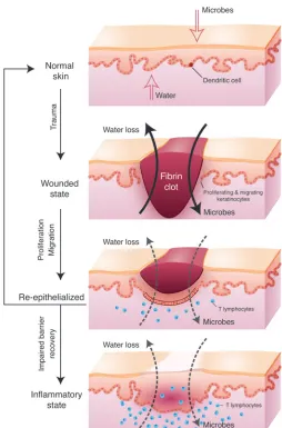
human psoriatic plaques in transgenic mice resulted in congeni-tal barrier deficiency (76). As described previously, the barrier is established in utero, maintained throughout life, and reestab-lished after trauma such as abrasion or wounding. Even when the epidermis has fully re-epithelialized, the ensuing scar at the site of trauma continues to exhibit mild barrier deficiency compared with uninvolved skin for as long as 1 year (77). Wounding is an initiating event in a significant percentage of psoriatic patients, which is clinically referred to as the isomorphic phenomenon. Interestingly, wounds on the mice with the increased gap junction-al communication re-epithelialized at a normal rate. However, the inability to restore the barrier arrested the wounds in the hyper-proliferative state and resulted in lymphocyte infiltration (76). These studies suggest that the signal to restore the homeostatic balance between proliferation and differentiation after re-epithe-lialization is restoration of the barrier and that in its absence the skin enters a pathologic state (Figure 2). Untangling some of these complex pathways will be a major area of future research, requiring the examination of tissue-specific genetic changes in both animal models and human patient samples.
Even if impaired barrier recovery contributes to psoriasis and AD, there must be additional factors that determine the type of immune response elicited since these disorders are characterized by distinct Th1/Th2 lymphocyte profiles. Beyond identifying the causative polymorphisms that under-lie the common susceptibility to both AD and psoriasis, we will need to understand the complexity of how epidermal and immune cells interact to elicit both the keratinocyte- and the T cell–specific manifestations of AD and psoriasis. One obvious possibility is that additional genetic or envi-ronmental risk factors modulate and specify the immune response. Much research focuses on the distinct Th1/Th2 lymphocyte infiltrations into psoriatic and AD skin.
The epidermal permeability barrier also has 2 distinct func-tions: retaining the water within the body and preventing the entry of dangerous substances. An animal model with an epi-dermal-specific targeted ablation of the adhesion molecule E-cadherin has a selective defect in water loss without any demonstrable defect in the entry of small molecules (41). These E-cadherin–deficient newborn mice have an increased rate of transepidermal water loss, but they demonstrate a normal ability to exclude the penetration of small molecules through their skin. As described above, many of these animal models of congenital barrier deficiencies die perinatally. Future studies of the immune response to a barrier deficiency evoked in adult skin will require either allogenic grafts or postnatally induced gene ablation.
Still, some hints about the type of immune stimulation that might result from the impaired barrier in these genetically distinct animal models can be gleaned from examining their neonatal skin tran-scriptional profile. For example, barrier-deficient Klf4–/– newborn
[image:7.585.52.306.78.463.2]mice already exhibit high levels of the mRNA encoding the cytokine thymic stromal lymphopoietin (Tslp), which activates dendritic cells and is highly upregulated in the epithelial cells of involved skin from patients with AD as well as in bronchial epithelium of asthmatics (78, 79). Recent experiments in animal models have shown that transgenic expression of Tslp in skin and lung epithelium is suf-ficient to induce AD and allergic airway inflammation, respectively (80, 81). Moreover, TSLP receptor knockout mice do not develop an inflammatory response in the lung to inhaled antigen (82). The high level of expression of TSLP in keratinocytes of AD patients and barrier-deficient mouse skin predicts that the keratinocytes signal to the immune cells to aid in combating the influx of invasive patho-gens. This model also predicts that a failure to repair the breach in
Figure 2
the barrier would continue to activate the local immune response. Consistent with the role of TSLP in immune cell activation is a pos-sible direct role for this cytokine in epidermal differentiation and barrier acquisition. Altogether, along with understanding the dis- tinct immune cell responses of psoriasis and AD, we need to investi-gate more specifically the nature of the epidermal barrier deficiency associated with these skin inflammatory disorders.
Future research directions: genomic analysis of the barrier breach stress response
The manifestation of a congenital ichthyosis disorder, transition-ing from the collodion membrane to the hyperkeratotic plaques, suggests that barrier-deficient skin adapts to the terrestrial envi-ronment. Molecular experiments discussed in this Review suggest that the skin senses the barrier loss and induces a stress response to repair this breach. The discovery of antibiotics and the use of spe-cialized emollients in the last 100 years have significantly altered our ability as a species to cope with a breach in barrier. However, one century is insignificant in evolutionary time. Our genomes have not adapted to the lower risk posed by a breach of barrier and continue to respond severely. We may be better served to damp-en the body’s inherent heightened stress response or to develop therapies selectively targeted against the strong stress response in order to improve quality of life. We may already be affecting barrier acquisition with standard treatment of inflammatory skin disorders that often includes the use of corticosteroids and emol-lients. As described above, corticosteroids not only act as immune suppressants but also accelerate epidermal barrier acquisition in utero (12, 13). However, corticosteroids elicit a complex response from keratinocytes and barrier function when administered short- term versus long-term and in high versus low doses (83). Emol-lients serve not only to provide a moist environment for wound healing, but also decrease the “sensing” of barrier perturbation. Either dampening the epidermal response to barrier disruption or utilizing drug therapies that enhance barrier recovery may amelio-rate some epithelial disorders that are more commonly perceived as inflammatory disorders.
Elucidation of the multiple ways in which a keratinocyte sig-nals a barrier disruption to other cell types and maintains a stress response is a daunting task. Although a keratinocyte’s initial response to a stressor may not occur at a transcriptional level, the changes necessary to maintain a stress response will be reflected in gene expression changes. Future experiments should define the expression profile changes that accompany a keratinocyte’s response to individual stresses that are often sensed simultane-ously, such as microbe infection, hypoxia, water loss, or oxidative stress. It will be equally instructive to monitor how the keratino-cyte responds when restored to normal homeostatic conditions and “turns off” the stress response. Existing algorithms can be utilized to determine both specific and common transcriptional responses elicited by stresses. Although this is an ambitious proj-ect, this type of model has been assembled to examine how the yeast Saccharomyces cerevisiae responds to changes in environment, food availability, and developmental state (84).
To gain a more comprehensive understanding of the stress response, investigators must elucidate further how these changes
are regulated at the transcription level. Toward this end, future experiments need to identify both the transcription factors and the DNA sequences to which they bind that regulate a stress response. Transcriptional profiling of keratinocytes deficient in a transcription factor essential to epidermal differentiation under stressed conditions may establish the hierarchy of some regulat-ed pathways. RNA interference, a novel technique that interferes with transcription of a specific RNA in mammalian cells, can be used to deplete keratinocytes of a putative regulatory protein and thus determine a specific protein’s necessary role in maintaining the stress response (85).
Although this is also a challenging project, novel technologies are being developed as part of the Human Genome Project’s goal to identify all functional elements in the human genome sequence (86). Analysis of the complete sequence of human, mouse, chimpanzee, and dog genomes has revealed that approximately 5% of our DNA is under strong positive selection (87). Approximately half of this conserved sequence encodes proteins or RNA genes. The function of the other half, the conserved noncoding sequences, is hypothesized to regulate the complex expression of nearby genes (86).
Multiple examples have been published demonstrating the role of conserved noncoding sequences resident in the proximal pro-moter or intron sequences of a gene that regulate its temporal or spatial expression. One intriguing example of this is the identifica-tion of a single nucleotide polymorphism in a runt-related transcrip-tion factor 1 (RUNX1 ) binding site that is associated with suscepti-bility to psoriasis (88). Some conserved noncoding sequences exert long-range effects on gene transcription, such as the locus control region regulating the developmentally regulated switching in the
b -globin gene complex (89). Another example of a conserved non-coding sequence is a long-range enhancer of the Sprr genes that has no activity during normal homeostatic growth of keratinocytes but serves as a strong enhancer when the stress response is activated to upregulate Sprr gene expression (90). It is intriguing to speculate that polymorphisms in the regulatory sequences that modulate the barrier recovery response may determine the susceptibility to AD and psoriasis. Perhaps a limited amount of the conserved non-coding sequence in our genomes regulates gene expression under homeostatic conditions, and a significant proportion encodes the complex stress response. Keratinocytes can be grown as monolayer or organotypic cultures or grafted as full-thickness skin onto mice, making this one of the most tractable systems to investigate com-plex gene regulation. With further analysis, we can gain insight into a multitude of common and rare human skin disorders.
Acknowledgments
This work is supported by the National Human Genome Research Institute Intramural Program. Darryl Leja prepared the illustra-tions. I am deeply indebted to David Nathan, Leonard Milstone, Jonathan Vogel, Mark Udey, and Cristina Strong for critical evalu-ation and suggestions.
Address correspondence to: Julie A. Segre, Building 49, Room 4A26, 49 Convent Drive, MSC 4442, Bethesda, Maryland 20892-4442, USA. Phone: (301) 402-2314; Fax: (301) 402-4929; E-mail: jsegre@nhgri.nih.gov.
1. Ito, M., et al. 2005. Stem cells in the hair follicle bulge contribute to wound repair but not to homeo-stasis of the epidermis. Nat. Med. 11:1351–1354. 2. Fuchs, E., and Raghavan, S. 2002. Getting under
the skin of epidermal morphogenesis. Nat. Rev. Genet. 3:199–209.
3. Niemann, C., and Watt, F.M. 2002. Designer skin: lineage commitment in postnatal epidermis. Trends
Cell Biol. 12:185–192.
review series
Epithelial barrier function: assembly and structur-al features of the cornified cell envelope. Bioessays.
24:789–800.
6. Nemes, Z., and Steinert, P.M. 1999. Bricks and mor-tar of the epidermal barrier. Exp. Mol. Med. 31:5–19.
7. Furuse, M., et al. 2002. Claudin-based tight junc- tions are crucial for the mammalian epidermal bar-rier: a lesson from claudin-1-deficient mice. J. Cell Biol. 156:1099–1111.
8. Steinert, P.M. 2000. The complexity and redundancy of epithelial barrier function. J. Cell Biol. 151:F5–F8. 9. Segre, J. 2003. Complex redundancy to build a
simple epidermal permeability barrier. Curr. Opin. Cell Biol. 15:776–782.
10. Morita, K., et al. 2002. Molecular architecture of tight junctions of periderm differs from that of the maculae occludentes of epidermis. J. Invest. Derma-tol. 118:1073–1079.
11. Kalia, Y.N., Nonato, L.B., Lund, C.H., and Guy, R.H. 1998. Development of skin barrier function in pre-mature infants. J. Invest. Dermatol. 111:320–326. 12. Aszterbaum, M., Feingold, K.R., Menon, G.K., and
Williams, M.L. 1993. Glucocorticoids accelerate fetal maturation of the epidermal permeability barrier in the rat. J. Clin. Invest. 91:2703–2708. 13. Hardman, M.J., Sisi, P., Banbury, D.N., and Byrne, C.
1998. Patterned acquisition of skin barrier function during development. Development. 125:1541–1552. 14. Hanley, K., et al. 1998. Glucocorticoid deficiency
delays stratum corneum maturation in the fetal mouse. J. Invest. Dermatol. 111:440–444.
15. Segre, J.A., Bauer, C., and Fuchs, E. 1999. Klf4 is a transcription factor required for establishing the barrier function of the skin. Nat. Genet. 22:356–360. 16. Jaubert, J., Cheng, J., and Segre, J.A. 2003. Ectopic
expression of kruppel like factor 4 (Klf4) acceler- ates formation of the epidermal permeability bar-rier. Development. 130:2767–2777.
17. Marshall, D., Hardman, M.J., Nield, K.M., and Byrne, C. 2001. Differentially expressed late con-stituents of the epidermal cornified envelope. Proc. Natl. Acad. Sci. U. S. A. 98:13031–13036.
18. Richard, G. 2004. Molecular genetics of the ich-thyoses. Am. J. Med. Genet. C Semin. Med. Genet.
131C:32–44.
19. Huber, M., et al. 1995. Mutations of keratinocyte transglutaminase in lamellar ichthyosis. Science.
267:525–528.
20. Russell, L.J., et al. 1995. Mutations in the gene for transglutaminase 1 in autosomal recessive lamellar ichthyosis. Nat. Genet. 9:279–283.
21. Lefevre, C., et al. 2003. Mutations in the transport-er ABCA12 are associated with lamellar ichthyosis type 2. Hum. Mol. Genet. 12:2369–2378.
22. Lefevre, C., et al. 2006. Mutations in a new cyto-chrome P450 gene in lamellar ichthyosis type 3.
Hum. Mol. Genet. 15:767–776.
23. Jobard, F., et al. 2002. Lipoxygenase-3 (ALOXE3) and 12(R)-lipoxygenase (ALOX12B) are mutated in non-bullous congenital ichthyosiform eryth-roderma (NCIE) linked to chromosome 17p13.1.
Hum. Mol. Genet. 11:107–113.
24. Lefevre, C., et al. 2004. Mutations in ichthyin a new gene on chromosome 5q33 in a new form of auto-somal recessive congenital ichthyosis. Hum. Mol. Genet. 13:2473–2482.
25. Williams, M.L., and Elias, P.M. 1985. Heterogene-ity in autosomal recessive ichthyosis. Clinical and biochemical differentiation of lamellar ichthyosis and nonbullous congenital ichthyosiform erythro-derma. Arch. Dermatol. 121:477–488.
26. Kelsell, D.P., et al. 2005. Mutations in ABCA12 underlie the severe congenital skin disease harle-quin ichthyosis. Am. J. Hum. Genet. 76:794–803.
27. Akiyama, M., et al. 2005. Mutations in lipid trans- porter ABCA12 in harlequin ichthyosis and func-tional recovery by corrective gene transfer. J. Clin. Invest. 115:1777–1784. doi:10.1172/JCI24834.
28. Chavanas, S., et al. 2000. Mutations in SPINK5, encoding a serine protease inhibitor, cause Neth-erton syndrome. Nat. Genet. 25:141–142. 29. Raghunath, M., et al. 2003. Self-healing collodion
baby: a dynamic phenotype explained by a particu-lar transglutaminase-1 mutation. J. Invest. Dermatol.
120:224–228.
30. Smith, F.J., et al. 2006. Loss-of-function mutations in the gene encoding filaggrin cause ichthyosis vul-garis. Nat. Genet. 38:337–342.
31. Matsuki, M., et al. 1998. Defective stratum corneum and early neonatal death in mice lacking the gene for transglutaminase 1 (keratinocyte transgluta-minase). Proc. Natl. Acad. Sci. U. S. A. 95:1044–1049.
32. Kuramoto, N., et al. 2002. Development of ichthyo-siform skin compensates for defective permeability barrier function in mice lacking transglutamin-ase 1. J. Clin. Invest. 109:243–250. doi:10.1172/ JCI200213563.
33. Koch, P.J., et al. 2000. Lessons from loricrin-defi-cient mice: compensatory mechanisms maintaining skin barrier function in the absence of a major cor-nified envelope protein. J. Cell Biol. 151:389–400.
34. Presland, R.B., et al. 2000. Loss of normal profilag-grin and filaggrin in flaky tail (ft/ft) mice: an animal model for the filaggrin-deficient skin disease ichthy-osis vulgaris. J. Invest. Dermatol. 115:1072–1081. 35. Wakabayashi, N., et al. 2003. Keap1-null mutation
leads to postnatal lethality due to constitutive Nrf2 activation. Nat. Genet. 35:238–245.
36. Takagi, S., et al. 2003. Alteration of the 4-sphin-genine scaffolds of ceramides in keratinocyte-specific Arnt-deficient mice affects skin bar-rier function. J. Clin. Invest. 112:1372–1382. doi:10.1172/JCI200318513.
37. Ting, S.B., et al. 2005. A homolog of Drosophila grainy head is essential for epidermal integrity in mice. Science. 308:411–413.
38. Indra, A.K., et al. 2005. TAF10 is required for the establishment of skin barrier function in foe-tal, but not in adult mouse epidermis. Dev. Biol.
285:28–37.
39. Leyvraz, C., et al. 2005. The epidermal barrier func-tion is dependent on the serine protease CAP1/ Prss8. J. Cell Biol. 170:487–496.
40. List, K., et al. 2002. Matriptase/MT-SP1 is required for postnatal survival, epidermal barrier function, hair follicle development, and thymic homeostasis.
Oncogene. 21:3765–3779.
41. Tunggal, J.A., et al. 2005. E-cadherin is essential for in vivo epidermal barrier function by regulating tight junctions. EMBO J. 24:1146–1156. 42. Moskowitz, D.G., et al. 2004. Pathophysiologic
basis for growth failure in children with ichthyo- sis: an evaluation of cutaneous ultrastructure, epi-dermal permeability barrier function, and energy expenditure. J. Pediatr. 145:82–92.
43. Esche, C., de Benedetto, A., and Beck, L.A. 2004. Keratinocytes in atopic dermatitis: inflammatory signals. Curr. Allergy Asthma Rep. 4:276–284.
44. Ghadially, R., Reed, J.T., and Elias, P.M. 1996. Stra-tum corneum structure and function correlates with phenotype in psoriasis. J. Invest. Dermatol.
107:558–564.
45. Leung, D.Y., Boguniewicz, M., Howell, M.D., Nomura, I., and Hamid, Q.A. 2004. New insights into atopic dermatitis. J. Clin. Invest. 113:651–657. doi:10.1172/JCI200421060.
46. Nickoloff, B.J., and Nestle, F.O. 2004. Recent insights into the immunopathogenesis of pso-riasis provide new therapeutic opportunities.
J. Clin. Invest. 113:1664–1675. doi:10.1172/ JCI200422147.
47. Johansson, S.G., et al. 2001. A revised nomencla-ture for allergy. An EAACI position statement from the EAACI nomenclature task force. Allergy.
56:813–824.
48. Sator, P.G., Schmidt, J.B., and Honigsmann, H.
2003. Comparison of epidermal hydration and skin surface lipids in healthy individuals and in patients with atopic dermatitis. J. Am. Acad. Derma-tol. 48:352–358.
49. Capon, F., et al. 2001. Fine mapping of the PSORS4 psoriasis susceptibility region on chromosome 1q21. J. Invest. Dermatol. 116:728–730.
50. Cookson, W.O., et al. 2001. Genetic linkage of childhood atopic dermatitis to psoriasis suscepti-bility loci. Nat. Genet. 27:372–373.
51. Palmer, C.N.A., et al. 2006. Common loss-of-fun- tion variants of the epidermal barrier protein fil-aggrin are a major predisposing factor for atopic dermatitis. Nat. Genet. doi:10.1038/ng1767.
52. Spergel, J.M., and Paller, A.S. 2003. Atopic derma-titis and the atopic march. J. Allergy Clin. Immunol.
112:S118–S127.
53. Spergel, J.M., et al. 1998. Epicutaneous sensitiza-tion with protein antigen induces localized allergic dermatitis and hyperresponsiveness to methacho-line after single exposure to aerosolized antigen in mice. J. Clin. Invest. 101:1614–1622.
54. Walley, A.J., et al. 2001. Gene polymorphism in Netherton and common atopic disease. Nat. Genet.
29:175–178.
55. Descargues, P., et al. 2005. Spink5-deficient mice mimic Netherton syndrome through degradation of desmoglein 1 by epidermal protease hyperactiv-ity. Nat. Genet. 37:56–65.
56. Cookson, W. 2004. The immunogenetics of asthma and eczema: a new focus on the epithelium. Nat. Rev. Immunol. 4:978–988.
57. Zenz, R., et al. 2005. Psoriasis-like skin disease and arthritis caused by inducible epidermal deletion of Jun proteins. Nature. 437:369–375.
58. Broome, A.M., Ryan, D., and Eckert, R.L. 2003. S100 protein subcellular localization during epi-dermal differentiation and psoriasis. J. Histochem. Cytochem. 51:675–685.
59. Sun, C., et al. 2006. Peptidoglycan recognition pro-teins Pglyrp3 and Pglyrp4 are encoded from the epidermal differentiation complex and are candi-date genes for the Psors4 locus on chromosome 1q21. Hum. Genet. 119:113–125.
60. Patel, S., Kartasova, T., and Segre, J.A. 2003. Mouse Sprr locus: a tandem array of coordinately regu-lated genes. Mamm. Genome. 14:140–148. 61. Zhou, X., et al. 2003. Novel mechanisms of T-cell
and dendritic cell activation revealed by profiling of psoriasis on the 63,100-element oligonucleotide array. Physiol. Genomics. 13:69–78.
62. Zimmermann, N., et al. 2005. Expression and regula- tion of small proline-rich protein 2 in allergic inflam-mation. Am. J. Respir. Cell Mol. Biol. 32:428–435. 63. Hooper, L.V., et al. 2001. Molecular analysis of
commensal host-microbial relationships in the intestine. Science. 291:881–884.
64. Nozaki, I., et al. 2005. Small proline-rich proteins 2 are noncoordinately upregulated by IL-6/STAT3 signaling after bile duct ligation. Lab. Invest.
85:109–123.
65. Pradervand, S., et al. 2004. Small proline-rich pro-tein 1A is a gp130 pathway- and stress-inducible cardioprotective protein. EMBO J. 23:4517–4525. 66. Ghadially, R., Brown, B.E., Sequeira-Martin, S.M.,
Feingold, K.R., and Elias, P.M. 1995. The aged epi-dermal permeability barrier. Structural, functional, and lipid biochemical abnormalities in humans and a senescent murine model. J. Clin. Invest.
95:2281–2290.
67. Nickoloff, B.J., and Naidu, Y. 1994. Perturbation of epidermal barrier function correlates with ini-tiation of cytokine cascade in human skin. J. Am. Acad. Dermatol. 30:535–546.