human hair follicle stem cells
George Cotsarelis
J Clin Invest.
2006;
116(1)
:19-22.
https://doi.org/10.1172/JCI27490
.
Hair follicle stem cells sustain growth and cycling of the hair follicle and are located in the
permanent portion of the follicle known as the bulge. In this issue of the
JCI
, Ohyama et al.
report the characterization of global gene expression patterns of human hair follicle stem
cells after their isolation using sophisticated laser capture techniques to microdissect out
bulge cells. They discovered a panel of cell surface markers useful for isolating living hair
follicle stem cells, a finding with potential therapeutic implications since isolated stem cells
in mice can generate new hair follicles when transplanted to other mice. The findings of
Ohyama et al. validate the use of the mouse for studying hair follicle biology but also
underscore critical differences between mouse and human stem cell markers. In particular,
CD34, which delineates hair follicle stem cells in the mouse, is not expressed by human
hair follicle stem cells, while CD200 is expressed by stem cells in both species. Ultimately,
this information will assist efforts to develop cell-based and cell-targeted treatments for skin
disease.
Commentary
Find the latest version:
Division of Cardiology, Department of Medicine, Weill Medical College of Cor-nell University, 525 East 68th Street, New York, New York 10021, USA. Phone: (212) 746-2201; Fax: (212) 746-2222; E-mail: ctbasson@med.cornell.edu.
1. Stauffer, B.L., Konhilas, J.P., Luczak, E.D., and Leinwand, L.A. 2006. Soy diet worsens heart disease in mice. J. Clin. Invest. 116:209–216.doi:10.1172/ JCI24676.
2. Maron, B.J., et al. 1995. Prevalence of hypertrophic cardiomyopathy in a general population of young adults. Echocardiographic analysis of 4111 subjects in the CARDIA Study. Coronary Artery Risk Devel-opment in (Young) Adults. Circulation. 92:785–789. 3. Davies, M.J., and McKenna, W.J. 1995. Hypertro-phic cardiomyopathy–pathology and pathogenesis.
Histopathology. 26:493–500.
4. Klues, H.G., Schiffers, A., and Maron, B.J. 1995. Phenotypic spectrum and patterns of left ventricu-lar hypertrophy in hypertrophic cardiomyopathy: morphologic observations and significance as assessed by two-dimensional echocardiography in 600 patients. J. Am. Coll. Cardiol. 26:1699–1708. 5. Maron, B.J. 2002. Hypertrophic cardiomyopathy: a
systematic review. J. Am. Med. Assoc. 287:1308–1320.
6. Maron, B.J., et al. 2003. American College of Car-diology/European Society of Cardiology clinical expert consensus document on hypertrophic car-diomyopathy. A report of the American College of Cardiology Foundation Task Force on Clinical Expert Consensus Documents and the European Society of Cardiology Committee for Practice Guidelines. J. Am. Coll. Cardiol. 42:1687–1713. 7. Spirito, P., Seidman, C.E., McKenna, W.J., and
Maron, B.J. 1997. The management of hypertro-phic cardiomyopathy. N. Engl. J. Med. 336:775–785. 8. Ahmad, F., Seidman, J.G., and Seidman, C.E. 2005. The genetic basis for cardiac remodeling. Annu. Rev. Genomics Hum. Genet. 6:185–216.
9. Horn-Ross, P.L., et al. 2001. Phytoestrogen con-sumption and breast cancer risk in a multiethnic population: the Bay Area Breast Cancer Study.
Am. J. Epidemiol. 154:434–441.
10. Sirtori, C.R., Arnoldi, A., and Johnson, S.K. 2005. Phy-toestrogens: end of a tale? Ann. Med. 37:423–438. 11. Cornwell, T., Cohick, W., and Raskin, I. 2004.
Dietary phytoestrogens and health. Phytochemistry.
65:995–1016.
12. Squadrito, F., et al. 2002. The effect of the phytoes- trogen genistein on plasma nitric oxide concentra- tions, endothelin-1 levels and endothelium depen-dent vasodilation in postmenopausal women.
Atherosclerosis. 163:339–347.
13. Vega-Lopez, S., et al. 2005. Plasma antioxidant capacity
in response to diets high in soy or animal protein with or without isoflavones. Am. J. Clin. Nutr. 81:43–49. 14. Dewell, A., Hollenbeck, C.B., and Bruce, B. 2002. The
effects of soy-derived phytoestrogens on serum lip- ids and lipoproteins in moderately hypercholester-olemic postmenopausal women. J. Clin. Endocrinol. Metab. 87:118–121.
15. Vikstrom, K.L., Factor, S.M., and Leinwand, L.A. 1996. Mice expressing mutant myosin heavy chains are a model for familial hypertrophic cardiomyopathy.
Mol. Med. 2:556–567.
16. Maass, A.H., Ikeda, K., Oberdorf-Maass, S., Maier, S.K., and Leinwand, L.A. 2004. Hypertrophy, fibro- sis, and sudden cardiac death in response to patho-logical stimuli in mice with mutations in cardiac troponin T. Circulation. 110:2102–2109.
17. Dubey, R.K., Gillespie, D.G., Jackson, E.K., and Keller, P.J. 1998. 17Beta-estradiol, its metabolites, and progesterone inhibit cardiac fibroblast growth.
Hypertension. 31:522–528.
18. Thigpen, J.E., et al. 1999. Phytoestrogen content of purified, open- and closed-formula laboratory animal diets. Lab. Anim. Sci. 49:530–536. 19. Iwasaki, K., et al. 1988. The influence of dietary
protein source on longevity and age-related disease processes of Fischer rats. J. Gerontol. 43:B5–B12. 20. Shimokawa, I., et al. 1993. Diet and the suitability
of the male Fischer 344 rat as a model for aging research. J. Gerontol. 48:B27–B32.
Gene expression profiling gets to the root of
human hair follicle stem cells
George Cotsarelis
University of Pennsylvania School of Medicine, Philadelphia, Pennsylvania, USA.
Hair follicle stem cells sustain growth and cycling of the hair follicle and
are located in the permanent portion of the follicle known as the bulge.
In this issue of the
JCI
, Ohyama et al. report the characterization of global
gene expression patterns of human hair follicle stem cells after their
iso-lation using sophisticated laser capture techniques to microdissect out
bulge cells (see the related article beginning on page 249). They
discov-ered a panel of cell surface markers useful for isolating living hair follicle
stem cells, a finding with potential therapeutic implications since isolated
stem cells in mice can generate new hair follicles when transplanted to
other mice. The findings of Ohyama et al. validate the use of the mouse
for studying hair follicle biology but also underscore critical differences
between mouse and human stem cell markers. In particular, CD34, which
delineates hair follicle stem cells in the mouse, is not expressed by human
hair follicle stem cells, while CD200 is expressed by stem cells in both
spe-cies. Ultimately, this information will assist efforts to develop cell-based
and cell-targeted treatments for skin disease.
Advances in stem cell biology have resulted in major clinical benefits. Bone marrow transplantation for treatment
of cancers and corneal transplantation for treatment of blindness resulting from chemical burns were both possible because stem cells were identified and isolated from the affected tissues (1, 2). Many disorders of the skin, such as can-cer, chronic wounds, skin atrophy and fragility, hirsutism, and alopecia, can be viewed as disorders of adult stem cells.
Because stem cells in the epidermis and
hair follicle serve as the ultimate source of cells for both of these tissues, under- standing the control of their prolifera- tion and differentiation is key to under-standing disorders related to disruption in these processes. Furthermore, the isolation, cultivation, and propagation of epithelial stem cells are important for tissue-engineering approaches to treating skin disorders (3).
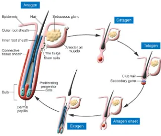
The skin possesses an outer covering produced by the epidermis that protects us from dehydration and from external environmental insults. The outer layer of the hair follicle is contiguous with the epidermis, forming invaginations that, in the case of scalp follicles, penetrate deeply into the fat underlying the skin. Approximately 5 million follicles that are spread over the body generate hair in a cyclical fashion. The duration of anagen (the period of hair growth), which varies from less than 60 to more than several thousand days on different body sites, determines the length of the hair shaft. After anagen, the follicle enters a stage of involution (catagen) and then a stage of
Nonstandard abbreviations used: K15, cytokeratin 15; LRC, label-retaining cell.
Conflict of interest: The author has declared that no conflict of interest exists.
rest (telogen), when the hair that was pro-duced is now dead yet remains anchored in the follicle until it is shed during exo-gen (Figure 1).
Eventually, stem cells at the base of the resting follicle, in an area known as the bulge, proliferate and regenerate a new lower follicle including the highly prolif-erative bulb cells that produce a new hair (Figure 1). After giving rise to the new hair-producing cells, the stem cells return to a quiescent state that is a hallmark of these cells. Despite being surrounded by highly proliferating epidermal and hair follicle cells, the cells in the bulge rarely undergo mitosis. The extended state of dormancy is in line with other stem-like characteristics of these cells, including a prolonged lifespan — probably as long as that of the organism. This characteristic is thought to predispose these cells to
tumor formation, since their long lifes-pan allows for accumulation of genetic mutations resulting from environmental insults such as UV irradiation or chemical carcinogens (4).
Mouse versus human hair follicles The majority of the previous work on hair follicle stem cells was performed on mouse or rat hair follicles (5–8). How-ever, major differences exist between rodent and human follicles, especially those on the scalp. Human scalp follicles are significantly larger, reaching lengths of 5 mm, compared with mouse pel-age follicles, which are only 1 mm long. Rodent vibrissa (whisker) follicles have also been extensively studied. These fol-licles are larger than pelage follicles but are highly unusual in their structure and cycling capabilities. They are surrounded
by cavernous blood-filled sinuses and are innervated by large nerves that track to the brain cortex and are organized into large “barrel fields,” each corresponding to one vibrissa follicle. Because the pri- mary purpose of vibrissa follicles is tac-tile sensation (known as whisking), they do not cycle in the same manner as other follicles. Rather than entering a true rest-ing phase, these follicles only briefly stop proliferating, without regressing, before generating a new hair shaft. The old shaft is retained in the follicle until the new shaft comes in beside it so that the follicle is never devoid of a long hair, and thus the animal maintains its sensory perception. This stands in stark contrast to human follicles, which undergo dramatic short- ening, losing up to 80% of their lower vol- ume during catagen, the stage of regres-sion (Figure 1), and also possess only 1 hair per follicle, while growing for years rather than weeks. Thus, the cellular and molecular characteristics of stem cells in the human follicle could be quite differ-ent from those in the rodent.
Identification of stem cells
Putative stem cells in mouse pelage (back skin) and vibrissa follicles were previ-ously identified as quiescent cells in vivo by label-retaining cell (LRC) studies (5). Bulge cells in the adult mouse were then shown, through genetic labeling studies, to generate all of the cell lineages within the follicle in vivo and after isolation, and thus to function as stem cells (4). By transplantation of human scalp tissue to immunodeficient mice, LRCs in human follicles were localized to the basal layer of the outer root sheath in the area of the bulge (9). Because the human bulge is not morphologically apparent, markers were needed for the study of this area. Cyto- keratin 15 (K15), an intracellular inter-mediate filament protein, was identified as one marker preferentially expressed by the human bulge (9). Ideally, however, cell surface proteins against which anti-bodies could be raised would be valuable for isolating the human bulge cells, test-ing their stem cell characteristics, and defining global gene expression patterns important for understanding control mechanisms for stem cell growth and proliferation.
[image:3.585.42.367.81.356.2]In this issue of the JCI, Ohyama et al. (10) report having defined the bulge area of the human hair follicle using a com-bination of proliferative characteristics, Figure 1
histological landmarks, and biochemical features. The location of quiescent LRCs in human scalp tissue grafted to immu-nodeficient mice was compared with the location of attachment of the arrector pili muscle (responsible for “goose bumps” upon contraction), which was identified using antibodies against smooth muscle. The LRCs were found at and above the site of muscle insertion up to the level of the sebaceous gland (Figure 1). The area containing the LRCs was also positive for K15, in contrast to the remainder of the outer root sheath, which is generally negative for this marker. K15 expression correlates with an immature phenotype within the skin, since it occurs in neona-tal but not adult interfollicular epidermal cells (11), and thus reflects the distinct biochemical makeup of the bulge cells compared with the rest of the epidermis and hair follicle.
Searching for stem cell genes
Defining gene expression patterns that control the quiescent, noncycling nature of hair follicle stem cells is a major goal
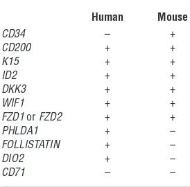
in epithelial stem cell biology, since it could lead to a better understanding of the abnormal proliferation that these cells undergo in the development of skin cancer (4). In addition, elucidation of genes involved in hair follicle stem cell proliferation and self-renewal may pro-vide insights into alopecia and abnor-mal wound healing, since bulge cells are important for hair growth and normal wound healing(12). In order to identify genes related to the stem cell phenotype, Ohyama et al. (10) compared microarray geneexpression profiles of bulge cells with those of other closely related cell populations within the hair follicle. Unlike in many other microarray studies in which heterogeneous mixtures of cell types were used to generate mRNA, the use of laser capture microdissection, in which individual cells or groups of cells are dissected out under a microscope from tissue sections on a glass slide and captured using a laser, permitted isola-tion of a specific population of mRNA highly enriched for messages found in stem cells. Ohyama et al. discovered the presence of many of the same differen-tially expressed genes in the human bulge cells that were previously described in mouse bulge cells (4, 8); however, several key differences between the mouse and the human data were noted (Table 1). In particular, CD34, which is an excellent bulge cell marker in the mouse (13), is not present on human bulge cells. Rath- er, CD34 is expressed in slightly more dif-ferentiated outer root sheath cells below the bulge in the human follicle; therefore, antibodies against CD34 cannot be used to isolate human hair follicle stem cells. To circumvent this problem, Ohyama et al. (10) identified a panel of bulge stem cell surface markers, including some previously identified in the mouse (e.g., CD200; ref. 4). Using a combination of antibodies against these markers as well as negative markers for nonbulge cells, human hair follicle stem cells were suc-cessfully isolated and then propagated in culture, a feat that is a prerequisite to using these cells for tissue-engineering purposes and that up to now has been technically impossible (3).
Outside influences
The stem cell microenvironment or niche in the bulge appears to possess potent signals for maintaining stem cells in an
undifferentiated state. In addition to epi-thelial stem cells, the bulge also contains other stem cell types, including melano-cyte stem cells (14). That the stem cell niche is a unique microenvironment is supported by several lines of evidence. Interestingly, after an injury to the bulge cells causes apoptosis, the bulge can be repopulated by cells that are slightly more differentiated and reside at the base of the follicle, called the secondary germ (15). Presumably this is a function of the microenvironment in an effort to maintain the integrity of the bulge area by actually inducing cells that are direct descendents of stem cells to revert to a stem cell phenotype. Furthermore, the power of the niche evidently may instruct bone marrow–derived dendritic APCs to remain relatively undifferentiated (16), and even influence mast cells to remain in a precursor state (17).
The findings reported by Ohyama et al. (10) suggest that CD200 is an impor-tant regulator of the immune-privileged state of the hair follicle. This is in line with a previous study demonstrating that CD200 inhibits autoimmune inflamma-tion in the hair follicle (18). This suggests that certain types of alopecia, such as lichen planopilaris, discoid lupus erythe- matosus, alopecia areata, and even andro- genetic alopecia, with which inflamma-tion appears to be associated, eventually could be treated with strategies aimed at augmenting CD200 expression.
The use of mouse models for study-ing human biology has been validated by these studies, with the caveat that findings from mouse and other animal models require confirmation in human systems. Because Ohyama et al.’s find-ings (10) are derived directly from human tissue, the significance of this line of research is greatly enhanced. By defining unique sets of cell surface markers for adult stem cells and other cell popula- tions within the human follicle, investi-gators can now readily extract and study very specific follicular cell types for their ability to regenerate hair and skin, with the hope of developing novel methods for treating skin and hair disease.
[image:4.585.53.190.134.269.2]Address correspondence to: George Cot-sarelis, University of Pennsylvania School of Medicine, M8 Stellar-Chance Labora-tories, 422 Curie Boulevard, Philadelphia, Pennsylvania 19104, USA. Phone: (610) 902-2400; Fax: (215) 573-9102; E-mail: cotsarel@mail.med.upenn.edu.
Table 1
Selected genes upregulated in bulge cells compared with nonbulge epi-thelial cells
Human Mouse
CD34 – +
CD200 + +
K15 + +
ID2 + +
DKK3 + +
WIF1 + +
FzD1 or FzD2 + +
PHLDA1 + –
FOLLISTATIN + –
DIO2 + –
CD71 – –
Human and mouse bulge cells preferen-tially express many of the same genes, but critical differences were also discov-ered, as reported in this issue of the JCI
by Ohyama et al. (10). In general, inhibi-tors of the WNT pathway, which is impor-tant for hair follicle cycling and differentia-tion, are increased. ID2, a gene previously associated with relatively undifferentiated cells, is upregulated in both species. Sur-prisingly, CD34, which is a specific bulge cell marker in the mouse, is lacking in
human follicle stem cells. FOLLISTATIN
and DIO2 are both in human but not
1. Bernstein, I.D., Andrews, R.G., and Rowley, S. 1994. Isolation of human hematopoietic stem cells. Blood Cells. 20:15–24.
2. Tsai, R.J., Li, L.M., and Chen, J.K. 2000. Recon-struction of damaged corneas by transplantation of autologous limbal epithelial cells. N. Engl. J. Med.
343:86–93.
3. Stenn, K.S., and Cotsarelis, G. 2005. Bioengineering the hair follicle: fringe benefits of stem cell technology.
Curr. Opin. Biotechnol. 16:493–497.
4. Morris, R.J. 2000. Keratinocyte stem cells: targets for cutaneous carcinogens. J. Clin. Invest. 106:3–8. 5. Cotsarelis, G., Sun, T.T., and Lavker, R.M. 1990.
Label-retaining cells reside in the bulge area of pilosebaceous unit: implications for follicular stem cells, hair cycle, and skin carcinogenesis. Cell.
61:1329–1337.
6. Morris, R.J., et al. 2004. Capturing and profiling adult hair follicle stem cells. Nat. Biotechnol. 22:411–417. 7. Oshima, H., Rochat, A., Kedzia, C., Kobayashi,
K., and Barrandon, Y. 2001. Morphogenesis and
renewal of hair follicles from adult multipotent stem cells. Cell. 104:233–245.
8. Tumbar, T., et al. 2004. Defining the epithelial stem cell niche in skin. Science. 303:359–363.
9. Lyle, S., et al. 1998. The C8/144B monoclonal anti-body recognizes cytokeratin 15 and defines the location of human hair follicle stem cells. J. Cell Sci.
111:3179–3188.
10. Ohyama, M., et al. 2006. Characterization and iso-lation of stem cell–enriched human hair follicle bulge cells. J. Clin. Invest. 116:249–260. doi:10.1172/ JCI26043.
11. Liu, Y., Lyle, S., Yang, Z., and Cotsarelis, G. 2003. Keratin 15 promoter targets putative epithelial stem cells in the hair follicle bulge. J. Invest. Dermatol.
121:963–968.
12. Ito, M., et al. 2005. Stem cells in the hair follicle bulge contribute to wound healing but not to homeostasis of the epidermis. Nat. Med. 11:1351–1354. 13. Trempus, C., et al. 2003. Enrichment for living
murine keratinocytes from the hair follicle bulge
with the cell surface marker CD34. J. Invest. Dermatol.
120:501–511.
14. Nishimura, E.K., et al. 2002. Dominant role of the niche in melanocyte stem-cell fate determination.
Nature. 416:854–860.
15. Ito, M., Kizawa, K., Toyoda, M., and Morohashi, M. 2002. Label-retaining cells in the bulge region are directed to cell death after plucking, followed by healing from the surviving hair germ. J. Invest. Dermatol. 119:1310–1316.
16. Gilliam, A.C., et al. 1998. The human hair follicle: a reservoir of CD40+ B7-deficient Langerhans cells that repopulate epidermis after UVB exposure.
J. Invest. Dermatol. 110:422–427.
17. Kumamoto, T., et al. 2003. Hair follicles serve as local reservoirs of skin mast cell precursors. Blood.
102:1654–1660.
18. Rosenblum, M.D., et al. 2004. Expression of CD200 on epithelial cells of the murine hair follicle: a role in tissue-specific immune tolerance? J. Invest. Der-matol. 123:880–887.
Delving deeper into MALT lymphoma biology
Francesco Bertoni and Emanuele Zucca
Laboratory of Experimental Oncology and Lymphoma Unit, Oncology Institute of Southern Switzerland, Bellinzona, Switzerland.
Mucosa-associated lymphoid tissue (MALT) lymphomas can arise in a
variety of extranodal sites. Interestingly, at least 3 different, apparently
site-specific, chromosomal translocations, all affecting the NF-κB
path-way, have been implicated in the development and progression of MALT
lymphoma. The most common is the translocation t(11;18)(q21;q21),
which results in a fusion of the
cIAP2
region on chromosome 11q21 with
the
MALT1
gene on chromosome 18q21 and is present in more than
one-third of cases. The frequency of this translocation is site-related:
com-mon in the gastrointestinal tract and lung, rare in conjunctiva and orbit,
and almost absent in salivary glands, thyroid, liver, and skin. In this issue
of the
JCI
, Hu et al. add to our understanding of the molecular
conse-quences of this translocation, showing that its fusion product,
cIAP2-MALT1, may concomitantly contribute to lymphomagenesis both as a
tumor suppressor gene and as an oncogene (see the related article
begin-ning on page 174).
MALT lymphoma: the clinical background
Extranodal marginal zone B cell lympho-ma (EMZL) of mucosa-associated lym-phoid tissue (MALT), also known as MALT lymphoma, is a neoplastic disease that constitutes approximately 8% of all non-Hodgkin lymphomas (1–4). It can arise in a variety of extranodal sites and occurs
most often in organs such as the stomach, the salivary glands, or the thyroid, where lymphocytes are normally absent. MALT is the result of chronic phlogistic events in response to either infectious condi-tions such as Helicobacter pylori –associ-ated chronic gastritis, or autoimmune disorders like Hashimoto thyroiditis and myoepithelial sialadenitis. In these condi- tions abnormal B cell clones can progres-sively replace the normal B cell population of the inflammatory tissue, giving rise to the EMZL. Over 10 years ago, H. pylori was identified as an etiologic factor in gastric MALT lymphomas after the demonstra-tion of tumor regression in the majority of early-stage cases treated with anti- Heli-cobacter antibiotic therapy, and this tumor therefore became a popular model of the
close pathogenetic link between chronic inflammation and lymphoma develop-ment. Other bacterial infections were later possibly implicated in the pathogenesis of MALT lymphomas arising in the skin (Borrelia burgdorferi), in the ocular adnexa (Chlamydia psittaci ), and in the small intes-tine (Campylobacter jejuni) (1, 4).
The prognosis for patients with MALT lymphomas is good; these tumors usually have an indolent course with overall 5-year survival rates greater than 80%, but rare cases with histologic transformation in cases of aggressive diffuse large-cell lym- phoma have been described. It is nowa-days generally accepted that eradication of H. pylori with antibiotics should be used as the sole initial treatment of localized gastric MALT lymphoma, while the use of anti-infectious treatment in nongastric locations is still under investigation. Other effective treatment approaches include radiotherapy, chemotherapy, and anti-CD20 mAbs (2, 3).
Many chromosomal translocations affecting the same pathway
Four main recurrent chromosomal trans-locations have been associated with the pathogenesis of EMZLs: t(11;18)(q21;q21), t(1;14)(p22;q32), t(14;18)(q32;q21), and t(3;14)(p14.1;q32) (5–8) (Table 1). The lat- ter is the most recently described and estab-lishes the juxtaposition of the transcription factor FOXP1 next to the enhancer region
Nonstandard abbreviations used: BIR, baculoviral IAP repeat; CARD, caspase recruitment domain; cIAP2, cellular inhibitor of apoptosis protein 2; DD, death domain; EMZL, extranodal marginal zone B cell lym-phoma; IAP, inhibitor of apoptosis protein; Iκ B, inhibi-tory κB; IKK, IκB kinase; MALT, mucosa-associated lymphoid tissue; NES, nuclear export signal.
Conflict of interest: The authors have declared that no conflict of interest exists.