HIV-1 kills renal tubular epithelial cells in vitro
by triggering an apoptotic pathway involving
caspase activation and Fas upregulation.
P G Conaldi, … , G Camussi, A Toniolo
J Clin Invest.
1998;
102(12)
:2041-2049.
https://doi.org/10.1172/JCI3480
.
HIV-infected patients suffer several renal syndromes, which can progress rapidly from renal
insufficiency to end-stage renal disease. Histologically, HIV-induced nephropathy is
characterized by prominent tubulopathy with apoptosis of tubular cells. Clinical and
experimental evidence suggests that renal injury may be directly related to virus infection.
Although HIV-1 is a polytropic and not solely lymphotropic pathogen, the susceptibility of
renal cells to HIV-1 remains to be determined. This paper demonstrates in vitro the
permissiveness of proximal tubular epithelial cells (PTEC) to HIV-1 and describes the
effects of PTEC infection to explain the pathogenesis of tubular damage in vivo. The results
indicate that PTEC express HIV-specific receptor and coreceptors and sustain virus
replication. We observed that HIV-1 infection causes the death of tubular cells by triggering
an apoptotic pathway involving caspase activation. Fas upregulation but not Fas ligand
expression was found in the infected PTEC. However, after HIV-1 infection, tubular cells
became susceptible to apoptosis induced through Fas stimulation. Caspase inhibition
prevented the death of the infected PTEC in spite of persistent viral replication. These
findings may explain the prominent histopathology of HIV-associated nephropathy and
demonstrate that the apoptosis of nonlymphoid cells can be directly induced by HIV-1.
Research Article
Find the latest version:
J. Clin. Invest.
© The American Society for Clinical Investigation, Inc. 0021-9738/98/12/2041/09 $2.00
Volume 102, Number 12, December 1998, 2041–2049 http://www.jci.org
HIV-1 Kills Renal Tubular Epithelial Cells In Vitro by Triggering an Apoptotic
Pathway Involving Caspase Activation and Fas Upregulation
Pier Giulio Conaldi,* Luigi Biancone,‡ Antonella Bottelli,*§ Alison Wade-Evans,iLorraine C. Racusen,¶
Mariarosaria Boccellino,‡ Viviana Orlandi,* Caterina Serra,** Giovanni Camussi,‡ and Antonio Toniolo*
*Division of Microbiology and ‡Division of Nephrology, Department of Clinical and Biological Sciences, University of Insubria, Varese 21100, Italy; §Institute of Forensic Medicine, University of Brescia, Brescia 25123, Italy; iDivision of Retrovirology, National Institute for
Biological Standards and Control, South Mimms, Potters Bar, Herts, United Kingdom EN6 3QG; ¶Department of Pathology, The Johns Hopkins University School of Medicine, Baltimore, Maryland 21205; and **Institute of Microbiology and Virology, University of Sassari, Sassari 07100, Italy
Abstract
HIV-infected patients suffer several renal syndromes, which
can progress rapidly from renal insufficiency to end-stage
renal disease. Histologically, HIV-induced nephropathy is
characterized by prominent tubulopathy with apoptosis of
tubular cells. Clinical and experimental evidence suggests
that renal injury may be directly related to virus infection.
Although HIV-1 is a polytropic and not solely lymphotropic
pathogen, the susceptibility of renal cells to HIV-1 remains
to be determined. This paper demonstrates in vitro the
per-missiveness of proximal tubular epithelial cells (PTEC) to
HIV-1 and describes the effects of PTEC infection to
ex-plain the pathogenesis of tubular damage in vivo. The
re-sults indicate that PTEC express HIV-specific receptor and
coreceptors and sustain virus replication. We observed that
HIV-1 infection causes the death of tubular cells by
trigger-ing an apoptotic pathway involvtrigger-ing caspase activation. Fas
upregulation but not Fas ligand expression was found in the
infected PTEC. However, after HIV-1 infection, tubular
cells became susceptible to apoptosis induced through Fas
stimulation. Caspase inhibition prevented the death of the
infected PTEC in spite of persistent viral replication. These
findings may explain the prominent histopathology of
HIV-associated nephropathy and demonstrate that the apoptosis
of nonlymphoid cells can be directly induced by HIV-1. (
J.
Clin. Invest.
1998. 102:2041–2049.) Key words:
HIV-associ-ated nephropathy
•tropism
•apoptosis
•caspases
•Fas
sen-sitization
Introduction
HIV-1 is a polytropic pathogen that, in addition to cells of the lymphoid organs, may infect cells of the gastrointestinal, vas-cular, and nervous systems (1–4). AIDS is characterized by cell loss in several organs and tissues, but the mechanisms of HIV-induced cytopathicity are still unclear (1, 5). The infected
pa-tients may suffer a spectrum of renal damages (6). HIV-associ-ated nephropathy (HIVAN)1 is a unique syndrome that affects
up to 10% of the seropositive individuals and is character-ized by glomerulosclerosis accompanied by prominent tubulo-interstitial injury. Clinically, patients with HIVAN have neph-rotic-range proteinuria with renal insufficiency, which does often rapidly progress to end-stage renal disease (6, 7). Acute renal failure secondary to tubular injury is also frequent in AIDS (6). In cases of HIVAN, viral antigens and genome have been demonstrated in microdissected renal tissues (8), but the permissiveness of renal cells to replication of HIV-1 is still con-troversial (9–14). The finding of apoptotic cells in glomeruli and especially in tubuli of HIV patients may suggest a possible mechanism of HIV-induced renal damage (15). This hypothe-sis is supported by the fact that renal tubular apoptohypothe-sis occurs in transgenic mice expressing HIV genes (16). It is well estab-lished that apoptosis plays a relevant role in the pathogenesis of AIDS by contributing to CD41 T cell depletion (17, 18). The enhanced susceptibility to apoptosis of T cells from HIV-positive individuals appears secondary to upregulation of Fas signaling or downregulation of Bcl-2 (19–21). Role and mecha-nisms of apoptosis in HIV-induced damage of nonlymphoid tissues are largely unknown.
The aim of this study was to investigate in vitro the permis-siveness of human proximal tubular epithelial cells (PTEC) to HIV-1 productive infection and characterize the cytopathic ef-fects (CPEs) of the infection, in order to evaluate the direct role of HIV-1 in the pathogenesis of tubular injury occurring in vivo. Our results demonstrate that PTEC express HIV-1 re-ceptors and are permissive to virus replication, and indicate that HIV-1 infection causes apoptosis of tubular cells through activation of caspase proteases.
Methods
Reagents.Reagents were obtained from the following sources: mAb against human Fas (IgM, clone CH-11, apoptosis inducing; Upstate Biotechnology, Lake Placid, NY); anti–human Fas mAb (IgG, clone ZB4, neutralizing; Upstate Biotechnology); anti–human Fas mAb (IgG, clone DX2; PharMingen, San Diego, CA); anti–human Fas ligand (FasL) mAb (clone NOK-1; PharMingen); anti-FasL rabbit IgG (N-20 and C-20; Santa Cruz Biotechnology, Santa Cruz, CA); FITC-conjugated goat anti–mouse IgG (Sigma Chemical Co., St. Louis, MO); anti-CD4 mAb (Sigma); FITC-conjugated anti-CD4 mAb (Becton Dickinson, San Jose, CA); anti-CXCR4 mAb (clone Address correspondence to Pier Giulio Conaldi, M.D., Ph.D.,
De-partment of Clinical and Biological Sciences, University of Insubria, Viale L. Borri 57, 21100 Varese, Italy. Phone: 39-332-278-311; FAX: 39-332-260-017; E-mail: pgiulio@working.it
Received for publication 18 March 1998 and accepted in revised form 21 October 1998.
12G5; kindly provided by Dr. J. Hoxie, University of Pennsylvania, Philadelphia, PA [22]); phycoerythrin (PE)-labeled anti-CXCR4 mAb (clone 12G5; PharMingen); anti-CCR3 mAb (clone 7B11, ob-tained from NIBSC AIDS Reagent Project, ARP3099); anti-CCR5 mAb (clone 2D7; obtained from NIBSC AIDS Reagent Project, ARP3099); PE-labeled anti-CCR5 mAb (clone 2D7; PharMingen); anti–caspase-3 rabbit IgG (PharMingen); caspase-3 inhibitor (Ac-DEVD-CHO peptide; PharMingen); IL-1b–converting enzyme (ICE) inhibitor (z-VAD-CH2F peptide; Kamiya Biomedical, Seattle, WA);
caspase inhibitor negative control (z-FA-CH2F peptide; Kamiya
Bio-medical); and HIV-1 recombinant Tat, gp120, and gp41 (Intracel, Lon-don, UK); p24 antigen detection ELISA (DuPont, Cologno Monzese, Italy). Unless otherwise specified, chemicals and tissue culture re-agents were from Sigma and molecular biology rere-agents were from Perkin-Elmer (Monza, Italy).
Renal cell cultures. Primary cultures of mesangial cells (MC) and PTEC were obtained from surgically removed kidneys of six different Caucasian patients, as described (23). The purity of the primary cul-tures was assessed on the basis of cell characterization, according to published criteria (24, 25). In brief, MC cultures grew as stellate cells in interwoven bundles and were positive for F-actin, detected with FITC-labeled phalloidin, and for smooth muscle–type myosin (Im-munotech, Marseille, France). MC were negative for HLA-DR and leukocyte common antigen (CD45; Becton Dickinson), and for factor VIII–related antigen (FVIIIr:Ag; Nordic Immunology, Tilburg, The Netherlands). The ability of MC to contract after stimulation with an-giotensin II and to phagocytize IgG-coated latex beads was analyzed as described (23). PTEC showed negative staining for FVIIIr:Ag, minimal staining for desmin, and marked staining with antibodies to cytokeratins and actin. PTEC were positive for alkaline phosphatase (determined by the naphtol AS-MX method) and showed enhanced cAMP production (Biotrak cAMP assay system; Amersham Interna-tional, Buckinghamshire, UK) after stimulation with 100 nM parathy-roid hormone, but not with 1 mM antidiuretic hormone.
Differentiated immortalized MC were obtained by infection of a pure primary culture with a hybrid Adeno5/SV40 virus (3, 26). Indi-vidual foci of immortalized cells were picked up, subcultured, and characterized according to phenotype. A differentiated MC line was selected and used between passages 20 and 35. An immortalized cell line of human PTEC, which has been characterized previously (25), was used (clone 5) to confirm and extend the experiments carried out with primary PTEC.
HIV infection. HIV-1P1 is a syncytium-inducing T-tropic virus
strain adapted to growth in T lymphoblastoid C8166 cells (3, 26). Vi-rus stocks were prepared from clarified supernatants and titrated as syncytium-forming units (SFU) per milliliter. Cultures of primary and immortalized MC and PTEC were infected with HIV-1 (moi of 0.1) or mock-infected with control supernatant, at 48 h after plating. The inoculum was allowed to adsorb for 2 h at 378C; monolayers were washed three times and refed with fresh medium. A 50% medium change was performed every 4 d. HIV-1 infection was monitored by p24 antigen and infectivity testing in culture supernatants, and by de-tection of proviral DNA or viral RNA in infected cultures. Cell via-bility of the infected cells was evaluated by trypan blue exclusion test. Purified HIV-1P1 was obtained by clarification (3,000 g for 15 min at
48C) and precipitation of the virus (10% polyethylene glycol-8000 in PBS for 18 h at 48C). The pelleted virus was layered on a double su-crose cushion (1:2 of 60% susu-crose and 20% susu-crose) and centrifuged at 27,000 rpm for 120 min at 48C in a SW27 rotor (Beckman, Fuller-ton, CA). Banded virus was collected and frozen at 2708C. Thawed virus was diluted in medium and titrated as SFU/ml in C8166 cells (26).
PCR. Detection of HIV-1 long terminal repeat (LTR) and gag
sequences was carried out as described (3, 26). PCR analysis of LTR sequences was carried out by amplification of 0.5 mg of DNA of in-fected PTEC with primers SK28/29 (Research Genetics, Huntsville, AL). gag-specific sequence was amplified with primers SK100/104 (Research Genetics) from cDNA obtained by RT of 1 mg of total
RNA treated with RNase-free DNase (Promega, Madison, WI). De-tection of CD4 transcripts was performed by RT-PCR of a 438-bp fragment, as reported (26). cDNA was synthesized using a CD4-spe-cific 39 primer and subsequently amplified with 59 and 39 CD4 primers (Clontech, Palo Alto, CA) according to the manufacturer’s instruc-tions. RT-PCR analysis of CXCR4 transcripts in immortalized MC and PTEC was carried out using mRNA extracted from 107 cells with
PolyATract GTC™ extraction buffer (Promega) and treated with RNase-free DNase. cDNA was synthesized using oligo d(T) primers and AMV-RT (Promega), again according to the manufacturer’s in-structions. PCR was performed on the cDNA using CXCR4-specific primers taken from the published human sequence (27; accession number M99293), which amplify a fragment of 1,078 bp. The program used for PCR was 948C for 4 min, followed by 40 cycles of 948C for 1 min, 628C for 1 min, and 728C for 1 min, with a final step of 728C for 20 min. Expression of FasL mRNA in uninfected or infected PTEC was evaluated by RT-PCR, as described (28; sense primer: 59 -CTGGGGATGTTTCAGCTCTTC-39; antisense primer: 59 -CTT-CACTCCAGAAAGCAGGAC-39). cDNAs were synthesized by ex-tension of antisense primer in a reaction mixture containing 2 mg of total RNA digested by RNase-free DNase. The amplification was carried out by 35 cycles of 948C for 30 s, 558C for 1 min, and 728C for 1.5 min. b-actin primers were used to check the integrity of all RNA samples. One-fifth of each reaction mixture was analyzed by electro-phoresis through agarose gels and visualized by staining with ethid-ium bromide. The DNA size markers used were either the DNA marker V from Boehringer Mannheim (Mannheim, Germany) or the low-mass DNA ladder from GIBCO BRL (Life Technologies A.G., Basel, Switzerland).
Expression of membrane molecules. CD4 expression in primary PTEC and MC was tested by indirect immunofluorescence assay. Iso-lated and suspended renal cells were incubated with anti-CD4 mAb (1 mg/106 cells) for 30 min at room temperature. After washings, the
cells were stained with FITC-labeled goat anti–mouse IgG for 30 min at room temperature, and examined with an epifluorescence Olym-pus BX60 microscope (OlymOlym-pus Optical Co., Tokyo, Japan). Flow cytometry of CD4 expression was carried out by incubating 106 renal
cells with 1 mg of FITC-conjugated specific antibody in PBS/0.1% so-dium azide for 30 min at room temperature. After washings, cell sus-pensions were fixed in 0.2% paraformaldehyde and analyzed with a flow cytometer (FACScan®; Becton Dickinson). Surface expression
of CXCR-4 was determined by incubating immortalized PTEC or MC (3 3 106) with 2 mg of 12G5 mAb for 15 min at room temperature
in 100 ml of PBS 1 1% BSA. The same procedure was used to evalu-ate CCR3 and CCR5 expression using 7B11 and 2D7 mAbs, respec-tively. The cells were washed twice with PBS and incubated with FITC-labeled goat anti–mouse IgG for 15 min at room temperature. After two washings with PBS, the cells were analyzed (FACScan®).
To assess coexpression of CD4 and chemokine receptors, immortal-ized PTEC were stained with FITC-labeled CD4 mAb and anti-bodies specific for CXCR-4 and CCR5 directly conjugated with PE. For analysis of surface Fas expression, 106 mock-infected and
HIV-infected PTEC were incubated with 0.5 mg of anti–human Fas IgG (DX2 mAb) in PBS/0.1% sodium azide for 60 min at 48C. Then, the cells were washed and incubated with FITC-conjugated goat anti– mouse IgG for 30 min at 48C. After washings, cell suspensions were fixed in 0.2% paraformaldehyde and flow cytometric analysis was performed. The same procedure was used to detect membrane-bound FasL by using a specific mAb (NOK-1) at the concentration of 1 mg/ 106 cells (29). In all experiments, isotype-matched irrelevant
antibod-ies were included as control.
Apoptosis measurement. PTEC were incubated with propidium iodide (PI) followed by flow cytometry analysis, as described (30). In brief, 106 cells were incubated for 4 h at 48C in 2 ml hypotonic
solu-tion containing 50 mg/ml PI, 0.1% sodium citrate, 0.1% Triton X-100, and 20 mg/ml DNase-free RNase A. Cells with subdiploid DNA con-tent (sub-G0/G1 peak) were considered apoptotic cells.
(31). Cells were fixed with 2% formaldehyde in PBS for 10 min at 48C, treated with PI solution for 20 min at room temperature, and then examined with an epifluorescence Olympus BX60 microscope. The apoptotic state of the cells was evaluated by differential uptake of acridine orange and ethidium bromide (31). Cells (5 3 104 in 25 ml
of PBS) were stained with 1 ml of a mixture of 100 mg/ml acridine or-ange and 100 mg/ml ethidium bromide (1:1). Observed through wide-band FITC filters, cells in the early stages of apoptosis are character-ized by condensed or fragmented bright green chromatin, and late apoptotic cells by condensed or fragmented bright orange chromatin.
Cell viability assay. PTEC were seeded in 24-well microtiter plates at 5 3 104 cells/well. The cultures were infected or mock-infected as
described above. After incubation under different conditions, dried monolayers were treated with 0.75% crystal violet in a solution of 50% ethanol, 0.25% NaCl, and 1.75% formaldehyde (32). After washing, the dye was eluted with 1% SDS in PBS and the absorbance was read at 595 nm with an ELISA reader. Cell viability was ex-pressed as percentage of uninfected controls. Assays were also per-formed with PTEC cultures treated with Tat (1 mg/ml), gp120 (1 mg/ ml), or gp41 (1 mg/ml) to evaluate the effect of these viral molecules on PTEC viability. For apoptosis induction experiments (33), anti-Fas agonistic IgM (100 ng/ml) or supernatants of COS cells producing human FasL (4 d after transfection) and mock-transfected COS cells were added to PTEC at day 2 post-infection (p.i.). For apoptosis inhi-bition experiments, infected PTEC were treated with anti-Fas neu-tralizing IgG (1 mg/ml) at days 2 and 4 p.i., or with caspase inhibitors (caspase-3 inhibitor: 100 mM; ICE protease blocking peptide: 0.1–100
mM; caspase inhibitor negative control: 100 mM) at days 1 and 4 p.i.
Western blot analysis. To identify the expression of FasL, tran-sient expression of cDNA encoding full-length human FasL, kindly provided by Dr. H. Yagita (Juntendo University, Tokyo, Japan [29]), was performed in COS cells transfected by electroporation at 250 V/960
mF (Electroporator II; Invitrogen, Carlsbad, CA). The cells were lysed 5–7 d after transfection at 48C for 1 h in a lysis buffer (50 mM Tris-HCl, pH 8.3, containing 1% Triton X-100, 10 mM PMSF, 10 mg/ ml leupeptin, and 100 U/ml aprotinin). Similarly, cell pellets were ob-tained from uninfected or HIV-infected PTEC cultures and then lysed. After centrifugation of the lysates at 15,000 g, the supernatants were quantitated for protein content by the Bradford method. Aliquots containing 100 mg of protein per lane were subjected to SDS/10%
PAGE under nonreducing or reducing (5% 2-mercaptoethanol) con-ditions and electroblotted onto nitrocellulose membrane filters. The blots were blocked with 5% nonfat milk in 20 mM Tris-HCl, pH 7.5, 500 mM NaCl plus 0.1% Tween (TBS-T). The membranes were sub-sequently incubated overnight at 48C with polyclonal rabbit antibody against amino terminus (N-20) or carboxyl terminus (C-20) of FasL, at a concentration of 500 ng/ml. After extensive washing with TBS-T, the blots were incubated for 1 h at room temperature with peroxi-dase-conjugated protein A (200 ng/ml; Amersham), washed with TBS-T, developed with ECL detection reagents (Amersham) for 1 min and exposed to X-Omat film (Eastman Kodak Co., Rochester, NY). To reveal caspase-3 activation, aliquots of the lysates obtained from uninfected or infected PTEC were subjected to SDS-PAGE and electroblotting, as described. Western blot analysis of proenzyme and cleavaged subunits was performed by using a 1:1,000 dilution of a polyclonal rabbit anti–caspase-3 antibody and revealed by ECL, as reported above.
Results
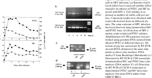
HIV-1 induces productive infection of renal tubular cells. Primary cultures of PTEC and MC were infected with HIV-1P1, a
syn-cytium-inducing virus strain adapted to T cell lines but able to infect nonlymphoid cells (3, 26). HIV-1 replication in PTEC was demonstrated by measurement of p24 content in culture supernatants (Fig. 1 a) and by detection of both proviral DNA and viral genomic RNA in the infected cells (Fig. 1 b). The es-tablishment of HIV-1 productive infection was confirmed by titration of infectious viral progeny released by PTEC cultures (mean titer of 102.5 SFU/ml at peak level). Active replication
of HIV-1P1 was also detected in MC; kinetics and grade of
vi-rus production were similar to those found in PTEC (Fig. 1 a). However, the effect of HIV-1 infection was different in the two cell types. Whereas viral replication in MC caused only minor CPEs, HIV-infected PTEC showed, in contrast, a pro-gressive reduction of cell viability and underwent death within 7–14 d p.i. (Fig. 1 a). The evidence achieved with primary
[image:4.612.65.554.485.737.2]tures of MC and PTEC was confirmed by infecting renal cell lines that retained phenotypic and functional characteristics associated with differentiated glomerular mesangial and tubu-lar epithelial cells, respectively (23, 25).
RT-PCR analysis of primary cultures revealed that both PTEC and MC can synthesize mRNAs specific for CD4, the major HIV receptor, and for CXCR-4, a chemokine receptor identified as the main fusion cofactor of T lymphotropic HIV-1 strains (34, 35). This evidence was confirmed using immortal-ized cell lines of PTEC and MC to avoid contamination of nonrenal cells (Fig. 1, c and d). By immunofluorescence tests, we found that 15–25% of suspended cells from primary PTEC and MC cultures showed positive staining for CD4. Flow cy-tometry confirmed the presence of HIV receptors on the sur-face of tubular cells and MC. Figs. 2 and 3 show FACScan®
analysis of immortalized PTEC, that were 55% (611) positive for CD4 and 15% (62) positive for CXCR-4. MC resulted as positive 45% (615) and 14% (63), respectively. It must be noted that lower levels of CCR3 (662%) and CCR5 (764%) chemokine receptors were detected on PTEC surface (Fig. 3). By two-color staining technique, we found that 8% (64) of PTEC are double positive for CD4 and CXCR-4, whereas 5% (63) of these cells coexpress CD4 and CCR5.
HIV-1 infection induces apoptosis of renal tubular cells. Fea-tures of the HIV-infected PTEC observed by light micros-copy were consistent with changes characteristic of apoptosis: rounding and detachment of the cells with fragmentation and shrinking of nuclei. To determine whether HIV-1 does induce apoptosis of renal tubular cells, PTEC were infected with HIV-1P1, harvested at different time points p.i., and examined
for DNA content in comparison with mock-infected cells. Cytofluorometric analysis revealed the presence of hypodip-loid cells from days 4–5 p.i. in the infected cultures (Fig. 4). By day 10 p.i., the percentage of apoptotic cells was 60–80% and by 14 d p.i. all the cells of infected PTEC cultures underwent detachment and death. Evidence of CPE was not found in mock-infected PTEC cultures. Apoptosis was confirmed by di-rect observation of chromatin condensation and nuclear
frag-mentation with PI staining (Fig. 5 a). Time course studies of the differential uptake of fluorescent DNA binding dyes re-vealed the presence of early apoptotic cells from day 4 p.i. and showed the progressive increase of the apoptotic changes in parallel with the lasting of PTEC infection (Fig. 5 b). The acti-vation of caspases is a critical event in the proteolytic cascade elicited by apoptotic stimuli (36). Indeed, by Western blot analysis of HIV-infected PTEC, we detected activated cas-pase-3 (Fig. 6), an enzyme implicated in the effector phase of apoptosis of different cell types (37). The exposure of tubular cells to gp120, gp41, and Tat (viral components that are known to induce apoptosis in other experimental models [30, 38, 39]) failed to impair the viability of PTEC cultures, whereas tubu-lar cells underwent apoptosis when the infection was per-formed with purified HIV-1P1 as the inoculum (data not
shown). These results indicate that apoptosis is a direct effect of HIV-1 productive infection of renal tubular epithelial cells.
[image:5.612.319.504.56.394.2]HIV-1 infection induces overexpression of Fas and sensitiza-tion to Fas signaling in renal tubular cells.Several members of the TNF receptor superfamily regulate cell survival (40). Re-cently, it has been reported that human tubular epithelial cells express Fas, a 43-kD member of TNF receptor family (41, 42). In this study we found that 70–80% of the cells of PTEC line
[image:5.612.56.298.502.678.2]Figure 2. Cytofluorometric analysis of membrane CD4 in renal tubu-lar epithelial cells. Immortalized PTEC were stained with FITC-con-jugated anti-CD4 mAb (shaded histogram) as reported in Methods. Negative control (empty histogram) was PTEC incubated with FITC-conjugated isotype-matched irrelevant mAb.
Figure 3. Cytofluorometric analysis of chemokine receptors in renal tubular epithelial cells by indirect immunofluorescence. (A) FAC-Scan® profile of immortalized PTEC incubated with isotype-matched
exhibited a positive staining for Fas, but HIV-1 infection in-duced in the tubular cells a prominent upregulation of Fas from days 2–3 p.i. (Fig. 7 a). On the contrary, no evidence of membrane FasL (43) was found in HIV-infected PTEC with FACScan® and Western blot analysis (Fig. 7 b), and neither
expression of FasL mRNA was detected by RT-PCR in tubu-lar cells after infection (not shown). The treatment of PTEC cultures with anti-Fas blocking antibody did not cause a signif-icant reduction of HIV-induced CPE. In fact, the mortality of HIV-infected PTEC at day 10 p.i. was 70615% in the case of
[image:6.612.59.403.57.297.2]untreated cultures, and 60612% (P5 ns) in cultures of tubu-lar cells added with anti-Fas mAb. According to a recent re-port (42), uninfected PTEC were resistant to Fas stimulation, as determined by measurement of cell viability after treatment with specific IgM-inducing Fas ligation (Fig. 8). In contrast, the addition of the anti-Fas agonistic antibody to the infected PTEC cultures at day 2 p.i. did relevantly increase and precipi-tate apoptotic changes of tubular cells, in comparison with the spontaneous kinetics of HIV-induced apoptosis (Fig. 8). Simi-lar results have been obtained treating HIV-positive cultures
Figure 4. HIV-induced apoptosis of renal tubular epithelial cells. DNA histograms of infected PTEC taken at the indicated time points p.i. and stained with PI to evaluate the DNA content. Apoptotic cells are char-acterized by low DNA stainability and ap-pear below the G1 peak in the distribution.
At day 10 p.i., the proportion of hypodip-loid cells in HIV-infected PTEC cultures was 70% (range 60–85%) versus 1–3% in mock-infected PTEC cultures.
[image:6.612.61.433.464.739.2]with supernatants of COS cells transfected with human FasL cDNA, but not with mock-transfected COS supernatants (data not shown). These findings indicate that HIV-1 productive in-fection can sensitize the tubular epithelial cells to Fas-medi-ated programmed cell death.
Caspase inhibition prevents apoptosis of HIV-infected renal tubular cells without affecting viral replication.Since the inhi-bition of caspase functions has been shown to block the development of programmed cell death (44), we evaluated whether HIV-induced PTEC apoptosis could be prevented by treatment with z-VAD-CH2F peptide, an irreversible caspase
inhibitor (45). As shown in Fig. 9 a, the death of HIV-infected tubular cells was prevented by peptide addition to the infected cultures at day 1 p.i. Similar results have been obtained using Ac-DEVD-CHO peptide specific for caspase-3 (46). Western blot analysis of HIV-infected PTEC treated with this peptide demonstrated an effective inhibition of caspase-3 activation (data not shown). By flow cytometry or light microscopy,
apop-totic changes or manifestations of overt CPEs were not de-tected in the infected PTEC cultures treated with caspase in-hibitors. Modifications of growth rate and cell viability were not observed in uninfected PTEC cultures after inhibition of caspase activity. Fig. 9 b demonstrates that treatment with z-VAD-CH2F peptide blocks HIV-induced apoptosis in a
dose-dependent fashion. The addition of z-FA-CH2F peptide,
caspase inhibitor negative control, did not influence PTEC commitment to death caused by HIV infection. Upon inhibi-tion of caspases, we did not observe relevant effects on virus replication, measured in terms of both extracellular p24 levels and production of infectious progeny from PTEC cultures at the early phase (up to day 4 p.i.) of the infection. Instead, at day 10 p.i. reduced contents of p24 were detected in HIV-infected cultures undergoing apoptosis (24 pg/ml) in respect to infected cultures treated with caspase inhibitors (155 pg/ml). This evidence can be explained by the reduction of the viable cells actively replicating HIV-1. These findings indicate that caspase inhibition can prevent the death of HIV-infected tubu-lar cells by blocking the apoptotic pathway activated by HIV infection without interfering with virus replication.
Discussion
[image:7.612.315.530.56.211.2]HIV-associated renal diseases affect up to 10% of the infected individuals, but their prevalence is probably underestimated and is going to increase, since prolonged survival of HIV pa-tients is being achieved with antiviral therapy (47). Tubular damage is a hallmark of renal injury in HIV-1 infection. Both acute renal failure and HIVAN developing end-stage renal disease are characterized by severe tubulopathy with degener-ative and apoptotic changes of tubular cells (6, 15). Studies on virus detection in renal tubuli of HIVAN patients reported conflicting results (8, 9, 12, 48). Recently, renal epithelial cells were infectable in particular experimental conditions, i.e., after exposure to high titers of primary HIV-1 isolates from children with HIVAN (14). Indeed, our results demonstrate that PTEC are susceptible to HIV-1 replication and undergo apoptosis as a consequence of the virus infection.
Figure 6. Western blot analysis of caspase-3 ex-pression in renal tubu-lar epithelial cells. Cell lysates were collected and processed by West-ern blotting as reported in Methods. Lane 1, HIV-infected PTEC at day 1 p.i.; lane 2, HIV-infected PTEC at day 4 p.i.; lane 3, uninfected PTEC. Inactive proenzyme (32 kD) was found in all samples. The activated form of caspase-3, consisting of large (17–22 kD) and small (10–12 kD) subunits, was detected in HIV-infected PTEC undergoing apoptosis.
Figure 7. Evaluation of the involvement of Fas/ FasL pathway in HIV-induced apoptosis of infected renal tubular epithelial cells. (a) Cytofluorometric anal-ysis of Fas expression in uninfected (empty histo-gram) and HIV-infected (shaded histogram) PTEC by indirect immunofluorescence. Negative controls were cells stained with iso-type-matched irrelevant mAb as primary anti-body (not shown). (b) Western blot analysis of FasL expression. Cell lysates of COS cells transfected with human FasL full-length cDNA were used as positive control (lane 1). Cell ly-sates of uninfected PTEC and HIV-infected PTEC at day 4 p.i. were run in lanes 2 and 3, respectively. Samples of HIV-infected PTEC taken at different times p.i. were constantly negative for FasL expres-sion. SDS-PAGE was performed in reducing conditions; Western blots were stained with anti-FasL C-20 antibody (see Methods).
[image:7.612.58.297.457.672.2]Generally, HIV-1 tropism is restricted at the level of the vi-rus entry (49). Recent studies demonstrate that expression of a high-affinity receptor and an appropriate coreceptor is neces-sary to render target cells permissive for attachment and pene-tration of the different HIV-1 strains (50). The permissiveness of PTEC and MC to HIV-1P1 can be explained by cell
expres-sion of CD4 and CXCR-4, the major coreceptor for T-tropic virus strains (34, 35). The detection of CCR5 and CCR3 chemokine receptors on the cell surface of PTEC suggests that tubular cells can also be susceptible to M-tropic or dual-tropic HIV-1 strains (51, 52). This finding accounts for the recent re-port that all the primary viral isolates capable of infecting renal epithelial cells could infect macrophages (14).
In this study we observed that HIV-1 can actively replicate both in PTEC and MC, but the outcome of viral infection is different in these two renal cell types. Whereas infected MC did not develop overt CPE, HIV-1 infection did irreversibly cause the death of PTEC by apoptosis. This finding may ex-plain the histopathological features of HIV-induced kidney damage. In fact, both acute renal failure and HIVAN are char-acterized by severe tubulopathy with prominent apoptotic fea-tures, tubular atrophy and formation of microcystic dilatation of tubuli (6, 7, 15). Our results demonstrate that apoptosis and
loss of tubular cells can occur as a direct effect of HIV-1 infec-tion of these cells.
AIDS is characterized by cell injury in several organs and tissues (1, 53). A body of evidence shows that programmed cell death plays a pivotal role in CD41 lymphocyte depletion (17, 18). Recent studies point out that apoptosis of nonlymphoid cells (neurons, astrocytes, and endothelial cells) may also con-tribute to the development of prominent pathologies of AIDS, such as brain damage and thrombotic thrombocytopenic pur-pura (54, 55). In these cases, the apoptotic changes have been related to soluble factors produced during virus infection. Our results indicate that HIV-1 can directly induce apoptosis of pa-renchymal epithelial cells susceptible to productive infection.
Programmed cell death is considered a mechanism of tubu-lar damage in many different renal diseases, but the pathways involved in the apoptotic response of renal cells are still under investigation (56). In this study we tested the possibility that Fas/FasL apoptotic pathway was operative in HIV-induced killing of renal tubular cells. Our results confirmed that PTEC express Fas (43), a membrane protein transducing a death sig-nal in permissive cells upon ligation, but FasL activity was not found in HIV-infected cultures, neither Fas blockade with a neutralizing antibody significantly reduced apoptosis of HIV-infected PTEC. However, whereas normal tubular cells were resistant to Fas stimulation as reported (43), HIV-infected PTEC showed Fas overexpression and sensitization to apop-totic pathway elicited by FasL or Fas cross-linking antibodies. It is of note that a similar condition may occur in vivo. In HIV-infected patients FasL-bearing lymphoid cells may estab-lish trans-interaction with Fas-upregulated tubular cells (21). Moreover, the enhanced glomerular permeability of HIVAN patients may expose PTEC to a high concentration of filtered immunoglobulins. Anti-Fas autoantibodies and anti-gp120 IgG cross-reacting with Fas were detected recently in HIV pa-tients (57, 58). Thus, Fas agonistic antibodies could represent the luminal apoptotic triggers for PTEC already sensitized to Fas signaling after HIV entry through basolateral surfaces. Our results indicate that HIV infection of renal tubular cells causes intracellular events allowing Fas sensitization and lead-ing to the activation of programmed cell death. This conclu-sion is consistent with the finding of tubular apoptosis in trans-genic mice expressing HIV genes (16). Self-association of death domain of Fas can lead to spontaneous signaling, inde-pendent of ligand, in case of failure of the intracellular mecha-nisms restricting Fas aggregation (59). Further studies are needed to clarify whether apoptosis of HIV-infected PTEC overexpressing Fas is caused by this phenomenon or by in-duction of other apoptotic events like ceramide proin-duction (60, 61).
[image:8.612.58.269.58.385.2]Caspases play a central role in the core signaling pathway of apoptosis (36). It was reported recently that the differential expression of a member of this protease family can modulate the susceptibility to apoptosis of lymphocytes taken from HIV-positive patients (62). In our study we detected caspase-3 acti-vation in HIV-infected PTEC undergoing apoptosis. On the other hand, we found that the timely inhibition of caspase ac-tivity prevents the development of apoptotic changes and pre-serves the viability of the infected tubular cells. Virus production was not impaired in PTEC cultures treated with peptide-based inhibitors, in agreement with the results obtained blocking apoptosis of HIV-infected lymphoid cells (63, 64). Thus, the inhibition of caspase appears to prevent virus-induced death of
Figure 9. Prevention of HIV-induced apoptosis of infected renal tu-bular epithelial cells by caspase inhibition. (a) Effect of caspase-inhibiting peptide z-VAD-CH2F (100 mM) on viability of
tubular cells without altering virus replication. The beneficial effect of cell survival obtained by arresting apoptosis in HIV-infected cell populations may provide a rationale for combin-ing antiviral drugs with antiapoptotic agents in order to clear infection and also to preserve tissue integrity and functions at the same time (65).
In conclusion, we found that both human mesangial and tu-bular cells are permissive to HIV-1 entry and are able to sus-tain active replication of the virus. However, a distinct effect on cell viability was observed in these two renal cell types after infection. HIV-1 kills tubular cells by triggering apoptosis with activation of caspases. Fas/FasL interactions are not involved in this phenomenon, but HIV-infected PTEC are sensitized to Fas-mediated apoptosis. Caspase inhibition rescues the in-fected PTEC from commitment to death in spite of the persis-tence of HIV replication. Since these findings are consistent with the histopathological features of tubulopathy in HIV-induced renal injury, our results may explain the pathogenesis of this common condition in the infected patients and suggest novel therapeutic hints.
Acknowledgments
The authors would like to thank Dr. H. Yagita (Juntendo University) for the generous gift of human FasL full-length cDNA, Dr. J. Hoxie (University of Pennsylvania) for providing 12G5 mAb, and the NIBSC AIDS Reagent Project (Resource Manager, Dr. H. Holmes), funded by EU Programme EVA, for providing the anti-CCR3 and anti-CCR5 mAbs.
This work was supported by grants from the Istituto Superiore di Sanità, Rome, Italy (AIDS Grant 40A.1.03 and AIDS Grant 30A.0.09) and by the Italian Ministry for Scientific Research (funds 40%).
References
1. Levy, J.A. 1993. Pathogenesis of human immunodeficiency virus infec-tion. Microbiol. Rev. 57:183–289.
2. Yahi, N., S. Baghdiguian, H. Moreau, and J. Fantini. 1992. Galactosyl cer-amide (or a closely related molecule) is the receptor for human immunodefi-ciency virus type 1 on human colon epithelial HT29 cells. J. Virol. 66:4848–4854. 3. Conaldi, P.G., C. Serra, A. Dolei, B. Basolo, V. Falcone, G. Mariani, P. Speziale, and A. Toniolo. 1995. Productive HIV-1 infection of human vascular endothelial cells requires cell proliferation and is stimulated by combined treat-ment with interleukin-1b plus tumor necrosis factor-a. J. Med. Virol. 47:355– 363.
4. He, J., Y. Chen, M. Farzan, H. Choe, A. Ohagen, S. Gartner, J. Busciglio, X. Yang, W. Hofmann, W. Newman, et al. 1997. CCR3 and CCR5 are co-recep-tors for HIV-1 infection of microglia. Nature. 385:645–649.
5. Fauci, A.S., G. Pantaleo, S. Stanley, and D. Weissman. 1996.
Immuno-pathogenic mechanisms of HIV infection. Ann. Intern. Med. 124:654–663.
6. D’Agati, V., and G.B. Appel. 1997. HIV infection and the kidney. J. Am. Soc. Nephrol. 8:138–152.
7. Rao, T.K.S. 1991. Human immunodeficiency virus (HIV) associated
nephropathy. Annu. Rev. Med. 42:391–401.
8. Kimmel, P.L., A. Ferreira-Centeno, T. Farkas-Szaliasi, A.A. Abraham, and C.T. Garrett. 1993. Viral DNA in microdissected renal biopsies of HIV in-fected patients with nephrotic syndrome. Kidney Int. 43:1347–1352.
9. di Belgiojoso, G.B., A. Genderini, L. Vago, C. Parravicini, S. Bertoli, and N. Landriani. 1990. Absence of HIV antigens in renal tissue from patients with HIV-associated nephropathy. Nephrol. Dial. Transplant. 5:489–492.
10. Alpers, C.E., J. McClure, and S.L. Bursten. 1992. Human mesangial cells are resistant to productive infection by multiple strains of human immuno-deficiency virus types 1 and 2. Am. J. Kidney Dis. 2:126–130.
11. Green, D.F., L. Resnick, and J.J. Bourgoignie. 1992. HIV infects glo-merular endothelial cells and mesangial but not epithelial cells in vitro. Kidney Int. 41:956–960.
12. Nadasdy, T., O. Hanson-Painton, L.D. Davis, K.W. Miller, L.E. De-bault, D.K. Burns, and F.G. Silva. 1992. Conditions affecting the immunohis-tochemical detection of HIV in fixed and embedded renal and non renal tis-sues. Mod. Pathol. 5:283–291.
13. Rappaport, J., J.B. Kopp, and P.E. Klotman. 1994. Host virus interac-tions and the molecular regulation of 1: role in the pathogenesis of HIV-associated nephropathy. Kidney Int. 46:16–27.
14. Ray, P.E., X.H. Liu, D. Henry, L. Dye III, L. Xu, J.M Orenstein, and T.E. Schuztbank. 1998. Infection of human primary renal epithelial cells with
HIV-1 from children with HIV-associated nephropathy. Kidney Int. 53:1217–
1229.
15. Bódi, I., A. Andrew, and P.L. Kimmel. 1995. Apoptosis in human immu-nodeficiency virus-associated nephropathy. Am. J. Kidney Dis. 26:286–291.
16. Bruggeman, L.A., S. Dikman, C. Meng, S.E. Quaggin, T.M. Coffman, and P.E. Klotman. 1997. Nephropathy in human immunodeficiency virus-1 transgenic mice is due to renal transgene expression. J. Clin. Invest. 100:84–92.
17. Ameisen, J.C., J. Estaquier, T. Idziorek, and F. De Bels. 1995. Pro-grammed cell death and AIDS pathogenesis: significance and potential mecha-nisms. Curr. Top. Microbiol. Immunol. 200:195–211.
18. Finkel, T.H., G. Tudor-Williams, N.K. Banda, M.F. Cotton, T. Curiel, C. Monks, T.W. Baba, R.M. Ruprecht, and A. Kupfer. 1995. Apoptosis occurs pre-dominantly in bystander cells and not in productively infected cells of HIV- and SIV-infected lymph nodes. Nat. Med. 1:129–134.
19. Katsikis, P.D., E.S. Wunderlich, C.A. Smith, L.A. Herzenberg, and L.A. Herzenberg. 1995. Fas antigen stimulation induces marked apoptosis of T
lym-phocytes in human immunodeficiency virus-infected individuals. J. Exp. Med.
181:2029–2036.
20. Gehri, R., S. Hahn, M. Rothen, M. Steuerwald, R. Neusch, and P. Erb. 1996. The Fas receptor in HIV infection: expression on peripheral blood lym-phocytes and role in the depletion of T cells. AIDS. 10:9–16.
21. Kaplan, D., and S. Sieg. 1998. Role of the Fas/Fas ligand apoptotic path-way in human immunodeficiency virus type 1 disease. J. Virol. 72:6279–6282.
22. Endres, M.J., P.R. Clapham, M. Marsh, M. Ahuja, J.D. Turner, A. Mc-Knight, J.F. Thomas, B. Stoebenau-Haggerty, S. Choe, P.J. Vance, et al. 1996. CD4-independent infection by HIV-2 is mediated by fusin. Cell. 87:745–746.
23. Conaldi, P.G., L. Biancone, A. Bottelli, A. De Martino, G. Camussi, and A. Toniolo. 1997. Distinct pathogenic effects of group B coxsackieviruses on human glomerular and tubular kidney cells. J. Virol. 71:9180–9187.
24. Stricker, G.E, and L.I. Stricker. 1985. Glomerular cell culture. Lab. In-vest. 53:122–131.
25. Racusen, L.C., C. Monteil, A. Sgrignoli, M. Lucskay, S. Marouillat, J.G.S. Rhim, and J.P. Morin. 1997. Cell lines with extended in vitro growth po-tential from human renal proximal tubule: characterization, response to induc-ers, and comparison with established cell lines. J. Lab. Clin. Med. 129:318–329.
26. Toniolo, A., C. Serra, P.G. Conaldi, F. Basolo, V. Falcone, and A. Dolei. 1995. Productive HIV-1 infection of normal human mammary epithelial cells. AIDS. 9:859–866.
27. Federsppiel, B., I.G. Melhado, A.M. Duncan, A. Delaney, K. Schappert, I. Clark-Lewis, and F.R. Jirik. 1993. Molecular cloning of the cDNA and chro-mosomal localization of the gene for a putative seven-transmembrane segment
(7-TMS) receptor isolated from human spleen. Genomics. 16:707–712.
28. Shiraki, K., N. Tsuji, T. Shioda, K.J. Isselbacher, and H. Takahashi. 1997. Expression of Fas ligand in liver metastases of human colonic adenocarci-nomas. Proc. Natl. Acad. Sci. USA. 94:6420–6425.
29. Kayagaki, N., A. Kawasaki, T. Ebata, H. Ohmoto, S. Ikeda, S. Inoue, K. Yoshino, K. Okumura, and H. Yagita. 1995. Metalloproteinase-mediated re-lease of human Fas ligand. J. Exp. Med. 182:1777–1783.
30. Accornero, P., M. Radrizzani, D. Delia, F. Gerosa, R. Kurrle, and M.P. Colombo. 1997. Differential susceptibility to HIV-gp120-sensitized apoptosis in
CD41 T-cell clones with different T helper phenotypes: role of CD95/CD95L
interactions. Blood. 89:558–569.
31. McGahon, A.J., S.J. Martin, R.P. Bissonnette, A. Mahboubi, Y. Shi, R.J. Mogil, W.K. Nishioka, and D.R. Green. 1995. The end of the (cell) line: methods for the study of apoptosis in vitro. Methods Cell Biol. 46:153–185.
32. Clement, M.V., and I. Stamenkovic. 1994. Fas and tumor necrosis factor receptor–mediated cell death: similarities and distinctions. J. Exp. Med. 180: 557–567.
33. Biancone, L., A. De Martino, V. Orlandi, P.G. Conaldi, A. Toniolo, and G. Camussi. 1997. Development of inflammatory angiogenesis by local stimula-tion of Fas in vivo. J. Exp. Med. 186:147–152.
34. Bleul, C.C., M. Farzan, H. Choe, C. Parolin, I. Clark-Lewis, J. Sodroski, and T.A. Springer. 1996. The lymphocyte chemoattractant SDF-1 is a ligand for
LESTR/fusin and blocks HIV-1 entry. Nature. 382:829–833.
35. Feng, Y., C.C. Broder, P.E. Kennedy, and E.A. Berger. 1996. HIV-1 en-try cofactor: functional cDNA cloning of seven-transmembrane, G protein-cou-pled receptor. Science. 272:872–877.
36. Salvesen, G.S., and V.M. Dixit. 1997. Caspases: intracellular signaling by proteolysis. Cell. 91:443–446.
37. Nicholson, D.W., A. Ali, N.A. Thornberry, J.P. Vaillancourt, C.K. Ding, M. Gallant, Y. Gareau, P.R. Griffin, M. Labelle, Y.A. Lazebnik, et al. 1995. Identification and inhibition of the ICE/CED 3 protease necessary for mamma-lian apoptosis. Nature. 376:37–43.
38. Li, C.J., D.J. Friedman, C. Wang, V. Metelev, and A.B. Pardee. 1995.
Induction of apoptosis in uninfected lymphocytes by HIV-1 Tat protein.
Sci-ence. 268:429–431.
Walczak, K.M. Debatin, and P.H. Krammer. 1995. Sensitization of T cells to
CD95-mediated apoptosis by HIV-1 Tat and gp120. Nature. 375:497–500.
40. Smith, C.A., T. Farrah, and R.G. Goodwin. 1994. The TNF receptor su-perfamily of cellular and viral proteins: activation, costimulation, and cell death. Cell. 76:959–962.
41. Itoh, N., S. Yonehara, A. Ishll, M. Yonehara, S. Mizushima, M. Sameshima, A. Hasa, Y. Seto, and S. Nagata. 1991. The polypeptide encoded by the cDNA for human cell surface antigen Fas can mediate apoptosis. Cell. 66: 233–243.
42. Boonstra, J.G., F.J. van der Woude, P.C. Wever, J.C. Laterveer, M.R. Daha, and C. van Kooten. 1997. Expression and function of Fas (CD95) on re-nal tubular epithelial cells. J. Am. Soc. Nephrol. 8:1517–1524.
43. Nagata, S. 1997. Apoptosis by death factor. Cell. 88:355–365.
44. Nicholson, D.W. 1996. ICE/CED-3-like proteases as therapeutic targets for the control of inappropriate apoptosis. Nat. Biotech. 14:297–301.
45. Enari, M., H. Hug, and S. Nagata. 1995. Involvement of an ICE-like protease in Fas-mediated apoptosis. Nature. 375:78–81.
46. Kermer, P., N. Klocker, M. Labes, and M. Bahr. 1998. Inhibition of CPP32-like proteases rescues axotomized retinal ganglion cells from secondary cell death in vivo. J. Neurosci. 18:4656–4662.
47. Winston, J.A., and P.E. Klotman. 1996. Are we missing an epidemic of HIV-associated nephropathy? J. Am. Soc. Nephrol. 7:1–7.
48. Cohen, A.H., N.C.J. Sun, P. Shapshak, and D.T. Imagawa. 1989. Dem-onstration of human immunodeficiency virus in renal epithelium in HIV-associ-ated nephropathy. Mod. Pathol. 2:125–128.
49. Hwang, S.S., T.J. Boyle, H.K. Lyerly, and B.R. Cullen. 1991. Identifica-tion of the envelope V3 loop as the primary determinant of cell tropism in HIV-1.
Science. 253:71–74.
50. D’Souza, D., and V.A. Harden. 1996. Chemokines and HIV-1 second receptors. Nat. Med. 2:1293–1300.
51. Alkhatib, G., C. Combadiere, C.C. Broder, Y. Feng, P.E. Kennedy, P.M. Murphy, and E.A. Berger. 1996. CC CKR5: A RANTES, MIP-1 alpha, and MIP-1 beta receptor as a fusion cofactor for macrophage-tropic HIV-1. Sci-ence. 272:1955–1958.
52. Choe, H., M. Farzan, Y. Sun, N. Sullivan, B. Rollins, P.D. Ponath, L. Wu, C.R. Mackay, G. LaRosa, W. Newman, et al. 1996. The beta-chemokine receptors CCR3 and CCR5 facilitate infection by primary HIV-1 isolates. Cell.
85:1145–1148.
53. Everall, I.P., P.J. Luthert, and P.L. Lantos. 1991. Neuronal loss in the frontal cortex in HIV-infection. Lancet. 337:1119–1121.
54. Laurence, J., D. Mitra, M. Steiner, L. Staiano-Coico, and E. Jaffe. 1996. Plasma from patients with idiopathic and human immunodeficiency virus-asso-ciated thrombotic thrombocytopenic purpura induces apoptosis in microvascu-lar endothelial cells. Blood. 87:3245–3254.
55. Shi, B., U. De Gerolami, J. He, S. Wang, A. Lorenzo, J. Busciglio, and D. Gabuzda. 1996. Apoptosis induced by HIV-1 infection of the central ner-vous system. J. Clin. Invest. 98:1979–1990.
56. Savill, J. 1994. Apoptosis and the kidney. J. Am. Soc. Nephrol. 5:12–21. 57. Silvestris, F., S. Nagata, P. Cafforio, N. Silvestris, and F. Dammacco. 1996. Cross-linking of Fas by antibodies to a peculiar domain of gp120 V3 loop can enhance T cell apoptosis in HIV-1 infected patients. J. Exp. Med. 184:2287– 2300.
58. Stricker, K., E. Knipping, T. Böhler, A. Benner, P.H. Krammer, and K.-M. Debatin. 1998. Anti-CD95 (APO-1/Fas) autoantibodies and T cell
deple-tion in human immunodeficiency virus type 1 (HIV-1)-infected children. Cell
Death Different. 5:222–230.
59. Boldin, M.P., I.L. Mett, E.E. Varfolomeev, I. Chumakov, Y. Shemer-Avni, J.H. Camonis, and D. Wallach. 1995. Self-association of the “death do-mains” of the p55 tumor necrosis factor (TNF) receptor and Fas/APO1 prompts signaling for TNF and Fas/APO1 effects. J. Biol. Chem. 270:387–391.
60. Tepper, C.G., S. Jayadev, B. Liu, A. Bielawska, R. Wolff, S. Yonehara, Y.A. Hannun, and M.F. Seldin. 1995. Role for ceramide as an endogenous me-diator of Fas-induced cytotoxicity. Proc. Natl. Acad. Sci. USA. 92:8443–8447.
61. Yoshimura, S., Y. Banno, S. Nakashima, K. Takenaka, H. Sakai, Y. Nishimura, N. Sakai, S. Shimizu, Y. Eguchi, Y. Tsujimoto, and Y. Nozawa. 1998. Ceramide formation leads to caspase-3 activation during hypoxic PC12 cell death. Inhibitory effects of Bcl-2 on ceramide formation and caspase-3 acti-vation. J. Biol. Chem. 273:6921–6927.
62. Sloand, E.M., J.P. Maciejewski, T. Sato, J. Bruny, P. Kumar, S. Kim, F.F. Weichold, and N.S. Young. 1998. The role of interleukin-converting en-zyme in Fas-mediated apoptosis in HIV-1 infection. J. Clin. Invest. 101:195–201. 63. Lu, W., R. Salerno-Goncalvez, J. Yuan, D. Sylvie, D.S. Han, and J.M.
Andrieu. 1995. Glucocorticoids rescue CD41 T cells from activation-induced
apoptosis triggered by HIV-1: implications for pathogenesis and therapy.
AIDS. 9:35–42.
64. Chinnaiyan, A.M., C. Woffendin, V.M. Dixit, and G.J. Nabel. 1967. The inhibition of pro-apoptotic ICE-like proteases enhances HIV replication. Nat. Med. 3:333–337.