Proinflammatory functions of vascular
endothelial growth factor in alloimmunity
Marlies E.J. Reinders, … , Mohamed H. Sayegh, David M.
Briscoe
J Clin Invest.
2003;
112(11)
:1655-1665.
https://doi.org/10.1172/JCI17712
.
Vascular endothelial growth factor (VEGF), an established angiogenesis factor, is
expressed in allografts undergoing rejection, but its function in the rejection process has not
been defined. Here, we initially determined that VEGF is functional in the trafficking of
human T cells into skin allografts in vivo in the humanized SCID mouse. In vitro, we found
that VEGF enhanced endothelial cell expression of the chemokines monocyte
chemoattractant protein 1 and IL-8, and in combination with IFN-
g
synergistically induced
endothelial cell production of the potent T cell chemoattractant IFN-inducible protein-10
(IP-10). Treatment of BALB/c (H-2
d) recipients of fully MHC-mismatched C57BL/6 (H-2
b) donor
hearts with anti-VEGF markedly inhibited T cell infiltration of allografts and acute rejection.
Anti-VEGF failed to inhibit T cell activation responses in vivo, but inhibited intragraft
expression of several endothelial cell adhesion molecules and chemokines, including
IP-10. In addition, whereas VEGF expression was increased, neovascularization was not
associated with acute rejection, and treatment of allograft recipients with the angiogenesis
inhibitor endostatin failed to inhibit leukocyte infiltration of the grafts. Thus, VEGF appears to
be functional in acute allograft rejection via its effects on leukocyte trafficking. Together,
these observations provide mechanistic insight into the proinflammatory function of VEGF in
immunity.
Article
Transplantation
Find the latest version:
Introduction
It is well established that leukocyte recruitment into sites of inflammation is associated with angiogenesis, and there is considerable evidence to suggest that angiogenesis and inflammation occur in an interac-tive and overlapping manner (1–4). Vascular endothe-lial growth factor (VEGF) is a potent angiogenesis fac-tor that has important roles in both normal physiological as well as pathological vasculogenesis and angiogenesis (5–7). VEGF stimulates endothelial cell proliferation and migration in vitro, and has also been found to reprogram endothelial cell gene expres-sion and to prevent endothelial cells from undergoing apoptosis and senescence (7, 8). In addition, VEGF
acts as a proinflammatory cytokine by increasing endothelial cell permeability, by inducing the expres-sion of endothelial cell adheexpres-sion molecules, and via its ability to act as a monocyte chemoattractant (9–11). Thus, VEGF is likely a key intermediary between cell-mediated immune inflammation and the associated angiogenesis reaction.
VEGF is produced by endothelial cells, macrophages, activated T cells, and a variety of other cell types (7, 12, 13). It is a heparin-binding, homodimeric glycoprotein with several protein variants resulting from alternative mRNA splicing (5). VEGF binds to three high-affinity tyrosine kinase receptors, Flt-1 (VEGFR-1), KDR (VEGFR-2) (7) and neuropilin (14), expressed almost exclusively by endothelial cells, although Flt-1 is also expressed by monocytes (15). The major stimulus for VEGF expression is hypoxia (16), but other factors that can upregulate VEGF expression include the degree of cell differentiation; local concentrations of glucose and serum; hormones; prostaglandins; modulators of protein kinase C; nitric oxide; and stimulators of adenylate cyclase (7, 17–20). In addition, we recently demonstrated that ligation of CD40 on endothelial cells and monocytes by CD40 ligand (expressed on platelets and activated T cells) is potent for the induc-tion of VEGF, thereby linking immune inflammainduc-tion with angiogenesis (12, 21).
Received for publication December 30, 2002, and accepted in revised form September 30, 2003.
Address correspondence to: David M. Briscoe, Division of Nephrology, Children’s Hospital, 300 Longwood Avenue, Boston, Massachusetts 02115, USA. Phone: (617) 355-6129;
Fax: (617) 232-4315; E-mail: david.briscoe@tch.harvard.edu. Marlies E.J. Reinders and Masayuki Sho contributed equally to this work.
Conflict of interest: The authors have declared that no conflict of interest exists.
Nonstandard abbreviations used: IFN-inducible protein-10 (IP-10); human umbilical vein endothelial cell (HUVEC); peripheral blood lymphocyte (PBL); mixed lymphocyte reaction (MLR); humanized SCID (huSCID).
Proinflammatory functions of vascular endothelial
growth factor in alloimmunity
Marlies E.J. Reinders,
1,2Masayuki Sho,
1,2Atsushi Izawa,
1,2Ping Wang,
2,3Debabrata Mukhopadhyay,
2,3Kerith E. Koss,
1Christopher S. Geehan,
1Andrew D. Luster,
2,4Mohamed H. Sayegh,
1,2and David M. Briscoe
1,21Division of Nephrology, Department of Medicine, Children’s Hospital, Boston, Massachusetts, USA 2Harvard Medical School, Boston, Massachusetts, USA
3Department of Pathology, Beth Israel Deaconess Medical Center, Boston, Massachusetts, USA
4Division of Rheumatology, Allergy and Immunology, Department of Medicine, Massachusetts General Hospital,
Boston, Massachusetts, USA
Vascular endothelial growth factor (VEGF), an established angiogenesis factor, is expressed in allo-grafts undergoing rejection, but its function in the rejection process has not been defined. Here, we initially determined that VEGF is functional in the trafficking of human T cells into skin allografts in vivo in the humanized SCID mouse. In vitro, we found that VEGF enhanced endothelial cell expres-sion of the chemokines monocyte chemoattractant protein 1 and IL-8, and in combination with IFN-γ synergistically induced endothelial cell production of the potent T cell chemoattractant IFN-inducible protein-10 (IP-10). Treatment of BALB/c (H-2d) recipients of fully MHC-mismatched
C57BL/6 (H-2b) donor hearts with anti-VEGF markedly inhibited T cell infiltration of allografts and
acute rejection. Anti-VEGF failed to inhibit T cell activation responses in vivo, but inhibited intra-graft expression of several endothelial cell adhesion molecules and chemokines, including IP-10. In addition, whereas VEGF expression was increased, neovascularization was not associated with acute rejection, and treatment of allograft recipients with the angiogenesis inhibitor endostatin failed to inhibit leukocyte infiltration of the grafts. Thus, VEGF appears to be functional in acute allograft rejection via its effects on leukocyte trafficking. Together, these observations provide mechanistic insight into the proinflammatory function of VEGF in immunity.
Little is known about the function of VEGF in alloimmunity. However, VEGF has been observed in human renal and cardiac allografts, and its expression is associated with acute and chronic rejection (22–25). Here, we provide evidence that VEGF functions in the local trafficking of allogeneic leukocytes in vivo and that anti-VEGF is potent in inhibiting the development of acute allograft rejection. These observations provide insight into a general role for VEGF in inflammation and provide compelling evidence that VEGF is of importance in alloimmunity.
Methods
Reagents. The following antibodies were used for immunohistochemistry in these studies: anti–mouse CD3, -CD4, -CD8, -CD45, and -CD31 (BD Pharmin-gen, San Diego, California, USA); anti–mouse IFN-inducible protein-10 (IP-10) (Antigenix America Inc., Huntington Station, New York, USA); Pan Macrophage Marker (Biosource International, Camarillo, California, USA); anti–human CD3, -CD68, and -vWF (Dako, Car-penteria, California, USA); and anti–human VEGF (Santa Cruz Biotechnology Inc., Santa Cruz, California, USA). Neutralizing monoclonal anti–human VEGF for in vivo studies was a gift from Genentech Inc. (South San Francisco, California, USA) (26, 27), and neutral-izing monoclonal hamster anti–murine IP-10 for in vivo studies was generated as described (28). The recombinant human cytokines IFN-γ and human VEGF165 were purchased from R&D Systems
(Min-neapolis, Minnesota, USA). Human IgG was purchased from Sigma Aldrich (St. Louis, Missouri, USA), and cyclosporine was purchased from Novartis (Basel, Switzerland). Endostatin (29–31) was a gift from Kashi Javaherian and Judah Folkman (Children’s Hospital, Boston, Massachusetts, USA).
Generation of anti-VEGF antiserum. Anti–murine VEGF antiserum was prepared by Maine Biotechnology Ser-vices (Portland, Maine, USA) according to the methods of Tilton et al. (32). We used the N-terminal sequence of secreted VEGF, CAPTTEGEQKSHEVIKFMD-VYQRSY, coupled with keyhole limpet hemocyanin using the maleimidobenzyl-N-hydroxylsuccinimide ester crosslinker. New Zealand White rabbits were then immunized with 500 µg of peptide in complete Fre-und’s adjuvant by subcutaneous injection. Rabbits received further immunizations of 250 µg peptide in incomplete Freund’s adjuvant every 3 weeks. After 6 weeks, anti-VEGF titers were tested by standard ELISA. High titers of anti-VEGF were present in antisera up to a dilution of 1:156,000. Neutralizing activity was assessed in vitro by evaluation of the ability of the anti-serum to inhibit murine VEGF–induced proliferation of human umbilical vein endothelial cells (HUVECs) as described (12). The VEGF-neutralizing activity of the antiserum was also assessed in vivo using a modified version of a standard VEGF-induced angiogenesis assay (33). Briefly, Chinese hamster ovary (CHO) cells (1 ×10⁵
cells) expressing VEGF, as described (34), were injected
into the ears of nude mice. The mice received control serum or anti-VEGF antiserum (0.8 ml) intraperi-toneally daily starting at day –1. Marked angiogenesis was evident at days 2–4 in control serum–treated mice, but not in mice that received anti-VEGF antiserum (Figure 1). Overall, using this in vivo assay, there was inhibition of about 80% of VEGF-induced angiogene-sis by the antiserum.
Animals. CB.17 SCID, C57BL/6 (H-2b), and BALB/c
(H-2d) mice were purchased from Taconic
(German-town, New York, USA) and were used at age 6–8 weeks. IP-10–/–mice on a pure 129Sv/J (H-2b) background were
generated as described (35).
Humanized SCID model. Full-thickness human neona-tal foreskin grafts were transplanted onto CB.17 SCID mice as described (4, 36) and were allowed to heal for 4–6 weeks. For humanization, peripheral blood lym-phocytes (PBLs) were isolated from leukapheresis packs obtained from the Blood Donor Centers at the Chil-dren’s Hospital and the Dana Farber Cancer Institute (Boston, Massachusetts, USA). All animals received 100
µl of anti–asialo GM1 at 24 hours prior to the transfer of 3 × 10⁸ PBLs by intraperitoneal injection. Anti–
[image:3.585.301.524.377.625.2]human VEGF (Genentech Inc.) was administered on day –2, at the time of humanization, and every other day (5 mg/kg intraperitoneally in 100 µl saline) as
Figure 1
Anti-VEGF antiserum neutralizes VEGF in vivo. The VEGF-neutraliz-ing activity of antiserum was assessed in vivo usVEGF-neutraliz-ing a modified ver-sion of a standard VEGF-induced angiogenesis assay. Briefly, CHO cells (1 ×10⁵cells) expressing VEGF were injected into the ears of
described (26, 27). Control mice received 100 µl saline or human IgG. Animals were sacrificed and skin grafts were harvested after 7 or 14 days. All protocols were in accordance with the Animal Care and Use Committee at Children’s Hospital (Boston, Massachusetts, USA).
Immunohistochemistry. Frozen specimens were fixed in acetone, and formalin-fixed specimens were deparaf-fined. Immunohistochemistry was performed with pri-mary and secondary horseradish peroxidase–conjugat-ed antibodies on frozen sections as describperoxidase–conjugat-ed (4, 12) and on paraffin-embedded sections using the VectaStain Kit (Vector, Burlinghame, California, USA) and the Tyra-mide Signal Amplification (TSA) Biotin system (NEN Life Science Products, Boston, Massachusetts, USA) according to the manufacturers’ instructions. All spec-imens were developed in 3-amino ethylcarbazole (4, 12) and were counterstained in Gill’s hematoxylin.
Grid counting. A standard calibrated grid method was used to quantitatively compare the amount of cellular infiltrate in skin grafts as described (36). Leukocytes were quantitated by counting the immunopositive cells within a calibrated grid at ×400 magnification. Four to six adjacent nonoverlapping fields of each specimen were analyzed by two experimenters blinded to sample identity, and mean counts were calculated.
Murine cardiac transplantation. BALB/c (H-2d) mice
were used as recipients of fully MHC-mismatched C57BL/6 (H-2b) or IP-10–/–(H-2b) donor hearts.
Vascu-larized intra-abdominal heterotopic heart transplanta-tion was performed as described (37). Recipients received intraperitoneal injections of (a) 0.8 ml of neu-tralizing anti–murine VEGF antiserum, or control
rab-bit serum, intraperitoneally (38) on days –1, 0, 2, 4, and 6; (b) neutralizing anti–IP-10 (200 µg) (39) intraperi-toneally on days –1, 1, 3, 5, and 7; or (c) endostatin, 20 mg/kg/day (30), subcutaneously from day 0 to day 7 as outlined in Results. Donor hearts were either harvest-ed after 7 days as indicatharvest-ed, or were monitorharvest-ed for the development of rejection. Harvested grafts were divid-ed into three parts and were either “snap frozen” in liq-uid nitrogen or fixed in formalin for later analysis. Spleen cells obtained from recipients 7 days after trans-plantation were used in ELISPOT assays as described (40). Briefly, responder cells from controls or anti-VEGF-treated recipients were cocultured with stimula-tor donor spleen cells in a mixed lymphocyte reaction (MLR) for 48 hours. ELISPOT for IFN-γ(BD Pharmin-gen) was performed to assess priming to alloantigen.
Cell culture. HUVECs were isolated from umbilical cords and were cultured as previously described (41). Murine endothelial cells were isolated from mouse heart by collagenase digestion and by subsequent sorting using magnetic beads (anti-CD31 and anti– ICAM-2) as described (42). The purity of endothelial cell cultures was determined by the assessment of baseline cell surface expression of vWF and CD31 and by the cytokine-inducible expression of E-selectin. PBLs or mouse splenocytes were isolated by Ficoll-Hypaque gradient centrifugation from blood ob-tained from healthy volunteers, and MLRs (human, 2 ×10⁵cells/well; mouse, 4 ×10⁵cells/well) were
[image:4.585.102.480.53.246.2]per-formed according to standard procedures (43). Pro-liferation was assessed after 3 days (mouse) or 5 days (human) by [3H]thymidine (1 µCi/ml) incorporation
Figure 2
Expression of VEGF in association with human leukocytic infiltration of skin. SCID mice bearing healed human skin transplants received 3 ×10⁸
for the last 18 hours of the coculture. Reagents or controls were added to cultures as indicated and ELISA was performed for analysis of IFN-γ(Endogen Woburn, Massachusetts), IL-2, or IP-10 (R&D Sys-tems) in coculture supernatants according to the manufacturers’ instructions.
RT-PCR and RNase protection assays. RNA isolated from cultured HUVECs, skin, and heart samples using the Ultraspec RNA Isolation System (Biotecx Laboratories, Houston, Texas, USA) was reverse-tran-scribed and PCR was performed using standard tech-niques (12). Sequence-specific primers for PCR were human VEGF (sense primer, 5′- TCACCGCCTCG-GCTTGTCACA-3′; antisense primer, 5′- ATGAACTTTCT-GCTGTCTTGG-3′), and β-actin (Stratagene, La Jolla, California, USA) was used as an internal control. PCR reactions were performed under the following condi-tions: 1 cycle at 94°C for 5 minutes followed by 35 cycles at 94°C for 1 minute, 60°C for 1 minute, and 72°C for 1 minute. The last cycle was extended to 7 minutes at 72°C. The amplified products were resolved by electrophoresis in an ethidium bro-mide–stained 1.5% agarose gel.
RNase protection assays were performed using the RiboQuant multiprobe template system (BD Pharmin-gen) according to the manufacturer’s instructions (12). Relative signals were detected by autoradiography with Kodak MR film, and expression was quantified by den-sitometry by means of an AlphaImager 2000 system (Alpha Innotech, San Leandro, California, USA). For quantification, signals were standardized to the inter-nal housekeeping gene GAPDH.
Statistical analysis. Data were compared by nonpara-metric analysis using the Log Rank Test for allograft survival studies, and the Mann-Whitney test was used for immunohistological studies. Pvalues less than 0.05 were considered statistically significant.
Results
Function of VEGF in allogeneic human leukocyte recruitment. We first used the huSCID mouse to determine the effect of VEGF on leukocyte trafficking into human skin. This model is unique, as circulating human leuko-cytes in the huSCID mouse selectively interact with human endothelial cells in the skin allograft (4, 36, 44), allowing for the analysis of molecules involved in endothelial cell–dependent mechanisms of human leukocyte trafficking. In our model, human foreskin is transplanted onto SCID mice and is allowed to heal for 4–6 weeks. After adoptive transfer of human PBLs into the mouse by intraperitoneal injection, CD4+and CD8+
T cells as well as CD68+monocytes/macrophages
[image:5.585.72.512.440.643.2]infil-trate the human skin over 7–14 days (4). As previously described (36), healed human skin in the huSCID mouse is vascularized by both human and mouse vas-culature, but human alloreactive leukocytes selectively interact and mediate vasculitis of only the human ves-sels. Thus, skin allografts do not slough in this model, as the murine vasculature maintains blood flow and integrity of the skin despite marked leukocyte infiltra-tion and marked vasculitis of the human vessels (4, 44). We initially evaluated the expression of VEGF in non-infiltrated and non-infiltrated human skin specimens from nonhumanized or huSCID mice, respectively, and
Figure 3
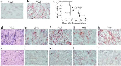
Function of VEGF in human leukocyte recruitment. Saline, IgG, or anti–human VEGF were received by huSCID mice bearing human skin transplants. (a–i) Skin grafts were harvested 14 days after humanization and infiltrates were identified by H&E staining (a–c), by immuno-staining with anti–human CD3 (d–f), and by immunostaining with anti–human CD68 (g–i). Treatment of huSCID mice with anti–human VEGF inhibited both CD3+T cell (f) and CD68+monocyte macrophage (i) infiltration of skin. (j) Quantitative assessment of CD3+T cell
found enhanced VEGF mRNA and protein expression in association with infiltrates (Figure 2). By immuno-histochemistry, VEGF protein was expressed by both inflammatory infiltrates and by vascular endothelial cells (Figure 2, d and e). In contrast, in skin specimens without infiltrates, VEGF was only found on keratin-ocytes and endothelial cells with minimal expression overall (Figure 2f). We treated huSCID mice with a neu-tralizing antihuman VEGF, human IgG, or saline. Con-sistent with its known function as a monocyte
chemo-attractant (11) anti-VEGF had a significant inhibitory effect on the recruitment of CD68+ monocytes/
macrophages (Figure 3). Few CD68+cells were evident
in skins harvested from animals treated with anti-VEGF (Figure 3i). Moreover, we unexpectedly found that anti-VEGF was most potent at inhibiting CD3+T
cell trafficking into human skin (Figure 3, d–f). By quantitative grid counting analysis, there was a statis-tically significant decrease in CD3+T cell infiltrates in
[image:6.585.55.535.51.496.2]anti-VEGF-treated huSCID animals both at day 7 and
Figure 4
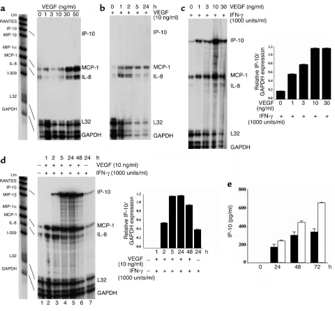
Effect of VEGF on endothelial cell chemokine expression. (a–d) Confluent cultures of human endothelial cells were treated for 4 hours (a
at day 14 (Figure 3j). Therefore, VEGF is functional for peripheral recruitment and for the trafficking of human allogeneic lymphocytes and monocytes in vivo. We also assessed the effect of anti-VEGF on the angiogenesis reaction, which we have previously report-ed to be associatreport-ed with mononuclear cell infiltration in this model (4). Our findings were that anti-VEGF markedly inhibited angiogenesis (data not shown), which is consistent with the interpretation that locally expressed VEGF is also of functional importance in leukocyte-associated angiogenesis.
In vitro analysis of VEGF-dependent chemokine production in endothelial cells. The function of VEGF as a monocyte chemoattractant is established in vitro (11). However, our finding that VEGF promotes T cell recruitment has not been previously reported in vitro or in vivo, to our knowledge. To study the mechanism of function of VEGF on T cell recruitment, we next assessed the effect of VEGF on human endothelial cell activation in vitro. Treatment of confluent cultures of endothelial cells with increasing concentrations of VEGF resulted in a dose-dependent increase in the expression of the endothelial cell adhesion molecules E-selectin, ICAM-1,
and VCAM-1 (not shown, and as reported by others [refs. 9, 10]). In addition, after treatment with VEGF, we found that the endothelial cell expression of the chemokines monocyte chemoattractant protein 1 (MCP-1) and interleukin-8 (IL-8) was also increased (Figure 4a). MCP-1 peaked in expression between 1 and 5 hours following treatment, and expression per-sisted for up to 24 hours (Figure 4b). In contrast, IL-8 peaked in expression at approximately 2 hours fol-lowing treatment, and decreased in expression by 24 hours. But there was no increase in the expression of the T cell–chemoattractant chemokines lympho-tactin, RANTES, IP-10 (Figure 4), or Mig (not shown) after VEGF treatment.
[image:7.585.54.529.340.600.2]Since IFN-γ-induced chemokines play a major role in the local recruitment of allogeneic lymphocytes, we also determined an effect of VEGF on IFN-γ-mediated activation of the potent T cell–chemoattractant chemokines IP-10 and Mig. We treated endothelial cells with IFN-γand found induced expression of IP-10 and Mig, as reported (45, 46). Moreover, we observed that a combination of VEGF with IFN-γresulted in a syner-gistic induction of endothelial cell IP-10 mRNA and
Figure 5
Blockade of VEGF in a fully MHC-mismatched murine model of acute cardiac allograft rejection. Fully MHC-mismatched C57BL/6 (H-2b)
donor hearts were transplanted into BALB/c (H-2d) mice as recipients. Untreated recipients develop marked leukocytic infiltrates and
rejec-tion by day 7. (aand b) Immunohistochemical analysis of VEGF in a normal nontransplanted heart (a) and in a rejecting allograft at day 7 (b), showing intense VEGF expression in association with allograft rejection. (c) Graft survival curves for recipients treated with anti-VEGF antiserum (filled squares; n= 6) or normal rabbit serum as a control (open squares; n= 5). Anti-VEGF–treated recipients show prolonged heart allograft survival (P < 0.001). (d–m) Histological analysis of cardiac allografts harvested at day 7 after transplantation from rejecting control serum–treated animals (d–h) or from animals treated with anti-VEGF (i–m). Control serum–treated animals had evidence of severe cellular rejection with extensive mononuclear cell infiltration (d), including CD45+, (e) CD3+(f), and macrophage (Mac) (g) infiltrates. In
protein expression, but not Mig expression (Figure 4, c–e, and not shown). In addition, we observed that IFN-γ inhibited the expression of IL-8, and that combinations of VEGF and IFN-γrestored IL-8 expression (Figure 4, c and d). Thus, VEGF may promote human allogeneic lymphocyte trafficking in vivo via regulation of endothelial cell production of chemokines including the potent T cell chemokine IP-10.
Blockade of VEGF in a fully MHC-mismatched murine model of acute cardiac allograft rejection. Our results also raise the possibility that VEGF is of importance in
allo-graft rejection. Alloallo-graft rejection is a complex multi-cellular process that is critically dependent upon T cell recognition of alloantigen processed and presented to T cells by either donor or recipient antigen-presenting cells (47). The subsequent recruitment of activated T cells and effector cells into an allograft has been shown to be mediated in part by locally expressed leukocyte-endothelial adhesion molecules (48) and chemokines (49). We used a physiological in vivo model of acute allograft rejection in which fully MHC-mismatched C57BL/6 (H-2b) donor hearts were transplanted into
BALB/c (H-2d) recipients. Similar to our findings in the
huSCID mouse (above), VEGF was expressed in associ-ation with graft infiltrates and acute rejection (Figure 5, a and b). Administration of our neutralizing anti-VEGF antiserum resulted in a significant prolongation of allograft survival up to 25 days after transplant ver-sus 11 days in control serum–treated animals (Figure 5c, P < 0.01). Severe cellular rejection with extensive mononuclear cell infiltration and myocyte necrosis was evident in sections of rejecting grafts at day 7 in
recipi-Figure 6
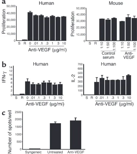
Function of VEGF in alloimmune T cell activation and allograft rejec-tion. (a) Anti–human VEGF or anti–murine VEGF antiserum was added into the human or the mouse MLR, respectively. Proliferation was assessed by [3H]thymidine incorporation for the last 18 hours of
[image:8.585.51.284.57.320.2]coculture. (b) The production of IFN-γand IL-2 was assessed by ELISA in coculture supernatants from a human MLR. As illustrated, block-ade of VEGF had no effect on proliferation or cytokine production in the MLR. Bars indicate the mean ± 1 SD for triplicate wells. Data are representative of three experiments with similar results. S, stimulators alone; R, responders alone. (c) Frequency of IFN-γ-producing cells in murine recipients of cardiac transplants as assessed by ELISPOT. Illus-trated is the production of IFN-γfrom a syngeneic, an untreated, and an anti-VEGF-treated animal. Representative of three such experi-ments performed in triplicate.
Figure 7
Function of angiogenesis in acute rejection. Fully MHC-mismatched C57BL/6 (H-2b) donor hearts
were transplanted into BALB/c (H-2d) mice, and
[image:8.585.225.530.489.733.2]ents treated with control rabbit serum (Figure 5, d–g). In contrast, we observed only a mild to moderate infil-trate, reduced numbers of CD3+T cells and
mono-cytes/macrophages, less myocyte damage, and minimal vasculitis in allografts from recipients treated with anti-VEGF antiserum (Figure 5, i–l). Moreover, the T cell chemoattractant IP-10 was observed in abundance on multiple cell types in rejecting grafts (Figure 5h), but there was minimal expression in grafts harvested from animals treated with anti-VEGF (Figure 5m).
Therefore, the intense expression of VEGF in allografts in vivo is of mechanistic significance in promoting the development of rejection.
Mechanism of function of VEGF in allograft rejection in vivo. The effect of anti-VEGF on leukocyte trafficking and allograft rejection in our in vivo studies could be explained by an effect of VEGF on T cell activation responses. However, the addition of our antibodies against VEGF to cocultures in human or mouse MLRs failed to inhibit T cell activation responses (Figure 6, a and b). Also, the in vivo priming of T cells to allo-antigen as manifested by ELISPOT analysis of IFN-γ in murine recipients of allografts (above) was unal-tered by anti-VEGF treatment (Figure 6c). Thus, VEGF does not function in alloimmune T cell activa-tion in vitro or in vivo.
We next assessed whether the intense intragraft expression of VEGF in acute rejection could mediate leukocyte recruitment in part via neovascularization. By immunohistochemistry, we found that endothe-lial CD31 was expressed diffusely on large vessels as well as microvessels throughout all grafts examined (Figure 7a). Moreover, the staining pattern of CD31 as well as the vascularization of isografts was very similar to that found in anti-VEGF-treated grafts. In contrast, we found fewer CD31-expressing vessels in control Ig–treated rejecting grafts; and some focal areas of rejecting grafts were devoid of any vascular staining (Figure 7a, and not shown). To assess the possibility of a role for angiogenesis in acute rejec-tion, we also treated animals with endostatin, a known endothelial cell–specific inhibitor of angio-genesis (29) (Figure 7, b–d). Our findings were that endostatin had a minimal effect on allograft survival, prolonging survival in three of eight animals; and failed to have any effect on leukocyte trafficking into the graft (Figure 7, b–d). Because the effect of endo-statin was so markedly different than that following treatment with anti-VEGF (Figure 5), we interpret these data as suggesting that VEGF mediates leuko-cyte trafficking into an allograft independent of an effect on angiogenesis.
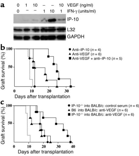
To further define the role of VEGF in T cell recruit-ment into cardiac allografts, we examined the effect of VEGF in primary cultures murine myocardial endothe-lial cells in vitro. In contrast to our findings in human endothelial cells, we found that VEGF alone induced the expression of IP-10 in murine endothelial cells (Fig-ure 8a). Thus, it is possible that VEGF may regulate T
cell trafficking primarily via IP-10-dependent mecha-nisms. Indeed, local intragraft endothelial cell expres-sion of IP-10 has been previously reported to be of crit-ical importance in the recruitment of T cells into allografts in rejection (39). Transplanted animals were next treated with neutralizing anti–IP-10 alone or in combination with anti-VEGF (Figure 8b). Our findings were that anti–IP-10 alone prolonged graft survival, and that combined treatment with anti–IP-10 and anti-VEGF significantly prolonged allograft survival versus treatment with anti–IP-10 alone or anti-VEGF alone (Figure 8b, P < 0.005 and P < 0.03, respectively). This is suggestive that a major effect of anti-VEGF in vivo is independent of IP-10. To further confirm this inter-pretation, we also transplanted allografts from IP-10–/–
[image:9.585.302.530.277.533.2]donors into wild-type mice and again found that anti-VEGF prolonged survival in the absence of local IP-10 production (Figure 8c, P < 0.04).
Figure 8
Function of VEGF-dependent regulation of IP-10 in allograft rejection. (a) Confluent cultures of murine myocardial endothelial cells were treated for 4 hours with recombinant murine VEGF or IFN-γalone or VEGF in combination with IFN-γas indicated. Total RNA was har-vested from endothelial cells and the expression of IP-10 was analyzed by RNase protection assay. (band c) The ability of VEGF to mediate IP-10-dependent trafficking and rejection was evaluated using anti-VEGF and anti–IP-10 in fully MHC-mismatched C57BL/6 (H-2b) or
IP-10–/–(H-2b) donor hearts transplanted into BALB/c (H-2d) mice.
Recipients of wild-type grafts were treated with anti-VEGF alone, or with anti-VEGF in combination with anti–IP-10. Recipients of IP-10–/–
donor grafts were treated with VEGF. Both anti–IP-10 and anti-VEGF were administered according to the schedule outlined in Meth-ods. As illustrated in b, we found that addition of anti-VEGF with anti–IP-10 significantly prolonged allograft survival in wild-type com-binations (P < 0.005); and in c, anti-VEGF prolonged survival in mice that received IP-10–/–donor hearts (P < 0.04). The survival of control
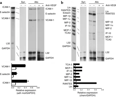
Lastly, we examined adhesion molecule and chemo-kine expression in vivo in cardiac allografts harvested on day 7 from control or anti-VEGF-treated wild-type mice. By RNase protection assay, we found a significant decrease in the expression of E-selectin, ICAM-1, and VCAM-1 in anti-VEGF-treated animals compared with that of controls (Figure 9a). In addition, anti-VEGF markedly inhibited the intragraft expression of several T cell–chemoattractant chemokines (including lym-photactin, RANTES, and IP-10) as well as the monocyte chemoattractant MCP-1 (Figure 9b). Thus, blockade of VEGF in vivo inhibits the local expression of several chemokines that are not directly regulated by VEGF. We interpret our findings as suggesting that VEGF is of importance in allograft rejection at early times after transplantation and prior to the induced expression and function of T cell–derived chemokines. Intragraft VEGF expression appears to promote early T cell and monocyte trafficking, and subsequent activation events result in additional VEGF expression as well as the expression of chemokines (including IP-10); both of these effects are additive in promoting rejection.
Discussion
VEGF is a most potent and critical mediator of physio-logical as well as pathophysio-logical angiogenesis. Its expres-sion and function have been reported in several chron-ic inflammatory processes; however, its mechanism of function in immunity has been unclear. Recent reports have defined the angiogenesis reaction mediated by VEGF to be proinflammatory in chronic inflammation (7). Here, we provide evidence that VEGF promotes endothelial cell chemokine production in vitro and in vivo and functions in the recruitment of monocytes and T cells into allografts. VEGF is thus an important proinflammatory cytokine in transplant rejection.
The expression of VEGF has been reported to be asso-ciated with allograft rejection (especially chronic rejec-tion) in both experimental models and following human transplantation (22, 24, 25). In addition, it has been reported that transplant recipients with poly-morphisms in the VEGF gene, encoding high VEGF production, are at increased risk for the development of acute renal allograft rejection (23). Here, we extend upon those observations and demonstrate that
intra-Figure 9
[image:10.585.88.492.53.391.2]graft VEGF has a biological effect in vivo in promoting rejection, as leukocyte infiltration is inhibited follow-ing treatment with neutralizfollow-ing anti-VEGF. Moreover, we demonstrate that induced intragraft VEGF is unlikely to be functional for the development of angio-genesis, as neovascularization is not a component of the rejection process. Rather, we show that the mecha-nism of function of VEGF appears to involve its ability to mediate intragraft leukocyte trafficking.
It is well established that the process of recruitment involves coordinate interactions among adhesion mol-ecules and chemokines (3, 49). Multiple individual molecules have been shown to be functional in the process of rejection (49–52), suggesting that there is some redundancy in the function of adhesion mole-cules and chemokines in vivo. It is known that VEGF induces the expression of adhesion molecules in human endothelial cells (9, 10), and two recent reports have pointed to an effect of VEGF on chemokine pro-duction (53, 54). Here, we extend upon those observa-tions and show that VEGF regulates IP-10, MCP-1, and IL-8, as well as the endothelial cell adhesion molecules E-selectin, ICAM-1, and VCAM-1, in vitro and in vivo. All of these molecules are expressed in association with rejection, and blockade of several molecules or their lig-ands individually (e.g., IP-10, MCP-1, ICAM-1, and VCAM-1) (49–52) has been shown to prolong allograft survival in murine models. Our data in this study imply that the ability of VEGF to regulate several of these molecules is the basis for its function in leukocyte traf-ficking. However, we have not fully addressed this com-plex question at an individual molecular level, and thus this interpretation will require further study. Never-theless, of all chemokine-chemokine receptor interac-tions studied to date, the most profound effects on graft survival are seen when the T cell chemoattractant IP-10 or its ligand CXCR3 are targeted (39, 50, 55). We found that VEGF-dependent T cell trafficking and rejection in vivo was not restricted to the function of IP-10. Our results therefore provide a framework for an analysis of the role of VEGF in the expression and func-tion of individual chemokines and adhesion molecules in leukocyte trafficking, in rejection, and in other cell-mediated immune inflammatory diseases (7, 38).
Another interpretation of our findings is that the major proinflammatory function of VEGF is in the ini-tiation of the inflammatory cascade, rather than in subsequent events resulting from T cell–mediated acti-vation responses. Consistent with this possibility, it is important to note that hypoxia is the major stimulus for VEGF expression (16). This suggests that VEGF will be produced locally within allografts immediately fol-lowing transplantation. Additional insults, including platelet and leukocyte recruitment into the graft, will further facilitate the expression of local VEGF (12) as well as other cytokines and chemokines (56) that are of importance in the rejection process (47, 49, 57). Here, we have provided evidence that early VEGF expression promotes T cell and monocyte recruitment. We suggest
that the subsequent induced expression of chemokines (e.g., IP-10) and VEGF are additive in promoting the development of rejection. As it is well established that early insults to allografts (such as prolonged ischemia) have long-term consequences (58), dysregulation of VEGF at early times following transplantation may have beneficial effects in promoting graft survival. Indeed, in support of this possibility, it has been noted that early (but not late) blockade of VEGF can limit the development of chronic arthritis (38).
In summary, our results provide compelling evidence that VEGF is a proinflammatory cytokine in allograft rejection. In addition, they provide insight into the role of VEGF in cell-mediated inflammatory reactions in immunity. We suggest that VEGF warrants considera-tion as a target for future therapeutic intervenconsidera-tion fol-lowing transplantation.
Acknowledgments
This work was supported by NIH grants AI46756 to D.M. Briscoe and CA069212 to A.D. Luster. The authors wish to thank Don Senger, Beth Israel Dea-coness Medical Center, for help with the generation of anti-VEGF antiserum; Kashi Javaherian and Judah Folkman for the gift of endostatin; and Stuart Robert-son, Evelyn Flynn, and William Wong for assistance with these studies. We also thank the maternity staff at South Shore Hospital for the supply of umbilical cords used for the generation of endothelial cells.
1. Sidky, Y.A., and Auerbach, R. 1975. Lymphocyte-induced angiogenesis: a quantitative and sensitive assay of the graft-vs.-host reaction. J. Exp. Med.141:1084–1100.
2. Auerbach, R., and Sidky, Y.A. 1979. Nature of the stimulus leading to lymphocyte-induced angiogenesis. J. Immunol.123:751–754. 3. Cotran, R.S. 1994. Inflammation and repair. In Pathologic basis of disease.
R.S. Cotran, V. Kumar, and S.L. Robbins, editors. WB Saunders, Philadelphia, USA. 51–92.
4. Moulton, K.S., et al. 1999. Angiogenesis in the huPBL-SCID model of human transplant rejection. Transplantation. 67:1626–1631. 5. Leung, D.W., Cachianes, G., Kuang, W.J., Goeddel, D.V., and Ferrara, N.
1989. Vascular endothelial growth factor is a secreted angiogenic mito-gen. Science. 246:1306–1309.
6. Folkman, J. 1995. Angiogenesis in cancer, vascular, rheumatoid and other disease. Nat. Med. 1:27–31.
7. Ferrara, N., and Davis-Smyth, T. 1997. The biology of vascular endothe-lial growth factor. Endocr. Rev. 18:4–25.
8. Gerber, H.P., Dixit, V., and Ferrara, N. 1998. Vascular endothelial growth factor induces expression of the antiapoptotic proteins Bcl-2 and A1 in vascular endothelial cells. J. Biol. Chem. 273:13313–13316. 9. Melder, R.J., et al. 1996. During angiogenesis, vascular endothelial growth factor and basic fibroblast growth factor regulate natural killer cell adhesion to tumor endothelium. Nat. Med. 2:992–997.
10. Kim, I., et al. 2001. Vascular endothelial growth factor expression of intercellular adhesion molecule 1 (ICAM-1), vascular cell adhesion mol-ecule 1 (VCAM-1), and E-selectin through nuclear factor-κB activation in endothelial cells. J. Biol. Chem.276:7614–7620.
11. Barleon, B., et al. 1996. Migration of human monocytes in response to vascular endothelial growth factor (VEGF) is mediated via the VEGF receptor flt-1. Blood.87:3336–3343.
12. Melter, M., et al. 2000. Ligation of CD40 induces the expression of vas-cular endothelial growth factor by endothelial cells and monocytes and promotes angiogenesis in vivo. Blood.96:3801–3808.
15. Clauss, M., et al. 1996. The vascular endothelial growth factor receptor Flt-1 mediates biological activities. Implications for a functional role of placenta growth factor in monocyte activation and chemotaxis. J. Biol. Chem.271:17629–17634.
16. Shweiki, D., Itin, A., Soffer, D., and Keshet, E. 1992. Vascular endothe-lial growth factor induced by hypoxia may mediate hypoxia-initiated angiogenesis. Nature.359:843–845.
17. Sato, K., et al. 1995. Stimulation by thyroid-stimulating hormone and Grave’s immunoglobulin G of vascular endothelial growth factor mRNA expression in human thyroid follicles in vitro and flt mRNA expression in the rat thyroid in vivo. J. Clin. Invest.96:1295–1302.
18. Shweiki, D., Neeman, M., Itin, A., and Keshet, E. 1995. Induction of vas-cular endothelial growth factor expression by hypoxia and by glucose deficiency in multicell spheroids: implications for tumor angiogenesis.
Proc. Natl. Acad. Sci. U. S. A.92:768–772.
19. Williams, B., Baker, A.Q., Gallacher, B., and Lodwick, D. 1995. Angiotensin II increases vascular permeability factor gene expression by human vascular smooth muscle cells. Hypertension.25:913–917. 20. Williams, B., Quinn-Baker, A., and Gallacher, B. 1995. Serum and
platelet-derived growth factor-induced expression of vascular perme-ability factor mRNA by human vascular smooth muscle cells in vitro.
Clin. Sci.88:141–147.
21. Reinders, M.E., Sho, M., Robertson, S.W., Geehan, C.S., and Briscoe, D.M. 2003. Proangiogenic function of CD40 ligand-CD40 interactions.
J. Immunol. 171:1534–1541.
22. Reinders, M.E.J., Fang, J.C., Wong, W., Ganz, P., and Briscoe, D.M. 2003. Expression patterns of vascular endothelial growth factor in human car-diac allografts: association with rejection. Transplantation. 76:224–230. 23. Shahbazi, M., et al. 2002. Vascular endothelial growth factor gene poly-morphisms are associated with acute renal allograft rejection. J. Am. Soc. Nephrol.13:260–264.
24. Pilmore, H.L., Eris, J.M., Painter, D.M., Bishop, G.A., and McCaughan, G.W. 1999. Vascular endothelial growth factor expression in human chronic renal allograft rejection. Transplantation. 67:929–933. 25. Torry, R.J., Labarrere, C.A., Torry, D.S., Holt, V.J., and Faulk, W.P. 1995.
Vascular endothelial growth factor expression in transplanted human hearts. Transplantation. 60:1451–1457.
26. Kim, K.J., et al. 1993. Inhibition of vascular endothelial growth factor-induced angiogenesis suppresses tumour growth in vivo. Nature.
362:841–844.
27. Presta, L.G., et al. 1997. Humanization of an anti-vascular endothelial growth factor monoclonal antibody for the therapy of solid tumors and other disorders. Cancer. Res. 57:4593–4599.
28. Khan, I.A., et al. 2000. IP-10 is critical for effector T cell trafficking and host survival in Toxoplasma gondiiinfection. Immunity. 12:483–494. 29. O’Reilly, M.S., et al. 1997. Endostatin: an endogenous inhibitor of
angio-genesis and tumor growth. Cell. 88:277–285.
30. Boehm, T., Folkman, J., Browder, T., and O’Reilly, M.S. 1997. Antiangio-genic therapy of experimental cancer does not induce acquired drug resistance. Nature. 390:404–407.
31. Bergers, G., Javaherian, K., Lo, K.M., Folkman, J., and Hanahan, D. 1999. Effects of angiogenesis inhibitors on multistage carcinogenesis in mice.
Science. 284:808–812.
32. Tilton, R.G., et al. 1997. Vascular dysfunction induced by elevated glu-cose levels in rats is mediated by vascular endothelial growth factor.
J. Clin. Invest. 99:2192–2202.
33. Basu, S., et al. 2001. The neurotransmitter dopamine inhibits angiogen-esis induced by vascular permeability factor/vascular endothelial growth factor. Nat. Med. 7:569–574.
34. Walter, D.H., et al. 1996. The in vivo bioactivity of vascular endothelial growth factor/vascular permeability factor is independent of N-linked glycosylation. Lab. Invest. 74:546–556.
35. Dufour, J.H., et al. 2002. IFN-γ-inducible protein 10 (IP-10; CXCL10)-deficient mice reveal a role for IP-10 in effector T cell generation and traf-ficking. J. Immunol. 168:3195–3204.
36. Briscoe, D.M., et al. 1999. The allogeneic response to cultured human skin equivalent in the hu-PBL-SCID mouse model of skin rejection.
Transplantation. 67:1590–1599.
37. Corry, R.J., Winn, H.J., and Russell, P.S. 1973. Primarily vascularized allo-grafts of hearts in mice. The role of H-2D, H-2K, and non-H-2 antigens in rejection. Transplantation. 16:343–350.
38. Lu, J., et al. 2000. Vascular endothelial growth factor expression and regulation of murine collagen-induced arthritis. J. Immunol.
164:5922–5927.
39. Hancock, W.W., et al. 2001. Donor-derived IP-10 initiates development of acute allograft rejection. J. Exp. Med. 193:975–980.
40. Yamada, A., et al. 2001. CD28-independent costimulation of T cells in alloimmune responses. J. Immunol. 167:140–146.
41. Gimbrone, M.A. 1976. Culture of vascular endothelium. Prog. Hemost. Thromb. 3:1–28.
42. Marelli-Berg, F.M., Peek, E., Lidington, E.A., Stauss, H.J., and Lechler, R.I. 2000. Isolation of endothelial cells from murine tissue. J. Immunol. Meth.
244:205–215.
43. Denton, M.D., Geehan, C., Alexander, S.I., Sayegh, M.H., and Briscoe, D.M. 1999. Endothelial cells modify the costimulatory capacity of trans-migrating leukocytes and promote CD28-mediated CD4+ T cell alloac-tivation. J. Exp. Med. 190:555–566.
44. Murray, A.G., et al. 1994. Human T-cell-mediated destruction of allo-geneic dermal microvessels in a severe combined immunodeficient mouse. Proc. Natl. Acad. Sci. U. S. A. 91:9146–9150.
45. Luster, A.D., Jhanwar, S.C., Chaganti, R.S., Kersey, J.H., and Ravetch, J.V. 1987. Interferon-inducible gene maps to a chromosomal band associat-ed with a (4;11) translocation in acute leukemia cells. Proc. Natl. Acad. Sci. U. S. A.84:2868–2871.
46. Farber, J.M. 1990. A macrophage mRNA selectively induced by γ -inter-feron encodes a member of the platelet factor 4 family of cytokines. Proc. Natl. Acad. Sci. U. S. A. 87:5238–5242.
47. Briscoe, D.M., and Sayegh, M.H. 2002. A rendezvous before rejection: where do T cells meet transplant antigens? Nat. Med. 8:220–222. 48. Briscoe, D.M., et al. 1995. Predictive value of inducible endothelial cell
adhesion molecule expression for acute rejection of human cardiac allo-grafts. Transplantation. 59:204–211.
49. Hancock, W.W. 2002. Chemokines and transplant immunobiology.
J. Am. Soc. Nephrol. 13:821–824.
50. Melter, M., Exeni, A., and Briscoe, D.M. 2002. Chemokines and their receptors in human clinical solid organ transplantation. Curr. Opin. Organ Transplant.7:77–84.
51. Winn, R., Vedder, N., Ramamoorthy, C., Sharar, S., and Harlan, J. 1998. Endothelial and leukocyte adhesion molecules in inflammation and dis-ease. Blood Coagul. Fibrinolysis. 9:S17–S23.
52. Denton, M.D., et al. 2000. The role of the graft endothelium in trans-plant rejection: evidence that endothelial activation may serve as a clin-ical marker for the development of chronic rejection. Pediatr. Transplant.
4:252–260.
53. Marumo, T., Schini-Kerth, V.B., and Busse, R. 1999. Vascular endothe-lial growth factor activates nuclear factor-κB and induces monocyte chemoattractant protein-1 in bovine retinal endothelial cells. Diabetes.
48:1131–1137.
54. Lee, T.H., Avraham, H., Lee, S.H., and Avraham, S. 2002. Vascular endothelial growth factor modulates neutrophil transendothelial migra-tion via up-regulamigra-tion of interleukin-8 in human brain microvascular endothelial cells. J. Biol. Chem. 277:10445–10451.
55. Hancock, W.W., et al. 2000. Requirement of the chemokine receptor CXCR3 for acute allograft rejection. J. Exp. Med. 192:1515–1520. 56. Denton, M.D., et al. 1998. Central role for CD40/CD40 ligand (CD154)
interactions in transplant rejection. Pediatr. Transplant. 2:6–15. 57. Sayegh, M.H. 1999. Why do we reject a graft? Role of indirect
allorecog-nition in graft rejection. Kidney Int. 56:1967–1979.