www.impactjournals.com/oncotarget/
Oncotarget, Vol. 7, No. 36
Advanced new strategies for metastatic cancer treatment by
therapeutic stem cells and oncolytic virotherapy
Geon-Tae Park
1and Kyung-Chul Choi
1,21 Laboratory of Biochemistry and Immunology, College of Veterinary Medicine, Chungbuk National University, Cheongju,
Chungbuk, Republic of Korea
2 TheraCell Bio & Science, Cheongju, Chungbuk, Republic of Korea
Correspondence to: Kyung-Chul Choi, email: kchoi@cbu.ac.kr
Keywords: metastatic cancer, therapeutic stem cell, oncolytic virotherapy, cancer treatment
Received: April 15, 2016 Accepted: May 29, 2016 Published: August 02, 2016
ABSTRACT
The field of therapeutic stem cell and oncolytic virotherapy for cancer treatment
has rapidly expanded over the past decade. Oncolytic viruses constitute a promising new class of anticancer agent because of their ability to selectively infect and destroy
tumor cells. Engineering of viruses to express anticancer genes and specific cancer
targeting molecules has led to the use of these systems as a novel platform of
metastatic cancer therapy. In addition, stem cells have a cancer specific migratory
capacity, which is available for metastatic cancer targeting. Prodrug activating enzyme or anticancer cytokine expressing stem cells successfully inhibited the proliferation of cancer cells. Preclinical models have clearly demonstrated anticancer activity of these two platforms against a number of different cancer types and metastatic cancer. Several systems using therapeutic stem cells or oncolytic virus have entered clinical trials, and promising results have led to late stage clinical development. Consequently, metastatic cancer therapies using stem cells and oncolytic viruses are extremely promising. The following review will focus on the metastatic cancer targeting mechanism of therapeutic stem cells and oncolytic viruses, and potential
challenges ahead for advancing the field.
INTRODUCTION
Cancer metastasis, which is a multiple process in
which malignant cells spread from the primary site to
colonize distant organs, is one of the greatest challenges
in cancer treatment. Although metastasis is responsible
for more than 90% of cancer associated mortality, it is
difficult to diagnose and treat [1]. For many patients,
metastasis has already occurred by the time when they
are diagnosed with primary cancer. Depending on the
type of cancer, metastasis shows various modalities. In
breast cancer, metastasis of the primary tumor is difficult
to detect and could remain latent for many years. Latent
cells are then activated by unknown factors, resulting in
formation of incurable lesions. Conversely, small cell
lung cancer has often metastasized to multiple organs at
the time of initial diagnosis [2]. Only a small number of
patients with metastatic cancer can be successfully treated
by conventional strategies such as surgical removal,
chemotherapy and radiotherapy [3]. Therefore, it is
necessary to develop a new strategy to prevent metastasis
or treat existing metastases.
Conventional strategies of cancer therapy, including
surgical resection, chemotherapy, radiotherapy and
immunotherapy, have made significant contributions
to cancer treatment. However, many people suffer from
side-effects such as insufficient anti-cancer effects that
involve drug resistance and systemic adverse reactions
due to off target effects [4]. For example, one of advanced
methods for metastatic breast cancer included monoclonal
antibody that binds to the breast cancer specific HER2/
neu receptor to interfere HER2 signaling pathway. As
results, inhibition of downstream signaling pathways,
cell cycle arrest and a reduction in angiogenesis occurred
[5]. This method is very effective for metastatic breast
cancer treatment, but it is only effective on breast cancer
that expresses the HER2/neu receptor. In addition, some
cancer patients acquired resistance to Trastuzumab during
the period of administration [6, 7]. Another major problem
associated with conventional cancer therapy is incomplete
elimination of the invasive primary tumor masses, which
cause metastasis to multiple organs, and tumor cell
dormancy that leads to disease recurrence [8, 9]. Therefore,
the recent goal of new therapies has been development of
cancer treatments that have sufficient therapeutic capacity
with little or no toxicity to normal cells. To achieve this
goal, a better understanding of the detailed mechanisms
of cancer using the latest biotechnology and innovative
anti-cancer technologies is needed.
A mechanistic understanding of the metastatic
process is important to development of anti-metastatic
therapies that could reduce patient mortality. The
metastatic process is initially derived from gene
mutations that correlate with proliferative ability. Cells
in normal tissues only divide when they receive growth
stimulatory signals from other cells and stop dividing
when they receive growth inhibitory signals; however,
gene mutations providing the ability to be split ignore
these signaling factors [10]. Additionally, these oncogenic
mutations cause the cells to maintain progenitor like
phenotypes and generate oncogenically transformed
cells from normal cells. However, many studies using
oncogene driven mouse models have shown that cancer
did not automatically metastasize to distant organs
and that oncogenic transformation is not sufficient for
metastatic potential [11]. This is because metastasis is a
very complicated process that involves a number of genes
associated with tumor cell invasion from the primary
tumor to the bloodstream, circulation and exit from the
circulatory system to distant organs, and then angiogenesis
and colonization of the distant organ [12]. Because of the
complexity, metastatic cancer treatment appears to be
difficult.
In this review, we will discuss recent strategies for
the treatment of metastatic cancer based on stem cells
and oncolytic viruses. Many stem cells have intrinsic
tumor tropic properties that originate from chemokine
interactions with cancer cells. Using this property, we can
make a specific delivery system of anticancer molecules.
Stem cells can migrate towards tumor microenvironments
and eliminate tumors, enabling site specific delivery.
Furthermore, stem cells can be modified to stably
express various anticancer agents including cytokines
and prodrug activating enzymes for induction of cancer
apoptosis and removal of specific tumors. In addition,
oncolytic viruses are a therapeutically useful system
that can be used to selectively infect and damage tumor
tissues without off target effects on normal tissues. Each
virus has a specific cellular tropism that determines which
tissues are preferentially infected. Viruses then increase
in the tumors and destroy them, after which they infects
another tumor cell. Viral oncotherapy can also be modified
to increase tumor selectivity and enhance oncolytic
activity. For example, some viruses have been modified
to express capsid proteins that bind with specific cancer
types and conditionally express the genes involved with
the activation of host immune system. These two strategies
will be able to complement the drawbacks of conventional
cancer therapy.
THERAPEUTIC STEM CELL FOR
METASTATIC CANCER
Specific tumor tropism of stem cells
Stem cells can trace cancer cells and tumor regions,
which makes them very useful for tracing metastatic
cancer and carrying anti-metastatic molecules. Various
chemokine-chemokine receptor interactions are important
to recognition of tumor cells and tumor tropism of stem
cells. Stromal cell derived factor 1 alpha (SDF-1α) and
its receptor, CSC chemokine receptor 4 (CXCR4), have
been identified as key molecules responsible for the
tropism of stem cells in many cancers [13]. According
to the results, the SDF1α -CXCR4 signaling pathway
plays a major role in the tumor specific migration of
commonly used stem cell types, including mesenchymal
SCs, embryonic SCs and induced pluripotent SCs [14-16].
Stem cell surface CXCR-4 binds with SDF-1α secreted
by cancer cells, which stimulates stem cells to express
more CXCR-4. Moreover, overexpression of the CXCR4
using gene transfection of human umbilical cord blood
derived MSCs increased the migratory capacity of MSCs
toward gliomas [15]. These results show the possibility
to further increase migration capacity toward metastatic
cancer
via
stem cell engineering. Other signaling pathways
have been found, including urokinase type plasminogen
activator (uPA) - uPA receptor (uPAR) and vascular
endothelial growth factor receptor 2 (VEGFR2) [17, 18].
The degree of migration of stem cells towards a tumor
is affected by diverse factors, including the nature of the
stem cell, type of cancer and tumor microenvironment.
Additional research is needed to better understand the
factors influencing the migratory capacity of stem cells
that allow the therapeutic potential for metastatic cancer
treatment to be increased while reducing side effects of
these stem cells.
Strategies for metastatic cancer treatment using
stem cells with anti-metastatic genes
anti-metastatic molecules. Stem cell secretion of therapeutic
molecules can initially be divided into two categories
depending on whether they directly target tumor cells
or support immune system. Direct targeting molecules
include the pro-apoptotic protein tumor necrosis factor
related apoptosis inducing ligand (TRAIL), which binds
to death receptor 4 (DR4) and DR5 and induces tumor
cell apoptosis [21]. CD40 ligand is another pro-apoptotic
molecule that binds to CD40 expressed on the tumor
cell surface [22-24]. Membrane bound CD40 ligand
triggered tumor cell apoptosis
via
activation of JNK/
activation protein-1 and stimulated the secretion of both
tumor necrosis factor alpha and interferon gamma, which
ultimately activated the caspase 3/7 pathway [25, 26].
Neural stem cells derived from induced pluripotent stem
cells transduced with baculovirus encoding CD40 ligand
sufficiently inhibited tumor development in a preclinical
model [27]. In addition, CD40 ligand expressing
endothelial progenitor cells (EPCs) successfully migrated
toward metastatic breast cancer lesions in the lung and
induced tumor apoptosis [28]. Using cytokines such as the
type I interferon family (IFN-α and β) to induce S-phase
accumulation and apoptosis of tumor cells is another
strategy for inhibition of proliferation pathways of the
cancer and associated cells [29]. Interferon expressing
stem cells have been shown to inhibit tumor growth in
various preclinical cancer models [30, 31]. Secretion of
interleukins that can stimulate immune system against
tumor microenvironments has also been tested. Human
MSCs have been engineered to secrete IL-12 and tested
in preclinical metastatic hepatoma models. These studies
revealed that the presence of IL-12 expressing stem
cells could modify the immune profile of the tumor
microenvironment. Moreover, the level of IFN-γ that
is critical for innate and adaptive immunity activation
increased. This change causes activation of natural killer
cells and recruitment of tumor specific CD8+ T cells [32]
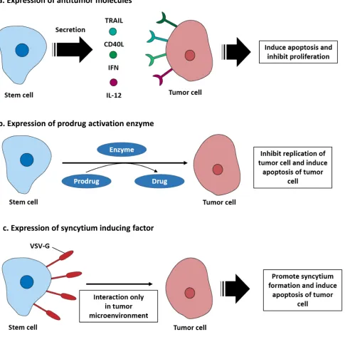
as shown in Figure 1a. In addition, Table 1 summarizes
the therapeutic gene transfer by stem cells for metastatic
cancer treatment.
Strategies for metastatic cancer treatment using
stem cells with prodrugs
Stem cell mediated suicide gene therapy is another
strategy for killing tumor cells. Stem cells are engineered
to express an enzyme that converts a non-toxic prodrug
into a cytotoxic drug that can efficiently kill tumor
cells
via
the bystander effect. Cytosine deaminase (CD)
and 5-fluorocytosine (5-FC) are well-known suicide
gene systems.
E. coli
cytosine deaminase can convert a
prodrug, 5-FC, into its active drug, 5-FU. The metabolite
of 5-FU (fluorodeoxyuridine monophosphate) binds to
the nucleotide binding site of the thymidylate synthase
and dNTP in tumor cells becomes imbalanced, which
can cause DNA damage and cell apoptosis [33]. In
addition, carboxylesterase converts the prodrug irinotecan
(CPT-11) to the potent topoisomerase I inhibitor SN-38.
Topoisomerase I catalyzes DNA unwinding, which is a
critical step in DNA replication and transcription. SN-38
binds to the DNA-Topoisomerase I complex, inhibiting
ligation of the nicked DNA strand. Moreover, the
SN-38-DNA-Topoisomerase I complex interrupts the movement
of DNA polymerase along the DNA strand and induces
tumor cell apoptosis [34]. The CD-5-FC system has
been used in modified MSCs and NSCs and applied in
metastasized preclinical models, where it could selectively
treat metastasized cancer and inhibit tumor growth [35,
36]. In addition, human NSCs expressing carboxylesterase
have been shown to be effective in preclinical models
of metastatic lung cancer [37]. Furthermore, stem
cell mediated suicide gene therapy has the additional
advantage of the stem cell being eliminated after its
therapeutic effect, which reduces side effects owing to
long term retention [38] (Figure 1b).
[image:3.612.56.562.58.216.2]Other strategies for inducing antitumor effects
have also been studied. For example, using the vesicular
stomatitis virus glycoprotein (VSV-G), which is one
of the fusogenic membrane glycoproteins (FMGs) in
neural stem cells, is a notable strategy for targeting tumor
microenvironments [39]. VSV-G expressed in the neural
Table 1: Therapeutic gene transfer by stem cells for metastatic cancer treatment
Gene
Function
References
TNF-related apoptosis inducing ligand (TRAIL)
Binds to death receptor and induces tumor cell
apoptosis
[18]
CD40 ligand
Stimulate the secretion of TNF-α and IFN-γ, which
activate the caspase 3/7 pathway
[24,25]
Type I interferon
Induce S-phase accumulation and apoptosis of tumor
cell
[27, 28]
Interleukin 12 (IL-12)
Stimulate the IFN-γ secretion and recruitment of tumor
specific T-cell
[29]
Cytosine deaminase
Convert prodrug (5-FC) to activated drug (5-FU)
[32, 33]
Carboxylesterase
Convert prodrug (CPT-11) to activated drug (SN-38) [34]
Vesicular stomatitis virus glycoprotein
stem cell membrane can induce rapid and extensive
cell-to-cell fusion and promote the formation of multinucleated
syncytia with cancer cells, eventually causing cell death.
To specifically kill tumor cells, they engineered a pH
sensor of VSV-G and generated a novel VSV-G mutant
that efficiently promotes syncytium formation at the
tumor extracellular pH (pH 6.8), but not at pH 7.4. In a
preclinical metastatic breast cancer model, this system
successfully inhibited cancer progression following
systemic stem cell administration [40] (Figure 1c).
Limitations of the stem cells based cancer therapy
[image:4.612.68.553.169.642.2]Cancer treatments using stem cells have made
improvements in regards to specific targeting of tumors,
but a few obstacles must still be overcome prior to clinical
application. The main concern is tumorigenicity of the
stem cell and cell fate after systemic administration. To
prevent therapeutic stem cells from forming tumors or
aberrantly differentiating in the host, the tumorigenicity
or differentiation potential should be tested in preclinical
models. In addition, immortalized stem cells with
therapeutic gene inserts may solve the difficulty of mass
culture of stem cells and enable their stability in tumors.
Therefore, it is important to develop a new strategy for
mass culture of stem cells to ensure the ability of a suicide
gene to perfectly eliminate stem cells after therapy.
Therapeutic modifications could adversely affect the safety
of stem cells. Therapeutic stem cells or secreted proteins
interrupt host tolerance to self-antigens, which provokes
additional complications in the patient and immune
responses that might impair therapy. Improvement of stem
cell specificity through detailed investigations of tumor
tropism could relieve possible side effects.
ONCOLYTIC VIROTHERAPY
The idea of oncolytic virotherapy originates from
clinical reports of cancer regression caused by viral
infection and is currently being developed by genetically
modifying viruses for the selective infection and
destruction of cancer cells [41].
Tumor specific targeting strategies
The specificity of oncolytic viruses for tumors is
very important to clinical trials. Many viruses have a
natural specific tissue tropism for cell surface proteins
that are overexpressed by cancer cells. This characteristic
is very useful for metastatic cancer tracing. For example,
measles virus recognizes the surface receptor CD46
(complement regulatory protein) for cell infection. CD46
is a cofactor for inactivation of complement components
that is often overexpressed in cancer cells [42]. Herpes
simplex virus uses the herpesvirus entry mediator
(HVEM) and nectins which are overexpressed on the
surface of cancer cells [43]. Coxsackievirus can recognize
cell surface glycoprotein (intracellular adhesion molecule
1; ICAM-1) and GPI-anchored membrane protein (decay
accelerating factor; DAF), which are overexpressed in
cancers including melanoma and breast cancer [44, 45].
The enterovirus family inhibits expression of CD155, a
key ligand in NK cell-mediated suppression of metastases
that is overexpressed by some cancer cells [46].
Oncolytic viruses can be engineered to directly
bind to unique surface molecules of cancer cells. This
modification could assign additional specificity for
metastasized tumor cells by improving infection of tumor
tissues and decreasing infection of healthy tissues. This
specificity can be achieved by modifying or combination
protein of virus that require for cancer cell recognition. For
example, glioma cells overexpress CD16 and CD80/86,
which bind with adenovirus serotype 3 [47, 48]. Based
on these findings, a chimeric adenovirus vector (Ad5/3)
contains the backbone of adenovirus serotype 5 fiber with
an adenovirus, serotype 3 knob. The Ad 5/3 chimeric
virus exhibits increased targeting capabilities for cell lines
analyzed
in vitro
by at least ten-fold. These data suggest
an improved safety of Ad 5/3 in the setting of malignant
glioma [49]. The adenovirus Ad5/3-Δ24 was modified to
bind to CD46, which are highly expressed in metastatic
renal cancer and significantly increased antitumor effects
in a preclinical model [50]. Other examples of engineered
specificities include lentiviruses pseudotyped with Sindbis
virus, which targeted human P-glycoprotein ectopically
expressed on the surface of melanoma cells. This oncolytic
virus successfully targeted metastatic melanoma cells after
systemic administration by tail vein injection [51].
[image:5.612.60.561.57.222.2]Another strategy is a tumor specific transcriptional
targeting using tumor specific promoters and microRNA
target sequences. This strategy can restrict virus replication
in off-target tissues. For example, adenovirus replication
is correlated with their ability to promote cell cycle entry
into the G1 phase through the viral immediate early
protein E1A. Therefore, tumor specificity is achieved by
Table 2: Therapeutic gene transfer by viruses for metastatic cancer treatment
Gene
Function
References
Granulocyte macrophage colony-stimulating
factor (GM-CSF)
Stimulate adaptive immunity against tumor associated
antigen
[56, 57]
Interleukin – 12 (IL-12)
Stimulate the IFN-γ secretion and recruitment of tumor
specific T-cell
[58, 59]
KAI1
Inhibit the EGFR signaling, which associated with cell
motility
[61]
NM23
Reduce the cell flexibility necessary for cytoskeleton
plasticity and cell motility
[62-65]
TGF-β receptor II (TGF-βRII)
Bind with TGF-β and inhibit the TGF-β signaling
pathway, which associated with cancer metastasis
[69]
Osteoprotegerin linked Immunoglobulin G
(OPG linked IgG)
Bind with nuclear factor kappa-B ligand (RANKL),
which associated with microenvironmental conditions to
placing the E1A gene under transcriptional regulation of a
tumor specific promoter [52], CXCR4 in breast cancer and
prostate specific antigen (PSA) promoter in prostate cancer
[53, 54]. Another strategy is the application of microRNA
targeting oncolytic viruses based on the discovery of
differential expression patterns of micro RNA in tumor
and normal tissues [55]. For example, the expression
of let-7 is functionally linked to tumors, regulating the
over-expression of proto-oncogenes, and reflecting the
differentiation state of tumors [56]. Incorporation of
let-7 microRNA complementary sequences within the
3’ untranslated region (UTR) of the wild-type matrix
protein of the VSV gene plays an essential role in viral
replication and eliminates unwanted replication and
associated toxicity in normal cells, but permits growth in
cancer cells [57]. In adenovirus, insertion of liver specific
microRNA (miR-122) binding sites in the 3’ UTR of the
gene encoding E1A of an oncolytic adenovirus decreased
its toxicity without sacrificing tumor killing activity in a
model of pancreatic cancer metastasis to the liver [58].
Strategies for metastatic cancer treatment using
viruses
After oncolytic virus infection, cancer cells are
destroyed by lysis. However, various strategies have been
studied to achieve the maximum therapeutic efficacy
for tumor cells. The therapeutic efficacy of oncolytic
viruses can be enhanced using strategies that enable
immunostimulatory factors to induce innate and adaptive
immune responses against tumors. One successful strategy
is the expression of granulocyte macrophage colony
stimulating factor (GM-CSF), which stimulates stem cells
to produce granulocytes and monocytes and stimulates
adaptive immunity against tumor associated antigens [59].
T-VEC is the most advanced herpes simplex virus based
oncolytic virus encoding the GM-CSF gene. In clinical
studies, T-VEC was shown to offer superior benefits
during treatment of metastatic melanoma [60]. A potent
anticancer molecule, IL-12, is an
interleukin
produced
by
dendritic cells
in immune response, and treatment of
cancer stimulates the production of
interferon-gamma
(IFN-γ) and
tumor necrosis factor-alpha
(TNF-α) from
T cells and NK cells [61]. In a preclinical model of
metastatic pancreatic cancer, the IL-12 gene inserted
adenovirus significantly inhibited tumor cell growth [62]
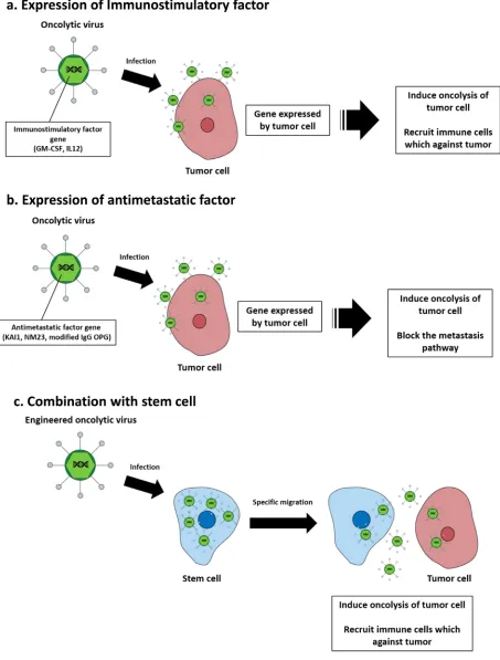
(Figure 2a).
[image:7.612.59.551.60.127.2]Transfer of metastatic suppressor genes or targeting
metastasis related molecules is an effective strategy of
targeting metastatic cancer. One of the tumor suppressor
proteins, KAI1, plays a key role in downregulation of
epidermal growth factor receptor (EGFR) signaling, which
is associated with increased receptor desensitization and
endocytosis [63]. An adenovirus expressing KAI1 is
applied in an orthotopic mouse model with non-small cell
lung cancer lymphatic metastasis, which has decreased
lymphatic metastasis but not decreased primary tumor
volume [64]. Reduced NM23 expression has been shown
to be significantly associated with metastatic behavior in
many cancer types [65-67]. Liver metastasis of ovarian
cancer and animal survival time were measured after
transfer of a recombinant adenovirus expressing NM23
into the preclinical model. A significant reduction in
the number of animals developing liver metastases and
prolongation of median survival time was observed
Table 3: Clinical trials for current stem cell cancer therapy
Stem cell
Name
Modification
Phase References *
Mesenchymal stem cell
GX-051
N/A
IL-12 expression
Loading oncolytic adenovirus (ICOVIR-5)
1
1
NCT02079324
NCT01864759
Neural stem cell
N/A
N/A
Cytosine deaminase expression
Carboxylesterase expression
1
1
NCT02015819
NCT02192359
*NCT number is the identifier number on ClinicalTrials.gov.
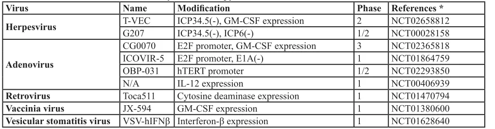
Table 4: Clinical trials for current oncolytic virotherapy
Virus
Name
Modification
Phase References *
Herpesvirus
T-VEC
G207
ICP34.5(-), GM-CSF expression
ICP34.5(-), ICP6(-)
2
1/2
NCT02658812
NCT00028158
Adenovirus
CG0070
E2F promoter, GM-CSF expression
3
NCT02365818
ICOVIR-5
E2F promoter, E1A(-)
1
NCT01864759
OBP-031
hTERT promoter
1/2
NCT02293850
N/A
IL-12 expression
1
NCT00406939
Retrovirus
Toca511
Cytosine deaminase expression
1
NCT01470794
Vaccinia virus
JX-594
GM-CSF expression
1
NCT01380600
Vesicular stomatitis virus
VSV-hIFNβ Interferon-β expression
1
NCT01628640
[image:7.612.56.565.162.295.2]relative to the untreated group [68]. Some studies have
suggested that overexpression of the transforming growth
factor beta (TGF-β) pathway is associated with breast
cancer bone metastases [69-71]. Based on these results,
modified adenoviruses expressing soluble form of
transforming growth factor-beta receptor II (TGF-βRII)
fused with human immunoglobulin Fc fragment could
bind with TGF-β and successfully inhibit breast cancer
with bone metastasis in a mouse model [72]. Similarly,
an adenovirus expressing soluble osteoprotegerin (OPG)
linked to the Fc portion of immunoglobulin G (IgG)
is also effective at inhibiting the progression of bone
metastasis of breast cancer [73]. Osteoprotegerin (OPG)
acts as a decoy receptor for receptor activator of nuclear
factor kappa-B ligand (RANKL), which allows sufficient
microenvironmental conditions to influence cancer cell
migration (Figure 2b) [74, 75].
Several studies have also explored the possibility of
combining oncolytic viruses with stem cells to improve
delivery [76]. The therapeutic efficacy of an oncolytic
virus is determined by clearance of the virus by the host
immune system following systemic and intratumoral
administration. Stem cells infected with oncolytic virus
migrated to the tumor and locally released undamaged
oncolytic viruses. hMSCs have recently been shown
to function as effective carriers to deliver oncolytic
viruses. In a preclinical model, hMSC transduced with
conditionally replicating adenoviruses significantly
suppressed pulmonary metastasis of breast cancer through
viral amplification in hMSCs [77]. In addition, stem
cell delivery of oncolytic viruses has been shown to be
effective in several preclinical cancer models, such as
ovarian cancer and hepatocellular carcinoma [78, 79].
Thus, hMSCs may be an effective platform for the targeted
delivery of oncolytic viruses to distant cancer sites such
as metastatic breast cancer (Figure 2c). In addition, the
therapeutic gene transfers by tumor-tropic viruses were
shown for metastatic cancer treatment (Table 2).
Limitations of the virotherapy
The development of oncolytic viruses as therapeutic
agents for metastatic cancer requires careful attention
to establish appropriate clinical trial designs, as well as
dosing regimens to minimize possible side effects. The
most important technical challenge to overcome is the
need to enhance tumor selectivity to decrease off target
effects after systemic delivery of the oncolytic virus. The
other major obstacle to successful application of viral
therapy is neutralization of the virus by the host antibody.
Many species of viruses are used in oncolytic virotherapy,
and most people have already been exposed to the virus
through previous vaccination or infection. Therefore,
circulating antibodies can inhibit the oncolytic virus before
it reaches the tumor site. Immune suppression agents and
the aforementioned stem cell transport strategy is currently
being investigated to solve this problem.
CONCLUSIONS
Metastasis of cancer is one of the main factors
leading to patient death. The unique properties of
metastatic cancer, including their small size, high
multiplicity and spread to multiple organs make it difficult
to treat. Although conventional cancer treatment strategies
have shown a lot of progress, they have been limited in
metastatic cancer by recurrence of cancer, induction of
drug resistance and systemic side effects after treatment.
Accordingly, new strategies are needed to treat metastatic
cancer.
Stem cell based and oncolytic virus strategies
have many potential benefits. Stem cell based therapies
are emerging as promising strategies to treat metastatic
cancer. Multiple types of stem cells have been shown
to exhibit natural tropism towards tumors. In addition,
when engineered to express therapeutic agents including
prodrug activation enzymes, cytokines and oncolytic
viruses, these vehicles can deliver treatments to target
sites of metastasized tumor lesions and effectively kill the
cancer cell. Many metastatic cancer models have shown
therapeutic stem cells to be safe and effective. In addition,
clinical trials using promising therapeutic stem cells are
under investigation and summarized in Table 3.
Oncolytic virotherapy has rapidly advanced in
a relatively short period through virological studies.
In the early stage, oncolytic viruses destroyed tumors
by their oncolysis ability alone; however, transduction
of therapeutic transgenes and combination with other
anti-tumor agents has enhanced the potency of the
oncolytic virus platform. In addition, tumor selectivity
has progressed
via
oncolytic virus particle modification,
serotype changes and use of tumor specific activated
promoters. Application of improved oncolytic viral
constructs that can be delivered systemically or
intratumorally will lead to effective treatments for
metastatic cancer patients. Table 4 summarized promising
clinical studies, which employ oncolytic viruses.
Despite these advances, additional research is
needed to develop safer strategies and a lot of validation
is required before preclinical models can be applied to
humans. By understanding of metastatic processes and
biological mechanisms that specifically drive each step
of metastasis, we can develop more advanced therapeutic
stem cells and strategies of oncolytic virotherapy, which
are highly promising approaches to the treatment of
metastatic cancer.
ACKNOWLEDGMENTS
Science and Technology (MEST) (2013R1A1A2059092).
CONFLICTS OF INTERESTS
The authors declare that there is no conflict of
interests to publish this paper.
REFERENCES
1. Gupta GP and Massague J. Cancer metastasis: building a framework. Cell. 2006; 127:679-695.
2. Weiss L. Metastasis of cancer: a conceptual history from antiquity to the 1990s. Cancer Metastasis Rev. 2000; 19:I-XI, 193-383.
3. Schroeder A, Heller DA, Winslow MM, Dahlman JE, Pratt GW, Langer R, Jacks T and Anderson DG. Treating metastatic cancer with nanotechnology. Nat Rev Cancer. 2012; 12:39-50.
4. Engel J, Lategahn J and Rauh D. Hope and Disappointment: Covalent Inhibitors to Overcome Drug Resistance in Non-Small Cell Lung Cancer. ACS Med Chem Lett. 2016; 7:2-5. 5. Jacot W, Pons E, Frenel JS, Guiu S, Levy C, Heudel PE,
Bachelot T, D’Hondt V, Darlix A, Firmin N, Romieu G, Thezenas S and Dalenc F. Efficacy and safety of trastuzumab emtansine (T-DM1) in patients with HER2-positive breast cancer with brain metastases. Breast Cancer Res Treat. 2016.
6. Shojaei S, Gardaneh M and Rahimi Shamabadi A. Target points in trastuzumab resistance. Int J Breast Cancer. 2012; 2012:761917.
7. Valabrega G, Montemurro F and Aglietta M. Trastuzumab: mechanism of action, resistance and future perspectives in HER2-overexpressing breast cancer. Ann Oncol. 2007; 18:977-984.
8. McCubrey JA, Abrams SL, Fitzgerald TL, Cocco L, Martelli AM, Montalto G, Cervello M, Scalisi A, Candido S, Libra M and Steelman LS. Roles of signaling pathways in drug resistance, cancer initiating cells and cancer progression and metastasis. Adv Biol Regul. 2015; 57:75-101.
9. Sosa MS, Bragado P and Aguirre-Ghiso JA. Mechanisms of disseminated cancer cell dormancy: an awakening field. Nat Rev Cancer. 2014; 14:611-622.
10. Hanahan D and Weinberg RA. The hallmarks of cancer. Cell. 2000; 100:57-70.
11. Klein CA. The systemic progression of human cancer: a focus on the individual disseminated cancer cell—the unit of selection. Adv Cancer Res. 2003; 89:35-67.
12. Fidler IJ. The pathogenesis of cancer metastasis: the ‘seed and soil’ hypothesis revisited. Nat Rev Cancer. 2003; 3:453-458.
13. Zhang S, Liu XZ, Liu ZL, Shang CZ and Hu QL. Tropism mechanism of stem cells targeting injured brain tissues by stromal cell-derived factor-1. Chin J Traumatol. 2009;
12:263-268.
14. Koizumi S, Gu C, Amano S, Yamamoto S, Ihara H, Tokuyama T and Namba H. Migration of mouse-induced pluripotent stem cells to glioma-conditioned medium is mediated by tumor-associated specific growth factors. Oncol Lett. 2011; 2:283-288.
15. Park SA, Ryu CH, Kim SM, Lim JY, Park SI, Jeong CH, Jun JA, Oh JH, Park SH, Oh W and Jeun SS. CXCR4-transfected human umbilical cord blood-derived mesenchymal stem cells exhibit enhanced migratory capacity toward gliomas. Int J Oncol. 2011; 38:97-103. 16. Shi M, Li J, Liao L, Chen B, Li B, Chen L, Jia H and
Zhao RC. Regulation of CXCR4 expression in human mesenchymal stem cells by cytokine treatment: role in homing efficiency in NOD/SCID mice. Haematologica. 2007; 92:897-904.
17. Gutova M, Najbauer J, Frank RT, Kendall SE, Gevorgyan A, Metz MZ, Guevorkian M, Edmiston M, Zhao D, Glackin CA, Kim SU and Aboody KS. Urokinase plasminogen activator and urokinase plasminogen activator receptor mediate human stem cell tropism to malignant solid tumors. Stem Cells. 2008; 26:1406-1413.
18. Schmidt NO, Przylecki W, Yang W, Ziu M, Teng Y, Kim SU, Black PM, Aboody KS and Carroll RS. Brain tumor tropism of transplanted human neural stem cells is induced by vascular endothelial growth factor. Neoplasia. 2005; 7:623-629.
19. Qiao L, Xu Z, Zhao T, Zhao Z, Shi M, Zhao RC, Ye L and Zhang X. Suppression of tumorigenesis by human mesenchymal stem cells in a hepatoma model. Cell Res. 2008; 18:500-507.
20. Schichor C, Albrecht V, Korte B, Buchner A, Riesenberg R, Mysliwietz J, Paron I, Motaln H, Turnsek TL, Jurchott K, Selbig J and Tonn JC. Mesenchymal stem cells and glioma cells form a structural as well as a functional syncytium in vitro. Exp Neurol. 2012; 234:208-219.
21. Stuckey DW and Shah K. TRAIL on trial: preclinical advances in cancer therapy. Trends Mol Med. 2013; 19:685-694.
22. Posner MR, Cavacini LA, Upton MP, Tillman KC, Gornstein ER and Norris CM, Jr. Surface membrane-expressed CD40 is present on tumor cells from squamous cell cancer of the head and neck in vitro and in vivo and regulates cell growth in tumor cell lines. Clin Cancer Res. 1999; 5:2261-2270.
23. Alexandroff AB, Jackson AM, Paterson T, Haley JL, Ross JA, Longo DL, Murphy WJ, James K and Taub DD. Role for CD40-CD40 ligand interactions in the immune response to solid tumours. Mol Immunol. 2000; 37:515-526. 24. Biagi E, Yvon E, Dotti G, Amrolia PJ, Takahashi S, Popat
25. Loskog A and Totterman TH. CD40L - a multipotent molecule for tumor therapy. Endocr Metab Immune Disord Drug Targets. 2007; 7:23-28.
26. Ullenhag G and Loskog AS. AdCD40L—crossing the valley of death? Int Rev Immunol. 2012; 31:289-298. 27. Zhu D, Chen C, Purwanti YI, Du S, Lam DH, Wu C,
Zeng J, Toh HC and Wang S. Induced pluripotent stem cell-derived neural stem cells transduced with baculovirus encoding CD40 ligand for immunogene therapy in mouse models of breast cancer. Hum Gene Ther. 2014; 25:747-758.
28. Purwanti YI, Chen C, Lam DH, Wu C, Zeng J, Fan W and Wang S. Antitumor effects of CD40 ligand-expressing endothelial progenitor cells derived from human induced pluripotent stem cells in a metastatic breast cancer model. Stem Cells Transl Med. 2014; 3:923-935.
29. Garrison JI, Berens ME, Shapiro JR, Treasurywala S and Floyd-Smith G. Interferon-beta inhibits proliferation and progression through S phase of the cell cycle in five glioma cell lines. J Neurooncol. 1996; 30:213-223.
30. Ren C, Kumar S, Chanda D, Chen J, Mountz JD and Ponnazhagan S. Therapeutic potential of mesenchymal stem cells producing interferon-alpha in a mouse melanoma lung metastasis model. Stem Cells. 2008; 26:2332-2338. 31. Ren C, Kumar S, Chanda D, Kallman L, Chen J, Mountz
JD and Ponnazhagan S. Cancer gene therapy using mesenchymal stem cells expressing interferon-beta in a mouse prostate cancer lung metastasis model. Gene Ther. 2008; 15:1446-1453.
32. Jeong KY, Lee EJ, Kim SJ, Yang SH, Sung YC and Seong J. Irradiation-induced localization of IL-12-expressing mesenchymal stem cells to enhance the curative effect in murine metastatic hepatoma. Int J Cancer. 2015; 137:721-730.
33. Longley DB, Harkin DP and Johnston PG. 5-fluorouracil: mechanisms of action and clinical strategies. Nat Rev Cancer. 2003; 3:330-338.
34. Kline CL and El-Deiry WS. Personalizing colon cancer therapeutics: targeting old and new mechanisms of action. Pharmaceuticals (Basel). 2013; 6:988-1038.
35. Yi BR, Kim SU and Choi KC. Synergistic effect of therapeutic stem cells expressing cytosine deaminase and interferon-beta via apoptotic pathway in the metastatic mouse model of breast cancer. Oncotarget. 2016;7:5985-99. doi: 10.18632/oncotarget.6719.
36. Zhao D, Najbauer J, Annala AJ, Garcia E, Metz MZ, Gutova M, Polewski MD, Gilchrist M, Glackin CA, Kim SU and Aboody KS. Human neural stem cell tropism to metastatic breast cancer. Stem Cells. 2012; 30:314-325. 37. Yi BR, Kim SU and Choi KC. Co-treatment with
therapeutic neural stem cells expressing carboxyl esterase and CPT-11 inhibit growth of primary and metastatic lung cancers in mice. Oncotarget. 2014; 5:12835-12848. doi: 10.18632/oncotarget.2547.
38. Stuckey DW and Shah K. Stem cell-based therapies for cancer treatment: separating hope from hype. Nat Rev Cancer. 2014; 14:683-691.
39. Bateman A, Bullough F, Murphy S, Emiliusen L, Lavillette D, Cosset FL, Cattaneo R, Russell SJ and Vile RG. Fusogenic membrane glycoproteins as a novel class of genes for the local and immune-mediated control of tumor growth. Cancer Res. 2000; 60:1492-1497.
40. Zhu D, Lam DH, Purwanti YI, Goh SL, Wu C, Zeng J, Fan W and Wang S. Systemic delivery of fusogenic membrane glycoprotein-expressing neural stem cells to selectively kill tumor cells. Mol Ther. 2013; 21:1621-1630.
41. Cattaneo R, Miest T, Shashkova EV and Barry MA. Reprogrammed viruses as cancer therapeutics: targeted, armed and shielded. Nat Rev Microbiol. 2008; 6:529-540. 42. Anderson BD, Nakamura T, Russell SJ and Peng KW. High
CD46 receptor density determines preferential killing of tumor cells by oncolytic measles virus. Cancer Res. 2004; 64:4919-4926.
43. Yu Z, Chan MK, P Oc, Eisenberg DP, Shah JP, Singh B, Fong Y and Wong RJ. Enhanced nectin-1 expression and herpes oncolytic sensitivity in highly migratory and invasive carcinoma. Clin Cancer Res. 2005; 11:4889-4897. 44. Guo P, Huang J, Wang L, Jia D, Yang J, Dillon DA,
Zurakowski D, Mao H, Moses MA and Auguste DT. ICAM-1 as a molecular target for triple negative breast cancer. Proc Natl Acad Sci U S A. 2014; 111:14710-14715. 45. Shafren DR, Au GG, Nguyen T, Newcombe NG, Haley
ES, Beagley L, Johansson ES, Hersey P and Barry RD. Systemic therapy of malignant human melanoma tumors by a common cold-producing enterovirus, coxsackievirus a21. Clin Cancer Res. 2004; 10:53-60.
46. Carlsten M, Norell H, Bryceson YT, Poschke I, Schedvins K, Ljunggren HG, Kiessling R and Malmberg KJ. Primary human tumor cells expressing CD155 impair tumor targeting by down-regulating DNAM-1 on NK cells. J Immunol. 2009; 183:4921-4930.
47. Ulasov IV, Rivera AA, Han Y, Curiel DT, Zhu ZB and Lesniak MS. Targeting adenovirus to CD80 and CD86 receptors increases gene transfer efficiency to malignant glioma cells. J Neurosurg. 2007; 107:617-627.
48. Ulasov IV, Tyler MA, Zheng S, Han Y and Lesniak MS. CD46 represents a target for adenoviral gene therapy of malignant glioma. Hum Gene Ther. 2006; 17:556-564. 49. Nandi S, Ulasov IV, Rolle CE, Han Y and Lesniak MS. A
chimeric adenovirus with an Ad 3 fiber knob modification augments glioma virotherapy. J Gene Med. 2009; 11:1005-1011.
Lee B, Wu L and Chen IS. Lentiviral vector retargeting to P-glycoprotein on metastatic melanoma through intravenous injection. Nat Med. 2005; 11:346-352.
52. Lu B, Makhija SK, Nettelbeck DM, Rivera AA, Wang M, Komarova S, Zhou F, Yamamoto M, Haisma HJ, Alvarez RD, Curiel DT and Zhu ZB. Evaluation of tumor-specific promoter activities in melanoma. Gene Ther. 2005; 12:330-338.
53. Latham JP, Searle PF, Mautner V and James ND. Prostate-specific antigen promoter/enhancer driven gene therapy for prostate cancer: construction and testing of a tissue-specific adenovirus vector. Cancer Res. 2000; 60:334-341.
54. Zhu ZB, Makhija SK, Lu B, Wang M, Kaliberova L, Liu B, Rivera AA, Nettelbeck DM, Mahasreshti PJ, Leath CA, 3rd, Yamamoto M, Alvarez RD and Curiel DT. Transcriptional targeting of adenoviral vector through the CXCR4 tumor-specific promoter. Gene Ther. 2004; 11:645-648.
55. Cawood R, Wong SL, Di Y, Baban DF and Seymour LW. MicroRNA controlled adenovirus mediates anti-cancer efficacy without affecting endogenous microRNA activity. PLoS One. 2011; 6:e16152.
56. Mayr C, Hemann MT and Bartel DP. Disrupting the pairing between let-7 and Hmga2 enhances oncogenic transformation. Science. 2007; 315:1576-1579.
57. Edge RE, Falls TJ, Brown CW, Lichty BD, Atkins H and Bell JC. A let-7 MicroRNA-sensitive vesicular stomatitis virus demonstrates tumor-specific replication. Mol Ther. 2008; 16:1437-1443.
58. Suzuki T, Sakurai F, Nakamura S, Kouyama E, Kawabata K, Kondoh M, Yagi K and Mizuguchi H. miR-122a-regulated expression of a suicide gene prevents hepatotoxicity without altering antitumor effects in suicide gene therapy. Mol Ther. 2008; 16:1719-1726.
59. Tong AW, Senzer N, Cerullo V, Templeton NS, Hemminki A and Nemunaitis J. Oncolytic viruses for induction of anti-tumor immunity. Curr Pharm Biotechnol. 2012; 13:1750-1760.
60. Senzer NN, Kaufman HL, Amatruda T, Nemunaitis M, Reid T, Daniels G, Gonzalez R, Glaspy J, Whitman E, Harrington K, Goldsweig H, Marshall T, Love C, Coffin R and Nemunaitis JJ. Phase II clinical trial of a granulocyte-macrophage colony-stimulating factor-encoding, second-generation oncolytic herpesvirus in patients with unresectable metastatic melanoma. J Clin Oncol. 2009; 27:5763-5771.
61. Kerkar SP, Leonardi AJ, van Panhuys N, Zhang L, Yu Z, Crompton JG, Pan JH, Palmer DC, Morgan RA, Rosenberg SA and Restifo NP. Collapse of the tumor stroma is triggered by IL-12 induction of Fas. Mol Ther. 2013; 21:1369-1377.
62. Poutou J, Bunuales M, Gonzalez-Aparicio M, Garcia-Aragoncillo E, Quetglas JI, Casado R, Bravo-Perez C, Alzuguren P and Hernandez-Alcoceba R. Safety and antitumor effect of oncolytic and helper-dependent
adenoviruses expressing interleukin-12 variants in a hamster pancreatic cancer model. Gene Ther. 2015; 22:696-706.
63. Odintsova E, Sugiura T and Berditchevski F. Attenuation of EGF receptor signaling by a metastasis suppressor, the tetraspanin CD82/KAI-1. Curr Biol. 2000; 10:1009-1012. 64. Takeda T, Hattori N, Tokuhara T, Nishimura Y, Yokoyama
M and Miyake M. Adenoviral transduction of MRP-1/ CD9 and KAI1/CD82 inhibits lymph node metastasis in orthotopic lung cancer model. Cancer Res. 2007; 67:1744-1749.
65. Mandai M, Konishi I, Koshiyama M, Mori T, Arao S, Tashiro H, Okamura H, Nomura H, Hiai H and Fukumoto M. Expression of metastasis-related nm23-H1 and nm23-H2 genes in ovarian carcinomas: correlation with clinicopathology, EGFR, c-erbB-2, and c-erbB-3 genes, and sex steroid receptor expression. Cancer Res. 1994; 54:1825-1830.
66. Muller W, Schneiders A, Hommel G and Gabbert HE. Expression of nm23 in gastric carcinoma: association with tumor progression and poor prognosis. Cancer. 1998; 83:2481-2487.
67. Sirotkovic-Skerlev M, Krizanac S, Kapitanovic S, Husnjak K, Unusic J and Pavelic K. Expression of c-myc, erbB-2, p53 and nm23-H1 gene product in benign and malignant breast lesions: coexpression and correlation with clinicopathologic parameters. Exp Mol Pathol. 2005; 79:42-50.
68. Li J, Zhou J, Chen G, Wang H, Wang S, Xing H, Gao Q, Lu Y, He Y and Ma D. Inhibition of ovarian cancer metastasis by adeno-associated virus-mediated gene transfer of nm23H1 in an orthotopic implantation model. Cancer Gene Ther. 2006; 13:266-272.
69. Akhtari M, Mansuri J, Newman KA, Guise TM and Seth P. Biology of breast cancer bone metastasis. Cancer Biol Ther. 2008; 7:3-9.
70. Kang Y, He W, Tulley S, Gupta GP, Serganova I, Chen CR, Manova-Todorova K, Blasberg R, Gerald WL and Massague J. Breast cancer bone metastasis mediated by the Smad tumor suppressor pathway. Proc Natl Acad Sci U S A. 2005; 102:13909-13914.
71. Shipitsin M, Campbell LL, Argani P, Weremowicz S, Bloushtain-Qimron N, Yao J, Nikolskaya T, Serebryiskaya T, Beroukhim R, Hu M, Halushka MK, Sukumar S, Parker LM, Anderson KS, Harris LN, Garber JE, et al. Molecular definition of breast tumor heterogeneity. Cancer Cell. 2007; 11:259-273.
73. Cody JJ, Rivera AA, Lyons GR, Yang SW, Wang M, Sarver DB, Wang D, Selander KS, Kuo HC, Meleth S, Feng X, Siegal GP and Douglas JT. Arming a replicating adenovirus with osteoprotegerin reduces the tumor burden in a murine model of osteolytic bone metastases of breast cancer. Cancer Gene Ther. 2010; 17:893-905.
74. Castellano D, Sepulveda JM, Garcia-Escobar I, Rodriguez-Antolin A, Sundlov A and Cortes-Funes H. The role of RANK-ligand inhibition in cancer: the story of denosumab. Oncologist. 2011; 16:136-145.
75. Koch L. Cancer: RANKL inhibition-a new weapon against breast cancer? Nat Rev Endocrinol. 2011; 7:2.
76. Kim J, Hall RR, Lesniak MS and Ahmed AU. Stem Cell-Based Cell Carrier for Targeted Oncolytic Virotherapy: Translational Opportunity and Open Questions. Viruses. 2015; 7:6200-6217.
77. Stoff-Khalili MA, Rivera AA, Mathis JM, Banerjee NS, Moon AS, Hess A, Rocconi RP, Numnum TM, Everts M, Chow LT, Douglas JT, Siegal GP, Zhu ZB, Bender HG, Dall P, Stoff A, et al. Mesenchymal stem cells as a vehicle for targeted delivery of CRAds to lung metastases of breast carcinoma. Breast Cancer Res Treat. 2007; 105:157-167.
78. Mader EK, Butler G, Dowdy SC, Mariani A, Knutson KL, Federspiel MJ, Russell SJ, Galanis E, Dietz AB and Peng KW. Optimizing patient derived mesenchymal stem cells as virus carriers for a phase I clinical trial in ovarian cancer. J Transl Med. 2013; 11:20.