Correction of renal tubular acidosis in carbonic
anhydrase II-deficient mice with gene therapy.
L W Lai, … , S J Hsu, Y H Lien
J Clin Invest.
1998;
101(7)
:1320-1325.
https://doi.org/10.1172/JCI1694
.
Carbonic anhydrase II (CAII) deficiency in humans is associated with a syndrome of renal
tubular acidosis, osteopetrosis, and cerebral calcification. A strain of mice of CAII deficiency
due to a point mutation also manifests renal tubular acidosis. We report here that retrograde
injection of cationic liposome complexed with a CAII chimeric gene, using a
cytomegalovirus (CMV) promoter/enhancer as an expression cassette to drive human CAII
cDNA, into the renal pelvis of CAII-deficient mice results in expression of CAII in the kidney.
The levels of both the CAII gene and its corresponding mRNA were highest by day 3 after
treatment, diminishing thereafter, but remaining detectable by 1 mo. After gene therapy,
CAII-deficient mice restored the ability to acidify urine after oral administration of ammonium
chloride. The ability to acidify urine was maintained at 3 wk after gene therapy, and was
eventually lost by 6 wk. Immunohistochemistry studies using anti-CAII antibodies showed
that CAII was expressed in tubular cells of the outer medulla and corticomedullary junction.
The gene therapy was not associated with nephrotoxicity as assessed by blood urea
nitrogen levels and renal histology. To our knowledge, this is the first successful gene
therapy of a genetic renal disease. Our results demonstrate the potential of gene therapy as
a novel treatment for hereditary renal tubular defects.
Research Article
Find the latest version:
J. Clin. Invest.
© The American Society for Clinical Investigation, Inc. 0021-9738/98/04/1320/06 $2.00
Volume 101, Number 7, April 1998, 1320–1325 http://www.jci.org
Correction of Renal Tubular Acidosis in Carbonic Anhydrase II–deficient Mice with
Gene Therapy
Li-Wen Lai,* Deva M. Chan,* Robert P. Erickson,‡ S. Jeff Hsu,‡ and Yeong-Hau H. Lien*
*Department of Medicine and ‡Department of Pediatrics, University of Arizona Health Sciences Center, Tucson, Arizona 85724
Abstract
Carbonic anhydrase II (CAII) deficiency in humans is asso-ciated with a syndrome of renal tubular acidosis, osteo-petrosis, and cerebral calcification. A strain of mice of CAII deficiency due to a point mutation also manifests renal tu-bular acidosis. We report here that retrograde injection of cationic liposome complexed with a CAII chimeric gene, us-ing a cytomegalovirus (CMV) promoter/enhancer as an ex-pression cassette to drive human CAII cDNA, into the renal pelvis of CAII-deficient mice results in expression of CAII in the kidney. The levels of both the CAII gene and its cor-responding mRNA were highest by day 3 after treatment, diminishing thereafter, but remaining detectable by 1 mo. After gene therapy, CAII-deficient mice restored the ability to acidify urine after oral administration of ammonium chloride. The ability to acidify urine was maintained at 3 wk after gene therapy, and was eventually lost by 6 wk. Immu-nohistochemistry studies using anti-CAII antibodies showed that CAII was expressed in tubular cells of the outer me-dulla and corticomeme-dullary junction. The gene therapy was not associated with nephrotoxicity as assessed by blood urea nitrogen levels and renal histology. To our knowledge, this is the first successful gene therapy of a genetic renal disease. Our results demonstrate the potential of gene therapy as a novel treatment for hereditary renal tubular defects. (J. Clin. Invest. 1998. 101:1320–1325.) Key words: liposome • gene transfer • kidney • renal tubules • inborn errors of me-tabolism • immunohistochemistry
Introduction
Carbonic anhydrase II (CAII)1 is an isozyme of the CA family
that catalyzes the reversible hydration of carbon dioxide (1). CAII is a cytoplasmic enzyme located in the proximal tubule, loop of Henle, and intercalated cells in the collecting duct of the kidney (2). Outside the kidney, CAII is found in a variety
of cells including osteoclasts and glia cells (3). Carbonic anhy-drase II deficiency in humans is an autosomal recessive disease characterized by renal tubular acidosis, osteopetrosis, cerebral calcification, and growth retardation (4). Lewis et al. (5) pro-duced a strain of CAII null mice by an inpro-duced mutation that resulted in mice with growth retardation and renal tubular aci-dosis. A point mutation was found at Gln154 that leads to
pre-mature termination of translation (1). It has been established that in CAII-deficient mice urine pH and chloride excretion is higher and titratable acid output is lower than in control mice (6). These mice are unable to acidify urine after challenge with an acid load (5, 6). Since the CAII gene is well characterized (7) and the role of CAII on urine acidification has been well studied, the CAII-deficient mouse serves as an ideal animal model for developing gene therapy targeting to the renal tu-bules.
Previously, we demonstrated that cationic liposomes are ef-fective for gene transfer in the kidney (8–10). Cationic lipo-somes, composed of a cationic lipid and a neutral lipid, have been used as vehicles for gene transfer both in vitro and in vivo. Since the initial report of lipofection in cultured cells by Felgner et al. in 1987 (11), the efficacy and safety of the in vivo use of liposomes have been demonstrated in studies of experi-mental animals (12, 13) and human gene therapy clinical trials (14, 15). Cationic liposomes are useful for gene delivery in the liver (16), lung (17), and cancers (13). When a mixture of Lipo-fectin (GIBCO BRL, Gaithersburg, MD), a cationic liposome preparation, and the cytomegalovirus (CMV)/b-galactosidase expression plasmid was injected into the renal artery or retro-gradely infused via the renal pelvis, b-galactosidase was ex-pressed only in renal tubular cells, but not in glomerular, vas-cular, or interstitial compartments of the kidney (8–10). In this study, we further evaluated the efficacy and safety of intrare-nal–pelvic liposomal delivery of the CAII gene on correction of renal tubular acidosis in the CAII-deficient mice model.
Methods
Animals. CAII-deficient mice were bred and maintained at the Uni-versity of Arizona animal care facility in accordance with the Na-tional Institute of Health guidelines. The original pair of heterozy-gous breeders, C57BL/6J CAR-2, were purchased from The Jackson Laboratories (Bar Harbor, ME). The resulting F1 male mutants were crossed to normal female C3H strain mice and the heterozygous F2 females were backcrossed to the F1 male mutants. The resulting ho-mozygous-deficient mice have a similar phenotype in terms of growth curves and urine pH values (both baseline and after acid load) com-pared with the original C57BL/6J CAR-2 mutant. Female CAII-defi-cient mice weighing16–20 g were used for liposome/DNA injection.
Construction of the chimeric CAII genes. The plasmid pCMVCAII was constructed by inserting the EcoRI–XbaI fragment of human CAII cDNA (kindly provided by Drs. P.J. Venta and R.E. Tashian Michigan State University, East Lansing, MI and University of Mich-igan, Ann Arbor, MI, respectively) into a pCI-neo vector (Promega Corp., Madison, WI) containing CMV promoter/enhancer region as an expression cassette, an upstream T7 RNA polymerase promoter Address correspondence to Yeong-Hau H. Lien, M.D., Ph.D.,
Sec-tion of Nephrology, Department of Medicine, University of Arizona Health Science Center, 1501 N. Campbell Ave., Tucson, AZ 85724. Phone: 520-626-6370; FAX: 520-626-2024; E-mail: lien@u.arizona.edu
Received for publication 10 September 1997 and accepted in re-vised form 28 January 1998.
site for in vitro transcription, and a simian virus 40 late polyadenyla-tion signal for polyadenylapolyadenyla-tion of transcribed mRNAs. An in vitro– coupled transcription/translation assay was performed using a cou-pled reticulocyte lysate system (Promega Corp.) that provides all the components required for transcription and translation of a coding se-quence initiated on an upstream RNA polymerase promoter site. The pCMVCAII produced a 29-kD protein equivalent to the molecular mass of human CAII (data not shown). Plasmid purification was per-formed using Qiagen endotoxin-free column (QIAGEN Inc., Chats-worth, CA) according to the manufacturer’s instruction.
Intrarenal–pelvic infusion. Injection of the gene was performed as described previously (8). After anesthesia with phenobarbital (0.06 mg/g body weight, i.p.), a midabdomen incision was made. Kidneys were exposed and the ureter and ureteropelvic junction freed of con-nective tissues and vascular structures. The ureter was clamped and pCMVCAII DNA/Lipofectin mixture (20 mg DNA and 50 ml Lipo-fectin in 200 ml of 5% dextrose) was injected into the kidney using a 30-gauge needle through the ureteropelvic junction. The ureter clamp was removed 5 min later, the incision was closed, and the mouse al-lowed to recover. For mock injections, a control plasmid, pCMVbgal, containing b-galactosidase cDNA, was injected in the same way as the experimental group.
RNA/DNA isolation. Mouse kidneys were harvested and imme-diately homogenized in TRI REAGENT (Molecular Research Cen-ter, Cincinnati, OH) for RNA isolation performed according to the manufacturer’s instruction. DNA isolation was performed using the QIAamp Tissue Kit (QIAGEN Inc.) according to the manufacturer’s instruction.
PCR. Isolated genomic DNA was subjected to PCR analysis us-ing a set of primers specific for the human CAII cDNA, (59 primer: 59-TTTCACTGGGGTTCACTTGATG-39 [exon 3] and 39 primer: 59-AAACACCCGAAGATTCATTATTTG-39 [exon 7]) that yields a 512-bp PCR fragment. PCR amplification was performed in a reac-tion volume of 25 ml containing 300 ng of genomic DNA, 10 mM Tris-HCl, pH 9.0, 50 mM KCl, 1.5 mM MgCl2, 0.1% Triton X-100, 200 mM
dNTPs, 2.0 mCi of a-[32P]dCTP; 20 pmol of primers, 1 U of Taq DNA
polymerase, and 220 ng of TaqStart antibody (CLONTECH Labora-tories, Inc., Palo Alto, CA). Amplification was performed using a PowerBlock cycler (Epicomp, San Diego, CA) with 35 cycles of dena-turation at 948C for 30 s, annealing at 608C for 30 s, extension at 728C for 40 s, and a final cycle at 728C for 5 min. In parallel, standards were amplified from purified plasmid in the amount of 0, 5, 19, 75, and 300 fg, with 300 ng of negative genomic DNA representing 0, 0.008, 0.031, 0.125, 0.5 copies of plasmids per genome, respectively. The amplified PCR fragments were resolved by 13 GeneAmp Detection Gel (Per-kin-Elmer Corp., Norwalk, CT) electrophoresis. The gel was dried and an autoradiogram developed after a 2–16-h exposure time. PCR products were quantitated by comparing intensities of the 512-bp band of the samples, measured by b-scope (Betagen, Waltham, MA), to plasmid standards run in parallel.
Reverse transcription–PCR. Total RNA samples were treated with RNase-free RQ1 DNase (1 U per mg of RNA; Promega Corp.) and extracted with phenol–chloroform to eliminate possible residual DNA. Reverse transcription (RT) was performed in a reaction vol-ume of 20 ml containing 2 mg of DNase-treated RNA, 60 pmol of 39 primer, 50 mM Tris-HCl, pH 8.3, 75 mM KCl, 3.0 mM MgCl2, 10 mM
DTT, 500 mM dNTPs, and 200 U Superscript II (GIBCO BRL) at 428C for 1 h. The cDNA generated by RT reaction (1 ml) was ampli-fied using the same PCR conditions as described above. Each DNase-treated RNA sample without conversion to cDNA was also used as a PCR template in parallel, to make sure that amplification of poten-tially contaminating plasmid DNA did not occur. RT-PCR products of the samples and standards were quantitated by using a b-scope to measure the intensity of product bands.
Acid loading test. Urine pH was measured by using pH reagent strips (Whatman Inc., Maidstone, UK) on voided droplets. Baseline urine pH values were recorded while the mice were on regular drink-ing water. The acid load was performed by replacdrink-ing drinkdrink-ing water
with a 0.6% NH4Cl and 2% sugar solution for 2 d. The weight of the
liquid and body weight of the mice were monitored daily to make sure that animals were not dehydrated.
Immunohistochemistry studies. Immunohistochemistry studies us-ing antibodies against human CAII were performed on renal sections from normal mice and CAII-deficient mice with or without gene ther-apy. Mice were anesthetized with pentobarbital and perfused through the left ventricle of the heart first with 60 ml of PBS and then with 60 ml of 4% paraformaldehyde–PBS. The kidney was removed and fixed in 4% paraformaldehyde–PBS for 4 h at room temperature. Af-ter the tissue was embedded in paraffin, 6-mm sections were deparaf-finized with xylene and alcohol, rehydrated, blocked with Tris-buff-ered saline (TBS; 150 mM NaCl, 50 mM Tris, pH 7.4) and 2% fetal bovine serum for 10 min. Primary antibodies, sheep anti–human CA II antibodies (The Binding Site Inc., San Diego, CA) were diluted (1:1,000) using TBS, 1% BSA, 0.1% sodium azide, and 0.1% Tween 20 and incubated with kidney sections overnight at 48C. After wash-ing with TBS and 0.1% Tween 20 and blockwash-ing with TBS and 2% fe-tal bovine serum, secondary antibodies conjugated with alkaline phosphatase were applied for 1 h at room temperature. The color re-action was carried out in nitro blue tetrazolium chloride/5-bromo-4-chloro-3-indolyl-phosphate solution (Boehringer Mannheim Corp., Indianapolis, IN) according to the manufacturer’s instruction, with 10 min pretreatment in 2 mM levamisole. The counterstain was per-formed using Nuclear Fast Red.
Part of the kidney tissues were fixed with 10% buffered formalde-hyde and the sections were stained with hematoxylin–eosin for detec-tion of possible tissue injuries due to gene therapy.
Blood urea nitrogen. Blood samples were collected in mice with or without gene therapy, at the time of death and blood urea nitrogen levels were measured using the urease method (18).
Results
Detection of human CAII DNA by PCR. The human CAII
cDNA was detected in CAII-deficient kidneys after gene ther-apy by PCR on day 3, 7, 14, 21, and 32 after injection (Fig. 1 a, lanes 6–10). Semiquantitative studies were performed using a set of standards that represent 0, 0.008, 0.031, 0.125, 0.5 copies of plasmids per genome, respectively (lanes 1–5). There were more than 0.5 plasmid copies per genome detected in the in-jected kidney harvested on day 3 and 7 after therapy (lanes 6
and 7). The plasmid levels decreased to 0.5–0.125 plasmid cop-ies per genome on day 14 (lane 8), and 0.03–0.008 plasmid cop-ies per genome on day 21 and 32 (lanes 9 and 10).
used as a PCR template in parallel, without conversion to cDNA (lanes 7, 9, 11, 13, and 15). No significant levels of PCR products were detected from the RT (2) samples, thus elimi-nating the possibility of amplification of potentially contami-nating plasmid DNA.
Effect of gene therapy on urine acidification ability.The short-term (1 wk) effects of gene therapy on urine pH are shown in Table I. In normal C3H strain mice, the baseline urine pH was 6.360.1 and after 2 d of acid loading test the urine pH dropped to 5.760.1. CAII-deficient mice had a baseline urine pH of 7.060.03 and were unable to acidify urine after acid load. Three groups of CAII-deficient mice received a retrograde in-jection of DNA/liposome mixture in the renal pelvis. The first and second groups received pCMVCAII in the left kidney and both kidneys, respectively. The third group received pCMV-bgal in both kidneys (mock injection). The post-therapy base-line urine pH was measured 5 d after treatment and the post-acid load urine pH measured on day 7 when a 2-d course of acid load was completed. Mice that received unilateral injec-tions and mock injecinjec-tions did not have significant changes in baseline urine pH. However, mice that received the bilateral
injections had a decreased baseline urine pH of 6.760.06 (P , 0.05 vs. pretherapy values). Mice that received unilateral andbilateral injections responded to the acid load by decreasing
urine pH levels to 6.460.2 and 6.260.09, respectively (both
P , 0.05 vs. pre-therapy values). In contrast, the animals that received mock injection were unable to acidify urine after acid load. These results indicated that the restored ability to acidify urine in the first and second groups is due to gene therapy with the CAII gene.
[image:4.612.314.556.82.209.2]Duration of therapeutic effects. Fig. 2 shows the time course of the changes in baseline and post-acid load urine pH after
Figure 1. Detection of the human CAII DNA (a) and RNA (b) in
CAII-deficient mice kidneys after gene therapy. a shows the detec-tion of the human CAII DNA by PCR. Lanes 1–5 are cDNA stan-dards that represent 0, 0.008, 0.031, 0.125, 0.5 copies of plasmids per genome, respectively (see text). Lanes 6–10 are kidney DNA from representative animals killed on days 3, 7, 14, 21, and 32 after gene therapy. b shows the detection of the human CAII mRNA by RT-PCR. Lanes 1–5 are RNA standards that represent a mix of CAII (1)/CAII (2) total kidney RNA in a ratio (wt/wt) of 0, 1:1,280, 1:320, 1:80, and 1:20, respectively (see text). Lanes 6, 8, 10, 12, and 14 are to-tal kidney RNA from representative animals killed on days 3, 7, 14, 21, and 32 after gene therapy. Lanes 7, 9, 11, 13, and 15 are corre-sponding samples without RT.
Table I. Urine Acidification After Gene Therapy in CAII-deficient Mice
Mouse group (n)
Urine pH
Baseline Post-acid load
Normal (6) 6.360.1 5.760.1 CAII deficient
Pre-therapy (18) 7.060.03* 7.060.03* Post-therapy
Unilateral (4) 6.960.1 6.460.2‡
Bilateral (10) 6.760.06‡ 6.260.09‡
Mock injection (4) 6.960.1 6.960.1
Values are mean6SE. Post-therapy baseline urine pH was measured 5 d after injection and post-acid load urine pH measured on day 7 when a 2-d course of acid load was completed. *P , 0.05 vs. normal; ‡P , 0.05 vs.
pretherapy or mock injection group.
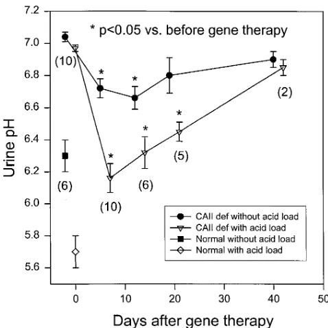
Figure 2. Time course of the changes in pre- and post-acid load urine
pH after gene therapy with bilateral injection. Values are means6SE.
n is the number of mice of each time point. The pre-and post-acid
[image:4.612.56.299.326.602.2] [image:4.612.314.553.419.658.2]gene therapy with bilateral injection. The baseline urine pH was 6.760.06, 6.760.07, 6.860.1, and 6.960.1, respectively, on days 5, 12, 19, and 40 (days 5 and 12, P , 0.05 vs. pre-therapy values). The post-acid load urine pH was 6.260.09, 6.360.1, 6.560.06, and 6.960.1, respectively, on days 7, 14, 21, and 42 (all P , 0.05 vs. pretherapy values, except day 42). Thus, the improvement of urine pH was observed up to 3 wk after gene therapy.
Tissue distribution of transgene expression. The distribution
of the human CAII derived from the transgene in the kidney was investigated using immunohistochemistry studies with an-tibodies against the human CAII. In the normal kidney, CAII staining was widely distributed, particularly in the inner med-ullary and cortical regions (Fig. 3 a). The CAII staining was absent in the kidney of CAII-deficient mice (Fig. 3 b). After gene therapy, CAII staining was found mainly in the outer stripe of medulla and corticomedullary junction (Fig. 3 c, 21 d after gene therapy). No staining of CAII was found in the in-ner medulla. In a higher power view, only renal tubular cells,
but not the vascular or interstitial cells, were positive for CAII (Fig. 3 d). The CAII staining was not detectable in the kidney 42 d after gene therapy (data not shown).
Blood urea nitrogen level and renal histology.
Nephrotox-icity of gene therapy was evaluated with blood urea nitrogen (BUN) and renal histology. BUN levels examined 21 or 42 d after gene therapy were within normal range (2162 mg/dl, n 5 7, vs. normal level, 2261 mg/dl, n 5 10). Renal histology stud-ies revealed no tubular damage or inflammatory reactions as-sociated with gene therapy. The histology of kidney after gene therapy was essentially indistinguishable from that of un-treated kidney.
Discussion
[image:5.612.58.397.303.736.2]In this report, we demonstrate that liposome-mediated gene therapy on CAII-deficient mice with the human CAII gene substantially corrects renal tubular acidosis for up to 3 wk. The time course of changes in the functional study paralleled to the
Figure 3. Immunohistochemical staining of
kidney sections using anti-human carbonic an-hydrase II (CAII) antibodies that recognize both human and mouse CAII. (A) Normal mouse; (B) CAII-deficient mouse; (C and D) representative CAII-deficient mouse 3 wk af-ter gene therapy. (A–C) Low power view; (D) higher magnification of indicated box in Fig. 2
C. In normal mice, CAII was found in the
quantitation of the transgene and its mRNA product. This is the first report showing that liposome-mediated gene therapy can be used for the treatment of renal tubular dysfunctions due to genetic defects.
The observation that urine pH decreased only after trans-fection with the human CAII gene but not with the mock gene, pCMVbgal, indicating that restoration of CAII gene leads to the correction of renal tubular acidosis. The ability to acidify urine was substantially but not completely restored after gene therapy. The effect of transgene appears to be dose dependent because urine pH decreased more in mice that received bilat-eral injection than those with unilatbilat-eral injection.
The transient effect of liposome-mediated gene therapy is expected because the transgene is not incorporated into the chromosome (19). The expression of the transgene lasted for about three weeks, a finding consistent with our previous study using b-galactosidase cDNA with the same delivery system (8). It is possible that this limitation is due to degradation of the delivered DNA over time (19). Recently, Thierry et al. (20) have shown that the duration of luciferase gene expres-sion after intravenous liposome-based delivery of a CMV-driven expression plasmid was increased from several weeks to several months by the inclusion of human papovavirus DNA sequences necessary for the episomal replication of the ex-pressed plasmid in mouse cells. This strategy may possibly be applied to increase the retention of DNA and the duration of gene expression in the kidney. Alternatively, since DNA–lipo-some complexes are not immunogenic, repeated administra-tion of these complexes over time into the renal pelvis could maintain longer term expression of the delivered gene. Re-cently, Song et al. (21) reported that repeated intravenous in-jection of luciferase–liposome complex in mice could maintain the same levels of transgene expression as the first injection. Similarly, Goddard et al. (22) reported that a second dose of a cystic fibrosis transmembrane conductance regulator (CFTR) cDNA–liposome complex is as effective as the first dose in re-storing cAMP-dependent chloride secretion to null CF mice trachea. This double treatment was well tolerated with no dis-cernible inflammation of lung tissues (22). The safety issue of intrarenal–pelvic delivery of gene therapy has been addressed previously (8) and in this study. The BUN levels in mice re-ceived intrarenal–pelvic injection of either pCMVbgal (8) or pCMVCAII in combination with Lipofectin were within the normal range. There were no apparent histological abnormali-ties in the injected kidney. These results suggest that a success-ful gene transfer with DNA–Lipofectin is not associated with any significant renal damage.
Our methodology of gene therapy in mice can be easily adopted to humans. Therapeutic genes can be delivered to the renal pelvis with a flexible ureterorenoscopy. The tissue distri-bution of gene expression after intrarenal–pelvic gene therapy is quite unique: the transgene was only expressed in the outer medulla and corticomedullary junction. These results are simi-lar to our previous studies using the b-galactosidase plasmid (8). It is not clear why there is no transgene expression in the inner medulla. We speculate that the high salt and urea con-tent in the inner medulla may affect the entry of DNA–lipo-some complex. Since this retrograde approach mainly trans-fects the outer medulla, the genetic detrans-fects affecting these regions may benefit from this type of treatment. Diseases with such characteristics include congenital nephrogenic diabetes insipidus, Bartter’s syndrome, Gitelman’s syndrome,
medul-lary cystic disease, familial juvenile nephronophthisis, and oth-ers. The genetic defects in the first three syndromes have been well characterized: congenital nephrogenic diabetes insipidus is caused by a defective vasopressin (V2) receptor (23) or wa-ter channel (24), Bartwa-ter’s syndrome by a defective potassium channel (25) or the furosemide-sensitive Na/K/2Cl cotrans-porter (26), and Gitelman’s syndrome by a defective thiazide-sensitive Na/Cl cotransporter (27). Our experimental model serves as a prototype of gene therapy targeting to renal tubular cells. It will be useful for testing various strategies to improve transfection efficiency and duration of gene expression. Once refined, this gene delivery system can be applied to patients with a variety of renal tubular dysfunction due to genetic de-fects.
Acknowledgments
We thank Drs. Patrick Venta and Richard Tashian for providing the CAII cDNA and Mr. Landon Inge for immunochemistry studies.
This work was supported by the Arizona Elk’s Foundation Trans-plant Research Grant and the Southern Arizona Foundation grant to L. Lai, and National Institutes of Health grant RO1DK52358 and a grant from the Dialysis Clinic, Inc., a nonprofit organization, to Y.H. Lien.
References
1. Tashian, R.E. 1992. Genetics of the mammalian carbonic anhydrases.
Adv. Genet. 30:321–355.
2. Lonnerholm, G., and Y. Ridderstrale. 1980. Intracellular distribution of carbonic anhydrase in the rat kidney. Kidney Int. 17:162–174.
3. Tashian, R.E. 1989. The carbonic anhydrases: widening perspectives on their evolution, expression and function. BioEssay. 10:186–192.
4. Sly, W.S., D. Hewett-Emmett, M.P. Whyte, Y.-S.L. Yu, and R.E. Tash-ian. 1983. Carbonic anhydrase II deficiency identified as the primary defect in the autosomal recessive syndrome of osteopetrosis with renal tubular acidosis and cerebral calcification. Proc. Natl. Acad. Sci. USA. 80:2752–2756.
5. Lewis, S.E., R.P. Erickson, L.B. Barnett, P.J. Venta, and R.E. Tashian. 1988. N-ethyl-N-nitrosourea-induced null mutation at the mouse Car2 locus: an animal model for human carbonic anhydrase II deficiency syndrome. Proc.
Natl. Acad. Sci. USA. 85:1962–1966.
6. Brechue, W.F., E. Kinne-Saffran, R.K.H. Kinne, and T.H. Maren. 1991. Localization and activity of renal carbonic anhydrase (CA) in CAII deficient mice. Biochim. Biophys. Acta. 1066:201–207.
7. Curtis, P.J. 1983. Cloning of mouse carbonic anhydrase mRNA and its in-duction in mouse erythroleukemic cells. J. Biol. Chem. 258:4459–4463.
8. Lai, L., G.W. Moeckel, and Y.H. Lien. 1997. Kidney-targeted liposome-mediated gene transfer in mice. Gene Ther.4:426–431.
9. Lien, Y.H., and L. Lai. 1997. Liposome-mediated gene transfer into the tubules. Exp. Nephrol. 5:132–136.
10. Lien, Y.H., and L. Lai. 1997. Gene therapy for renal diseases. Kidney
Int. 52(Suppl. 61):S85–S88.
11. Felgner, P.L, T.R. Gadek, M. Holm, R. Roman, H.W. Chan, M. Wenz, J.P. Northrop, G.M. Ringold, and M. Danielsen. 1987. Lipofection: a highly ef-ficient, lipid-mediated DNA-transfection procedure. Proc. Natl. Acad. Sci.
USA. 84:7413–7417.
12. Zhu, N., D. Liggitt, Y. Liu, and R. Debs. 1993. Systemic gene expression after intravenous DNA delivery into adult mice. Science.261:209–211.
13. Stewart, M.J, G.E. Plautz, L.D. Buono, Z.Y. Yang, L. Xu, X. Gao, L. Huang, E.G. Nabel, and G.L. Nabel. 1992. Gene transfer in vivo with DNA-liposome complexes: safety and acute toxicity in mice. Hum. Gene Ther. 3:267– 275.
14. Nabel, G.L, E.G. Nabel, Z. Yang, B.A. Fox, G.E. Plautz, X. Gao, S. Shu, D. Gordon, and A.E. Chang. 1993. Direct gene transfer with DNA-lipo-some complexes in melanoma: expression, biologic activity, and lack of toxicity in humans. Proc. Natl. Acad. Sci. USA. 90:11307–11311.
15. Caplen, N.J., E. Alton, P.G. Middleton, I.R. Dorin, B.J. Stevenson, X. Gao, S.R. Durham, P.K. Jeffery, M.E. Hodson, C. Coutelle, et al. 1995. Lipo-some-mediated CFTR gene transfer to the nasal epithelium of patients with cystic fibrosis. Nat. Med.1:39–46.
16. Alino, S.F., M. Bobadilla, J. Crespo, and M. Lejarreta. 1996. Human al-pha 1antitrypsin gene transfer to in vivo mouse hepatocytes. Hum. Gene Ther. 7:531–536.
Hartikka, J. Nietupski, M. Manthorpe, M. Nichols, et al. 1996. A novel cationic lipid greatly enhances plasmid DNA delivery and expression in mouse lung.
Proc. Natl. Acad. Sci. USA. 26:11454–11458.
18. Bernt, E., and H.U. Bergmeyer. 1965. Urea. In Methods of Enzymatic Analysis. H.U. Bergmeyer, editor. Academic Press, New York. 401–406.
19. Ledley, F.D. 1995. Nonviral gene therapy: the promise of genes as phar-maceutical products. Hum. Gene Ther. 6:1129–1144.
20. Thierry, A.R., Y. Lunardi-Iskandar, J.L. Bryant, P. Rabinovich, R.C. Gallo, and L.D. Mahan. 1995. Systemic gene therapy: biodistribution and long-term expression of a transgene in mice. Proc. Natl. Acad. Sci. USA. 92:9742– 9746.
21. Song, Y.K., F. Liu, S. Chu, and D. Liu. 1997. Characterization of cat-ionic liposome-mediated gene transfer in vivo by intravenous administration.
Hum. Gene Ther. 8:1585–1594.
22. Goddard, C.A., R. Ratcliff, J.R. Anderson, E. Glenn, S. Brown, D.R. Gill, S.C. Hyde, L.J. MacVinish, L. Huang, C.F. Higgins, et al. 1997. A second dose of a CFTR cDNA-liposome complex is as effective as the first dose in re-storing cAMP-dependent chloride secretion to null CF mice trachea. Gene
Ther. 4:1231–1236.
23. Rosenthal, W., A. Seibold, A. Antaramian, M. Lonergan, M.F. Arthus,
G.N. Hendy, M. Birnbaumer, and D.G. Bichet. 1992. Molecular identification of the gene responsible for congenital nephrogenic diabetes insipidus. Nature. 359:233–235.
24. van Lieburg, A.F. M.A. Verdijk, V.V. Knoers, A.J. van Essen, W. Proesmans, R. Mallmann, L.A. Monnens, and B.A. van Oost. 1994. Patients with autosomal nephrogenic diabetes insipidus homozygous for mutations in the aquaporin 2 water-channel gene. Am. J. Hum. Gene. 55:648–652.
25. Simon, D.B., F.E. Karet, J. Rodriguez-Soriano, J.H. Hamdan, A. DiPi-etro, H. Trachtman, S.A. Sanjad, and R.P. Lifton. 1996. Genetic heterogeneity of Bartter’s syndrome revealed by mutations in the K1 channel, ROMK. Nat.
Genet. 14:152–156.
26. Simon, D.B., F.E. Karet, J.M. Hamdan, A. DiPietro, S.A. Sanjad, and R.P. Lifton. Bartter’s syndrome, hypokalaemic alkalosis with hypercalciuria, is caused by mutations in the Na-K-2Cl cotransporter NKCC2. Nat. Gene. 13: 183–188.