Copyright © 1998, American Society for Microbiology. All Rights Reserved.
Serratia ficaria: a Misidentified or Unidentified Rare Cause of
Human Infections in Fig Tree Culture Zones
T. ANAHORY, H. DARBAS,* O. ONGARO, H. JEAN-PIERRE,ANDP. MION
Laboratoire de Bacte´riologie, Hoˆpital Arnaud de Villeneuve, CHU de Montpellier, F-34295 Montpellier Cedex 5, France
Received 12 December 1997/Returned for modification 31 March 1998/Accepted 18 August 1998
Serratia ficaria, an enterobacterium involved in the fig tree ecosystem, has been isolated from human clinical samples in rare instances, and its role as a pathogen is unclear. In 7 years, we have isolated S. ficaria from seven patients; it was the only pathogen in 4 patients, including a patient with septicemia described previously and three patients with gallbladder empyemas described in the present report. From March 1995 to July 1997, the incidence of biliary infections due to S. ficaria was 0.7%. We discuss the digestive carriage of this bacterium and its epidemiology with respect to the fig tree life cycle. Since fig trees grow around the Mediterranean as well as in the United States (California, Louisiana, Hawaii), S. ficaria should be more frequently isolated. In our experience, various strains have been misidentified or unidentified by commercial systems. Incorrect identi-fication could be an additional explanation for the paucity of reported cases. S. ficaria produces nonpigmented, lactose-negative colonies which give off a potatolike odor. This odor is the primary feature of S. ficaria and must prompt reexamination of the identifications proposed by commercial systems. We tested 42 novel strains using three commercial systems: Vitek gram-negative identification (GNI) cards and API 20E and ID 32E strips (bioMe´rieux, Marcy-l’Etoile, France). The percentages of positivity that we have obtained were lower than those published previously for the following characteristics: lipase, gelatinase, DNase, and rhamnose. The best system for the recognition of S. ficaria is ID 32E, which correctly identified 27 of 42 strains. The API 20E system gave correct identifications for only two strains. S. ficaria was not present in the Vitek GNI card system database.
Serratia ficaria was first described in 1979 by Grimont et al.
(9) as part of the fig tree ecosystem. Since then, this bacterium has been isolated from human clinical samples in relatively few instances (1, 2, 7, 14, 15), and its role as a pathogen was always questionable. Since 1990, in Montpellier, France, a city located in the Mediterranean area, we have isolated S. ficaria from seven patients. In four patients, three patients with gallbladder empyemas and one patient with septicemia originating from the gut, its pathogenic role was clear (3, 4). To date, no other reports of such infections have been published, even though the fig tree grows throughout the Mediterranean area and in the United States (California [7], Louisiana [7], and Hawaii [14]). Is S. ficaria misidentified? As yet, the available data on the biochemical characteristics of this species concern fewer than 20 strains (5, 9). Are all S. ficaria biotypes known? The two principal publications (5, 9) report characteristics which were studied by conventional methods. Are these methods still used in routine laboratories? As far as we are concerned, we use API 20E strips for the identification of enterobacteria, and in our work we have encountered some problems with the identification of S. ficaria. Consequently, we have tested three commercial systems, gram-negative identification cards (Vitek GNI cards) and API 20E and ID 32E strips (bioMe´rieux, Marcy-l’Etoile, France), for their abilities to identify 42 novel strains of S. ficaria. We report on the biochemical character-istics that we obtained and discuss the main deficiencies in the three systems for the identification of this species.
CASE REPORTS
Patient 1. On 7 October 1990, a 70-year-old man was ad-mitted to the visceral surgery service for acute cholecystitis. His body temperature was 37.8°C. The leukocyte count was 18,000/ mm3. The patient was given ceftriaxone (2 g per day) and
underwent cholecystectomy on the next day. The operation revealed an important local inflammation: the gallbladder was full of multiple stones, was large and purplish, and was covered with numerous false membranes; its wall was exceptionally thick; and an associated odditis was found. On 11 October, the persistence of a subfebrile state (37.9°C) led to a change from ceftriaxone to amoxicillin plus clavulanic acid (1 g twice daily). Apyrexia was achieved 4 days later. The patient returned home on 17 October.
This man had eaten figs in season, but his fig consumption was poorly related to the timing of the beginning of clinical signs.
Patient 2. An 81-year-old man suffering from an acute hy-drocholecystis with fever (39°C) and leukocytosis (29,000 poly-mophonuclear leukocytes/mm3) was admitted on 20
Septem-ber 1993 to the visceral surgery service. On the previous day, he had been given amoxicillin plus clavulanic acid (2 g per day) by perfusion. An operation was performed on 21 September. The operation showed a necrotic gallbladder full of pus and infected mud, a sample of which was sent to our laboratory.
Under antibiotic treatment, the body temperature and the leukocyte count (9,800/mm3) returned to normal on 27
Sep-tember. From 29 September, the antibiotic was administered per os and the patient was discharged.
It was not possible to ask the patient about his fig consump-tion.
Patient 3.On 22 May 1996, a 59-year-old woman was re-ferred to the emergency service for acute cholecystitis revealed
* Corresponding author. Mailing address: Laboratoire de Bacte´ri-ologie, Hoˆpital Arnaud de Villeneuve, F-34295 Montpellier Cedex 5, France. Phone: 33 467 33 58 88. Fax: 33 467 33 61 25. E-mail: michel.brun@cge-ol.fr.
3266
on May 15, 2020 by guest
http://jcm.asm.org/
by pain in the right hypochondrium and biliary vomiting. On admission, the patient was febrile (38°C). Abdominal palpation detected a “guard” reaction at the right side. The leukocyte count was 15,380/mm3. Therapy with intravenous amoxicillin
plus clavulanic acid (1 g three times daily) was started. On the next day, an operation revealed gangrenous cholecystitis with a purulent perivesicular discharge. The gallbladder had a thick wall and contained one calculus of cholesterol.
A couple of days after surgery, oral antibiotic treatment (1 g twice daily) replaced administration by perfusion. The body temperature returned to normal on 26 May. The patient was discharged 1 day later.
In May, edible fresh figs do not grow locally. We asked the patient about the presence of a fig tree in her garden or neighborhood. She said that no fig trees were in proximity to her home but that perhaps there were fig trees in the country-side where she frequently walked.
Laboratory data for patients 1 to 3.Microscopic examina-tion of biliary fluid from the three patients showed numerous polymorphonuclear cells but no bacterium. After incubation for 24 h at 37°C under aerobic and anaerobic conditions,
S. ficaria was isolated, in pure culture, on MacConkey medium
and chocolate agar (bioMe´rieux) from intraoperative samples from the three patients. The antibiotic susceptibilities of the isolates were tested by disk diffusion and the results were read according to the standards of the French Antibiogram Com-mittee. Except for tetracycline, to which one isolate was resis-tant, all strains had similar susceptibility patterns: resistance to cephalothin and susceptibility to all other beta-lactams, ami-noglycosides, chloramphenicol, colistin, trimethoprim-sulfa-methoxazole, nalidixic acid, and fluoroquinolones.
MATERIALS AND METHODS
Strains.The 42 strains studied comprised 11 clinical isolates (the sources of which are given in Table 1) and 31 strains from the fig tree ecosystem. Between February and June 1994, we collected figs, buds, and insects from fig trees growing within or around the precinct in which our hospital is located. This sampling allowed us to recover 11 S. ficaria isolates from male figs, isolate 1 from a pollinated female fig, 14 isolates from Blastophaga psenes (a fig tree-specific pollinator that breeds in male figs), and 5 isolates from Philotrypesis caricae (a parasitic insect which also breeds in figs but which is not implicated in their pollination). Because all of the environmental strains were isolated from fig trees located in an area covering 2.5 km2, it seemed possible that these strains had the same clonal origin. By means of pulsed-field gel electrophoresis (the restriction enzyme XbaI was obtained from Appligene Oncor, Illkirch, France), we looked for clonal strains. Among human strains, the two Belgian isolates constituted a clone. Among the environmental strains we detected four clones, and these clones were essentially among strains isolated from insects collected from the same fig. However, three or four different clones could cohabitate in the same fruit. The isolates from different figs and, a fortiori, from different fig trees were
different. Thus, of 11 human strains, 10 were genetically different, and of 31 environmental strains, 23 had different genomes.
At the time of their isolation, our strains were identified as follows. Biochem-ical characteristics were obtained on API 20E strips with, if necessary, control of the utilization of citrate as the sole carbon source by culture on Simmons citrate agar and examination for a potatolike odor. For environmental strains, isolation from figs or insects that breed in figs was also considered. All human isolates and doubtful environmental strains were sent to P. A. D. Grimont (Unite´ des En-te´robacte´ries, Institut Pasteur, Paris, France), who confirmed our identification by means of a carbon source utilization study with Biotype 99 carbon source strips (bioMe´rieux).
Identification.In the opinion of Grimont and Grimont (8), the methodology of carbon source utilization tests is essential to Serratia identification. Conse-quently, they have set up the special gallery of tests mentioned above (Biotype 99; bioMe´rieux). The supplier of this system presents it as a dedicated tool for research laboratories. Furthermore, the interpretation of results requires special software sold by another group (Institut Pasteur Taxolab, Paris, France), and this program can be loaded only on Macintosh computers. These reasons excluded the use of Biotype 99 since we wished to work under the same routine conditions used by nonspecialized laboratories in order to point out the difficulties encoun-tered in S. ficaria identification.
After overnight incubation at 37°C on MacConkey agar, cultures were tested for oxidase, catalase, and DNase reactions and were used to inoculate three systems for the identification of gram-negative organisms: Vitek GNI cards and API 20E and ID 32E strips. These systems were used according to the recom-mendations of the supplier.
The Vitek GNI cards were introduced into a reader-incubator. The incubation cycle is 4 to 18 h. At the completion of the incubation cycle, the biochemical pattern was printed for each card in the reader-incubator.
The ID 32E system requires incubation at 37°C for 24 h under aerobic con-ditions. Before strip reading, the indole reaction was revealed by the addition of one drop of James reagent. The automatic reading uses the ATB Expression instrument; the reader records the color of each tube and transmits the data to the computer.
Also, the API 20E strips require incubation at 37°C for 24 h in a humid atmosphere under aerobic conditions. Then, reagents were added as appropriate to the tryptophan desaminase (TDA), Voges-Proskauer, and indole (IND) tubes. Nitrate reduction was revealed in a glucose tube with Griess reagents. All reaction tubes were read visually. When the color interpretation was not clear, the reactions were noted as doubtful.
For DNase detection, a sample of each cultured strain was streaked onto DNA-toluidine blue agar (Sanofi-Diagnostics Pasteur, Marnes la Coquette, France). This medium was incubated for 24 h at room temperature (about 22°C). If DNase is present, the agar shows a pink halo extending several millimeters around the streak.
Catalase reactions were studied with hydrogen peroxide.
The cytochrome oxidase test was performed with cultures from trypto-casein soy agar and disks impregnated with dimethyl-paraphenylenediamine oxalate (Sanofi-Diagnostics Pasteur).
RESULTS
[image:2.612.53.549.82.210.2]Frequency of biliary infections due to S. ficaria. The fre-quency of occurrence of biliary infections due to S. ficaria was estimated from March 1995 to July 1997 (inclusive). During this period, 385 biliary samples were seeded under aerobic and anaerobic conditions. Growth was obtained from 166 of the
TABLE 1. Sources of human strains
Sexa Age (yr) Geographic
location Isolation date Source Reference
M 62 Belgiumb January 1982 Expectoration 2
M 62 Belgiumb January 1982 Tracheal secretions 2
M ? Francec July 1988 Extra-articular knee wound 10
M ? Francec October 1992 Bronchial secretions
M 70 France October 1990 Bile 3
F 42 France May 1991 Stools
M France June 1993 Drainage tube of ankle wound
M 83 France July 1993 Four blood cultures 4
M 81 France September 1993 Bile
F 56 France September 1993 One blood culture
F 59 France May 1996 Bile
aM, male; F, female.
bFrom J. A. Brouillard, Department of Bacteriology, Institut Provincial d’Hygie`ne et de Bacte´riologie, Mons, Belgium. cFrom C. Suc, Laboratoire de Bacte´riologie, Hoˆpital Rangueil, Toulouse, France.
on May 15, 2020 by guest
http://jcm.asm.org/
cultured samples from 142 infected patients. Eighty-two infec-tions were monomicrobial (the infecinfec-tions were due to an an-aerobic bacterium in three patients). Among the 60 remaining mixed infections, 15 were caused by flora that included anaer-obes. One enterobacterium or a diverse combination of en-terobacteria were responsible for 84 infections, Serratia spp. were responsible for 3 infections, and S. ficaria was responsible for 1 infection (0.7% of infected patients).
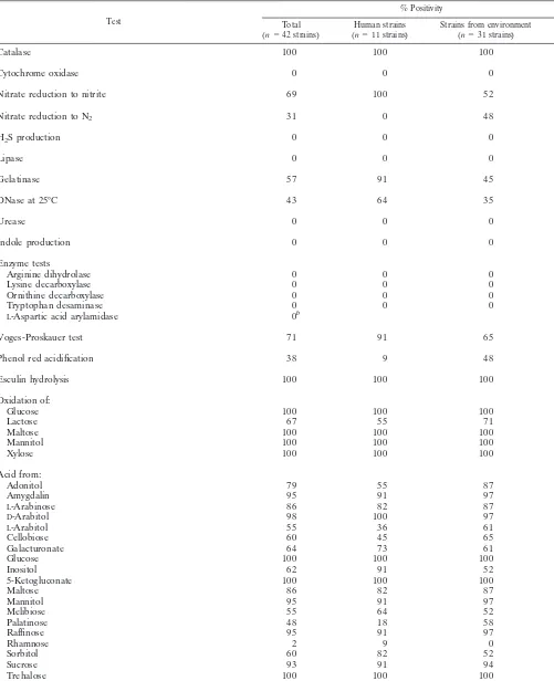
Identification.Table 2 presents our results expressed as the percentages of strains positive by the various tests. The three identification systems do not include the same tests. When a biochemical test exists only in one identification system, the result in Table 2 is the result for that system. In adverse situations, the same strain may give different or concordant results in the various systems, but this depends on the specific characteristic. Tests for H2S production, urease, and indole
and other tests for enzymes implicated in the metabolism of amino acids were constantly negative, whereas tests forb -ga-lactosidase expression and carbohydrate acidifications were much more variable. For both b-galactosidase and carbohy-drate acidifications, the percentages reported in Table 2 were established from the number of strains showing positive reac-tions in all three systems.
Table 2 also presents the characteristics of bacteria isolated from patients and strains obtained from local fig trees and the relatively significant difference as determined by the chi-square test (P,0.05). Human strains reduced nitrate only to nitrite, whereas about half of the environmental strains reduced ni-trate to nitrogen.b-Galactosidase was present in about twice as many environmental strains as human strains, and environ-mental strains were more often glucidolytic for adonitol, ino-sitol, and palatinose. Human strains were more often proteo-lytic.
With ID 32E strips, 27 strains were correctly identified. The biochemical profiles of other strains were considered unaccept-able. Among these strains, S. ficaria was proposed as the only choice (2 strains), as the first choice (8 strains), and as the second choice (3 strains) or was never proposed (2 strains);
Serratia rubidaea also appeared among the proposed
identifi-cations for 10 strains.
API 20E strips gave correct identifications for two S. ficaria strains. Sixteen strains were identified to the Serratia genus level: S. ficaria appeared as the first choice for three strains and as the second choice behind Serratia plymuthica for 13 strains. One strain was identified as S. plymuthica. Twenty-three strains had unacceptable profiles. Among these, S. ficaria and/or S.
ply-muthica appeared with various other possible identifications
(20 strains); the genus Serratia never appeared (3 strains). Vitek GNI cards did not allow us to identify S. ficaria be-cause this species was not included in the system’s database. For 27 strains, the proposed identification agreed with Serratia genus, for 14 other strains the proposed identification was
Klebsiella ozaenae, and 1 strain remained unidentified.
DISCUSSION
Pathogenicity. All three intraoperative biliary samples yielded pure cultures of S. ficaria, and this bacterium was responsible for the local and general infectious state. Fever and leukocytosis were always present. Pus was observed mac-roscopically, and microscopic examination showed numerous polymorphonuclear cells. Thus, S. ficaria is able to cause severe infections such as these deep suppurations or septicemia as reported previously (4) and can clearly play a pathogenic role. However, the level of this pathogenicity seems to be low be-cause in the patients with septicemia and gallbladder
empy-ema, the course to recovery was both uncomplicated and speedy, even though the patients were elderly and even though the patient with septicemia was suffering from cancer.
Epidemiology. Gallbladder contamination is brought about by the bacteria from the small gut. On the other hand, the septicemia occurred after an antrectomy and anastomosis be-tween the remaining stomach and the first jejunal loop. Both cases of infection imply that S. ficaria must be a part (at least transiently) of the human intestinal flora. Consequently, using selective caprylate medium (16), we have looked for its pres-ence in feces, although that effort was in vain (13). Several things may explain this failure. (i) The clinical cases reflect S.
ficaria carriage at the duodenojejunal level of the intestine,
whereas stool culture investigates the colic flora. (ii) The screening method that we used was based on DNase, and we may have missed S. ficaria in stools. Indeed, among the 11 clinical strains of S. ficaria from our own collection, all except 4 isolated from bile and stools were DNase positive. (iii) Fi-nally, S. ficaria is very rarely isolated (in our laboratory, seven isolates in 7 years), and digestive carriage must also be rare and likely depends on both environmental and climatic factors which are difficult to determine considering our present state of knowledge about the epidemiology of S. ficaria. All these reasons make the intestinal carriage of S. ficaria difficult to probe and prove.
For some investigators it seems logical to explain S. ficaria digestive carriage by fresh fig consumption (1, 2, 7, 14, 15). We believe the explanation to be less simple, particularly when some of the details of fig tree biology are considered. The fig tree (Ficus carica) is a dioecious species. The male tree yields inedible figs, sometimes called caprifigs, in which breeds a specific pollinator (B. psenes), a hymenopteran the size of a midge. In the course of a year, there are usually two genera-tions of B. psenes, in May and in July, August, and September. This second generation pollinates female figs, which turn ripe and edible in October. This is the case for both wild F. carica and a kind of cultured F. carica (for example, the Smyrna variety called Calimyrna in California). Another kind of cul-tured fig tree gives, in July and October, two crops of parthe-nocarpic figs which ripen and become edible without pollina-tion. These cultivars are cultured in southern France both in home gardens and for commercial production. The partheno-carpic figs harvested in July do not contain S. ficaria; thus, the only edible figs able to harbor S. ficaria are those that are pollinated in July and that ripen in October (unpublished ob-servations). Then, if fig consumption was the only cause of human contamination, all clinical isolates should occur in Oc-tober, like in our patients 1 and 2 and the patient reported by Gill et al. (7). However, how is the infection in the third patient, which occurred in May when no figs were ripe, ex-plained? This month is the time when the first generation of B.
psenes is flying. In the same way, the case of septicemia (4)
occurred in July, when the second generation of hymenopter-ans leaves male trees to pollinate the female figs on the female trees. These flies can cover several kilometers and therefore can contribute to the spread of S. ficaria over a wide area. Grimont and colleagues (10, 11) isolated S. ficaria from figs, a fig leaf, and B. psenes and also from common grass, scilla, market mushrooms, and an ant. We were unable to consider the isolation of S. ficaria from market mushrooms because the season and geographic origin were not indicated. However, the scilla collected in Bordeaux grew 5 m away from a fig tree from which S. ficaria was isolated from a fig and a leaf, and all three isolated strains belonged to the same serovar (O2:H1) (11). The picking time was not mentioned, but the isolation of S.
ficaria from a leaf and from around a tree which bore figs could
on May 15, 2020 by guest
http://jcm.asm.org/
TABLE 2. Biochemical characteristics of S. ficaria: comparison between human and environmental strains
Test
% Positivity
Total
(n542 strains) (nHuman strains511 strains) Strains from environment(n531 strains) Pa
Catalase 100 100 100
Cytochrome oxidase 0 0 0
Nitrate reduction to nitrite 69 100 52 0.01
Nitrate reduction to N2 31 0 48 0.01
H2S production 0 0 0
Lipase 0 0 0
Gelatinase 57 91 45 0.01
DNase at 25°C 43 64 35 NS
Urease 0 0 0
Indole production 0 0 0
Enzyme tests
Arginine dihydrolase 0 0 0
Lysine decarboxylase 0 0 0
Ornithine decarboxylase 0 0 0
Tryptophan desaminase 0 0 0
L-Aspartic acid arylamidase 0b
Voges-Proskauer test 71 91 65 NS
Phenol red acidification 38 9 48 0.03
Esculin hydrolysis 100 100 100
Oxidation of:
Glucose 100 100 100
Lactose 67 55 71 NS
Maltose 100 100 100
Mannitol 100 100 100
Xylose 100 100 100
Acid from:
Adonitol 79 55 87 0.03
Amygdalin 95 91 97 NS
L-Arabinose 86 82 87 NS
D-Arabitol 98 100 97 NS
L-Arabitol 55 36 61 NS
Cellobiose 60 45 65 NS
Galacturonate 64 73 61 NS
Glucose 100 100 100
Inositol 62 91 52 0.03
5-Ketogluconate 100 100 100
Maltose 86 82 87 NS
Mannitol 95 91 97 NS
Melibiose 55 64 52 NS
Palatinose 48 18 58 0.03
Raffinose 95 91 97 NS
Rhamnose 2 9 0 NS
Sorbitol 60 82 52 NS
Sucrose 93 91 94 NS
Trehalose 100 100 100
Glucose fermentation in the presence of:
2,4,49-Trichloro-29-hydroxy-diphenylether (0.3 g/liter) 64 91 55 0.04
P-Coumaric (2 g/liter) 100 100 100
Continued on following page
on May 15, 2020 by guest
http://jcm.asm.org/
indicate whether the month was that of the first generation of
B. psenes fly (if a male tree is involved) or the season of
pollination, since Bordeaux is located in an area of B. psenes activity. The S. ficaria-carrying ant was also collected in Bor-deaux (11), but when? As far as we are concerned, we have not isolated any S. ficaria strains from cochineals or ants recovered from fig trees during a period that was not during the period of activity of B. psenes. Because a majority of clinical isolates occurred between May and October (corresponding to the activity period of B. psenes) and were related to the geographic area of Blastophaga activity (California, Louisiana, Hawaii for U.S. cases and below the 46th parallel of northern latitude for French cases) (1, 4, 7, 14, 15), we think that Blastophaga plays a part, perhaps the major part, in S. ficaria epidemiology. This hypothesis is further supported by reports from Grimont and colleagues (8, 11). Californian figs could mature only after B.
psenes from male trees imported from Greece and Algeria
established its life cycle. Strains of S. ficaria from the Mediter-ranean region were not antigenically uniform, while all U.S. strains studied (isolated from the fig wasp or from human patients) belonged to the same serotype (O1:H1) and were likely taken to the United States by the Mediterranean B.
psenes. Arguing against a role for B. psenes, we must cite the
two unexplainable Belgian cases of S. ficaria infection that occurred in January (2) and the isolation of S. ficaria from common grass picked up in Saint-Remy-le`s-Chevreuse, France (11). These isolates are situated well above the 46th parallel of northern latitude. January is the time of fig pollination in the austral hemisphere.
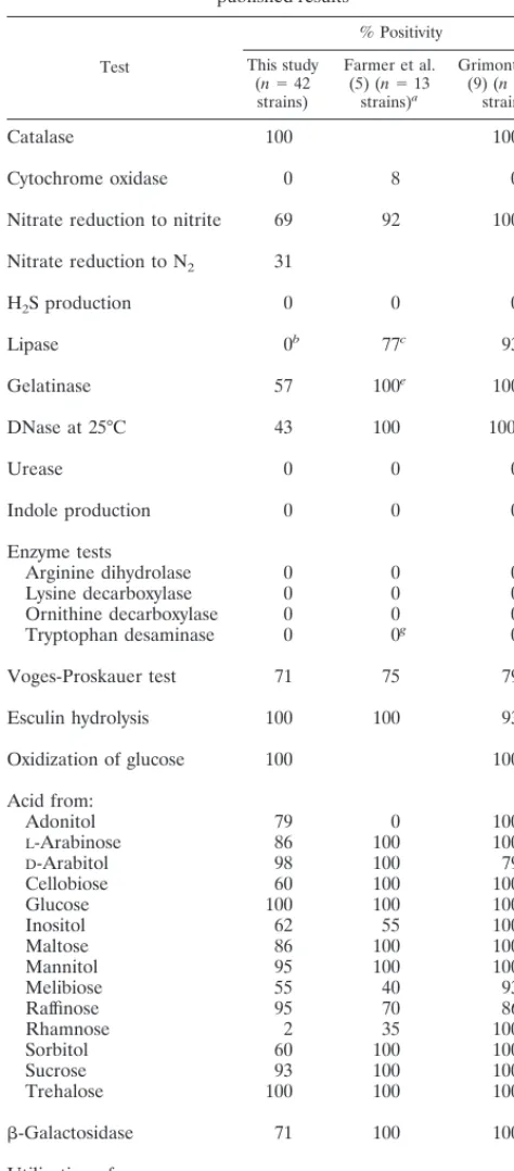
Identification.Table 3 compares the results obtained with 3 commercial systems (from Table 2) with those obtained by Farmer et al. (5) and Grimont et al. (9) by conventional meth-ods. In their princeps publication, Grimont et al. (9) report on the characteristics of 14 original strains issued from the envi-ronment (figs and B. psenes). Farmer et al. (5) studied 13 strains, including 10 strains supplied by Grimont and 3 clinical isolates. Thus, the distribution of strains (clinical and environ-mental) in the series of Farmer et al. (5) is similar to ours (3 of 13 and 11 of 42, respectively). We tested 42 other strains by three commercial systems. However, all results are consistent
with respect to the following characteristics: catalase; cyto-chrome oxidase; nitrate reduction; H2S production; urease
production; indole production; decarboxylases and tryptophan desaminase production; Voges-Proskauer test; esculin hydro-lysis; acidification of L-arabinose, D-arabitol, cellobiose, glu-cose, inositol, maltose, mannitol, melibiose, raffinose, sorbitol, sucrose, and trehalose;b-galactosidase production; and utili-zation of malonate and citrate. Citrate utiliutili-zation was always positive when it was tested with Simmons citrate medium or by the Vitek GNI card, but the API 20E system did not allow the correct expression of this characteristic.
Farmer et al. (5) describe the genus Serratia as usually being colistin resistant and producing the extracellular enzymes DNase, gelatinase, and lipase. The resistance to the polymyxin group concerns all the molecules of this group, but, unexpect-edly, none of our S. ficaria strains tested with the GNI Vitek card grew in the presence of polymyxin B (300mg/ml). Is this concentration too high to allow the expression of colistin re-sistance (the disk for antibiotic susceptibility testing contains 50mg of colistin)? Is colistin resistance an usual characteristic for members of the genus Serratia or only for the species
Serratia marcescens? Among the 33 strains of S. ficaria whose
antibiograms have been published, only 8 were resistant (4). Concerning lipase activity, the use of different substrates may explain the different results. Farmer et al. (5) and Grimont et al. (9) proved the lipase activity using two conventional substrates, corn oil and Tweens, respectively, which revealed the enzyme in 77 and 93% of strains, respectively (not a sig-nificant difference). Apparently, S. ficaria lipase is not able to lyse 5-bromoindoxyl ester, the substrate selected by bio-Me´rieux for its lipase test (ID 32E).
[image:5.612.61.548.81.269.2]A total of 57 and 43% of our strains were positive for gelatinase and DNase, respectively. Moreover, clinical strains tend to be more proteolytic than strains recovered from the fig tree ecosystem. The difference is significant for gelatinase but not for DNase. All human strains are DNase positive except the four strains isolated from bile and stools, both of which contain biliary salts (we do not yet have any explanation for this phenomenon). This observation is very surprising if one compares these results with the results obtained by Farmer et
TABLE 2—Continued
Test
% Positivity
Total
(n542 strains) (nHuman strains511 strains) Strains from environment(n531 strains) Pa
Enzyme tests
a-Galactosidase 93 100 90 NS
b-Galactosidase 71 45 81 0.03
a-Glucosidase 60 82 52 NS
b-Glucosidase 100 100 100
b-Glucuronidase 0 0 0
a-Maltosidase 43 18 52 0.05
N-Acetyl-b-glucosaminidase 0 0 0
Utilization of:
Acetamide 0 0 0
Citratec 100 100 100
Malonate 0 0 0
Growth in the presence of polymyxin B (0.3 g/liter) 0 0 0
aP values were determined by thex2test. NS, not significant.
bDoubtful for 12% of strains.
cA rate of positivity of 100% on Simmons citrate agar and also with Vitek GNI cards. This characteristic is very difficult to read when API 20E galleries are used
(positive, 27%; negative, 19%; doubtful, 17%).
on May 15, 2020 by guest
http://jcm.asm.org/
al. (5) or Grimont et al. (9) with environmental strains (100% of strains produce both enzymes) but is not abnormal if one considers that in various bacteria proteolytic enzymes play a part as virulence factors. In the same way, it seems logical that environmental strains show strong nitrate reductase activity up to the nitrogen step and that strains cultured from fruits dis-play strong saccharoclastic metabolism.
With regard to sugar fermentation, the only significant dif-ferences between the results of Grimont et al. (9), those of Farmer et al. (5), and those from our present study concern the fermentation of adonitol and rhamnose. Table 4 presents the results provided by the three identification systems compared with those obtained by Farmer et al. (5) and Grimont et al. (9) by conventional methods. The GNI Vitek card gave the best results and results that were nearer the reported ones. With the API 20E and ID 32E strips, most sugar fermentations were difficult to read. Grimont and Grimont (8) noted previously “that utilization tests are preferable to fermentation tests since strains able to utilize a polyalcohol sometimes fail to produce enough acid products to give a positive reaction in fermenta-tion tests.” It is a pity that utilizafermenta-tion tests useful under routine conditions are not yet available.
[image:6.612.303.546.82.382.2]Therefore, how should S. ficaria be identified? On Mac-Conkey medium, S. ficaria looks like a lactose-negative entero-bacterium (or a weakly lactose-positive enteroentero-bacterium, de-pending on the supplier of medium). The colonies are smooth, beige, transparent, or opaque and can turn pinkish after sev-eral days. The culture gives off a strong potatolike odor. Avoid the GNI Vitek card because it does not include S. ficaria in its database. API 20E strips do not differentiate S. ficaria from S.
TABLE 3. Biochemical characteristics of S. ficaria: comparison with published results
Test
% Positivity
This study (n542 strains)
Farmer et al. (5) (n513
strains)a
Grimont et al. (9) (n514
strains)
Catalase 100 100
Cytochrome oxidase 0 8 0
Nitrate reduction to nitrite 69 92 100
Nitrate reduction to N2 31
H2S production 0 0 0
Lipase 0b 77c 93d
Gelatinase 57 100e 100
DNase at 25°C 43 100 100f
Urease 0 0 0
Indole production 0 0 0
Enzyme tests
Arginine dihydrolase 0 0 0
Lysine decarboxylase 0 0 0
Ornithine decarboxylase 0 0 0
Tryptophan desaminase 0 0g 0g
Voges-Proskauer test 71 75 79
Esculin hydrolysis 100 100 93
Oxidization of glucose 100 100
Acid from:
Adonitol 79 0 100
L-Arabinose 86 100 100
D-Arabitol 98 100 79
Cellobiose 60 100 100
Glucose 100 100 100
Inositol 62 55 100
Maltose 86 100 100
Mannitol 95 100 100
Melibiose 55 40 93
Raffinose 95 70 86
Rhamnose 2 35 100
Sorbitol 60 100 100
Sucrose 93 100 100
Trehalose 100 100 100
b-Galactosidase 71 100 100
Utilization of:
Citrate 100h 100i 100i
Malonate 0 0 0
aIncluding 10 strains supplied by P. A. D. Grimont. bSubstrate, 5-bromo-indoxyl ester.
cSubstrate, corn oil.
dSubstrate, Tweens 40, 60, and 80. eAt 22°C.
fTemperature not indicated. gPhenylalanine desaminase.
hA rate of positivity of 100% on Simmons citrate agar and also with Vitek
GNI cards. This characteristic is very difficult to read when API 20E galleries are used (positive, 27%; negative; 19%; doubtful, 17%).
[image:6.612.50.287.96.635.2]iSimmons citrate.
TABLE 4. Comparison of some results given by the three identification systems
Test
API 20E ID 32E GNI Vitek % Positive
No.a % No. % No. % Farmeret al.
(5)
Grimont et al. (9)
Adonitol 132 76 141 98b 0 100
27 21
?2
L-Arabinose 137 88 141 98 142 100 100 100
22 21
?3
Inositol 126 62 128 67 129 69 55 100
214 214 213
?2
Mannitol 141 98 141 98 100 100
21 ?1
Rhamnose 17 17 14 10 117 40 35 100
227 236 225
?8 ?2
Sorbitol 135 83 126 62 142 100 100 100
214
?7 ?2
Sucrose 141 98 139 93 142 100 100 100
21
?1 ?2
aNo., number of positive (1), negative (2), or doubtful (?) results. bBoldface numbers are closely related results.
on May 15, 2020 by guest
http://jcm.asm.org/
plymuthica very well. The biochemical characteristics
impli-cated in this inadequate differentiation are rhamnose acidifi-cation and utilization of citrate. In the API 20E database, 100 and 92% of S. ficaria strains are expected to be citrate and rhamnose positive, respectively, whereas 65 and 8% of S.
plymuthica strains, respectively, are expected to be positive. In
our experience, few strains of S. ficaria can acidify rhamnose. On the other hand, we mentioned above the difficulties en-countered with reading the API 20E citrate tubes. The easiest control is to smell the plates. If the culture smells of potato, the bacterium is S. ficaria; it is also necessary to control citrate positivity on Simmons citrate agar. With ID 32E strips, a pos-sible misidentification is between S. ficaria and S. rubidaea. Unfortunately, a few strains of S. rubidaea (6) give off a musty or potatolike odor. The latter species, however, appears to be lactose positive on MacConkey medium, and most strains pro-duce prodigiosin. Besides, S. rubidaea is “rarely isolated both in the natural environment and in human patients” (12), whereas S. ficaria isolates are relatively more frequently iso-lated (in 7 years, we have isoiso-lated S. ficaria from seven patients but have isolated S. rubidaea from only one patient) and occur in the period and zone of activity of B. psenes. Season and geographic area are also very important criteria for consider-ation in the diagnosis of an S. ficaria infection.
Up to now, published reports about characteristics for the identification of S. ficaria concerned a maximum of 17 strains. The database of commercial systems was probably established with data for a few strains and must evolve. The aim of the study described in this report, based on 42 strains, is to con-tribute to such an update.
Conclusion.In the area of activity of B. psenes, S. ficaria can be responsible for human infections such as gallbladder empy-ema or even septicemia. In the region surrounding Montpellier (Mediterranean France), the frequency of biliary infections due to S. ficaria was estimated to be about 0.7%. However, this bacterium can escape identification by various commercial sys-tems. GNI Vitek cards are inappropriate because S. ficaria is not included in the system’s database. API 20E strips gave the correct identification for 2 of 42 strains, and ID 32E strips gave correct identifications for 27 of 42 strains. Up to now, reports on the biochemical characteristics of S. ficaria have been based on data for 14 or 13 strains, including only 3 clinical isolates and 10 redundant environmental strains. Our results, obtained with data for 42 new strains (11 human strains plus 31 envi-ronmental isolates) are in concordance with published data except those for lipase, gelatinase, DNase, and rhamnose. The
revision of positivity percentages for these substrates could improve the recognition of S. ficaria. In the present state, the best system for S. ficaria identification is the ID 32E system.
Our study of 42 novel strains shows interesting differences between human strains, which are the most proteolytic, and environmental strains, which are the most glucidolytic and which are better nitrate reducers.
REFERENCES
1. Bollet, C., J. Freney, P. de Micco, F. Grimont, and P. A. D. Grimont. 1990. Un nouveau cas de contamination par Serratia ficaria. Revue de la litte´rature et e´cologie. Med. Mal. Infect. 20:97–100.
2. Brouillard, J. A., W. Hansen, and A. Compere. 1984. Isolation of Serratia
ficaria from human clinical specimens. J. Clin. Microbiol. 19:902–904.
3. Darbas, H., H. Jean-Pierre, G. Boyer, and M. Riviere. 1993. Pyochole´cyste a`
Serratia ficaria. Med. Mal. Infect. 23:269–270.
4. Darbas, H., H. Jean-Pierre, and J. Paillisson. 1994. Case report and review of septicemia due to Serratia ficaria. J. Clin. Microbiol. 32:2285–2288. 5. Farmer, J. J., III, B. R. Davis, F. W. Hickman-Brenner, A. Mc Whorter, G. P.
Huntley-Carter, M. A. Asbury, C. Riddle, H. G. Wathen-Grady, C. Elias, G. R. Fanning, A. G. Steigerwalt, C. M. O’Hara, G. K. Morris, P. B. Smith, and D. J. Brenner. 1985. Biochemical identification of new species and biogroups of Enterobacteriaceae isolated from clinical specimens. J. Clin. Microbiol. 21:46–76.
6. Gallois, A., and P. A. D. Grimont. 1985. Pyrazines responsible for the pota-tolike odor produced by some Serratia and Cedecea strains. Appl. Environ. Microbiol. 50:1048–1051.
7. Gill, V. J., J. J. Farmer III, P. A. D. Grimont, M. A. Asbury, and C. L.
McIntosh. 1981. Serratia ficaria isolated from human clinical specimen. J. Clin. Microbiol. 14:234–236.
8. Grimont, F., and P. A. D. Grimont. 1992. The genus Serratia, p. 2822–2848.
In A. Balows, H. G. Tru¨per, M. Dworkin, W. Harder, and K. H. Schleifer
(ed.), The prokaryotes, 2nd ed. Springer-Verlag, New York, N.Y. 9. Grimont, P. A. D., F. Grimont, and M. P. Starr. 1979. Serratia ficaria sp. nov.,
a bacterial species associated with smyrna figs and the fig wasp Blastophaga
psenes. Curr. Microbiol. 2:277–282.
10. Grimont, P. A. D., F. Grimont, and M. P. Starr. 1981. Serratia species isolated from plants. Curr. Microbiol. 5:317–322.
11. Grimont, P. A. D., and C. Deval. 1982. Somatic and flagellar antigens of
Serratia ficaria from the United States and the Mediterranean region. Curr.
Microbiol. 7:363–366.
12. Grimont, P. A. D., and F. Grimont. 1984. Genus Serratia, p. 477–484. In N. R. Krieg and J. G. Holt (ed.), Bergey’s manual of systematic bacteriology. The Williams & Wilkins Co., Baltimore, Md.
13. Ongaro, O. 1995. Ph.D. thesis. University of Montpellier I, Montpellier, France.
14. Pien, F. D., and J. J. Farmer III. 1983. Serratia ficaria isolated from a leg ulcer. South. Med. J. 76:1591–1592.
15. Richard, C., J. de Coquet, and C. Suc. 1989. Serratia ficaria: mise au point a` propos du premier isolement chez l’homme en France. Med. Mal. Infect.
19:45–47.
16. Starr, M. P., P. A. D. Grimont, F. Grimont, and P. B. Starr. 1976. Caprylate-thallous agar medium for selectively isolating Serratia and its utility in the clinical laboratory. J. Clin. Microbiol. 4:270–276.