Accepted
Article
This article has been accepted for publication and undergone full peer review but has not been through the copyediting, typesetting, pagination and proofreading process, which may lead to differences between this version and the Version of Record. Please cite this article as doi: 10.xxxx/jah3.5167
This article is protected by copyright. All rights reserved
Current perspectives on Coronavirus 2019 (COVID-19) and cardiovascular disease: A white paper by the JAHA editors
Ajay K. Gupta, MD, MRCP, PhD, (1,2,3); Hani Jneid, MD (4); Daniel Addison, MD (5), Hossein Ardehali, MD, PhD (6); Amelia K. Boehme, PhD, MSPH (7,8); Sanket Borgaonkar, MD (4); Romain Boulestreau, MD, (9); Kevin Clerkin, MD (10); Nicolas Delarche, MD (9); Holli A. DeVon, PhD, RN, FAAN, FAHA (11); Isabella M. Grumbach, MD, PhD (12); Jose Gutierrez, MD, MPH (7); Daniel A. Jones, MBBS, MRCP, PhD (1, 3); Vikas Kapil, MBBS, PhD, FRCP, (1,2); Carmela Maniero, MD, PhD(1,2); Amgad Mentias, MD (13); Pamela S. Miller, PhD, RN, CNS(14); Sher May Ng, MBBChir, MA, MRCP(3); Jai D. Parekh (12); Reynaldo H. Sanchez, MD (5); Konrad Teodor Sawicki, MD, PhD (6); Anneline S.J.M. te Riele, MD, PhD (15); Carol Ann Remme, MD, PhD(16); Barry London, MD, PhD (12)
1) William Harvey Research Institute, Barts and the London School of Medicine and Dentistry, Queen Mary University of London, London, UK
2) Barts BP Centre of Excellence, Barts Heart Centre, London, UK 3) Royal London and St Bartholomew’s Hospital, Barts Health NHS Trust 4) Division of Cardiology, Baylor College of Medicine, Houston TX
5) Division of Cardiovascular Medicine, Department of Medicine, The Ohio State University, Columbus, OH 43210
6) Feinberg Cardiovascular and Renal Research Institute, Northwestern University, Chicago, IL 7) Department of Neurology, Vagelos College of Physicians and Surgeons, Columbia University, New York, NY
8) Department of Epidemiology, Mailman School of Public Health, Columbia University, New York, NY
9) Department of Cardiology, Pau Hospital, Pau, France
10) Department of Medicine, Division of Cardiology, Vagelos College of Physicians and Surgeons, Columbia University, New York, NY
11) University of California Los Angeles, School of Nursing, Los Angeles, CA
12) Division of Cardiovascular Medicine, Department of Medicine, University of Iowa, Carver College of Medicine, Iowa City, IA
13) Division of Cardiology, Department of Internal Medicine, University of Iowa, Iowa City, IA 14) Center for Nursing Excellence, UCLA Health, Los Angeles, CA
15) Department of Cardiology, Division of Heart & Lungs, University Medical Center Utrecht, Utrecht, The Netherlands
16) Department of Clinical and Experimental Cardiology, Heart Centre, Amsterdam UMC, Location Academic Medical Center, Amsterdam, The Netherlands
Accepted
Article
This article is protected by copyright. All rights reserved
Correspondence to:
Ajay K. Gupta, MD, MRCP, PhD Queen Mary University of London William Harvey Research Institute
Barts & The London School of Medicine & Dentistry Charterhouse Square London, EC1M 6BQ United Kingdom ajay.gupta@qmul.ac.uk And Barry London, MD, PhD
Division of Cardiovascular Medicine Department of Medicine
University of Iowa, Carver College of Medicine 200 Hawkins Drive
Iowa City, IA 52242 barry-london@uiowa.edu
Key Words: Coronavirus disease 2019; COVID-19; SARS-CoV-2; Cardiovascular disease;
cardiovascular risk factors, management; treatment
Accepted
Article
This article is protected by copyright. All rights reserved
Coronavirus Disease 2019 (COVID‐19), caused by the Severe Acute Respiratory Syndrome Coronavirus-2 (SARS-CoV-2), has infected more than 3.0 million people worldwide and killed more than 200,000 as of April 27, 2020, making it the most lethal
pandemic since the Spanish flu of 1918.1, 2 COVID-19 may preferentially infect individuals
with cardiovascular conditions, is more severe in subjects with cardiovascular comorbidities,
may directly or indirectly affect the heart and may interact with cardiovascular medications.3
In addition, the widespread effects of the pandemic on the global healthcare system affects the routine and emergency cardiac care for patients who are, may be, or are not infected with COVID-19. In this White Paper authored by the Physicians and Scientists on the Editorial
Board of the Journal of the American Heart Association (JAHA), we address the
cardiovascular comorbidities of COVID-19 infection; the diagnosis and treatment of standard cardiovascular conditions during the pandemic; and the diagnosis and treatment of the
cardiovascular consequences of COVID-19 infection. In addition, we will touch on the safety of healthcare workers and on ethical issues related to patient care in the COVID-19 era.
COVID-19 disease and Troponin
Take Home Points:
• Elevated troponin levels are frequently seen in patients with COVID-19 disease; and
are associated with increased severity of disease and risk of death.
• In the absence of a specific etiology, elevated levels of troponins are likely due to
myocardial injury from inflammation or a direct effect of SARS-CoV-2 infection.
Research questions:
• The role of troponin in clinical risk stratification, and as a prognostic factor of disease
severity and mortality, needs to be further explored, particularly after accounting for other confounders.
Accepted
Article
This article is protected by copyright. All rights reserved
• Mechanistic studies are needed to evaluate the cause of myocardial injury, and
whether there is a potential for therapeutic options.
Between 7 and 27.8% of COVID-19 patients may have elevated troponin levels4–7. In this
section, we discuss the implications of elevated troponins, both in terms of etiology and clinical interpretation.
Foremost, even during the on-going COVID-19 pandemic, the basic clinical tenets do
not change: common causes of elevated troponin, such as type 1 myocardial infarction (MI)8,
should be clinically excluded in all patients. If clinical suspicion for spontaneous MI arises, the modified pathways for ST-elevation myocardial infarction and acute coronary syndrome
in the COVID-19 pandemic era can provide guidance to clinicians.9 It is also vital to
understand that in the setting of SARS-Cov-2 infection, there is a greater possibility of type 2 MI, due to a mismatch between myocardial oxygen demand and supply. Aside from these causes, COVID-19 patients should be clinically evaluated to exclude other common causes of troponin elevation, such as decompensated heart failure, arrhythmia, renal failure, hypoxemia and hypotension etc.
SARS-CoV-2 infection is associated with systemic inflammation, and that may, in theory, contribute to the excess risk of type 1 myocardial infarction by destabilization of coronary atheromatous plaques, increased platelet aggregation and higher risk of stent thrombosis. However, to date there are no reports of an increase in STEMI risk associated with COVID-19 disease, although increased risk of myocardial infarction has been
demonstrated in similar respiratory viral infections, such as influenza.10
Other reasons for elevated troponin levels, more specific to SARS-COV-2 infection are: first, the virus appears to evoke a cytokine storm, resulting in intense activation of
Accepted
Article
This article is protected by copyright. All rights reserved
inflammatory proteins.6 An association of high troponin levels has been seen with the
elevated inflammatory markers in several cohorts,6, 7 suggesting direct myocardial
inflammatory damage due to myocarditis.4 This could also reflect a hypercoagulable state
causing microvascular thrombi and secondary MI.11 Second, it is also possible that the
elevated troponin levels are due to coronary microvascular ischemia mediated by SARS-CoV-2 binding of the endothelial ACE-2 receptor. Third, it could be due to direct myocarditis through cardiac viral infection. A specific section is dedicated to this issue in this article.
Elevated troponin levels also have a strong prognostic implication in those with COVID-19 disease. Several studies have shown that those with elevated troponin levels at
baseline have a greater risk of having a severe disease,11 increased intensive care unit
admissions and significantly higher mortality.6, 7 In a cohort study,7 presence of elevated
troponin levels were second to the presence of ARDS in the strength of association with mortality: hazard ratios were 4.26 (95% CI 1.92-9.49) and 7.89 (95% CI 3.73-16.66), respectively. Guo et al, in a single-center retrospective analysis of 187 COVID-19 patients,
studied the relationship of baseline troponin levels and other comorbidities with mortality.6
They reported that the risk of death can be stratified according to the presence of elevated troponin and/or previous history of cardiovascular disease. The risk of death in these patients increased linearly, with 7.62% of those dying with no history of cardiovascular disease compared with 13.3% of those with presence of only previous history of cardiovascular disease, 37.5% in those with presence of elevated troponin levels only, and 69.4% in those with both elevated troponin levels and history of cardiovascular disease. Notably, elevated troponin level carried a strong prognostic value even in the absence of cardiovascular disease history. In addition, the authors reported that in survivors, during the hospitalization period, the troponin levels remained stable and within normal limits. On the other hand,
non-survivors showed a trend of gradual and progressive increase in troponin levels. This suggests
Accepted
Article
This article is protected by copyright. All rights reserved
that troponin elevation may reflect progression of the disease to a severe stage,12 notably
through a continual inflammatory surge.
If troponin elevation occurs in the absence of clinical symptoms, ECG changes and other indications, extensive investigations such as echocardiography and coronary
angiography are not recommended routinely to exclude acute coronary event. Similarly, while it is crucial to ensure adherence to long-term prescribed cardiovascular therapies, it is unclear whether isolated elevation of troponin warrants any cardiovascular therapy. This topic is discussed extensively in a separate section.
In conclusion, elevated troponin levels are frequently seen in patients with COVID-19 disease. The reasons are multifactorial, and routine causes should be excluded in the first step. In the absence of specific etiology, SARS-CoV-2 infection induced inflammation with myocardial injury may be the cause. Elevated troponin levels are associated with higher risk of severe disease and death. Several gaps in knowledge persists, and it will be interesting to evaluate whether there is a linear association of troponin levels with risk of death, after adjusting for other confounders and elevated inflammatory response. Mechanistic studies are needed to evaluate for the cause of myocardial injury, and whether there is a potential
therapeutic option available. The role of troponin as a prognostic factor and in stratification
of risk needs further elaboration.13
Accepted
Article
This article is protected by copyright. All rights reserved
Figure 1: Etiology of troponin elevation in patients with SARS-COV-2 infection and its prognostic implication
AMI: Acute myocardial infarction, PE: pulmonary embolism, AHF: acute heart failure, MI: myocardial infarction, ICU: intensive care unit
COVID-19 and Hypertension
Take Home Points:
• Hypertension is associated with a higher risk of severe COVID-19 disease and greater
mortality rates.
• Until further studies reveal the impact of pre-existing or de novo RAS blockade on
COVID-19 disease progression or severity, there is no justification to omit RAS blockers in COVID-19 patients.
Research questions
• What is the association of a pre-existing hypertension diagnosis and/or blood pressure
level itself, with COVID-19 susceptibility and prognosis in fully adjusted analyses?
• What is the association of pre-existing or de novo RAS blockade with COVID-19
outcomes in both hypertensive and non-hypertensive patients?
Accepted
Article
This article is protected by copyright. All rights reserved
Early, epidemiological analyses have suggested an association between COVID-19 disease (and its associated mortality) and cardiovascular risk factors, such as hypertension. This focused section summarizes current understanding and key missing information regarding hypertension and COVID-19 disease.
SARS-CoV-2, similar to SARS-CoV that caused severe acute respiratory syndrome (SARS) in 2003, enters cells through an endosomal pathway, with its spike protein binding to
angiotensin-converting enzyme 2 (ACE2).14 ACE2 is a mono-carboxypeptidase which
cleaves and generates several peptides within the renin–angiotensin system (RAS), including angiotensin II (AngII). It is widely expressed in different tissues including lungs, heart and kidneys and SARS-Cov-2 internalization down-regulates surface expression of ACE2, resulting in increased AngII signaling. While animal models have shown that ACE inhibitors (ACEi) and angiotensin receptor blockers (ARBs) can increase ACE2 tissue mRNA levels, poor evidence exists in human tissues and there are no consistent clinical data. RAS is fundamental in the pathogenesis and continuation of human hypertension (and as a target for first-line therapeutics) and therefore, concern has arisen regarding the possibility of
hypertension as a risk factor for, and predictor of, negative outcomes with COVID-19. Furthermore, scrutiny has been placed on the widespread concomitant use of RAS drugs (such as ACEi and ARBs) in patients with CVD and/or hypertension and the effects thereof on outcomes from COVID-19 disease.
Various reports have shown that between 15-35% of patients with COVID-19 have
coexisting hypertension15, 16 (Table 1). A meta-analysis of 8 recent reports, including 46248
confirmed COVID-19 patients, suggests that hypertension is the most common comorbidity
(18%) seen in these patients.26 Similar relationships between pre-existing hypertension and
other novel coronavirus infections such as Severe Acute Respiratory Syndrome (SARS) and
Accepted
Article
This article is protected by copyright. All rights reserved
Middle East Respiratory Syndrome (MERS) have been identified previously. For example,
meta-analysis of >600 MERS cases revealed hypertension prevalence of up to 50%;27 thus so
far, the prevalence of hypertension in the COVID-19 pandemic appears to be lower. This apparent association of COVID-19 with hypertension could be confounded by two factors: a) the high prevalence of hypertension in the general population, as in China at least
23% of the adult population is hypertensive;28 and b) the association of hospitalized
COVID-19 patients with age. For example, in a study on COVID-191 COVID-COVID-19 patients (30% of them hypertensive), after multivariable adjustments for all other demographic and clinical
parameters, hypertension was no longer an independent risk factor.2
A background of hypertension seems to correlate with severity of the disease and mortality. Wu et al. found that, among COVID-19 patients, hypertension was twice more common in those with Acute respiratory distress syndrome (ARDS) and, among these, more common
among those who died (HR 1.82 and 1.70, respectively).18 A review of 3200 Italian
COVID-19 hospital deaths reported hypertension was present in 73.8 % of cases.29 In another large
study, hypertension was an independent risk factor, associated with 50% excess risk of ICU
admission or death even after controlling for age and smoking status.19
It is unclear how hypertension itself, or blood pressure level, could correlate with severity of COVID-19 disease. Some hypertensive patients may have high ACE2 tissue expression (thus facilitating the virus entry in target cells), to counteract RAS activation and high Ang II level (latter contributes to the lung injury with inflammation and fibrosis and also causes direct
myocardial damage.30
Indeed, if the above hypothesis is true then it is possible that ARBs may be
protective.31, 32 Recent retrospective data may be consistent with this hypothesis.33 In this
study, the use of ARBs in hypertensive patients with COVID-19 was associated with lower
risk of adverse outcomes (OR of severe disease=0·343).Another study in their retrospective
Accepted
Article
This article is protected by copyright. All rights reserved
evaluation reported that amongst COVID-19 patients, those with hypertension and on ACEi or ARB treatment (compared to the hypertensive patients on other treatments) were
associated with numerically lower ICU admissions and deaths.34 However, both reports are
retrospective and have not had a peer-review yet (pre-prints), and clearly have several issues with confounding and bias. Pending further evidence, for now, it is difficult to be certain in
which direction co-existent treatment with ARBs or even starting ARBs de novo would lead
to in COVID-19 disease given the potential opposing effects of ACE2 upregulation on viral entry and RAS blockade preventing further lung and cardiac injury. This is under
investigation in at least one RCT (NCT04312009). To date, all international hypertension and
cardiac societies have recommended continuation of RAS drugs in COVID-19 disease in patients with CVD and/or hypertension.
The current knowledge gap regarding the role of hypertension as a risk factor independent from age or other comorbidities will require large epidemiological studies including non-hospitalized patients with milder forms of COVID-19 infections, and a comparison with the prevalence in the general population, which may also reveal the impact of concomitant RAS blocking drugs on infection incidence and progression. There is currently a knowledge gap regarding both the effect of human RAS blockade on ACE2 expression and on the effect of ACE2 or other RAS genotype (i.e. low renin status) variations that may alter angiotensin II levels, and thereby predispose to viral infection and/or to more severe lung disease. Another question that needs further elaboration is whether prevalent BP level per se has an
independent role on severity of infection or not. Lastly, the impact of RAS blocking drugs in COVID-19 affected patients with or without hypertension needs to be urgently investigated.
Accepted
Article
This article is protected by copyright. All rights reserved
Table 1. Prevalence of hypertension in case series studies of Patients with COVID-19 disease Article Total number of patients Proportion (%) of patients with HT at baseline Proportion (%) of those in ICU with HT Proportion (%) of those with severe disease or ARDS with HT Deaths (%) amongst those with HT Composite (%) of death, ICU admission or intubation in those with HT Notes Huang C et al.12 41 15% 15% - - - Two-thirds were exposed to the Huanan seafood market
Zang J et al.17 140 30% - 37.9% - - Severe vs non
severe disease Wu C et al.18 201 19.4% - 27.4% 36.4% - ARDS and Deaths Shi S et al.7 416 30.5% - - - Cardiac injury and mortality Guan W et al.19 1590 16.9% - 32.7% - 35.8% Comorbidities and outcomes Chen T et al.20 274 34% - - 48% - Clinical characteristics of deceased patients Guan W et
al.15 1099 15% - 23.7% - 35.8% Patients across
Mainland China Wang et al.5 138 31.2% 58.2% - - - Critically ill vs non-critically ill patients Liu K et al.21 137 9.5% - - - - Patients admitted in 9 tertiary hospitals in Hubei Province (Dec 2019-Jan 2020) Du Y et al.22 85 - - - 37.6% - Clinical features of fatal cases Wang L et al.23 339 40.8% - - 50% - Consecutive cases over 60 years old Zhou F et al.2 191 30% - - 48% - Risk factors associated to in-hospital death. Mo P et al.24 155 23.9% - - - - Study on refractory COVID-19 patients
Cao J et al.25 102 27.5% - - 64.7% - Short-term
outcomes Summary
(total/mean[S D])
4908 24.9 [9.1%] 36.5% 30.4 [6.2]% 47.4 [10.2]% 35.8%
Accepted
Article
This article is protected by copyright. All rights reserved
HT: hypertension. ARDS: Acute Respiratory Distress Syndrome. ICU: Intensive Care Unit SD: standard deviation
COVID-19 infection and the risk of vascular events
Take Home Points
• Vascular events appear to be a common complication of COVID-19 infection.
• The increased burden of vascular comorbidities among people with severe infection is
only a partial explanation or such increased risk of events.
As the COVID-19 pandemic strains the medical system of the United States and other
countries, there exists a need to understand the cause of inpatient mortality in this population. The risk of severe COVID-19 is associated with older age and cardiovascular comorbidities
including hypertension, coronary artery disease, stroke, and diabetes mellitus.2 Additionally,
COVID-19 may in turn cause cardiovascular disease. The SARS-CoV-2 utilizes the ACE2 receptor to enter cells, which is highly expressed in the heart, potentially explaining the increased risk of poor outcomes among people with cardiovascular disease (CVD) as well as increasing the risk of myocardial injury and cardiovascular disease after infection. Recent reports indicate up to 22% of COVID-19 patients who required ICU care had evidence of myocardial injury, and 12% of patients who did not have prior CVD had elevated troponin
levels or cardiac arrest during their hospitalization.3 Acute cardiac injury is a predictor of
mortality and occurs in a significant proportion of COVID-19 patients.3 In a study of 187
patients confirmed to have COVID-19, a third of the patients had prior cardiovascular disease. There exists a biological gradient between history of prior cardiac disease and plasma evidence of T-troponin including a much higher mortality noted among patients with
elevated plasma levels of T-troponin and prior cardiovascular disease.6 Interestingly, in this
study there was a linear association between elevated troponin levels and the inflammatory
Accepted
Article
This article is protected by copyright. All rights reserved
biomarker C-reactive protein. Increased levels of T-troponin, leukocytosis and elevated
D-dimer are markers of increased mortality in this population.20 To date, there is one study on
risk of stroke after COVID-19.35 Stroke occurred in ~6% of severe COVID-19 patients, and
patients with cardiovascular disease were at higher risk for stroke after COVID-19, similar to other studies on stroke risk after infections.
There is a paucity of good quality data regarding the rates of vascular disease in the setting of acute COVID-19 infection. A small case series from Washington state reported that
up to one third of those severely affected with COVID-19 had an acute vascular event.36 To
date, among the 932 deaths from COVID-19 in New York City (NYC), 748 patients had an underlying illness including diabetes, lung disease, cancer, immunodeficiency, heart disease, hypertension, asthma, kidney disease, and GI/liver disease. One of our top priorities is to investigate the predictors and rates of vascular events in NYC, given the increased risk of death among COVID-19 patients with vascular events and the unfortunate role of NYC as the latest epicenter of the pandemic in the world.
Infections have long been identified as risk factors and/or triggers for stroke and myocardial infarction (MI). The role of sepsis as a risk factor for stroke, myocardial infarction, and new-onset atrial fibrillation has been described, and illustrated that patients with concomitant coagulopathy, congestive heart failure, renal failure and other circulation disorders had increased the risk of stroke after sepsis with the risk remaining up to a year
after the sepsis event.37 More common infections, such as respiratory tract infections or
influenza-like illness (ILI), have been identified as both a potential chronic risk factor and an acute trigger for stroke and myocardial infarction. Moreover, risk of infections and stroke/MI share several similarities. First, endothelial dysfunction plays an important role in both stroke
and sepsis pathophysiology.38 Second, higher baseline biomarkers of inflammation are related
to both an increased risk of infections and stroke.39, 40 Third, independent risk factors for
Accepted
Article
This article is protected by copyright. All rights reserved
stroke are similar to independent risk factors for infections, with increased inflammatory biomarker activity during a baseline stable phase of health associated with future risk of both
stroke and sepsis.39, 40
The data so far available for acute COVID-19 infection suggests a role for acute systemic inflammation in the pathophysiology of the myocardial injury. The inflammatory response to the virus causes an acute influx of inflammatory cytokines, which in turn
activates the endothelium and may cause widespread vascular damage.41 At this time, there
seem to be two patterns of myocardial injury with COVID-19: cytokine storm mediated or secondary hemophagocytic lymphohistiocytosis vs viral myocarditis or stress
cardiomyopathy.42 The specific mechanism of cardiac involvement is also still under
investigation, but is potentially ACE2 mediated. It is imperative to better understand the pathophysiology of COVID-19-related vascular events so that adequate therapies may be implemented to curb the associated vascular morbidity and mortality.
COVID-19 and myocardial injury: possible mechanisms
Take Home Point
• Broad elevations of chemokines and cytokines occur in SARS-CoV2 infection, similar
to cytokine release syndrome (CRS) seen in cancer patients on immune-modulating therapy. Yet, some overlap with troponin elevation has been seen.
Research Questions
Could the elevation in inflammatory markers (ex. IL-6) play a role in COVID-19 associated cardiac toxicity. And if so, could this be a potential target for treatment?
Accepted
Article
This article is protected by copyright. All rights reserved
A growing number of reports have described cardiac injury, absent of coronary obstruction,
during severe COVID-19 infection.43, 44 In a recent single-center study of 416 patients with
confirmed COVID-19, cardiac troponin I (cTnI) elevation was seen in 19.7% and corresponded
to higher in-hospital mortality.7 Similarly, in another study of 187 confirmed COVID-19
patients, 52 (27.8%) had evidence of myocardial injury, reflected by elevated troponin-T.6 In
addition to elevations in troponin, a study of 138 hospitalized COVID-19 patients also revealed
a high prevalence of arrhythmias, occurring in 16.7% (23) of patients.5 Despite these
observations, the exact mechanism(s) by which COVID-19 induces cardiac damage (Figure 2)
remain unclear.
Viral myocarditis with inflammatory infiltrates or myocyte necrosis and injury by a systemic inflammatory response (SIRS) have been implicated as potential causes of cardiac injury.
Here, we used the criteria for clinically suspected myocarditis by the ESC45 for a critical
appraisal of reports on cardiac injury by SARS, MERS and other coronavirus infections as well as emerging data on COVID-19. Moreover, we discuss the concept of broad systemic inflammatory response as an alternative or additional mechanism for myocardial injury in COVID-19.
Coronaviruses are not regarded as cardiotrophic viruses, and reports on cardiac involvement with coronavirus-induced respiratory illness are rare. The criteria for clinically suspected myocarditis were met in one patient with coronavirus OC43 infection (chest pain,
ECG changes, pericardial effusion, Table 2).46 In one patient with MERS who presented with
chest pain and dyspnea, the diagnosis was corroborated by elevated cardiac biomarkers,
diagnostic ECG changes and findings by echocardiography and CMR.47 Of note, LV
dysfunction persisted at three months after diagnosis. In the literature on SARS, we identified no cases of myocarditis or evidence of other major cardiac involvement. In one SARS patient
with LV dysfunction, myocyte necrosis or inflammatory infiltrates was absent at autopsy,48
Accepted
Article
This article is protected by copyright. All rights reserved
whereas SARS-CoV RNA and macrophages were detected in 7 out 20 autopsy samples in
another report.49
The two viruses causing SARS (SARS-CoV) and COVID-19 (SARS-CoV2), both
attach to ACE2 as the cellular entry receptor.14 It may be reasonable to postulate that those
with elevated ACE2 receptors are at increased risk for infection and more severe disease response, including arrhythmic events. In most studies on COVID-19, cardiac injury was defined by lab parameters, such as troponin and creatinine kinase-MB. As of this writing, few studies have reported additional ECG or cardiac imaging data needed to apply of the ESC criteria. So far, two cases of myocarditis have been reported, each supported by biomarkers,
LV dysfunction with pericardial effusion and normal coronary anatomy.43, 44 The patients
symptomatically improved on guideline-directed heart failure treatment. No substantial
pathology was seen in the myocardium of another patient at autopsy,50 raising concerns over
this hypothesized mechanism of COVID-associated myocardial injury.
Concurrently, increasing data have shown the presence of a broad systemic
inflammatory response after COVID-19 confirmation.50–53 Notably, similar presentations
have been observed among cancer patients with cytokine release syndrome (CRS) following initiation of novel immune-modulating therapies, even manifesting with heart failure and
arrhythmias.54, 55 In these patients, high levels of circulating cytokines, including IL-6 have
been observed.55 In patients with severe CRS, elevation of ACE2 and other markers (ex.
interferon) have been linked to organ toxicity manifestations.56 Among COVID-19 patients,
broad elevation of chemokines and cytokines have also been reported, with emerging reports
suggesting potential efficacy with IL-6 blockade after infection.57, 58 In accordance to a Phase
III study, randomizing patients to the IL-6 receptor antagonist, Tocilizumab, after COVID-19
infection is currently underway.59 These observations raise the potential that much of the
troponin release may actually be driven by a broader inflammatory response.
Accepted
Article
This article is protected by copyright. All rights reserved
In summary, the pathophysiology of COVID-19 induced cardiac clinical manifestations remains unknown, and may relate to a direct viral-induced myocarditis, a broader systemic inflammatory response, or even another process not described here such as hypoxia or catecholamine induced cardiac injury. Given the tsunami of COVID-19 infections and the potential profound implications on cardiovascular health, additional studies are needed.
Figure 2. Balance of the evidence to guide current understanding of mechanisms of clinical cardiac events after COVID-19 infection.
Accepted
Article
This article is protected by copyright. All rights reserved
Table 2. Coronavirus-associated myocarditis.
Author Year Age/Sex Symptoms Cardiac
biomarkers
ECG Imaging Virology
Riski46 1980 46/male Chest pain,
fatigue Not provided ST elevations, T wave inversions Pericardial effusion (TTE) CoV OC43 titer
Alhogbani47 2016 60/male Chest pain,
fever, dyspnea Troponin-T, proNT-BNP Sinus tachycardia with T wave inversions Severely decreased LVEF, pericardial effusion (TTE); LGE and T2w images: interstitial edema (CMR) MERS sputum RT-PCR
Hu43 2020 37/male Chest pain,
diarrhea, dyspnea Troponin-T, CK-MB, BNP Inferior ST elevations LVEF 27%, pericardial effusion (TTE) SARS-CoV2 sputum RT-PCR
Inciardi44 2020 53/female Fatigue,
fever, cough Troponin-T, proNT-BNP Diffuse ST elevations LVEF 27%, biventricular diffuse hypokinesis (TTE); LGE andT2w images: interstitial edema (CMR) SARS-CoV2 sputum RT-PCR
TTE = transthoracic echocardiogram; CMR = cardiac MRI; LGE = late gadolinium enhancement
COVID-19 and cardiac arrhythmias
Take Home Points
• COVID-19 is associated with a high inflammatory burden that may cause arrhythmias
due to increased metabolic demand, hypoxia and/or sympathetic stimulation in patients with and without pre-existing cardiovascular disease.
• New-onset ventricular arrhythmias combined with elevated troponin-T levels in the
setting of COVID-19 should raise suspicion of myocarditis.
Accepted
Article
This article is protected by copyright. All rights reserved
• Antiviral therapy for COVID-19 may lead to electrical disturbances (most often QTc
prolongation) and increased arrhythmic risk.
While cardiovascular complications of COVID-19 are increasingly recognized, the incidence of cardiac arrhythmias in affected patients is infrequently reported. Given the paucity of data, it is tempting to assess precedent among similar viruses including severe acute respiratory syndrome (SARS) coronavirus (SARS-CoV). Yu et al described that 87 (72%) of 121 hospitalized SARS-CoV patients had tachycardia which could not be solely explained by
hypotension, while transient bradycardia occurred in 18 (15%) patients.60 Apart from one
patient with atrial fibrillation, no arrhythmias were observed, but the cohort existed mostly of relatively young (mean age 38 years) and healthy healthcare workers. For COVID-19 (SARS-CoV-2), a literature search to determine the prevalence of arrhythmias yielded three reports
(date of search 4 April, 2020), which are summarized in Table 3. All studies were retrospective
case series in hospital settings in China, with arrhythmias reported in 6-60% of patients. Wang et al were the first to report that 23 (17%) of 138 COVID-19 patients developed arrhythmias,
although the types of arrhythmia were not specified.5 In addition, these authors showed that 16
(44%) of 36 intensive care unit admissions experienced arrhythmias as complication. A lower prevalence of arrhythmias was reported by Guo et al, who showed that 11 (6%) of 187 COVID-19 patients developed ventricular tachycardia or fibrillation, most often (n=9/11) in the setting
of myocardial injury (defined as troponin-T >99th percentile of normal).6 Indeed, it seems likely
that the occurrence of arrhythmias is related to severity of disease, as the proportion of patients with reported arrhythmias was much higher (n=51, 60%) in a study of 85 fatal COVID-19
cases.22 Of note, none of these reports compare the prevalence of arrhythmias among
cardiovascular disease-naïve versus cardiac comorbid patients.
Accepted
Article
This article is protected by copyright. All rights reserved
As with all viral infections, the high inflammatory burden caused by COVID-19 stresses the heart and vascular system. This leads to increased metabolic demand, hypoxia, and neurohumoral stress, which (alone or in combination) may trigger arrhythmias. In addition to such “indirect” pro-arrhythmic effects, COVID-19 infection may also directly affect the myocardium. For instance, COVID-19 induced myocarditis may set the stage for arrhythmias; the latter may also occur in the absence of overt respiratory failure, i.e. either in the initial phase of the disease or after improvement of respiratory failure. Indeed, some publications have reported sudden cardiac arrest early in the disease course (e.g. immediately after hospital
admission),4 and anecdotal reports in the media describe relatively young and healthy
individuals who died suddenly and were later found to test positive for SARS-CoV-2. Whether these sudden deaths are the consequence of myocarditis, other COVID-19 related effects on the heart, or e.g. massive pulmonary embolism, remains elusive. In rare cases, it is possible that the presence of SARS-CoV-2 uncovers an underlying cardiac condition. For instance, fever associated with COVID-19 infection may unmask Brugada syndrome, an inherited
disorder associated with increased arrhythmia risk.61 Accordingly, specific COVID-19 related
recommendations have been proposed for patients with inherited arrhythmia syndromes.62
Lastly, several of the proposed drugs for COVID-19 have pro-arrhythmic potential. In addition to a modest effect on the QT-interval and increased risk of torsades de pointes, (hydroxy)chloroquine may induce conduction abnormalities, particularly AV-conduction
problems which may be consequent to sodium channel inhibition.63 Lopinavir/ritonavir is also
reported to potentially affect repolarization, although QT prolongation was only rarely
observed (n=1/99, 1%) in a recent open-label trial in COVID-19 patients.64 Nonetheless, both
(hydroxy)chloroquine and lopinavir/ritonavir should be avoided in patients with congenital or acquired long QT syndrome, and particular care should be taken to prevent electrolyte disturbances (especially hypokalemia) and the concomitant use of other QT-interval
Accepted
Article
This article is protected by copyright. All rights reserved
prolonging drugs.62 The combination of (hydroxy)chloroquine with azithromycin or
lopinavir/ritonavir may prove particularly harmful since these all inhibit CYP3A4 and potentially affect (hydroxy)chloroquine metabolism. In addition, other proposed drugs for
COVID-19 such as remdesivir, interferon, and ribavarine may have cardiac side-effects.65
Given that the level of evidence for a potential beneficial effect of these drugs in COVID-19 patients is as yet limited, caution should be exercised when prescribing these medications to those with pre-existing cardiovascular disease.
Table 3. Summary of clinical reports describing arrhythmias in COVID-19 patients. Search strategy: (“SARS-CoV-2” OR “COVID-19” OR “novel coronavirus”) AND (“arrhythmia” OR “tachycardia” OR “bradycardia” OR “cardiac arrest”), date of search 4 April 2020.
Reference Location Type of study Setting N N with
arrhythmia Remarks Guo et al6 Wuhan, China Single-center retrospective case series Hospitalized patients 187 11 (6%) VT/VF
Only VT/VF reported. Almost all (9/11 patients with VT/VF) had increased troponin-T levels.
Du et al22 Wuhan, China Multi-center retrospective case series Fatal cases 85 51 (60%) type of arrhythmia unknown
Report on 85 fatal cases. No information on type of arrhythmia. Wang et al5 Wuhan, China Single-center retrospective case series Hospitalized patients 138 23 (17%) type of arrhythmia unknown No information on type of arrhythmia. Arrhythmic occurrence relates to severity of disease: 44% of 36 ICU patients had arrhythmias.
ICU: intensive care unit, VF: ventricular fibrillation, VT: ventricular tachycardia
COVID-19 and the use of statin and other cardiovascular protective therapies
Take Home Points
• A significant proportion of patients with COVID-19 have evidence of myocardial
injury, which portends a higher risk of ICU admission and death.
Accepted
Article
This article is protected by copyright. All rights reserved
• All patients with COVID-19, where clinically indicated, should be commenced on
statins and antiplatelet therapy, if not already on them. Currently, there is no evidence to stop cardioprotective therapy, assuming no contra-indications.
Research questions:
• Is there a role for early use of the cardioprotective therapies in patients with
COVID-19 who have either cardiovascular risk factors or evidence of myocardial injury?
• What is the pathophysiology of troponin rise in the patients with COVID-19? - is there
an argument for increased plaque rupture and microvascular thrombosis, on top of the hypothesized direct myocardial infiltration and inflammation?
Early observations from China found a higher prevalence of cardiovascular comorbidities among non-survivors and patients requiring ICU, with hypertension, diabetes and cardio-cerebrovascular disease increasing overall case fatality rates (CFR) of 2.3% to 6%, 7.3% and
10.5% respectively.66 Other studies suggest that serum troponin is an independent predictor of
prognosis, showing patients with pre-existing cardiovascular disease but normal troponins had lower mortality rate (13.3%) compared to patients with no known cardiovascular comorbidities
and elevated troponin (37.5%).6 A review of serum troponin in critically ill patients with a wide
range of presentations previously suggested an association between elevated serum troponin
and increased mortality (OR 2.5, CI 1.9-3.4, p<0.001) and length of ICU stay.67 A list of
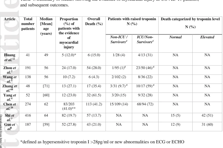
published studies suggesting myocardial injury in patients with COVID-19 is summarized in
Table 4.2, 4–7, 12, 20, 68
Whilst it is reasonable to assume that patients with pre-existing cardiovascular co-morbidities are more susceptible to COVID-19 related myocardial injury, only 30% and 15% of patients with cardiac injury had coronary heart disease and chronic heart failure,
respectively.7 This suggests that the elevated troponin may be a marker of overall disease
Accepted
Article
This article is protected by copyright. All rights reserved
severity, regardless of underlying cardiovascular status. Data is lacking in this regard as less
than 27% of patients with troponin elevation had electrocardiograms (ECGs) in one study,7 and
there are few studies linking imaging correlates of myocardial injury (echocardiography,
cardiac magnetic resonance) with troponin elevation.43, 44 Troponin elevation itself also
correlated strongly with other systemic inflammatory markers such as interleukin-6, C-reactive protein, ferritin as well as aggressive disease on chest radiography, findings which support the notion that troponin may be one of many markers of disease severity.
Further, there is accumulating evidence that patients with severe COVID-19 may have a cytokine storm syndrome and that mortality may be due to virally driven hyperinflammation. This is supported by observations of patients admitted to ICU recording higher plasma cytokine levels including interleukin (IL)-2, IL-7, IL-10 and granulocyte-colony stimulating factor
(GCSF).12 The close correlation between troponin levels, inflammatory markers and
triglyceride levels (akin to secondary haemophagocytic lymphangiohistiocytosis) may support indirect effect of SARS-CoV-2 on the myocardium through immune-mediated mechanisms. Whether there is a role for standard therapies for troponin elevation such as statin or dual antiplatelet therapy is unknown.
Development of new therapeutic agents to a novel disease such as COVID-19 is unrealistic in a timely manner. Instead, many have used existing pharmaceuticals such as corticosteroids, hydroxychloroquine, azithromycin and tocilizumab, primarily addressing the systemic inflammatory response. Given that myocardial injury is a strong predictor of severe disease, one may reasonably consider maximal optimization of treatment options, particularly in those with previous cardiovascular disease or who at a higher cardiovascular risk, as a potential therapeutic target. To date, there is no evidence on the effect of statins in COVID-19 independent of CAD or otherwise. One possible mechanism for myocardial injury in COVID-19 could be through plaque instability and rupture triggered by the profound cytokine activity
Accepted
Article
This article is protected by copyright. All rights reserved
and inflammation. Past clinical trials have shown that statins reduce cardiovascular events, with evidence suggesting this is mediated through reduction of inflammation and plaque
stabilization.69, 70 That may suggest a preventative role of statin therapy.
From a theoretical perspective, statins play a role in regulating the innate immune response through post-translational modification of intracellular signaling molecules. This leads to suppression of the transcription factor NFkB, decreased chemokine levels including
IL-1β, TNFα, IL-6 and decreased monocyte activation.71 This reduces inflammatory cell
infiltrates and macrophage accumulation in atherosclerotic plaques. This is relevant in SARS-like infections, which pre-clinically seem to upregulate a pro-inflammatory gene, MYD88 which results in activation of the NFkB pathway leading to marked inflammation. Transgenic mice with an attenuated NFκB pathway were more likely to survive a SARS infection. Statins play a role in regulating MYD88 expression levels in stress conditions and early, high dose
statins appeared to suppress NF-kB activation.72 However, the use of statins in COVID-19
without any obvious clinical indication may be problematic, given its interactions with supportive treatment, including macrolides and potential antiretrovirals, such as lopinavir and ritonavir combination.
The increased risk of plaque rupture in severe illness poses the question if ant iplatelets and other cardioprotective therapies such as beta-blockers are beneficial. A retrospective study including 20,000 patients in ICUs showed that patients taking aspirin, beta-blockers and/or statins had a 30-day mortality reduction in a troponin-dependent fashion. In particular, aspirin
and beta-blockers reduced 30-day mortality only if the serum troponin was elevated.73 The
utility of continuation or de-novo commencement of RAS blockers in these patients have been discussed in the section on hypertension in this manuscript.
There have been reports of COVID-19 patients presenting with chest pain and dyspnoea with ST-segment elevation on electrocardiography, later found to have non-obstructed
Accepted
Article
This article is protected by copyright. All rights reserved
coronary arteries. This has led to the proposition of widespread microvascular thrombosis
although myocarditis remains a possibility.43, 44 The role of aspirin and beta-blocker in
fulminant myocarditis is however, conflicting. Historic experimental data have shown selected beta-blockers were cardioprotective through suppression of inflammatory cytokines in
autoimmune myocarditis, whilst others had deleterious effects.74
Indeed, studies have suggested high levels of D-dimer (>1 µg/ml) are associated with
higher mortality risk indicating a hypercoagulable state.2 This is supported by findings of small
vessel lumen stenosis and occlusion on lung dissections in COVID-19 patients.75 Further, a
recent retrospective study found better prognosis associated with heparin anticoagulation.76
However, whether there is a similar role of oral anticoagulants and/ or antiplatelets in those with less severe disease, who are treated in outpatient or ambulatory settings, is unknown. In summary, we need a better understanding of the mechanisms underlying cardiac injury in COVID-19. Further histo-pathological studies may distinguish between plaque rupture, microvascular thrombosis or direct myocardial infiltration and inflammation as a cause of troponin elevation. Moving forward, prospective, randomized clinical trials are needed to determine the role of cardioprotective therapies and anticoagulation in COVID-19 patients. Pending these, patients on pre-existing cardioprotective therapies should continue on them if possible. Patients with COVID-19 who meet conventional indications for these cardiovascular therapies should be commenced on them in the absence of contraindications. At present, there is no data to support expanding the role of these protective therapies to other COVID-19 patients, but that may change with rapidly evolving studies.
Accepted
Article
This article is protected by copyright. All rights reserved
Table 4: Summary of studies showing the evidence of myocardial injury in COVID-19 patients, and subsequent outcomes.
Article Total number patients Median [Mean] age (years) Proportion (%) of patients with the evidence of myocardial injury Overall Death (%)
Patients with raised troponin N (%)
Death categorized by troponin level N (%) Non-ICU / Survivors♯ ICU/Non-Survivors♯ Normal Elevated Huang et al.12 41 49 5 (12.0)* 6 (15.0) 1/28 (4) 4/13 (31) NA NA Zhou et al.2 191 56 24 (17.0) 54 (28.0) 1/95 (1)♯ 23/50 (46) ♯ NA NA Wang et al.5 138 56 10 (7.2) 6 (4.3) 2/102 (2) 8/36 (22) NA NA Zhang et al.68 48 [71] 13 (27.1) 17 (35.4) 3/31 (9.7) ♯ 10/17 (59) ♯ NA NA Yang et al.4 52 [60] 12 (23.0) 32 (61.5) 3/20 (15) 9/32 (28) NA NA Chen et al.20 274 62 83/203 (41.0)** 113 (41.2) 15/109 (14) 68/94 (72) NA NA Shi et al.7 416 64 82 (19.7) 57 (13.7) NA NA 15 (5) 42 (51) Guo et al6 187 [59] 52 (27.8) 43 (21.0) NA NA 12 (9) 31 (60)
*defined as hypersensitive troponin I >28pg/ml or new abnormalities on ECG or ECHO ** defined as >15.6 pg/mL
♯ survivors or non-survivors
NA: not available; NB: most proportion are rounded off to the nearest integer
Accepted
Article
This article is protected by copyright. All rights reserved
Acute Coronary Syndrome in the COVID-19 Pandemic Era: how to triage and when to resort to invasive strategies
Take Home Points:
• Timely primary percutaneous coronary intervention (PCI) remains the mainstay treatment
for ST-elevation myocardial infarction (STEMI).
• In case of patient- or system-related delays in mechanical reperfusion in the
contemporary COVID-19 era, fibrinolytic therapy within door-to-balloon of 30 minutes may be an alternative treatment for STEMI in the absence of contraindications.
• An invasive strategy is highly recommended for patients with non-ST-elevation acute
coronary syndrome (NTE-ACS) who are at high risk.
• In the COVID-19 era, and especially when the local community outbreak is increasing
and the healthcare system is overwhelmed, moderate- and low-risk patients with NSTE-ACS can be treated with an ischemia-guided approach.
Acute coronary syndromes (ACS) encompass a spectrum of clinical entities ranging from ST-elevation myocardial infarction (STEMI) to non-ST-ST-elevation myocardial infarction and unstable angina. The latter two, often coined collectively as non-ST-elevation ACS (NSTE-ACS), differ in their pathophysiology from STEMI in that they predominantly result from an acute
non-occlusive thrombus overlying a disrupted plaque.77 On the other hand, STEMI is usually
attributable to an acute thrombosis overlying a disrupted plaque, which is completely occlusive
of the epicardial coronary artery.78 STEMI manifests with an acute myocardial injury pattern on
Accepted
Article
This article is protected by copyright. All rights reserved
ECG and needs to be aborted immediately to prevent irreversible myocardial damage. Therefore,
a timely reperfusion strategy is the mainstay treatment after STEMI.78 Of the two available
reperfusion modalities, primary percutaneous coronary intervention (PCI) is preferable to
fibrinolytic therapy because it is safer and more effective.79 On the other hand, moderate- and
high-risk NSTE-ACS patients who are medically stabilized can be treated with an urgent - but not necessarily emergent - invasive strategy (i.e. coronary angiography with intent to
revascularize).77
The swiftly spreading COVID-19 pandemic in the United States is placing an
unparalleled pressure on the healthcare system. There is currently a rapid depletion of resources in many medical centers including shortages of hospital and ICU beds, personal protective equipment (PPE), and even healthcare workers (HCWs). Hospitalized patients and HCWs are at increased risk of infection with SARS-CoV2. Revisiting the modality of reperfusion after STEMI and the merits of an invasive strategy and its timing after NSTE-ACS is therefore critically important.
The initial assessment of any patient with suspected ACS should include a history, physical examination (PE) and a 12-lead ECG. Measurements of cardiac biomarkers of necrosis (e.g. Troponins) should be performed serially, but reperfusion therapy should be decided upfront based on the clinical presentation and ECG. In the COVID-19 era, a focused but meticulous history and PE should be conducted assuming that every patient may be – at a minimum - an asymptomatic SARS-CoV2 carrier.
In the current section, we propose categorizing ACS patients into three categories: critically ill ACS patients and non-critically ill STEMI and NSTE-ACS patients. In the COVID-19 era, triaging patients to optimal medical therapy (OMT) alone versus invasive approach
Accepted
Article
This article is protected by copyright. All rights reserved
(added to OMT) will depend on many factors, including patients’ ACS category, their COVID-19 status, and safety issues (e.g. PPE availability, shortage of HCWs).
All ACS patients who are critically ill (e.g. cardiogenic shock, mechanical complications, refractory heart failure) should undergo emergent coronary angiography with intent to
revascularize (Figure 3). In general, this life-saving strategy should take place irrespective of the patients’ COVID-19 status and other considerations.
For STEMI patients who are not critically ill, timely primary PCI remains the treatment of choice (Figure 3). Mechanical reperfusion, within a first medical contact-to-device
(FMC-device) time of 90 minutes when presenting to a PCI-capable hospital, is highly recommended.80
Timely reperfusion is critically important to salvaging myocardium. However, in a small cohort of 7 STEMI patients from Hong Kong undergoing primary PCI in the COVID-19 era, longer
median times in all components of STEMI care were observed.81 Difficulty of achieving timely
mechanical reperfusion, especially in geographies severely affected by COVID-19, should be recognized.
Accordingly, it is reasonable to administer fibrinolytic therapy, in the absence of
contraindications,82 to appropriate STEMI patients in COVID-19 affected communities (Figure
3). This is particularly relevant to STEMI patients who do not have high-risk features and are either known or suspected to have COVID-19. A bedside echocardiogram can be readily available and will add incremental value for risk stratification. An example of a STEMI patient eligible for fibrinolytic would be the case of a COVID-19 (+) patient who is hemodynamically stable and who is presenting with ST elevation in aVL alone and echocardiographic evidence of small lateral hypokinesis. Experts from the Sichuan Provincial People’s Hospital in China proposed the use of fibrinolytic therapy in COVID-19 (+) patient who are presenting with
Accepted
Article
This article is protected by copyright. All rights reserved
STEMI, are hemodynamically stable and have no contraindication to fibrinolysis.83 Fibrinolytic
therapy is most effective when administered within the first 2-3 hours of symptom onset. When chosen, timely pharmacological reperfusion within door-to-needle (DNT) of 30 minutes and
preferably using the newer generation tenecteplase (TNK), is recommended.80 In case of failed
fibrinolytic reperfusion, rescue PCI is a reasonable subsequent strategy.84 As for STEMI patients
presenting to non-PCI capable hospitals, timely pharmacologic reperfusion is strongly encouraged, especially when the receiving PCI center is tackling a COVID-19 surge. In our opinion, transfer of successfully reperfused STEMI patients to a PCI-capable hospital, as part of a pharmacoinvasive approach, should not be routinely implemented but rather individualized depending on many factors (e.g. status of the receiving hospital and outbreak in the community).
For NSTE-ACS patients who are not critically ill, we believe the majority of those may be safely treated with an ischemia-guided strategy in the COVID-19 era (Figure 3). A routine invasive strategy is superior to an ischemia-guided strategy after NSTE-ACS and reduces the
risk of recurrent MI.85 However, the largest benefit appears to be confined to high-risk patients,
and only modest benefit was observed in moderate-risk patients.85 These studies were largely
done more than a decade ago when dual antiplatelet therapy (especially the novel P2Y12 receptor
inhibitors), intensive lipid lowering and OMT were not routinely implemented or available. We therefore believe that treating moderate-risk NSTE-ACS patients in COVID-19-affected medical communities with an ischemia-guided strategy is reasonable. All NSTE-ACS patients need to be
risk stratified adequately using objective risk scores.86 Those treated with an ischemia-guided
approach may cross over to an invasive strategy if they manifest significant spontaneous or
provoked ischemia or become unstable despite OMT.87 On the other hand, high-risk NSTE-ACS
patients should still receive an invasive strategy.85 An early invasive strategy within ≤ 24 hours is
Accepted
Article
This article is protected by copyright. All rights reserved
preferred, as it usually reduces recurrent ischemia compared to a delayed invasive strategy.88
This is best done if COVID-19 testing is achieved prior to the procedure. Otherwise, when high level of suspicion for COVID-19 exists, a delayed invasive strategy (24-72 hours) may be selectively implemented in a controlled setting until the test result is available.
There are several considerations that need to be accounted for when entertaining invasive therapy in an ACS patient in the contemporary COVID-19 era. All patients should undergo clinical screening for COVID-19. Ideally, every ACS patient undergoing non-emergent invasive therapy should get tested for COVID-19 before the procedure. If not feasible, at least patients with high clinical suspicion should be tested. All procedures for patients with confirmed or suspected COVID-19 should be performed, if possible, in a dedicated/repurposed cardiac
catheterization laboratory with optimal protection strategies for HCWs and patients.9 Evaluation
of the patient’s airway should be assessed prior to transfer to the catheterization laboratory. In patients with concerning respiratory distress, intubation should be performed prior to arrival of
the patient to the catheterization lab if possible.89 All patients should be outfitted with a
facemask. Proper protection of HCWs with appropriate PPE is essential, including appropriate
facemask (N95 respirator), face-shield or goggles for eye protection, and gowns.90
When the healthcare setting is not safe for HCWs and patients (e.g. lack of PPE, shortage of ventilators), the risks and benefits of an invasive strategy should be revisited, irrespective of the ACS category. Members of the heart team should deliberate and engage the patient and family in a process of shared decision making and, if necessary, invoke the ethics committee. This also applies to critically ill patients whose instability is attributable to advanced COVID-19 illness rather than the ACS itself, and in whom an invasive therapy is likely futile.
Accepted
Article
This article is protected by copyright. All rights reserved
Overall, the aforementioned recommendations and proposed clinical algorithm provide general clinical guidance. In the absence of robust data specific to the COVID-19 era, they
represent our opinion and complement guidance provided by other societies.9, 89 As such, they
should be tailored to local medical institutions and communities. These recommendations are dynamic and should be constantly revised as the burden of the outbreak and the availability of resources continues to change over time.
Figure 3. Invasive Therapies for ACS Patients in the COVID-19 Era.
^ Acute MI with mechanical complications is best treated with surgical revascularization and concomitant repair (with adjunctive percutaneous ventricular assist devices). ^^ Risk stratification after NSTE-ACS can be performed using an objective risk score (e.g. GRACE, TIMI). * When pursuing an invasive approach, appropriate personal protective equipment (e.g. gowns, face shield/goggles, N95 masks) and set-up (e.g. negative pressure room) must be available for the safety of healthcare workers and patients. Otherwise, defaulting to the alternative approach (pharmacologic reperfusion or ischemia-guided strategy) after deliberation between the heart team members, invoking the ethics team when appropriate, and in a process of shared decision making with the patient and family.
Accepted
Article
This article is protected by copyright. All rights reserved
** Fibrinolytic therapy should be administered within DTN of 30 minutes. Upon failure of pharmacologic
reperfusion, rescue PCI is recommended. *** Patients treated with an ischemia-guided strategy may cross over to an invasive strategy in case of significant spontaneous or inducible ischemia, or any evidence of hemodynamic or electrical instability. **** Unsafe healthcare setting can be attributed to a myriad of factors (e.g. lack of PPE, lack of ventilators, shortage of healthcare workers, negative pressure cath lab not available). LV (Left Ventricle), OMT (Optimal Medical Therapy), GRACE (Global Registry of Acute Coronary Events), PPE (Personal Protection Equipment), TIMI (Thrombolysis in Myocardial Infarction), OMT (Optimal Medical Therapy), DTN (Door-To-Needle)
ECMO in the COVID-19 Pandemic Era
Take Home Points
• Venovenous and venoarterial extracorporeal membrane oxygenation (ECMO) may be
utilized as salvage therapies in COVID-19 patients with refractory respiratory or cardiorespiratory failure, respectively.
• When needed, it is best to institute ECMO early before multiorgan failure ensues.
• ECMO is a complex therapeutic modality that requires advanced expertise and intensive
resource utilization. In the absence of definitive evidence supporting its utility, it should be used judiciously after careful assessment of the benefits and risks by a multifaceted medical team and with shared decision making with patient and family.
The clinical presentation of patients with COVID-19 is variable.15 Hypoxemic respiratory failure
requiring ICU admission occurs in 5-6% of patients, of whom the majority will require
mechanical ventilation.91 The most common etiology for respiratory failure in these patients is
acute respiratory distress syndrome (ARDS).18 Mortality rates are significantly high in patients
who require mechanical ventilation and are reported to be 50%.18, 91 Furthermore, COVID-19
patients on mechanical ventilation require prolonged duration of respiratory support with a
Accepted
Article
This article is protected by copyright. All rights reserved
median of 17 days of ventilation.92 COVID-19 can also cause cardiovascular complications,
including cardiac injury, myocarditis, heart failure, cardiogenic shock, tamponade, and
pulmonary embolism.93–95 Patients with underlying cardiovascular disease (CVD) are more
likely to suffer from myocardial injury with COVID-19, and this combination (underlying CVD
and myocardial injury) portends the highest mortality (70%).6
Venovenous (VV) and venoarterial (VA) extracorporeal membrane oxygenation (ECMO) can offer respiratory or cardiorespiratory support for patients with refractory respiratory or
cardiorespiratory failure, respectively.96 ECMO is also used in cases of cardiac arrest in
conjunction with cardiopulmonary resuscitation (E-CPR).97 Thus, ECMO could potentially offer
a life-saving treatment to sick COVID-19 patients, as it did in prior Coronavirus epidemics.98
ECMO is a complex therapeutic modality that requires intensive resource utilization and is usually offered in tertiary and quaternary centers with a full ECMO team. It is therefore
important to understand its benefits and limitations, especially when resources are stretched and
healthcare systems are overwhelmed as is the case in the current COVID-19 pandemic.96, 98
There are conflicting reports regarding preliminary outcomes of ECMO use in sick
COVID-19 patients. In one study, patients treated with ECMO had a mortality rate of 86%.4 In
another study, 50% of patients treated with ECMO died, while 37.5% were successfully weaned
off ECMO but remained on mechanical ventilation.99 A potential marker of poor prognosis in
COVID-19 with ECMO is lymphopenia. Lymphopenia also occurred in prior Coronavirus
epidemics and was caused by lymphocyte apoptosis.100 Lymphopenia occurs in 70% of
symptomatic COVID-19 patients, and the degree of lymphopenia is associated with more severe
disease and ICU admission.5, 12 ECMO use is associated with a decline in lymphocytic count and
this potentially confounds the clinical course in COVID-19 patients.101 One possible explanation
Accepted
Article
This article is protected by copyright. All rights reserved
of poor prognosis is the significant rise in cytokines and inflammatory biomarkers, although there are conflicting opinions on whether it is an association or implicated in the causal
pathway.102 In COVID-19 patients, the cytokine release syndrome is associated with lower
probability of survival.16
Given the limited evidence for ECMO use in COVID-19 patients with ARDS, and the extensive resources and experience required to provide ECMO support, The Extracorporeal Life
Support Organization (ELSO) released a guidance document.96 Several important issues should
be taken into account when considering ECMO for COVID-19 patients (Table 5). First, it should be determined early which kind of support that patient would need, whether respiratory support only or whether there is evidence of myocardial dysfunction and cardiogenic shock requiring cardiorespiratory support. If ECMO is needed, it should be provided early in the course of mechanical ventilation, as prognosis is usually worse with longer duration on mechanical
ventilation. Second, if ECMO is likely to be required, the consensus recommendation is to insert it early before multiorgan failure ensues. Third, ECMO may be futile in elderly patients with significant comorbidities in which survival is unlikely. This is important at a time when
resources are limited and the healthcare system is strained, as is the case in the current COVID-19 outbreak in several parts of the United States, Italy, and Spain. Fourth, a strong relationship
between ECMO volume and outcome has been documented.103 It is highly advisable to refer
COVID-19 patients who might require ECMO support to experienced centers, as the best
survival outcomes were reported in centers performing at least 30 ECMO procedures annually.103
Fifth, given the risk of significant aerosolization and disease transmission during
cardiopulmonary resuscitation, and the number of personnel required in the room for E-CPR, it is probably ill-advised to utilize ECMO in COVID-19 patients with cardiac arrest. Finally, in
Accepted
Article
This article is protected by copyright. All rights reserved
COVID-19 patients with severe left ventricular (LV) dysfunction (e.g. myocarditis, takotsubo cardiomyopathy) complicating their ARDS, increased LV wall stress and consequent myocardial ischemia from ECMO may exacerbate cardiac dysfunction. In these patients, venting the heart using an intra-aortic balloon pump (IABP) or a percutaneous ventricular assist device (e.g. Impella) may be a reasonable strategy, as it has been successfully utilized in cardiogenic shock patients. However, this should be only performed judiciously with consideration of available resources and expertise in the middle of a pandemic.
It is probably too early in the pandemic to fully judge the utility of ECMO in the COVID-19 era. Currently, it remains an indispensable therapeutic modality in the armamentarium of clinicians attempting to salvage a decompensated COVID-19 patient with ARDS. It is laudable that ELSO encouraged early entry of COVID-19 patients treated with ECMO into its registry by
participating hospitals and waived membership fees during the pandemic.96 This will help the
medical community examine the role of ECMO and inform clinical practice with critically-needed evidence.
Accepted
Article
This article is protected by copyright. All rights reserved
Table 5. Clinical and Procedural Cons