Impact of whole-body rehabilitation in patients receiving chronic
mechanical ventilation
Ubaldo J. Martin, MD; Luis Hincapie, RPT; Mark Nimchuk, RPT; John Gaughan, PhD; Gerard J. Criner, MD
I
t is generally accepted that chronic ventilator-dependent pa-tients are weak or deconditioned; however, there are limited data showing this to be true or describing the magnitude and extent of weakness. In chronic ventilated patients, immobility and catabolism from the underlying med-ical/surgical illness, poor nutritional sta-tus, and exposure to pharmacologic agents (e.g., neuromuscular blocking agents and steroids) are all commonly present, all of which may adversely affect functional status and result in substantial weakness of limb, respiratory, and swal-lowing musculature. Although the bene-fit of rehabilitation in stable chronic ob-structive pulmonary disease (COPD) has been well described (1, 2), there is apau-city of data regarding the effects of reha-bilitation in patients receiving chronic mechanical ventilation. This is due to several factors: the high acuity of illness in ventilated patients, their frequent transfers to different institutions and lev-els of care, and the complexities and la-bor-intensiveness and need for specially trained personnel to provide rehabilita-tion in the ventilated patient. As a result, available studies tend to include small numbers of patients receiving a mix of invasive and noninvasive ventilation who have COPD as the primary cause of respi-ratory failure. Frequently, baseline char-acteristics are poorly characterized, and the rehabilitation treatment is not well described (3–5).
Currently there are no guidelines for rehabilitation of the chronically venti-lated patient. This is important, because these patients have extremely high mor-tality rates (1-yr survival 30 –70%) (6, 7), substantial morbidity (reduced quality of life and poor functional status), and high costs of care (ⱖ$9 million/day in the United States) (8). Because mechanical ventilation and its inherent constraints on mobility are superimposed on the pa-tient’s already impaired functional status
by the disease necessitating ventilation, a well-trained multidisciplinary team, as well as special equipment and physical plant requirements, is usually recom-mended, but the actual rehabilitative needs and the efficacy of treatment are unclear.
Therefore, we sought a) to characterize the level of whole-body weakness in chronic ventilated patients admitted to an inpatient ventilator-dependent rehabilita-tion unit; and b) to evaluate the impact of aggressive whole-body and respiratory muscle rehabilitation on improving their respiratory and peripheral muscle strength and functional status.
MATERIALS AND METHODS
Subjects. Forty-nine consecutive patients admitted to our chronic ventilator-dependent rehabilitation unit (VRU) from surgical, med-ical, and cardiac intensive care units (ICUs), without previous history of underlying neuro-muscular diseases, were studied. All patients met criteria for clinical stability as shown in Table 1. Patients admitted to this unit had been invasively ventilated forⱖ14 consecutive days and had failed at least two attempts at weaning from mechanical ventilation. Be-cause of the retrospective nature of the study and the use of standard therapy, the Institu-From the Division of Pulmonary and Critical Care
Medicine (UJM, GJC), Department of Physical Medicine and Rehabilitation (LH, MN) and Biostatistics (JG), Temple University School of Medicine, Philadelphia, PA.
No author disclosed any financial support or in-volvement with organizations with financial interest in the subject matter.
Copyright © 2005 by the Society of Critical Care Medicine and Lippincott Williams & Wilkins
DOI: 10.1097/01.CCM.0000181730.02238.9B
Objective:To evaluate the prevalence and magnitude of weak-ness in patients receiving chronic mechanical ventilation and the impact of providing aggressive whole-body rehabilitation on con-ventional weaning variables, muscle strength, and overall func-tional status.
Design:Retrospective analysis of 49 consecutive patients.
Setting: Multidisciplinary ventilatory rehabilitation unit in an academic medical center.
Patients:Forty-nine consecutive chronic ventilator-dependent patients referred to a tertiary care hospital ventilator rehabilita-tion unit.
Interventions:None.
Measurement and Main Results:Patients were 58ⴞ7 yrs old with multiple etiologies for respiratory failure. On admission, all patients were bedridden and had severe weakness of upper and lower extremities measured by a 5-point muscle strength score
and a 7-point Functional Independence Measurement. Postreha-bilitation, patients had increases in upper and lower extremity strength (p < .05) and were able to stand and ambulate. All weaned from mechanical ventilation, but three required subse-quent intermittent support. Six patients died before hospital dis-charge. Upper extremity strength on admission inversely corre-lated with time to wean from mechanical ventilation (Rⴝ.72,p<
.001).
Conclusions: Patients receiving chronic ventilation are weak and deconditioned but respond to aggressive whole-body and respiratory muscle training with an improvement in strength, weaning outcome, and functional status. Whole-body rehabilita-tion should be considered a significant component of their ther-apy. (Crit Care Med 2005; 33:2259 –2265)
KEYWORDS: pulmonary rehabilitation; weaning; ventilation; me-chanical; chronic; deconditioning
tional Review Board waived the need for writ-ten consent.
Ventilator Rehabilitation Unit (VRU).The VRU is an 18-bed respiratory care unit. It was one of the original clinical sites for the Health Care Financing Administration Chronic Ven-tilator Dependent Demonstration Project that examined costs and outcome in ventilated pa-tients receiving aggressive, multidisciplinary, whole-body and respiratory rehabilitation as well as weaning attempts (9, 10). The staff, medical direction, and project objectives have remained intact since completion of the project, and the unit size and staff number have quadrupled. The unit is staffed by a mul-tidisciplinary team, including board-certified pulmonary and critical care attendings and pulmonary/critical care fellows, a nurse coor-dinator, nurses with special training in respi-ratory patients and their management,
nutri-tionists, psychologists, respiratory, physical, occupational and speech therapists, and a so-cial worker. The physician staff, nurses, and therapists attend daily rounds. Weekly meet-ings are attended by all team members and are led by the medical director of the unit. The emphasis in this unit is placed on whole-body and respiratory rehabilitation for patients who suffer from chronic respiratory failure (9, 10). An outline of the daily and weekly treatment plan is shown in Figure 1.
Initial Assessment. On admission to the VRU, all patients underwent a detailed history and physical examination performed by the pulmonary/critical care physician attending and fellow. Causes for respiratory failure, weaning variables, ventilatory variables, and gas exchange variables were assessed on ad-mission to the unit. A physical therapist, speech pathology specialist, and psychologist
also assessed patients to establish a comphensive rehabilitation plan. Charts were re-viewed to determine whether patients had been exposed to steroids or neuromuscular blocking agents during their ICU stay. A qual-itative assessment of the use of steroids or neuromuscular agents was made in each pa-tient, but quantification of the exact amount of each of these agents was not performed. The use of other medications such as sedative agents, antibiotics, and psychotropic agents was also noted.
Weaning.All patients were weaned on tra-cheal collar with supplemental inspired oxy-gen concentrations adjusted to maintain a pulse oximetric oxygen saturation ⬎95%. Conventional weaning variables, including the rapid shallow breathing index, were performed before and when possible at the end of the weaning period. Weaning was initiated once the patient was clinically stable and the un-derlying reason for respiratory failure was re-solved. In clinically stable patients, a weaning trial was attempted even in the absence of adequate standardized weaning variables (such as rapid shallow breathing index and negative inspiratory force [NIF]), since these tend to be less accurate in this particular pop-ulation.
Patients were initially weaned anywhere from 30 mins to 1 hr. The weaning goal was extended daily as tolerated by the patient, pro-vided that there were no exacerbations of the underlying condition or development of a sig-nificant new disorder. A weaning trial was stopped if the patient exhibited severe agita-tion, a respiratory rate ⬎35, arterial oxygen saturation⬍90% despite an increment in FIO2
to 60%, heart rate⬎140, systolic blood pres-sure ⬎180 mm Hg, new onset hypotension (systolic blood pressure ⬍90 mm Hg), or marked increment in work of breathing as evidenced by additional recruitment of acces-sory respiratory muscles, diaphoresis, and anxiety.
Rehabilitation Program. Rehabilitation was conducted by physical therapists 5 days per week in a gym constructed onsite within the VRU. Rehabilitation was started on admis-sion to the VRU. Because a structured reha-bilitation program had not been developed for the ICU, none of these patients received reha-bilitation by physical therapists during their ICU stay. Initially, therapy sessions were 30 – 60 mins in duration, depending on the patient’s tolerance for physical activity. Phys-ical therapy sessions were not conducted dur-ing spontaneous breathdur-ing trials until the pa-tient could spontaneously breath for⬎4 hrs. Once the patient was able to do so, physical therapy did proceed simultaneously with the spontaneous breathing trials. In those who were unable to breath spontaneously, physical therapy was accomplished by using a portable ventilator. The physical therapists were in-structed to increase FIO2to maintain an
oxy-gen saturation⬎94% during the rehab period. In patients with airway obstruction, a
nebu-Figure 1.Outline of daily and long-term planning in the ventilatory rehabilitation unit.DME, durable medical equipment.
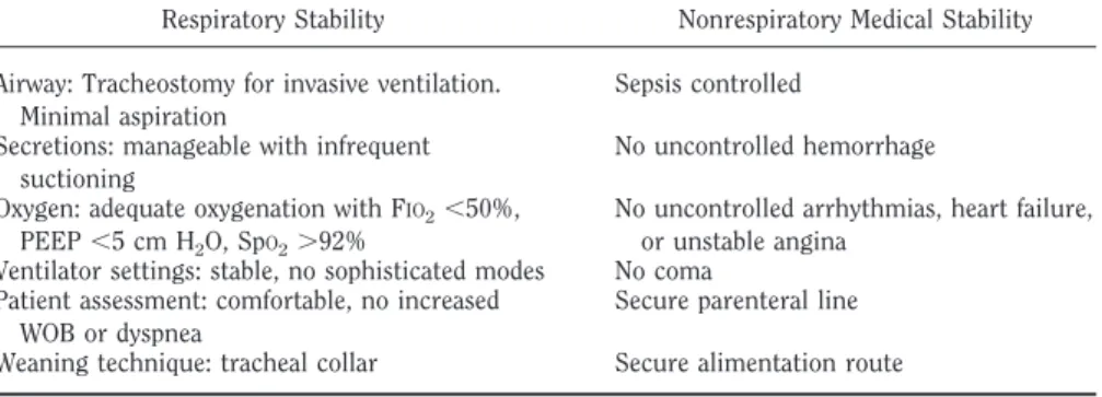
Table 1.Criteria for transfer of ventilator-dependent patients from intensive care unit to ventilator rehabilitation unit
Respiratory Stability Nonrespiratory Medical Stability Airway: Tracheostomy for invasive ventilation.
Minimal aspiration
Sepsis controlled Secretions: manageable with infrequent
suctioning
No uncontrolled hemorrhage Oxygen: adequate oxygenation with FIO2⬍50%,
PEEP⬍5 cm H2O, SpO2⬎92%
No uncontrolled arrhythmias, heart failure, or unstable angina
Ventilator settings: stable, no sophisticated modes No coma Patient assessment: comfortable, no increased
WOB or dyspnea
Secure parenteral line Weaning technique: tracheal collar Secure alimentation route
lized albuterol treatment with was given 30 mins before the rehabilitation session. All pa-tients were suctioned before a rehabilitation session. Respiratory therapists assisted physi-cal therapists and made changes in ventilator rate or tidal volume as needed for comfort; in most of cases, ventilator settings were not altered during the rehab period.
As the patient tolerated the sessions, the steps of the conditioning program were ad-vanced and the training interval was progres-sively lengthened. The patients progressed from one session per day to two sessions per day once they were able to consistently toler-ate⬎45 mins in one session. Oxygen satura-tion, heart rate, and blood pressure were mon-itored during sessions.
In the initial stages, the rehabilitation pro-cess concentrated on improving trunk control and patient maintenance of body posture. Pushing against resistance and using resis-tance bands and/or low weights followed pas-sive and active training of upper and lower extremities. As patients improved trunk con-trol, upper and lower extremity ergometry (magnetically or tension adjusted pedal
exer-ciser), standing and ambulation using parallel bars, stepping in place, and then staircase ex-ercises were introduced.
Inspiratory muscle training using a threshold device was instituted in patients ca-pable of breathing spontaneously for⬎2 hrs. Patients used the device twice daily for 15 mins set at one third of the patient’s maxi-mum NIF. NIF was measured early in the morning every day, via the tracheostomy tube, before weaning attempts or physical therapy sessions as previously described (11). Patients were considered successfully weaned from me-chanical ventilation once they tolerated 48 consecutive hours of unassisted breathing on tracheal collar.
Measurements.Extremity muscle strength was assessed by a physical therapist on admis-sion, daily during physical therapy sessions, and on discharge from the VRU using a 5-point motor score (MS) that considered strength and range of motion in all major muscle groups (Table 2) and a previously val-idated 7-point Functional Independence Mea-surement Scale (FIM) that evaluates indepen-dent functioning (Table 3) (12, 13). Upper
limb motor strength score was obtained by combining the mean scores for shoulder, el-bow, and wrist flexion and extension. Simi-larly, lower limb motor strength scores were obtained by combining the mean scores for hip, knee and plantar flexion, and extension.
Statistical Analysis.Unpaired Student’st -test was used to analyze pairwise comparisons in normally distributed variables. Wilcoxon’s log rank test or Mann-Whitney rank sum test was used in the absence of normally distrib-uted variables. Proportions were analyzed us-ing a Z-test. Linear regression analysis was used to establish correlations between vari-ables. Stepwise multiple regression analysis was used to establish correlates between wean-ing time and multiple intervenwean-ing variables. Significance was set atp⬍.05.
RESULTS
A total of 49 patients were enrolled. Primary causes for respiratory failure were pneumonia in 15 patients (31%), heart failure in nine (18%), acute respi-ratory distress syndrome in eight (16%), sepsis in eight (16%), acute on chronic renal failure in three (6%), and diaphrag-matic dysfunction in two (4%); the fol-lowing were seen once in individual pa-tients: cerebrovascular accident, pulmonary embolism, cardiac tampon-ade, drug overdose, pneumothorax, and ventricular arrhythmia. A total of nine patients had a previous history of COPD. Demographic data are shown in (Table 4). All patients were on assist-control ventilation and had a positive end-expiratory pressure⬍5 cm H2O. Patients were intubated for 18.1⫾7.7 days (range 9 –29 days) before tracheostomy. The time from tracheostomy placement to transfer to the VRU was 7⫾6 days. At the time of admission none of the patients were able to sustain a spontaneous breathing trial for ⬎2 hrs. The time to wean from mechanical ventilation, mea-sured from the initial day of admission to the VRU, was 16.0 ⫾ 9 days. Of 49 pa-tients, 15 (30%) weaned inⱕ7 days. Pa-tients were transferred to the VRU after achieving clinical stability. No significant differences in age or male to female ratio, Acute Physiology and Chronic Health Evaluation score on admission, or time on mechanical ventilation before trache-ostomy were noted between patients who weanedⱕ7 days and those who took⬎7 days to wean. A total of 16 patients (30%) were exposed to systemic corticosteroids, whereas 16 (30%) were exposed to neu-romuscular blocking agents. A total of nine patients (18%) were exposed to both agents for⬍5 days.
Table 3.Functional independence measurement score
Transfers (Supine to Sit, Sit to Stand) Ambulation, feet Stairs, steps Score
Total assistance (two people) ⬍50 ⬍4 1
Maximal assistance (two people) 50 4–12 2
Moderate assistance (two people) 150 12 3
Minimal assistance (two people) 150 12 4
Supervision 50 (indep) 12 5
Modified independence 150 12 (indep) 6
Complete independence ⬎150 (indep) ⬎12 7
Indep, independent in performance.
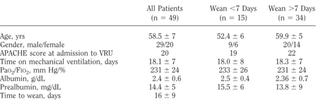
Table 4.Demographic data
All Patients (n⫽49) Wean⬍7 Days (n⫽15) Wean⬎7 Days (n⫽34) Age, yrs 58.5⫾7 52.4⫾6 59.9⫾5 Gender, male/female 29/20 9/6 20/14
APACHE score at admission to VRU 20 19 22
Time on mechanical ventilation, days 18.1⫾7 18.0⫾8 18.3⫾7 PaO2/FIO2, mm Hg/% 231⫾24 233⫾26 231⫾24
Albumin, g/dL 2.4⫾0.6 2.5⫾0.4 2.36⫾0.7
Prealbumin, mg/dL 14.4⫾5 15.5⫾6 13.8⫾9
Time to wean, days 16⫾9
APACHE, Acute Physiology and Chronic Health Evaluation; VRU, ventilatory rehabilitation unit.
Table 2.Muscle strength score
Muscle Activity Score
Ability to hold the test position against gravity and maximum pressure 5 Same as above, except holding against moderate to minimal pressure 4 Ability to hold the test position against gravity 3 Ability to move a body part through a partial arc of motion (with gravity lessened
by a the observer)
2 Contraction of muscle, prominence of tendon without visible motion of body part 1
On admission to the VRU, all patients were severely deconditioned as evidenced by low scores in motor strength and func-tional status scales (Tables 5).
Motor Strength. On admission, pa-tients exhibited marked weakness of both upper and lower extremities. Proximal muscle scores were lower (e.g., weaker) than distal muscle scores for both upper and lower limbs, but they did not reach statistical significance. A significant im-provement was noted in the combined motor strength score for upper and lower limbs at the time of discharge from the VRU (Table 5). Using a similar analysis, on admission, patients who weaned ⱕ7 days had higher upper limb motor strength scores than patients who took ⬎7 days to wean (Table 6). Although pa-tients who weaned quicker continued to have higher motor strength scores at the time of discharge, the difference between groups was not statistically significant (Table 6).
Functional Status and Ambulation. The ability to transfer from the supine-to-sit and sit-to-stand positions was scored according to the FIM scale and constitutes the transfer scores. A signifi-cant improvement was noted in the abil-ity to transfer from the supine to the sitting position and from the sitting to the standing position. (Table 5). There were no significant changes in the
ambu-lation and stair-climbing categories of the FIMS scale. All patients were able to sit and stand at the end of the rehabilita-tion period. All patients were bed-bound at the time of admission; ambulation went from 0 to 52⫾18 feet for the group (p ⫽.005). A total of 40 patients (81%) were able to ambulate at the time of dis-charge.
Weaning Variables. Several changes in respiratory variables were noted. A sig-nificant decrement in respiratory rate (f) and a concomitant increment in tidal vol-ume (VT) resulted in lower f/VTratio
(Ta-ble 7). An improvement in respiratory muscle strength was evidenced by a sig-nificant increment in maximal NIF.
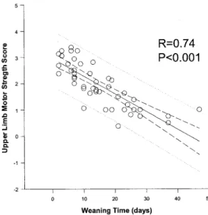
A significant correlation was found be-tween upper limb motor strength at the time of admission and time to wean (R⫽ .72, R2⫽.54,p⬍.001, Fig. 2). Conven-tional weaning variables at admission or on the day of successful weaning did not exhibit a significant correlation with weaning success. On admission, the f/VT
ratio was⬍105 in 40% of these patients. On the day of successful weaning, only 63% of all patients had an f/VT ratio
⬍105.
Stepwise Regression Analysis of Fac-tors Affecting Weaning Time. Stepwise multiple regression analysis was per-formed to establish predictors of weaning time. Gender, age, use of neuromuscular blocking agents and systemic steroids, presence of COPD, renal failure, heart failure, stroke pneumonia, prealbumin, f, f/VT, NIF, and lower and upper limb
mo-tor strength scores on VRU admission were included in the analysis. Three vari-ables were found to be statistically signif-icant in terms of weaning time: upper motor strength, exposure to neuromus-cular blocking, and systemic steroids (Ta-ble 8). According to the regression equa-tion, an increment of 1 point in the upper extremity motor strength scale results in a reduction ofⵑ7 days in weaning time. According to the same equation, the ex-posure to steroids and neuromuscular blocking agents independently adds ⵑ4 days to weaning time. The percent fit (R2) of the equation is .73. As can be seen from the standardized estimates in Table 8, the contribution of upper motor strength to weaning time appears to be twice as in-fluential as the exposure to neuromuscu-lar blocking agents or steroids.
Outcomes.Six (12%) patients died af-ter having successfully weaned (three with severe sepsis, two with respiratory failure due to pneumonia, and one with
worsening COPD and respiratory failure). Two patients required noninvasive me-chanical ventilation at the time of dis-charge, and one patient eventually devel-oped the need for nocturnal invasive ventilation via tracheostomy.
DISCUSSION
Our study shows that chronic venti-lated patients, when medically stable and transferred from the ICU, are severely weak and deconditioned, with limited ability to sit and stand, and are unable to ambulate. Following several weeks of whole-body and respiratory rehabilita-tion, patients have significant increases in whole-body and respiratory muscle strength and are ventilator independent as well as functionally improved. Upper extremity strength significantly corre-lates with weaning time, even more so than conventional variables used to pre-dict weaning outcome (e.g., f/VT). These
data suggest that patients undergoing prolonged periods of mechanical ventila-tion following an acute, devastating med-ical or surgmed-ical disorder suffer from sig-nificant global skeletal muscle weakness that limits their ability to wean and per-form the activities of daily living. Fortu-nately, favorable improvements are found in these patients in response to conven-tional whole-body and respiratory muscle rehabilitative techniques.
In this heterogeneous group of patients, skeletal muscle dysfunction was present in both the upper and lower limbs, a fact that contrasts with the available data in COPD patients in whom upper limb motor strength is frequently preserved (14). Our chronic ventilated patients also had signif-icantly decreased NIF indicating respiratory muscle weakness, again in contrast to pa-tients with COPD in whom diaphragm strength is preserved in comparison to nor-mal individuals when assessed at isovolume conditions (15). These data suggest that weakness occurs in the chronic ventilated patient in both the peripheral and respira-tory muscles and emphasize the impor-tance of reconditioning all muscle groups in this patient population. Overall, proxi-mal muscles appeared to be more affected than distal muscles, and larger muscle groups (hips, thighs) were more severely affected than smaller muscle groups (wrist, arm). Whether this occurs due to the greater muscle mass located in the proxi-mal regions affording easier assessment or due to the preferential effects of neuromus-Table 6.Motor strength for weaning groups upon
admission and at discharge Short Wean (⬍7 Days) Prolonged Wean (⬎7 Days) Admission
Upper limb score 3.2⫾0.4 1.5⫾0.6a Lower limb score 2.5⫾0.4 1.2⫾0.5a Discharge
Upper limb score 3.9⫾0.6 3.3⫾0.6b Lower limb score 2.4⫾0.4 3.0⫾0.6c
a
p⬍.01;b
p⬍.08;c
p⫽.1.
Table 5.Motor strength and functional indepen-dence measurement (FIM) score for all patients on admission and at discharge
Admission Discharge Motor strength
Upper limb score 1.9 3.6a Lower limb score 1.5 2.7a FIM score
Supine to sit 1.0 3.0a Sit to stand 1.0 3.0a
a
cular blocking agents, systemic corticoste-roids, and/or inactivity impairing these muscle groups is unclear and needs further assessment.
A significant correlation was found be-tween upper limb motor strength on ad-mission to the ventilatory rehabilitation unit and the time to wean from invasive mechanical ventilation, in contrast to more conventional variables, such as f/VT
(16). The pectoralis muscles are of large mass, are attached to an extensive area of the ventral bony thorax, and have both inspiratory (17–20) and expiratory ac-tions (17, 20, 21). Several studies in cys-tic fibrosis patients (22), elite collegiate swimmers (23), and C4 tetraplegic
pa-tients (24) have shown that pectoralis muscle training can result in improve-ments in ventilatory mechanics, both in-spiratory and expiratory actions. In our rehabilitative program, special training was done for the arms (e.g., weight lift-ing, arm cycllift-ing, and isometric exercises using an indistensible plastic band). Whether pectoralis muscle strength and thereby its potential respiratory mechan-ical action were responsible for the asso-ciation between weaning outcome and upper extremity strength in our study is a matter of speculation and needs further investigation.
Our results suggesting that weaning outcome in chronically ventilated
pa-tients is dependent on skeletal muscle strength fit well with the present under-standing of factors predicting a patient’s ability to sustain unassisted breathing loads. In the initial period when the pa-tient is at his or her weakest state, a decrease in maximum transdiaphrag-matic pressure (Pdimax) increases the Pdi/ Pdimax ratio and results in a decrease in the maximum time a breathing load can be sustained (Tlim) (25). As patients are reconditioned and increase skeletal mus-cle strength, Pdimaxincreases, Pdi/Pdimax decreases, and Tlim increases mirroring an increase in weaning time. That f/VT
does not correlate with weaning outcome in this group of patients is also not sur-prising in that rapid shallow breathing likely signifies a breathing strategy adopted by the central nervous system designed to avoid the development of re-spiratory muscle fatigue (e.g., central fa-tigue), rather than signifying the pres-ence of respiratory muscle fatigue (e.g., peripheral fatigue), itself (26).
In an attempt to determine whether there were any factors that may have been important in affecting weaning out-come, we divided patients into those who weaned in⬍7 days and those who weaned in⬎7 days. All patients had a significant improvement in motor strength at the time of discharge. As expected, patients who weaned in⬍7 days had greater up-per limb motor strength scores on admis-sion. The difference between groups per-sisted at the time of discharge, but it had significantly narrowed. This suggests that although certain patients may take a longer time to increase their strength, continuous whole-body rehabilitation results in similar positive outcomes for both groups. No sta-tistically significant differences were noted in age or male to female ratio, Acute Phys-iology and Chronic Health Evaluation score on admission, and PaO2/FIO2 ratio within
groups to explain the difference in weaning time.
Patients had a significant improvement in the transfer variables portion of the FIM scale; however, a significant difference was not observed in ambulation variables. We believe that this is related to the FIM scale used, where a distance improvement of 150 feet is required for statistical significance to be achieved. Nevertheless, a significant im-provement was seen in the distance that patients were able to ambulate at discharge compared with admission; 81% of all pa-tients were capable of assisted or unassisted ambulation at the time of discharge. This is similar to previous data reported by Nava Figure 2.A significant correlation was found between upper motor strength on admission and weaning
time in days (R⫽.72,p⬍.001).
Table 7.Breathing variables at admission and at discharge
Variables Admission Discharge p
Respiratory rate 25 20 .07a
Tidal volume, L 0.22 0.26 .002a
f/VT 104 80 ⬍.001a
NIF 24 35 ⬍.001a
f/VT, respiratory frequency/tidal volume; NIF, negative inspiratory force. a
Mann-Whitney rank-sum test.
Table 8.Variable estimates from stepwise regression analysis
Variable df Variable Estimate SE t Value p Standardized Estimate Intercept 1 25.4 3.05 8.3 ⬍.0001 0 NMBA 1 4.6 2.14 2.2 .03 0.21 Steroids 1 4.8 1.76 2.7 .0092 0.22
Upper motor strength score
1 ⫺7.1 1.19 ⫺5.9 ⬍.0001 ⫺0.59
(4), where 87% of patients had a similar outcome. It became increasingly clear to us that conventional scales adopted from reg-ular rehabilitation programs may not be adequate to evaluate improvement and progress in this particular group of pa-tients, and future efforts should be devoted to devising appropriate evaluation scores.
Our study has several disadvantages. It is a retrospective analysis of data col-lected prospectively without a control group. Our intent was not to determine whether rehabilitation should be used but to characterize the extent of weak-ness and need for rehabilitation in this unique patient group and evaluate the efficacy of treatment. A question that this study did not address, but that should be addressed in future work, is whether more intense or different forms of phys-ical reconditioning would be of greater value than the results we achieved. Fu-ture studies will have to address the ques-tion of the intensity and methods of re-habilitation in patients on chronic invasive mechanical ventilation.
Although the motor strength variables were performed by the same physical ther-apist, strength was not objectively quanti-fied. Unfortunately, the degree of decondi-tioning in these patients precludes the initial use of devices such as a dynamome-ter, which are ill suited in patients who are barely able to contract their muscles.
None of these patients had overt neu-romuscular disease by history or physical examination, but we did not perform rou-tine electromyograms to rule out the presence of critical illness polyneurop-athy. This may be particularly important since 15% of our patients had sepsis as
the precipitating factor for respiratory failure and prolonged mechanical venti-lation, and critical illness polyneuropathy is highly prevalent in this group (27, 28). In this group of patients, systemic ste-roids and paralytic agents independently resulted in additional days to wean as evidenced by the regression model. It is therefore possible that some of these pa-tients had less severe, albeit significant forms of critical illness myopathy, or polyneuropathy. However, if critical ill-ness polyneuropathy was present, it tem-porally responded to the rehabilitative program as outlined and would not change our final results and the efficacy of whole-body rehabilitation in this pa-tient group.
CONCLUSIONS
Chronic ventilated patients are weak and severely deconditioned following stabi-lization of their acute problems and deemed ready to wean from mechanical ventilation. Whole-body rehabilitation con-ducted by a multidisciplinary team appears to improve both motor strength and func-tional variables and should therefore be considered an important part in the care of chronically ventilated patients. Upper arm motor strength appears to be a simple yet significant predictor of weaning time in these patients. The benefit of rehabilitation appears to be applicable even in patients with the most severe forms of decondition-ing, since all patients have comparable de-grees of motor strength and functional sta-tus at the time of discharge. Randomized, controlled trials evaluating different inten-sities of whole body rehabilitation should be pursued.
ACKNOWLEDGMENTS
We acknowledge the efforts of the ded-icated team of physical and respiratory therapists as well as nurse coordinators who were able to provide the daily reha-bilitative care for these patients
REFERENCES
1. Celli BR: Pulmonary rehabilitation in pa-tients with COPD.Am J Respir Crit Care Med
1996; 152:861– 864
2. Foster S, Lopez D, Thomas HM: Pulmonary rehabilitation in lung disease other than chronic obstructive pulmonary disease.
Chest1990; 141:601– 604
3. Make B, Gilmartin M, Brody JS, Snider GL: Rehabilitation of ventilator-dependent
sub-jects with lung diseases: The concept and initial experience.Chest1984; 86:358 –365 4. Nava S: Rehabilitation of patients admitted to a
respiratory intensive care unit.Arch Phys Med Rehabil1998; 79:849 – 854
5. Zanotti E, Felicetti G, Maini M, et al: Periph-eral muscle strength training in bed-bound patients with COPD receiving mechanical ventilation.Chest2003; 124:292–296 6. Knaus WA: Prognosis with mechanical
ven-tilation: The influence of disease, severity of disease, age and chronic health status on survival from an acute illness.Am Rev Respir Dis1989; 140(Suppl):S8 –S13
7. Health Care Financing Administration: Ven-tilator-Dependent Demonstration, Technical Advisory Panel Meeting, Washington, DC, February 20, 1990
8. Milligan S: AARC and Gallup estimate num-bers and costs of caring for chronic mechan-ical ventilator patients. AARC Times1991; 15:30 –36
9. Criner GJ: Care of the patient receiving in-vasive mechanical ventilation. Respir Care Clin North America2002; 8:575–592 10. Criner GJ, Kreimer DT, Tomaselli M, et al:
Financial implications of noninvasive pres-sure ventilation.Chest1995; 108:475– 481 11. Marini JJ, Smith TC, Lamb: Estimation of
in-spiratory muscle strength in mechanically ven-tilated patients: The measurement of maximal inspiratory pressure.J Crit Care1986; 1:32–38 12. Guide for the Uniform Data Set for Medical Rehabilitation (Adult FIM) Version 4.0. Buf-falo, NY, State University of New York at Buffalo, 1993
13. Ottenbacher K, Hsu Y, Begranger C, et al: The reliability of the Functional Indepen-dence Measure: A quantitative review.Arch Phys Med Rehabil1996; 77:1226 –1232 14. Skeletal muscle dysfunction in chronic
ob-structive pulmonary disease: A statement of the ATS.Am J Respir Crit Care Med1999; 159:S1–S40
15. Similowski T, Yan S, Gauthier AP, et al: Con-tractile properties of the human diaphragm during chronic hyperinflation.N Eng J Med
1991; 325:917–923
16. Yang KL, Tobin MJ: A prospective study of indexes predicting the outcome of trials of weaning from mechanical ventilation.N Eng J Med1991; 324:1445–1450
17. Muza SR, Criner GJ, Kelsen SG: Effect of lung volume on the respiratory action of the canine deep pectoral muscles.J Appl Physiol
1992; 73:2408 –2418
18. Muza SR, Criner GJ, Kelsen SG: Pectoralis mus-cle recruitment during breathing in normal hu-mans.Am Rev Respir Dis143:A366
19. Muza SR, Criner GJ, Kelsen SG: Pectoralis muscle recruitment during weaning in pa-tients with chronic respiratory failure. Am Rev Respir Dis143:A163
20. Roussos C, Macklem PT: The respiratory muscles.N Eng J Med1982; 307:786 –797 21. De Troyer A, Estenne M, Heilporn A:
Mech-anisms of active expiration in tetraplegic subjects.N Eng J Med1986; 314:740 –744
W
hole-body
re-habilitation
conducted by
a multidisciplinary team
ap-pears to improve both motor
strength and functional
vari-ables and should therefore
be considered an important
part in the care of
chroni-cally ventilated patients.
22. Keens TG, Krastens IRB, Wanamaker IM: Ventilatory muscle endurance training in normal subjects and patients with cystic fi-brosis.Am Rev Respir Dis1977; 116:853– 860 23. Clanton T, Dixon G, Drake J, Gadek J: Effects of swim training on lung volumes and in-spiratory muscle condition. J Appl Physiol
1987; 62:39 – 46
24. Estenne M, Kroop C, Van Vaerehnbergh J, et al: The effect of pectoralis muscle training in tetraplegic patients.Am Rev Respir Dis1989; 139:1218 –1222
25. Roussos CT, Macklem PT: Diaphragmatic fa-tigue in man.J Appl Physiol1977; 43:189 –197 26. Laghi F, Cattapan SE, Jubran A, et al: Is weaning failure caused by low frequency
fa-tigue of the diaphragm? Am J Respir Crit Care Med2003; 167:120 –127
27. Marchetti N, Cordova FC, Criner GJ: Patterns of neurologic recovery and clinical outcome in patients with critical illness neuropathy.
Am J Respir Crit Care Med2002; 165:A688 28. Hund E: Critical illness polyneuropathy.