pp. 206–217
The immunoexpression of androgen receptor,
estrogen receptors
a
and
b
, vanilloid type 1 receptor
and cytochrome p450 aromatase in rats testis chronically
treated with letrozole, an aromatase inhibitor
Anna Pilutin
1, Kamila Misiakiewicz-Has
1, Agnieszka Kolasa
1,
Irena Baranowska-Bosiacka
2, Mariola Marchlewicz
3, Barbara Wiszniewska
11
Department of Histology and Embryology, Pomeranian Medical University, Szczecin, Poland
2Department of Biochemistry and Medical Chemistry, Pomeranian Medical University, Szczecin,
Poland
3
Department of Esthetic Dermatology, Pomeranian Medical University, Szczecin, Poland
Abstract: The function of testis is under hormonal control and any disturbance of hormonal homeostasis can lead to morphological and physiological changes. Therefore the aim of the study was to investigate the expres-sion of androgen and estrogen receptors (AR, ERs), vanilloid receptor (TRPV1), cytochrome P450 aromatase (P450arom), as well as apoptosis of cells in testis of adult rats chronically treated with letrozole (LT), a non-ste-roidal aromatase inhibitor, for 6 months. The testicular tissues were fixed in Bouin’s fixative and embedded in paraffin. Immunohistochemistry with monoclonal antibodies (abs) against AR, ERa, P450arom, and polyclonal abs against ERb, TRPV1, caspase-3 was applied. Long-lasting estradiol deficiency, as an effect of LT treatment, produced changes in the morphology of testis and altered the expression of the studied receptors in cells of the seminiferous tubules and rate of cell apoptosis. The immunostaining for AR was found in the nuclei of Sertoli cells and the cytoplasm of spermatogonia and spermatocytes in III–IV stages of the seminiferous epithelium cycle. The intensity of staining for P450arom was lower in the testis of LT-treated rats as compared to control animals. The immunofluorescence of ERa and ERb was observed exclusively in the nuclei of Leydig cells of LT-treated rats. There were no changes in localization of TRPV1, however, the intensity of reaction was stronger in germ cells of the seminiferous epithelium after LT treatment. The apoptosis in both groups of animals was observed within the population of spermatocytes and spermatids in II and III stages of the seminiferous epithe-lium cycle. In testis of LT-treated rats the immunoexpression of caspase-3 was additionally found in the germ cells in I and IV stages, and Sertoli, myoid and Leydig cells. In conclusion, our results underline the important role of letrozole treatment in the proper function of male reproductive system, and additionally demonstrate that hormonal imbalance can produce the morphological abnormalities in testis. (Folia Histochemica et Cytobio logica 2014, Vol. 52, No. 3, 206–217)
Key words: letrozole; testis; seminiferous epithelium; androgen and estrogen receptors; aromatase; TRPV1; apoptosis; immunohistochemistry
Introduction
The main function of the testis is the production of spermatozoa in the process of spermatogenesis. This complex process is precisely regulated by andro-gens, however, estrogens have also been shown to be involved [1, 2]. Estrogens are formed in reaction
Correspondence address: A. Pilutin, Ph.D., D.Sc.
Department of Histology and Embryology Pomeranian Medical University
Powstancow Wlkp. St. 72, 70–111 Szczecin, Poland e-mail: anna_kondarewicz@wp.pl
usly reported that germ cells have also possibility to convert androgens into estrogens [6, 8]. The aromatase transcript was detected in pachytene and late spermato-cytes, spermatids [9] and spermatozoa [10]. Androgens and estrogens exert their cellular effects via steroid receptors (ARs and ERs, respectively) of the nuclear receptor superfamily and act as ligand-dependent tran-scription factors [11]. After the binding of androgens, ARs undergo a conformational change, dimerization, translocation to the cell nucleus and binding to specific DNA sequence, thus modulating expression of target genes [12]. There is a general agreement that ARs can be detected within the seminiferous epithelium in nuclei of Sertoli cells, peritubular myoid cells, interstitial Ley-dig cells and perivascular smooth muscle cells [13]. The biological functions of estrogens are mediated by two types of estrogen receptors: a (ERa) and b (ERb) [6]. ERs regulate gene expression by direct interaction with estrogen-response elements (EREs) in the promoter of target genes [14]. The precise localization of ERs in male gonads remains the subject of numerous studies and topic of many debates [4]. In rat testes ERa was found to be localized in spermatocytes, round spermatids as well as in Leydig cells, while ERb — in Sertoli cells [15, 16]. In rat seminiferous tubules transcripts of transient receptor potential vanilloid 1 (TRPV1) were detected by RT-PCR [17]. Immunohistochemical and Western blot analyses localized TRPV1 in the plasma membra-ne of cultured two rat spermatogonial stem cells limembra-nes (Gc-5spg and Gc-6spg); the receptor was also immu-nolocalized in premeiotic germ cells both in undiffe-rentiated and diffeundiffe-rentiated spermatogonia, and early spermatocytes in adult rat testis [18]. It was suggested, that TRPV1 plays a crucial role in the protection of germ cells against heat stress, preventing sperma-togonia from undergoing massive cell death [19]. However, Grimaldi et al. [20] showed a strong incre-ase of TRPV1 mRNA expression in mouse meiotic spermatocytes and spermatids [20], therefore, TRPV1 is regarded to play protective role in meiotic pro-gression, in addition to regulatory function in sperm capacitation [21].
Proper progression of meiosis and the transition of spermatocytes into haploid round spermatids cannot be completed without adequate interactions between
morphological changes in seminiferous tubules and interstitial tissue in rats with low concentrations of circulating and intratesticular estradiol (E2) resulting from the prolonged administration letrozole (LT), a non-steroidal aromatase inhibitor [27, 28]. Therefo-re, the aim of our current study was to determine the expression and localization of steroid receptors (AR, ERs), P450arom, TRPV1 receptor as well as rate of apoptosis in adult rats testis after long-term estradiol deficiency caused by treatment with letrozole.
Material and methods
Animals and study design.Sexually mature 3-month old male Wistar rats were maintained under standard conditions of lighting (12L:12D) and nutrition. The animals were ran-domly divided into a control and experimental group (6 rats per each group). Rats of the experimental group received per os letrozole (Femara®; Novartis Pharma, Nuremberg, Germany) — non-steroidal inhibitor of cytochrome P450 aromatase in a dose 1 mg/kg b.w./day for 6 months as pre-viously described in details [28]. Afterward, animals were sacrificed under thiopental anesthesia (120 mg/kg b.w., i.p., Biochemie GmbH, Vienna, Austria).
The experiment was conducted in full accordance with Polish law and with the approval of the Ethics Committee of the Pomeranian Medical University in Szczecin. Morphological analysis and immunohistochemistry.The testes of control and experimental rats were fixed in Bouin’s fluid and subsequently embedded in paraffin, and a series of slides (3–5 μm) were prepared. For morphological analysis slides were stained with Azan Trichrome kit (Bio Optica-Milano, Optica-Milano, Italy).
To identify the presence of P450arom and androgen receptors in the seminiferous tubules and interstitial tissue, the immunohistochemical (IHC) reactions with specific antibodies: mouse monoclonal anti-cytochrome P450 aromatase (1:100, MCA 2077T, Serotec Ltd, Kidlington, Oxford, UK), mouse monoclonal antibody anti-AR (1:50, Clone AR441, DakoCorporation, Carpinteria, CA, USA) were carried out. To visualize transient receptor potential vanilliod 1, rabbit anti-TRPV1 polyclonal antibody (1:100, cat. no. sc-9163, Santa Cruz Biotechnology, Santa Cruz, CA, USA) and to identify apoptotic cells rabbit anti-caspase-3
active polyclonal antibody (1:400, cat. no. AF835, R & D Systems, Abingdon, UK) were used.
The deparaffinized sections were microwaved in citrate buffer (pH 6.0) for heat-induced epitope retrieval. After slow cooling to room temperature, the slides were washed in PBS twice for 5 min and then incubated for 60 min with mentioned above primary antibodies. Next, slides were stained with avidin-biotin-peroxidase system with 5,5’-dia-minobenzidine (DAB) as the chromogen (EnVision+ System- -HRP, code K4010, DakoCytomation, Glostrup, Denmark) in accordance with manufacturer’s staining protocol. The sections were washed in distilled H2O and counterstained with hematoxylin, excluding slides with the localization of AR. As a negative control, the specimens were processed in the absence of the primary antibodies. Positive staining was defined by visual identification of DAB brown pig-mentation in the light photomicroscope (Zeiss, Axioscope, Jena, Germany).
Immunofluorescent study.To identify ERa and ERb pre-sence in seminiferous tubules’ cells and interstitial tissue the immunofluorescent reactions, following incubation with specific primary antibodies — mouse monoclonal anti-ERa (1:50; F-10, Santa Cruz Biotechnology), polyclonal rabbit anti-ERb (1:50, H-150, Santa Cruz Biotechnology) — were carried out. The next step was the treatment of slides with secondary antibodies conjugated with proper fluorochromes: anti-mouse IgG conjugated with FITC (1:64; Sigma-Aldrich, St. Louis, MO, USA) for ERa visualization or anti-rabbit IgG conjugated with Texas Red (1:100; Vector Labs. Bur-lingame, CA, USA) for ERb visualization. After embedding, sections were evaluated in confocal microscope (FV500, Olympus, Tokyo, Japan).
Results
Histological structure of testis of letrozole-treated and control rats
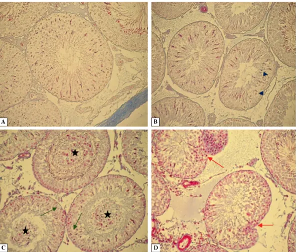
The testis of control rats presented normal morpho-logy with all generations of germ cells in the semini-ferous epithelium, corresponding to the stages of the seminiferous epithelium cycle (Figure 1A). In the XII stage of the seminiferous epithelium cycle, prolifera-ting germ cells were visible (Figure 1B).
Similarly to our earlier observations [27, 28] the general abnormalities in the structure of seminiferous tubules in the letrozole-treated rats were noticed. They included the presence in the lumen of premature sloughed germ cells — late pachytene spermatocytes, spermatids in different steps of differentiation, as well as irregular intercellular empty spaces, the effect of germ cells sloughing (Figure 1C). Additionally, multinucleated giant cells between the germ cells
of the seminiferous epithelium (Figure 5E), and in the lumen of tubules (Figure 5F) were observed in some seminiferous tubules. Long-lasting treatment of rats with letrozole resulted also in the irregularity of lamina propria of some seminiferous tubules, in form of the invaginations directed into the lumen of tubules (Figure 1D).
Immunohistochemical study
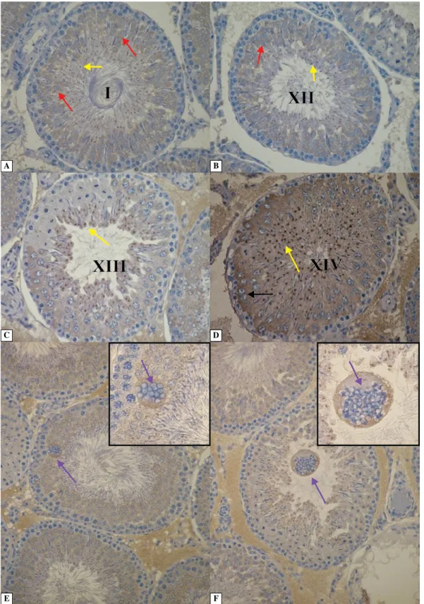
Androgen receptors. Immunolocalization of AR in the wall of seminiferous tubules of control rats testis was found in the nuclei of Sertoli cells. Expression of AR was also observed in the nuclei of myoid and Leydig cells (Figure 2A, Table 1).
As shown in Figure 2B, there was a change in the pattern of AR expression in the testes of letrozole--treated rats. Positive reaction was shown not only in the nuclei of Sertoli cells, but also in the cytoplasm of spermatogonia and spermatocytes in III/IV stages of the seminiferous epithelium cycle. Germ cells of other stages of the seminiferous epithelium cycle did not show immunoreactivity for AR. Expression of AR was also observed in the nuclei of myoid cells and the nuclei of Leydig cells (Figure 2B, Table 1). No AR-positive cells were detected when the primary antibody was omitted (Figure 2A, insert).
Cytochrome P450 aromatase. In the testis of control rats, P450arom was strongly expressed in germ cells, mainly spermatocytes and spermatids in X–XII stages of the seminiferous epithelium cycle. The cytoplasm of Sertoli and Leydig cells was P450arom immuno-positive (Figure 3A, B, Table 1).
In the testis of letrozole-treated rats, P450arom--positive reaction was detected in the cytoplasm of spermatogonia, spermatocytes, spermatids and Sertoli cells in X–XII stages of the seminiferous epithelium cycle (Figure 3C). The cytoplasm of germ cells, im-maturely sloughed into the lumen of seminiferous tubules did not show immunoreactivity for P450arom. The P450arom staining was detected in the cytoplasm of Leydig cells (Figure 3D). However, the intensity of P450arom-immunoreactivity in cells of seminiferous tubules of letrozole-treated rats was lower than in control animals (Table 1).
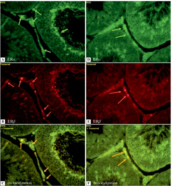
Estrogen receptors. The use of the immunofluorescent technique revealed the expression of ERs in cells of the seminiferous epithelium, cells of the lamina propria of seminiferous tubules and in the interstitial tissue in the testes of control rats. ERa -immuno-fluorescence was prominent in the nuclei of single spermatids, Leydig and myoid cells (Figure 4A). Nuclei of the Sertoli, myoid and Leydig cells exhibi-ted red fluorescence presenting expression of ERb
(Figure 4B). Co-expression of both ERa and ERb
was evident in the nuclei of Leydig and myoid cells (Figure 4C, Table 1).
Long-term treatment of the rats with letrozole cau-sed evident changes in the expression of ERs. There were no cells exhibiting green immunofluorescence Figure 1. The morphology of the testis of control (A, B) and letrozole-treated rats (C, D). Blue arrowheads — proliferating germ cells; black stars — prematurely sloughed germ cells; green arrows — intercellular empty spaces; red arrows — irre-gularities of lamina propria. Azan Trichrome staining. Magnification × 20
Figure 2. Immunoexpression of androgen receptor in the testis of control (A) and letrozole-treated rats (B). Black arrows — cytoplasm of Sertoli cells; red arrows — nuclei of Sertoli cells; green arrows — myoid cells; violet arrows — cytoplasm of germ cells; blue arrows — Leydig cells. Androgen receptors were visualized by immunohistochemistry (IHC) as descri-bed in Methods. Magnifications: A, B × 40. Insert — negative control, × 20
A B
C D
of ERa in the seminiferous epithelium and the wall of seminiferous tubules. Only the Leydig cells in the interstitial tissue were ERa-positive (Figure 4D). The lack of ERb immunofluorescence in the cells of the seminiferous epithelium and lamina propria of the seminiferous tubules was found in this group. Red fluorescence (ERb) with strong intensity was observed only in the nuclei of Leydig cells (Figure 4E), while the co-expression of ERa and ERb was prominent also only in the nuclei of Leydig cells (Figure 4F, Table 1). Transient receptor potential vanilloid 1. In the
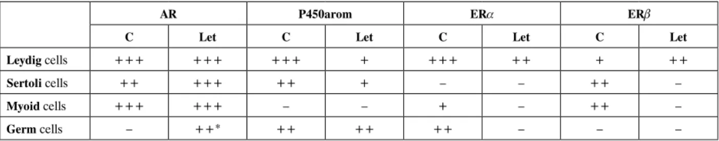
seminife-rous epithelium of control rats the immunoexpression of TRPV1 was observed in the cytoplasm of primary spermatocytes (Figure 5A), spermatids (Figure 5B) and cytoplasm of the residual spermatids (Figure 5C, D). No differences were observed in the expression of the TRPV1 in testis of letotrozole-treated rats. The po-sitive immunoreaction was identified in the cytoplasm of the same cell types as in control rats. However, the intensity of the reaction was stronger in germ cells of the seminiferous epithelium of letrozole-treated rats. The mature spermatids were TRPV1-immuno-Table 1. Summary of receptors and P450arom localization in the testis of control (C) and letrozole-treated (Let) rats presented as intensity of immunostaining and immunofluorescence
AR P450arom ERa ERb
C Let C Let C Let C Let
Leydig cells +++ +++ +++ + +++ ++ + ++
Sertoli cells ++ +++ ++ + – – ++ –
Myoid cells +++ +++ – – + – ++ –
Germ cells – ++* ++ ++ ++ – – –
Intensity of immunostaining and immunofluorescence scored as negative (–), weak positive (+), moderate positive (++) or strong positive (+++). *III/IV stages of the seminiferous epithelium cycle
Figure 3. Immunolocalization of cytochrome P450 aromatase in the testis of control (A, B) and letrozole-treated rats (C, D). Red arrows — Sertoli cells; black arrows — Leydig cells; yellow arrows — germ cells. IHC. Magnification × 40
A B
Figure 4. The immunoexpression of ERa (green fluorescence) and ERb (red fluorescence) in testis of control (A–C) and letrozole-treated rats (D–F). Green arrows — ERa; red arrows — ERb; orange arrows — co-localization of ERa and ERb. IHC. Magnification × 40
negative, similarly to the proliferating spermatocytes (Figure 5C). Moreover, the strong TRPV1 expression was observed in the cytoplasm of intraepithelial and luminal multinucleated giant cells (Figure 5E, F). Evaluation of apoptosis. In control rats, the apoptosis of germ cells, determined by the immunoexpression of caspase-3, was observed in the II and III stages of the seminiferous epithelium cycle. A strong positive reaction was present in spermatids, while there were no caspase-3-positive cells within the population of spermatogonia and spermatocytes (Figure 6A, B).
In the seminiferous epithelium of rats treated with letrozole immunoexpression of caspase-3 was observed
not only in the germ cells in II and III stages of the seminiferous epithelium cycle but also in the popula-tion of spermatids in the VI stage of the seminiferous epithelium cycle, and in spermatocytes and spermatids in I stage of the seminiferous epithelium cycle (Figure 6C, D). The immunoexpression of caspase-3 additio-nally was noticed in nuclei of Sertoli cells, myoid cells of lamina propria and Leydig cells (Figure 6C, D).
Discussion
The results of the study revealed that chronic treatment of rats with letrozole can affect not only
mor-A D B E C F ERa ERa ERb ERb co-localization co-localization
Figure 5. Immunolocalization of TRPV1 in testis of control (A, B) and letrozole-treated rats (C–F). Red arrows — cy-toplasm of spermatids; yellow arrows — residual cycy-toplasm; black arrow — cycy-toplasm of primary spermatocytes; violet arrows — cytoplasm of giant cells. IHC. Magnifications: A–D — × 40; E–G — × 20; inserts — × 40
A B
C D
phology but also the expression of studied receptors and proteins in testis. It has been showed in our earlier study, that letrozole significantly decreased level of circulating and intratesticular estradiol (E2) by 43% and 48%, re-spectively, which caused the morphological alterations in the seminiferous epithelium and elements of lamina propria of seminiferous tubules [27]. Moreover, the hormonal imbalance affected the immunoexpression of c-Kit receptor in spermatogonia and caused morphome-tric changes such as decreased diameter of seminiferous tubules, and decreased thickness of layer occupied by c-Kit-R-positive spermatogonia [28]. Therefore, we expected that the lower level of E2 in letrozole-treated rats also could produce abnormalities associated with the expression of key proteins in the germ cells responsible for proper spermatogenesis.
In the present study, the immunolocalization of AR in nuclei of Sertoli cells, myoid and Leydig cells of testis from control and experimental rats was in agreement with many previous studies [13, 16, 29–31]. However, we also found the cytoplasm of spermatogonia and spermatocytes in III/IV stages of the seminiferous epithelium cycle in testis of experimental rats was AR-immunopositive, while the cytoplasm of germ cells of other stages of the seminiferous epithelium cycle was immunonegative. The presence of ARs in germ cells is controversial.
However, some investigators demonstrated that nuc-lear ARs were detectable by immunohistochemistry in germ cells in human and animal testes [13, 29, 32]. The cytoplasmic localization of AR was shown in elongated spermatids in the adult rat testis [33], Ley-dig cells of patients with Klinefelter’s syndrome [34], and in mice with a Sertoli cells-specific knock-out of the connexin 43 gene [35]. Generally, a two-step model of steroid hormone action has been accepted; it assumes that steroid hormone receptors exist in two different forms: the unliganded receptor in the cytoplasm and the hormone-bound receptor complex in the nucleus [36]. Probably, ARs are localized in the lysosomal compartment of the cytoplasm involved in receptor degradation [34]. Physiological interplay between androgens and estrogens has been well esta-blished in male reproductive tract, and each hormone may affect the expression of the other’s receptor (AR of the ERs, mainly ERa, and vice versa) [37]. Therefore, we suggest that the cytoplasmic localiza-tion of ARs in germ cells of the seminiferous epithe-lium of letrozole-treated rats can be caused by lower level of circulating and intratesticular estradiol.
In male gonads, androgens undergo local conver-sion to estrogens thanks to P450arom activity. It is known that germ cells, mainly pachytene spermato-cytes and round spermatids in humans and animals Figure 6. Caspase-3 expression in testis of control (A, B) and letrozole-treated rats (C, D). Black arrows — cytoplasm of spermatids; green arrow — cytoplasm of spermatogonia and primary spermatocytes; red arrows — Sertoli cells; blue arrow — myoid cell; violet arrows — interstitial cells. IHC. Magnification × 40
A B
are the major places of aromatase localization in the seminiferous epithelium [9, 38, 39]. It was also postu-lated, that the aromatase gene had been expressed in all developmental stages of germ cells of rat testis [39]. Myoid cells are the only population of cells within the testicular tissue that do not express P450 aromatase [2, 40]. The long-lasting lower level of estradiol did not affect the immunolocalization of P450 aromatase in germ cells, however, at the same time, the P450 aro-matase immunoreactivity was lower. In vitro studies showed that both testosterone and dihydrotestostero-ne (DHT) stimulated the expression of the P450arom gene and the aromatase activity in pachytene sper-matocytes and round spermatids, while estradiol down-regulated the aromatase transcription in germ cells [41]. Additionally, we detected the presence of P450arom in the cytoplasm of sloughed immature germ cells in the lumen of seminiferous tubules in testes of experimental rats.
Estrogens exert their cellular effect via estrogen receptors scattered throughout whole male repro-ductive tract. It was reported that estrogen signaling is depended upon a balance between ERa and ERb
activities [11]. In this way, decreased estradiol level can affect expression of estrogen receptors. Interac-tions between estrogen or estrogen receptors and androgen receptor also occur. In this way reduced en-dogenous estrogen levels could affect AR as well [42]. In our study ERa-immunofluorescence was promi-nent in the nuclei of Leydig and myoid cells, similarly as was reported by others [1, 43]. Interestingly, we also showed the presence of ERa in germ cells. This finding was unexpected because the most consistent data across species reported the presence of ERa in Leydig cells. However, Lucas et al. [44] demonstrated the presence of ERa in Sertoli cells of immature and adult rats and also in some spermatids in the adult animals. Similar results were obtained by Cavaco et al. [45], who found immunofluorescence of ERa not only in Leydig cells, but also in Sertoli cells, sper-matogonia, spermatocytes, round spermatids and elongated spermatids/spermatozoa. Bois et al. [15] reported that ERa expression in germ cells depen-ded on stages of seminiferous epithelium cycle and occurred in stages VII to XIV. Moreover, both ERs mRNA levels were higher in round spermatids than in pachytene spermatocytes [15]. The cytoplasmic locali-zation of ERa was found in Sertoli and Leydig cells in the testes of men with normal spermatogenesis [46]. Thus, both our results and the cited references sug-gest that estrogens play one of the major roles in the haploid steps of spermatogenesis.
Our current finding of the ERb absence in germ cells differs from the reports of other authors who
found ERb-immunopositivity in gonocytes, sperma-togonia, pachytene spermatocytes, round and elon-gated spermatids [47, 48]. These discrepancies could be explained by the data of Carreau and Hess [1] who described testicular ERb-expression to be de-pendent on species, subjects within one species as well as preservation techniques and antibodies used for immunohistochemistry. The ERa and ERb are co-expressed in some regions of rat male reproductive tract and exist separately in other parts [49]. The ho-modimers ERa–ERa and heterodimers ERa–ERb
bind with higher affinity to a ERE (estrogen response element) than homodimers ERb–ERb [50]. This may explain our observation of the presence of both ERs in the same cell. As a result of adult male rats treat-ment with letrozole, we observed changes in ERa and ERb expression in testis. We concluded, according to results of other authors [2, 51, 52] and ours, that the chronical treatment of male rats with aromatase inhibitor, and in consequence estrogen deficiency, can follow ERs down-regulation.
The important role in the progression of the spermatogenesis plays transient receptor potential vanilloid 1 (TRPV1), one of molecular targets for en-docannabioids. TRPV1 is a ligand-gated, nonselective cation channel that first was found in neurons [53], and next in non-neuronal cells [19]. TRPV1 can be also activated by vanilloid compounds such as cap-saicin, and a number of other ligands [54], and by ambient temperature [55]. TRPV1 was also shown to be expressed in rat testis [17, 56]. Germ cells, from spermatogonia to spermatozoa, and Sertoli cells possess a complete biochemical machinery to synthesize, transport, degrade and bind endocanna-bioids, that can affect male reproductive functions [14, 57]. Our results on the immunolocalization of TRPV1 in germ cells, and in residual spermatids cy-toplasm of experimental rats are in agreement with the findings of other authors who detected TRPV1 channels in the seminiferous tubules of rat testis using RT-PCR [17, 20, 56, 58, 59]. Grimaldi et al. [20] showed elevated expression level of the TRPV1 gene in all stages of spermatogenesis, with the peak expression in meiotic and postmeiotic cells in mouse testis. However, data on the expression of TRPV1 in rat testis are controversial. Li et al. [60] did not find expression of TRPV1 in rat testicular tissue, however, they found TRPV5 in plasma membranes of sperma-togenic cells. Studies performed in male mice and rats indicated that TRPV1 expression was mostly found in premeitoic germ cells, whereas spermatocytes only weakly expressed this receptor, while no expression was observed in postmeiotic germ cells [18, 19]. Strong positive immunoreaction was visible in our study in
in female rats, and did not influence change in the TRPV1 expression in testis. The inhibitory effect of estradiol on activation of TRPV1 by capsaicin was observed in adult rat nociceptor neurons [61].
TRPV1 has been proposed to be associated with apoptosis of germ cells [19]. In our study sponta-neous apoptosis in control rats was observed in the population of spermatids, which is contrary to the results of other studies. In male rats and hamsters programmed cell death was noticed in a few diffe- rentiating spermatogonia and spermatocytes during their meiotic divisions [62], while in human testes germ cell apoptosis was observed in spermatogonia, spermatocytes and spermatids [63]. Moreover, incre-ased number of apoptotic germ cells is specific for par-ticular stage of the seminiferous epithelial cycle [64]. This finding was clearly observed by us. In testis of letrozole-treated rats, caspase-3-positive germ cells were seen in the population of spermatids also in the VI stage and in spermatocytes and spermatids in the I stage of the seminiferous epithelium cycle. Study by Omezzine et al. [65] indicated the immunolocali-zation of active caspase-3 exclusively in postmeiotic germ cells particularly in round spermatids in adult rat testes exposed in utero to different doses of an antiandrogen (flutamide). The presence of intense caspase-3 immunoreactivity in nuclei of many germ cells in rat testis in our study suggests that caspase-3 can be translocated from the cytoplasm to the nuclei and such translocation can be necessary for apoptosis to occur [66]. The presence of immunolabeling in Sertoli cells can be connected with the fact that these cells phagocytose fragments of neighboring apoptotic germ cells [67]. Additionally, it cannot be excluded that Sertoli cells themselves may undergo apoptosis in abnormal condition [63], as shown in cimetidine-tre-ated adult rats [68]. Leydig cells produce the primary male steroid hormone testosterone, and their function can be affected by hormonal imbalance. The apoptosis of the cells, identified as caspase-3-immunopositive cells was found in testes of pubertal mice exposed to different dose of bisphenol A (BPA). The effect resulted from decreased expression of steroidogenic enzymes in Leydig cells induced by the xenoestro - gen [69]. Peritubular myoid cells maintain the
mor-affected the expression of androgen receptor, es-trogen receptors, vanilloid receptor 1, aromatase cytochrome P450 and increased apoptosis of cells in the seminiferous tubules. These results underline the important role of estrogens in male reproductive system, and additionally demonstrate that hormonal imbalance can produce the morphological and func-tional abnormalities in testis.
References
1. Carreau S, Hess RA. Oestrogens and spermatogenesis. Philos Trans R Soc Lond B Biol Sci. 2010;365:1517–1535.
2. Carreau S, Bouraima-Lelong H, Delalande C. Role of estro-gens in spermatogenesis. Front Biosci (Elite Ed). 2012;4:1–11. 3. Akingbemi BT. Estrogen regulation of testicular function.
Reprod Biol Endocrinol. 2005;27:3–51.
4. Carreau S, Bouraima-Lelong H, Delalande C. Estrogen. A female hormone involved in spermatogenesis. Adv Med Sci. 2012;57:31–36.
5. Kotula-Balak M, Hejmej A, Lydka M, Cierpich A, Bilinska B. Detection of aromatase, androgen and estrogen receptors in bank vole spermatozoa. Theriogenology. 2012;78:385–392. 6. Carreau S, Bois C, Zanatta L, Silva FRMB, Bouraima-Lelong H,
Delalande C. Estrogen signaling in testicular cells. Life Sci. 2011;89:584–587.
7. Hess RA. Estrogen in the adult male reproductive tract: a review. Reprod Biol Endocrinol. 2003;1:52.
8. Bilińska B, Schmalz-Frączek B, Sadowska J, Carreau S. Immunolocalization of cytochrome P450 aromatase and estrogen receptors a and b in bank vole testicular cells. Acta Histochem. 2000;102:167–181.
9. Carpino A, Pezzi V, Rago V, Bilinska B, Ando S. Immunolo-calization of cytochrome P450 aromatase in rat testis during postnatal development. Tissue Cell. 2001;33:349–353. 10. Levallet J, Bilinska B, Mittre H, Genissel C, Fresnel J,
Car-reau S. Expression and immunolocalization of functional cytochrome P450 aromatase in mature rat testicular cells.
Biol Reprod. 1998;58:919 – 926.
11. Lee HR, Kim TH, Choi KC. Functions and physiological roles of two types of estrogen receptors, ERa and ERb, identified by estrogen receptors knockout mouse. Lab Anim Res. 2012;28:71–76.
12. Yong EL, Loy CJ, Sim KS. Androgen receptor gene and male infertility. Hum Reprod Update. 2003;9:1–7.
13. Wang RS, Yeh S, Tzeng CR, Chang C. Androgen receptor roles in spermatogenesis and fertility: lessons from testicular cell-specific androgen receptor knockout mice. Endocr Rev. 2009;30:119–132.
14. Grimaldi P, Pucci M, Di Siena S et al. The faah gene is the first direct target of estrogen in the testis: role of histone demethylase LSD1. Cell Mol Life Sci. 2012;66:4177–4790.
15. Bois C, Delalande C, Nurmio M et al. Age- and cell-related gene expression of aromatase and estrogen receptors in the rat testis. J Mol Endocrinol. 2010;45:147–159.
16. Pelletier G, Labrie C, Labrie F. Localization of oestrogen receptor a, oestrogen receptor b and androgen receptors in the rat reproductive organs. J Endocrionol. 2000;165:359–370. 17. Stein RJ, Santos S, Nagatomi J et al. Cool (TRPM8) and hot
(TRPV1) receptors in the bladder and male genital tract.
J Urol. 2004;172:1175–1178.
18. Mizrak SC, Gadella BM, Erdost H et al. Spermatogonial stem cell sensitivity to capsaicin: an in vitro study. Reprod Biol Endocrinol. 2008;6:52.
19. Mizrak SC, van Dissel-Emiliani FM. Transient receptor potential vanilloid receptor-1 confers heat resistance to male germ cells. Fertil Steril. 2008;90:1290–1293.
20. Grimaldi P, Orlando P, Di Siena S et al. The endocannabinoid system and pivotal role of the CB2 receptor in mouse sper-matogenesis. Proc Natl Acad Sci USA. 2009;106:11131–11136. 21. Maccarrone M, Barboni B, Paradisi A et al. Characterization of the endocannabinoid system in boar spermatozoa and implications for sperm capacitation and acrosome reaction.
J Cell Sci. 2005;118:4393–4404.
22. Smith LB, Walker WH. The regulation of spermatogenesis by androgens. Semin Cell Dev Biol. 2014;30:2–13.
23. Robertson KM, Sipmson ER, Lacham-Kaplan O et al. Chara-cterization of the fertility of male aromatase knockout mice.
J Androl. 2001;22:825–830.
24. Robertson MR, O’Donnell L, Jones MEE et al. Impairment of spermatogenesis in mice lacking a functional aromatase
(cyp 19) gene. Proc Natl Acad Sci USA. 1999;96:7986–7991. 25. Shalet SM. Normal testicular function and spermatogenesis.
Pediatr Blood Cancer. 2009;53:285–288.
26. Pentikäinen V, Erkkilä K, Suomalainen L, Parvinen M, Dun-kel L. Estradiol acts as a germ cell survival factor in the human testis in vitro. J Clin Endocrinol Metab. 2000;85:2057–2067. 27. Kondarewicz A, Kolasa A, Zawiślak B et al. Testis
morphol-ogy in rats chronically treated with letrozole, an aromatase inhibitor. Folia Histochem Cytobiol. 2011;49:677–684. 28. Misiakiewicz K, Kolasa A, Kondarewicz A, Marchlewicz M,
Wiszniewska B. Expression of the c-Kit receptor in germ cells of the seminiferous epithelium in rats with hormonal imbalance. Reprod Biol. 2013;13:333–340.
29. Berensztein EB, Baquedano MS, Gonzales CR et al. Ex-pression of aromatase, estrogen receptor a and b, androgen receptor, and cytochrome P-450scc in the human early pre-pubertal testis. Pediatr Res. 2006;60:740–744.
30. Suarez-Quian CA, Martinez-Garcia F, Nistal M, Regadera J. Androgen receptor distribution in adult human testis. J Clin Endocrinol Metab. 1999;84:350–358.
31. Timurkaan S, Gur FM, Karan M. Immunohistochemical distribution of androgen receptor in rat testis during postnatal development. Revue Med Wet. 2012;163:112–115.
32. Boukenaoui N, Moudilou E, Chevalier C, Amirat Z, Exbrayat JM, Khammar F. Postnatal changes in testicular development and androgen receptors immunolocalization in D’Man ram lambs. Folia Histochem Cytobiol. 2012;50:38–45.
33. Vornberger W, Prins G, Musto NA, Suarez-Quian CA. Androgen receptor distribution in rat testis: new implications for androgen regulation of spermatogenesis. Endocrinology
1994;134:2307–2316.
34. Kotula-Balak M, Bablok L, Frącki S, Jankowska A, Bilińska B. Immunoexpression of androgen receptors and aromatase in testes of patient with Klinefelter’s syndrome. Folia Histochem Cytobiol. 2004;42:215–220.
35. Chojnacka K, Brehm R, Weider K et al. Expression of the androgen receptor in the testis of mice with a Sertoli cell specific knock-out of the connexin 43 gene (SCCx43KO(–/–).
Reprod Biol. 2012;12:341–346.
36. Gasc JM, Baulieu EE. Steroid hormone receptors: intracel-lular distribution. Biol. Cell. 1986;56:1–6.
37. Panet-Raymond V, Gottlieb B, Beitel LK et al. Interactions between androgen and estrogen receptors and the effects on their transactivational properties. Mol Cell Endocrinol.
2000;167:139–150.
38. Bilińska B, Schmalz-Fraczek B, Sadowska J, Carreau S. Local-ization of cytochrome P450 aromatase and estrogen receptors alpha and beta in testicular cells — an immunohistochemical study of the bank vole. Acta Histochem. 2000;102:167–181. 39. Lambard S, Galeraud-Denis I, Saunders PTK, Carreau S.
Human immature germ cells and ejaculated spermatozoa contain aromatase and oestrogen receptors. J Mol Endocrinol. 2004;32:279–289.
40. Carreau S, Delalande C, Galeraud-Denis I. Mammalian sperm quality and aromatase expression. Microsc Res Tech. 2009;72:552–557.
41. Bourguiba S, Lambard S, Carreau S. Steroids control the aromatase gene expression in purified germ cells from the adult male rat. J Mol Endocrinol. 2003;31:83–94.
42. Ramesh R, Pearl CA, At-Taras E, Roser J, Berger T. Onto-geny of androgen and estrogen receptor expression in porcine testis: effect of reducing testicular estrogen synthesis. Animal Reprod Sci. 2007;102:286–299.
43. Akingbemi BT, Ge R, Rosenfeld CS et al. Estrogen recep-tor-alpha gene deficiency enhances androgen biosynthesis in the mouse Leydig cell. Endocrinology 2003;144: 84–93. 44. Lucas TF, Siu ER, Esteves CA et al. 17beta-estradiol induces
the translocation of the estrogen receptors ESR1 and ESR2 to the cell membrane, MAPK3/1 phosphorylation and pro-liferation of cultured immature rat Sertoli cells. Biol Reprod. 2008;78:101–114.
45. Cavaco JEB, Laurentino SS, Barros A, Sousa M, Socorro S. Estrogen receptors a and b in human testis: both isoform are expressed. Syst Biol Reprod Med. 2009;55:137–144.
46. Filipiak E, Suliborska D, Laszczynska M et al. Estrogen receptor alpha localization in the testes of men with normal spermatogenesis. Folia Histochem Cytobiol. 2013;50:340–345. 47. Sasso-Cerri E. Enhanced ERbeta immunoexpression and
apoptosis in the germ cells of cimetidine-treated rats. Reprod Biol Endocrinol. 2009;7:127–134.
48. Saunders PT, Fisher JS, Sharpe RM, Millar MR. Expression of oestrogen receptor beta (ER beta) occurs in multiple cell types, including some germ cells, in the rat testis. J Endocrinol. 1998;156:R13–R17.
49. Oliveira CA, Mahecha GAB, Carnes K et al. Differential hormonal regulation of estrogen receptors ERa and ERb and androgen receptor expression in rat efferent ductules.
Soc Reprod Fertil. 2004;128:73–86.
50. Carpino A, Rago V, Pezzi V, Carani C, Ando S. Detection of aromatase and estrogen receptors (ERa, ERb1, ERb2) in human Leydig cell tumor. Eur J Endocrinol. 2007;157: 239–244.
51. Hess RA, Bunick D, Lubahn DB, Zhou Q, Bouma J. Morpho-logic changes in efferent ductules and epididymis in estrogen receptor-a knockout mice. J Androl. 2000;21:107–121. 52. Joseph A, Hess R, Schaeffer DJ et al. Absence of estrogen
receptor alpha leads to physiological alterations in the mouse epididymis and consequent defects in sperm function. Biol Reprod. 2012;82:948–957.
2013;110–111:80–86.
57. Grimaldi P, Di Giacomo D, Geremia R. The endocannabi-noid system and spermatogenesis. Front Endocrinol (Lau sanne). 2013;4:192.
58. Rossato M, Ion Popa F, Ferigo M, Clari G, Foresta C. Human sperm express cannabinoid receptor Cb1, the activation of which inhibits motility, acrosome reaction, and mitochondrial function. J Clin Endocrinol Metab. 2005;90:984–991. 59. Sun X, Wang H, Okabe M et al. Genetic loss of Faah
com-promises male fertility in mice. Biol Reprod. 2009;80:235–242. 60. Li S, Wang X, Ye H, Gao W, Pu X, Yang Z. Distribution
profiles of transient receptor potential melastatin- and vanil-loid-related channels in rat spermatogenic cells and sperm.
Mol Biol Rep. 2010;37:1287–1293.
61. Xu S, Cheng Y, Keast JR, Osborne PB. 17beta-estradiol activates estrogen receptor beta-signalling and inhibits transient receptor potential vanilloid receptor 1 activation by capsaicin in adult rat nociceptor neurons. Endocrinology. 2008;149:5540–5548.
62. Sinha Hikim AP, Swerdloff RS. Hormonal and genetic control of germ cell apoptosis in the testis. Rev Reprod. 1999;4:38–47.
66. Kim J-M, Ghosh SR, Weil ACP, Zirkin BR. Caspase-3 and caspase-activated deoxyribonuclease are associated with testicular germ cell apoptosis resulting from reduced intra-testicular testosterone. Endocrinology. 2001;142:3809–3816. 67. Pentikäinen V, Erkkilä K, Dunkel L. Fas regulates germ
cell apoptosis in the human testis in vitro. Am J Physiol.
1999;276:E310–E316.
68. Sasso-Cerri E, Miraglia SM. In situ demonstration of both TUNEL-labeled germ cell and Sertoli cell in the cimeti-dine-treated rats. Histol Histopathol. 2002;17:411–417. 69. Li YJ, Song TB, Cai YY et al. Bisphenol A exposure induces
apoptosis and upregulation of Fas/FasL and caspase-3 ex-pression in the testes of mice. Toxicol Sci. 2009;108:427–436. 70. Liu XL, Chen XY, Wang ZC, Shen T, Zhao H. Effects of
exposure to bisphenol A during pregnancy and lactation on the testicular morphology and caspase-3 protein expression of ICR pups. Biomed Rep. 2013;1:420–424.
71. França LR, Leal MC, Sasso-Cerri E, Vasconcelos A, Debel-juk L, Russell LD. Cimetidine (Tagamet) is a reproductive toxicant in male rats affecting peritubular cells. Biol Reprod.
2000;63:1403–1412.
Submitted: 10 July, 2014 Accepted after reviews: 30 September, 2014