From
DEPARTMENT OF LABORATORY MEDICINE
Karolinska Institutet, Stockholm, Sweden
INFECTIONS IN SKIN CANCER
Laila Sara Arroyo Mühr
All previously published papers were reproduced with permission from the publisher. Paper III is an Open Access article distributed in accordance with the Creative Commons Attribution Non Commercial (CC BY-NC 4.0) license.
Cover photography: HPV 197 L1 protein. Predicted by The Phyre2 web portal for protein modeling, prediction and analysis. Kelley LA et al. Nature Protocols 10, 845-858 (2015). Published by Karolinska Institutet
Printed by Eprint AB 2016 © Laila Sara Arroyo Mühr, 2016 ISBN 978-91-7676-215-8
Department of Laboratory Medicine
Infections in Skin Cancer
AKADEMISK AVHANDLING
som för avläggande av medicine doktorsexamen vid Karolinska
Institutet offentligen försvaras i Föreläsningssal Solen 4U, Alfred
Nobels Allé 8, Karolinska Institutet, Huddinge.
Fredagen den 08 april 2016, kl 13.00
av
Laila Sara Arroyo Mühr
MSc Pharmacy
Stockholm 2016
Principal Supervisor:
Professor Joakim Dillner Karolinska Institutet
Department of Laboratory Medicine Division of Pathology
Co-supervisor(s):
PhD Emilie Hultin Karolinska Institutet
Department of Laboratory Medicine Division of Pathology
Associate Professor Ola Forslund Lund University
Department of Laboratory Medicine Division of Medical Microbiology
Professor Göran Andersson Karolinska Institutet
Department of Laboratory Medicine Division of Pathology
Opponent:
PhD Max Käller
Royal Institute of Technology Division of Gene Technology
Examination Board:
Professor Ingemar Ernberg Karolinska Institutet
Department of Microbiology, Tumor and Cell Biology
Examination Board:
Professor Lars Engstrand Karolinska Institutet
Department of Microbiology, Tumor and Cell Biology
Examination Board:
Professor Emeritus Jonas Blomberg Uppsala University
Department of Medical Science
“We only see what we know” (J.W. von Goethe)
ABSTRACT
The increasing prevalence of skin cancer results in that it will soon equal that of all other cancers combined. Sun exposure is a well-known risk factor for its development, but despite the growing public awareness of the harmful consequences of ultraviolet radiation, the cancer incidence continues to increase, implying that other factors might also have a role in promoting this disease.
Data from immunosuppressed patients reveals a 100-fold increased incidence of non-melanoma skin carcinoma (NMSC), but an infectious etiology has not been established. However, certain human papillomaviruses (HPVs) have previously been detected in this type of cancer.
We applied high throughput sequencing to different skin lesions in order to assess which organisms were present. Most viral reads (>95%) belonged to human papillomavirus. Traditionally, viral detection was performed using PCR methods. We used degenerate “general” HPV primers and multiplexed novel “specific” HPV primers in order to amplify a broad number of HPVs by PCR. This method showed a very high sensitivity, but the HPV types with low similarity to the primer sequences might have escaped amplification. Therefore, we performed an unbiased approach based on non-PCR whole genome amplification, independent of sequence information, in order to detect those “escaping” HPV types, as well as to determine if other viruses were present in the samples.
Overall, we identified almost 100 putative novel HPV types in total, and characterized 4 novel HPV types (HPV 197, 200, 201 and 202). Most of the HPV types were detected in very few patients each, and at a very low viral load (below 0.5 copies/cell), except for HPV 197, which was the most commonly found virus in skin tumors (37.4% of skin lesions). Despite the higher sensitivity of PCR methods, the unbiased approach detected HPV in 37/40 condyloma acuminata that had been reported as “HPV-negative” with specific PCR techniques. Certain HPV types, including HPV 197, were not detected by PCR and only by non-PCR based methods. Therefore, more unbiased PCR-independent methods are needed to describe which organisms are most commonly present in skin lesions.
The work in this thesis has expanded our knowledge of the wide genomic diversity of HPV on the skin, and finds that PCR-independent methods are needed to describe which organisms are most commonly present in skin lesions. Further studies are needed to assess any possible role of viral infections in skin cancer, elucidation of mechanistic effects and determine the direction of causality of any associations.
SAMMANFATTNING
Den ökande förekomsten av hudcancer resulterar snart i lika många fall som alla andra cancertyper tillsammans. Solexponering är en känd riskfaktor för utveckling av hudcancer, men trots allmän kännedom kring de skadliga konsekvenserna av ultraviolett strålning, så ökar frekvensen av sjukdomen, vilket tyder på att det även kan finnas andra bidragande faktorer.
Sjukdomsstatistik från patienter med nedsatt immunförsvar visar en 100-faldig ökad frekvens av icke-melanom hudcancer, men någon bakomliggande infektion har ännu inte kunnat fastställas. Dock har vissa typer av humant papillomvirus (HPV) hittats i denna typ av cancer.
Vi sekvenserade allt DNA i olika hudförändringar för att undersöka vilka mikroorganismer de innehöll. De flesta virussekvenserna (>95%) kom från HPV.
Traditionellt har virus detekterats med olika PCR-metoder. Vi använde oss av degenererade ”generella” HPV-primers och multiplexade nya ”specifika” HPV-primers för att möjliggöra PCR-amplifiering av många olika HPV-typer. Denna metod visade på en mycket hög känslighet, men HPV-typer med låg likhet till primersekvenserna kan ha undgått amplifiering. För ett mer objektivt tillvägagångssätt amplifierade vi allt DNA utan PCR och oberoende av någon sekvensinformation för att kunna detektera eventuella HPV-typer som kan ha undgått PCR-amplifieringen likväl som andra virus i proverna. Totalt identifierade vi nära 100 möjliga nya typer, samt karaktäriserade 4 nya HPV-typer (HPV 197, 200, 201 och 202). De flesta HPV-HPV-typerna detekterades bara i några få patienter var, med mycket låga virustal (mindre än 0,5 kopior/cell), förutom HPV 197, vilket var det vanligast förekommande viruset bland hudtumörer (37,4% av hudförändringarna). Trots den högre känsligheten hos PCR-baserade metoder, detekterade den mer objektiva PCR-oberoende metoden HPV i 37/40 condylomata acuminata som alla tidigare rapporterats som negativa med specifika PCR-metoder. Vissa HPV-typer, inklusive HPV 197, detekterades inte med PCR, utan enbart med metoder utan PCR. Därför behövs fler objektiva, PCR-oberoende, metoder för att beskriva vilka mikroorganismer som är vanligast förekommande i hudförändringar.
Arbetet i denna avhandling har utökat vår kunskap om den stora genetiska mångfalden av HPV i hud, samt konstaterar att PCR-oberoende metoder är nödvändiga för att beskriva vilka mikroorganismer som är vanligast förekommande i hudförändringar. Vidare studier är nödvändiga för att fastställa möjliga samband mellan virusinfektioner och hudcancer, klargöra mekanistiska effekter, samt avgöra orsaksriktning mellan funna samband.
RESUMEN
El cáncer de piel es el más frecuente de los cánceres en el ser humano. A pesar de que la exposición a la luz ultravioleta es un factor de riesgo bien conocido y de la creciente concienciación popular sobre sus efectos perjudiciales, la incidencia de este cáncer continúa aumentando. Se estima que no tardará mucho en sobrepasar en número a la suma del resto de cánceres. Esto sugiere que pueden existir otros factores que contribuyen al desarrollo de esta enfermedad.
Las personas inmunodeprimidas presentan una mayor incidencia en la mayoría de los cánceres, sobre todo en los causados por virus oncogénicos (consecuencia de la reducción general de su respuesta inmune). El cáncer de piel tipo no melanoma presenta la incidencia más elevada (>100 veces) en este tipo de pacientes, pero aún no se ha asociado ningún agente etiológico que justifique esta situación.
A lo largo de esta tesis, se han secuenciado (secuenciación masiva de nueva generación) diferentes lesiones de piel con el fin de determinar los organismos presentes en la epidermis. La mayoría de las secuencias virales obtenidas (>95%) correspondieron al virus del papiloma humano (HPV). Tradicionalmente, la detección de virus ha venido realizándose mediante la reacción en cadena de la polimerasa (PCR). En esta tesis se utilizaron múltiples pares de primers y primers degenerados con el objetivo de amplificar un gran número de HPVs, obteniendo una gran sensibilidad. Sin embargo, aquellos genotipos cuyas secuencias no fuesen similares a las secuencias de los primers, pudieron no haberse amplificado, y por tanto, no ser detectados. Para obviar esta limitación se optó por realizar un protocolo no sesgado (WGA), independiente de la secuencia a amplificar, para determinar si había más genotipos de HPV y/o otros virus presentes en las diferentes lesiones de piel.
Esta tesis ha permitido identificar hasta casi 100 secuencias pertenecientes a posibles nuevos tipos de HPV y caracterizar 4 nuevos genotipos (HPV 197, 200, 201 and 202). La mayoría de los HPVs se detectaron en muy pocos pacientes cada uno y en una concentración viral baja (<0.5 copias/célula), a excepción del HPV 197, que fue el genotipo encontrado con mayor frecuencia (presente en el 37,4% de las lesiones). A pesar de que los métodos basados en la PCR fueron más sensibles, el método basado en WGA fue capaz de detectar HPV en 37/40 condilomas, que habían sido previamente clasificados como HPV-negativos tras genotiparse vía PCR. Algunos HPV, como el tipo 197, fueron detectados solo con el protocolo basado en WGA. Por lo tanto, estimamos que son necesarios más métodos no sesgados, imparciales, para descubrir cuáles son los organismos presentes con mayor frecuencia en las lesiones de piel.
El trabajo realizado en esta tesis ha incrementado nuestro conocimiento sobre la gran diversidad genómica del HPV en la piel. Se necesitan más estudios para evaluar cualquier posible asociación de una infección viral con el cáncer, dilucidar los mecanismos para su desarrollo y determinar la dirección de causalidad.
LIST OF PUBLICATIONS
This thesis is based on the following papers:
I. Arroyo Mühr LS, Smelov V, Bzhalava D, Eklund C, Hultin E, Dillner J. Next generation sequencing for human papillomavirus genotyping.
J Clin Virology 2013;58:437-42.
II. Ekström J, Arroyo Mühr LS, Bzhalava D, Söderlund-Strand A, Hultin E, Nordin P, Stenquist B, Paoli J, Forslund O, Dillner J.
Diversity of human papillomaviruses in skin lesions. Virology 2013;447:300-11.
III. Bzhalava D, Arroyo Mühr LS, Lagheden C, Ekström J, Forslund O, Dillner J, Hultin E.
Deep sequencing extends the diversity of human papillomaviruses in human skin.
Scientific Reports 2014;4:5807.
IV. Arroyo Mühr LS, Hultin E, Bzhalava D, Eklund C, Lagheden C, EkströmJ, JohanssonH, Forslund O, Dillner J.
Human papillomavirus type 197 is commonly present in skin tumors. Int J Cancer 2015;136:2546-55.
V. Arroyo Mühr LS, Bzhalava D, Lagheden C, Eklund C, Johansson H, Forslund O, Dillner J, Hultin E.
Does human papillomavirus-negative condylomata exist? Virology 2015;485:283-8.
TABLE OF CONTENTS
1 INTRODUCTION 1
1.1 NON-MELANOMA SKIN CANCER 1
1.2 INFECTIONS AND NON-MELANOMA SKIN CANCER 4 1.3 POTENTIAL PATHOGENS IN SKIN CANCER 5
1.3.1 HPV 6
1.3.1.1 Nomenclature and classification 6 1.3.1.2 HPV in Skin Cancer 10
1.4 VIRAL DETECTION IN SKIN CANCER 12
1.4.1 Amplification techniques 13
1.4.1.1 PCR using “general” or “degenerated” primers 13 1.4.1.2 Unbiased approach: Whole genome amplification 14
1.4.2 Detection techniques 16
1.4.2.1 Hybridization to type-specific probes 17 1.4.2.2 High throughput sequencing 18
1.5 HIGH THROUGHPUT SEQUENCING INSTRUMENTS 18
1.5.1 454 GS technology (Roche) 19
1.5.1.1 Generation of a template DNA library 20 1.5.1.2 Emulsion-based clonal amplification of the library 20
1.5.1.3 Pyrosequencing 20
1.5.2 Genome Analyzer System technology (Illumina) 21
1.5.2.1 Generation of a template DNA library 22 1.5.2.2 Cluster generation 23 1.5.2.3 Sequencing by synthesis 23
1.6 HIGH THROUGHPUT SEQUENCING DATA ANALYSIS 24
2 SUMMARY OF PUBLICATIONS 32
2.1 AIMS 32
2.2 MATERIALS AND METHODS 33
2.2.1 Study material 33
2.2.2 Methods 35
2.2.2.1 Sample adequacy 35 2.2.2.2 HPV Amplification 35
2.2.2.3 HPV Detection 37
2.2.2.4 New SE types and Cloning HPV types 38 2.2.2.5 Summary of methods throughout the papers 39
2.3 RESULTS AND DISCUSSION 41
2.3.1 Paper I 41
2.3.2 Paper II 42
2.3.3 Paper III 45
2.3.4 Paper IV 47
2.3.5 Paper V 49
2.4 CONCLUDING REMARKS AND FUTURE PERSPECTIVES 50
3 ACKNOWLEDGEMENTS 53
4 REFERENCES 55
ABBREVIATIONS
Aa AK Amino acid Actinic keratosis BCC bp CIBasal cell carcinoma Base pairs Confidence interval CIN dNTP dsDNA EmPCR EV FFPE GAAS GASiC GRAMMy GS HPV
Cervical intraepithelial neoplasia Deoxyribonucleotide triphosphate Double-stranded DNA
Emulsion PCR
Epidermodysplasia verruciformis Formalin-fixed paraffin-embedded
Genome relative Abundance and Average Size Genome Abundance Similarity Correction
Genome Relative Abundance estimates based on Mixture Model theory Genome Sequencer Human papillomavirus HR HTS IARC High risk
High throughput sequencing
International Agency for Research on Cancer
KA Keratoacanthoma
LR Low risk
MDA MID NGS
Multiple displacement amplification Multiplex identifier
Next generation sequencing NMSC
nt OLC PCR PGM
Non-melanoma skin cancer Nucleotide
Overlap/Layout/Consensus Polymerase chain reaction Personal Genome Machine PV RT-PCR SCC SIR SNP ssDNA UV Papillomavirus Reverse-transcription PCR Squamous cell carcinoma Standardized incidence ratio Single nucleotide polymorphism Single-stranded DNA
Ultraviolet WGA
WHO
Whole genome amplification World Health Organization
1
1 INTRODUCTION
1.1 NON-MELANOMA SKIN CANCER
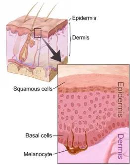
The skin provides protection and receives sensory stimuli from the external environment, being the largest organ in the body. It is composed of three primary layers: epidermis, dermis and hypodermis (Figure 1).
Most skin cancers arise from the epidermis and are named for the type of cells that become malignant. There are three major types of skin carcinoma:
- Basal cell skin cancer (BCC),
the most frequently occurring form of skin cancer. This carcinoma arises in the skin’s basal cells, which compose the deepest layer of the epidermis.
- Squamous cell skin cancer (SCC), the second most common form of skin cancer. This tumor is an uncontrolled growth of abnormal cells arising in the squamous cells, which line most of the epidermis´ upper layers.
- Melanoma, the most dangerous form of skin cancer. This type of cancer originates in the melanocytes, the pigment-producing cells located in the basal layer of the epidermis.
Figure 1: Schematic picture of a cross section of the skin. Image reprinted with permission from the National Cancer Institute, 2016. Illustrator: Don Bliss.
2
BCC and SCC are generally grouped together as non-melanoma skin cancers (NMSCs) to distinguish them from melanoma, which develops from very different cells and is treated differently, because it is more likely to metastasize.
About 80% of all NMSC are BCCs while SCCs constitute up to 20% [1]. There are a few other rarer types which represent only 1% of skin cancers [1], including keratoacanthomas (KA) - a benign tumor, characterized by a rapid onset followed by spontaneous regression within a few months, Merkel cell carcinomas, cutaneous lymphomas, Kaposi sarcomas and skin adnexal tumors and sarcomas, all of which are classified as non-melanoma skin cancers.
The worldwide incidence of skin cancers has been increasing over the past decades [2-4]. However, it is very difficult to gather overall statistics on NMSC as BCC commonly does not enter the cancer data collection system, since this type of skin cancer is generally treated successfully by dermatologists, and therefore, does not require hospitalization [2]. Consequently, national rates are not available for many countries and worldwide NMSC statistics are often estimates [2]. Most authors report that the average yearly increase in incidence of non-melanoma skin cancer since 1960 is about 3-8%, worldwide [5, 6]. It is estimated that one in every three cancers diagnosed is a skin cancer; between 2 and 3 million non-melanoma skin cancers and 132,000 melanoma skin cancers occur globally each year (http://www.who.int, accessed on 2016-01-15). These data suggest that the prevalence of these tumors will soon equal that of all other cancers combined.
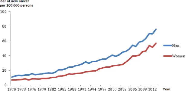
In the case of Sweden, according to regulations by the Swedish National Board of Health and Welfare, all pathology and cytology departments in Sweden must nowadays report all cases of SCC and BCC (SOSFS 2006:15) to the National Swedish Cancer Register. The registration of SCC started already in 1958 whereas registration of BCC started in 2003 [7]. The quality of the infrastructure of the Swedish Cancer Register in terms of completeness, width of information, and reliability of linkage using the personal identification number is internationally recognized. Nordic Registries are known to be very accurate with an overall completeness of over 95% (almost 100% for solid tumors) [8]. Data collected on NMSC in Sweden shows an increase over the past years in accordance with the overall estimated incidence (Figure 2, Table 1).
3
Figure 2: Total number of new skin cancer cases (ICD-7 191. Melanoma excluded) per 100,000 persons (crude rate) in Sweden, 1970-2013. (From the National Board of Health and Welfare´s statistical database, accessed on 2016-01-15).
Table 1: Total number of BCCs by gender, 2004-2011. (From the National Board of Health and Welfare´s statistical database, accessed on 2016-01-15).
The fact that NMSC occurs mainly on sun-exposed sites and that its prevalence can be reduced by sun-protection, provides indirect but crucial evidence for the etiology of ambient solar radiation. While the role of cumulative sun exposure in SCC is well established, the association between sun exposure and BCC seems to rely on intermittent sun exposure and exposure during childhood. Even though we have identified the most
4
important risk factor and despite growing public awareness of harmful consequences of sun exposure, NMSC incidence continues to increase.
The rising incidence of NMSC might partly be explained by increased patient and physician awareness of the disease, improved coding, as well as an age shift in the population. Furthermore, it might also suggest that other factors might have a role in promoting NMSC in addition to ultraviolet (UV) radiation.
Several complex genotypic, phenotypic and environmental factors contribute to pathogenesis of NMSC. Older age, male sex, fair skin, blond hair, blue eyes, weakened or suppressed immune system [9-12], and a number of inherited genetic skin conditions like epidermodysplasia verruciformis (EV), influence cancer development (http://www.cancer.net, accessed on 2016-01-15).
1.2 INFECTIONS AND NON-MELANOMA SKIN CANCER
The last few decades have led to the realization that a considerable proportion of cancers develop due to infections [13]. Considering infectious agents classified by International Agency for Research on Cancer (IARC) as carcinogenic to humans, 2.1 million (16.4%) of the total 12.7 million new cancer cases that occurred in 2008 in the world were attributable to infections[13].
To date, eight very different viruses have been identified as carcinogenic in humans, including retroviruses (Human T-cell leukaemia virus type I and Human immunodeficiency virus type I), RNA-viruses (Hepatitis C virus), DNA viruses with retroviral features (Hepatitis B virus), and both large double-stranded DNA viruses (Epstein-Barr virus and Kaposi Sarcoma-associated herpesvirus) and small double-stranded viruses (Human papillomavirus, HPV; and Merkel Cell Polyomavirus) [14, 15]. In addition, a bacterium (Helicobacter pylori) and some parasites are also clearly implicated in human cancer [13].
5 All oncogenic infectious agents identified so far have the ability to establish persistent infection in their host. The immune system controls the replication of infectious agents (particularly viruses) and/or expansion of infected cells. Immunosuppression is accompanied by a higher fraction of infection-associated cancers [11, 16-18].
Nordic, including Swedish, registry linkage studies were influential in establishing the large excess risk of cancer among transplant recipients more than 10 years ago [11, 19]. A few cancer types such as brain, breast, corpus uteri, and prostate cancers show no significantly increased incidence in immunosuppressed patients compared to the general population [15, 19]. However, majority of cancers (as well as the overall cancer incidence) is greatly increased among these patients [15, 19].
Most cancer types that are increased among transplant recipients are known to be caused by viruses, e.g. HPV-associated anogenital cancers, Epstein-Barr virus-associated lymphomas, Merkel cell carcinomas and HepatitisB/HepatitisC-associated liver cancers [18], implying that immunosuppression specifically induces an impaired ability to control tumorgenic viruses.
As was pointed out by the 2008 Nobel Laureate Harald zur Hausen, exploration of any further role of infections in cancer is likely to be particularly rewarding if it is focused on the cancer forms that are increased among the immunosuppressed, but that do not have an established microbiological etiology [14]. Non-melanoma carcinoma of the skin (NMSC), including squamous cell carcinoma (SCC) and basal cell carcinoma (BCC), is by far the most highly increased disease in this patient group (about 100-fold increased incidence) [18], but an infectious etiology has not been established.
1.3 POTENTIAL PATHOGENS IN SKIN CANCER
The extreme variety of infectious agents potentially involved in human cancer rules out the possibility of predicting promising candidates. However, human papillomaviruses and bacteria (Staphylococcus aureus) have previously been detected in SCC[20-23].
6
The experiments carried out in this thesis have searched for viruses in NMSC (HPVs as well as other viruses). Metagenomic sequencing found that >95% of the viral sequences present in skin samples belonged to the Papillomaviridae family [24] and no other specific virus was commonly detected in most skin cancer specimens. Most studies so far have been carried out after using general polymerase chain reaction (PCR) systems and therefore, they are biased to detect only viruses with sequences of high similarity to the PCR primers used. Viruses that present low similarity to the primer sequences may have remained undetected in previous studies.
1.3.1 HPV
1.3.1.1 Nomenclature and classification
The genus Papillomavirus is a group of small, non-enveloped DNA viruses known since antiquity but first described in the 1930’s [25].The name “Papilloma” comes from the Latin term “papilla” (pustule or nipple) and the Greek suffix “oma” (tumor). Papillomaviruses are identified by the abbreviation PV and one or two letters indicating the host species. For example, human papilloma virus is identified as HPV.
Papillomavirus isolates were traditionally described as “types”. The rapid increase in the number of isolates identified demonstrated a need for a taxonomic classification within the family [26]. The first attempt to classify all types relied on the ability of the viruses to infect the squamous epithelium (skin types) or the mucosal epithelium (mucosal types). However, this classification was found to be incorrect due to the possible presence of the same type in both types of epithelium.
HPV classification and nomenclature is based on sequence analysis, as inefficient cell culture systems have limited the possibilities for classification based on biological properties. Both the ability to obtain amplification products based on the PCR technique, and the high stability and conservation of HPV genomes over evolution, support the current classification system based on the differences found in the genome [26],
7 particularly in the L1 open reading frame, which is the most conserved gene in the genome and encodes for the major capsid protein.
Classifications are as follows (Figure 3, Table 2):
- Genus: different genera within a family share less than 60% nucleotide sequence identity. Currently, human papillomaviruses are divided in five different genera (alpha, beta, gamma, mu and nu).
- Species: different species within a genus share between 60% and 70% nucleotide identity. There are a total of 49 species, which are designated with a number.
- Genotype (type): genotypes within a species share between 71 and 89% nucleotide homology.
The International Committee on Taxonomy of Viruses is responsible for the papillomavirus nomenclature down to the species [26-28]. In order to establish a potential novel HPV type officially, the whole viral genome must be cloned (more than one plasmid can be used) and sent to the International HPV Reference Center together with the sequence. The International HPV Reference Center will confirm the DNA sequence and assign the submitted clones the novel HPV genotype number, if it is novel.
Today, 205 HPV different genotypes have been completely cloned, sequenced and given an official number at the International HPV Reference Center (http://www.hpvcenter.se, accessed on 2016-01-15). Four previously awarded HPV type numbers (HPV46, HPV55, HPV64 and HPV79) were withdrawn (mostly due to re-classification as subtypes of other HPV types). There are therefore 201 different HPV types established today.
The number of HPV types is continuously growing. The exact number of putative novel types is difficult to ascertain, mostly due to the possibility of different non-overlapping partial sequences representing the same virus type.
It is estimated that at least 400 different HPV types exist [29]. In the HPV center, 360 putatively novel HPV types have been already discovered [30-33]. The gamma genus is rapidly growing with now up to 81completely HPV established types, surpassing alpha
8
During the last five-year period, 77% of all new HPVs deposited in the International Reference Center belonged to the gamma genus. Surprisingly, mu and nu genera have almost not increased in number. Methods that are independent of sequence information have not revealed any additional members. An exception is the recently discovered HPV 204, isolated from the anal canal.
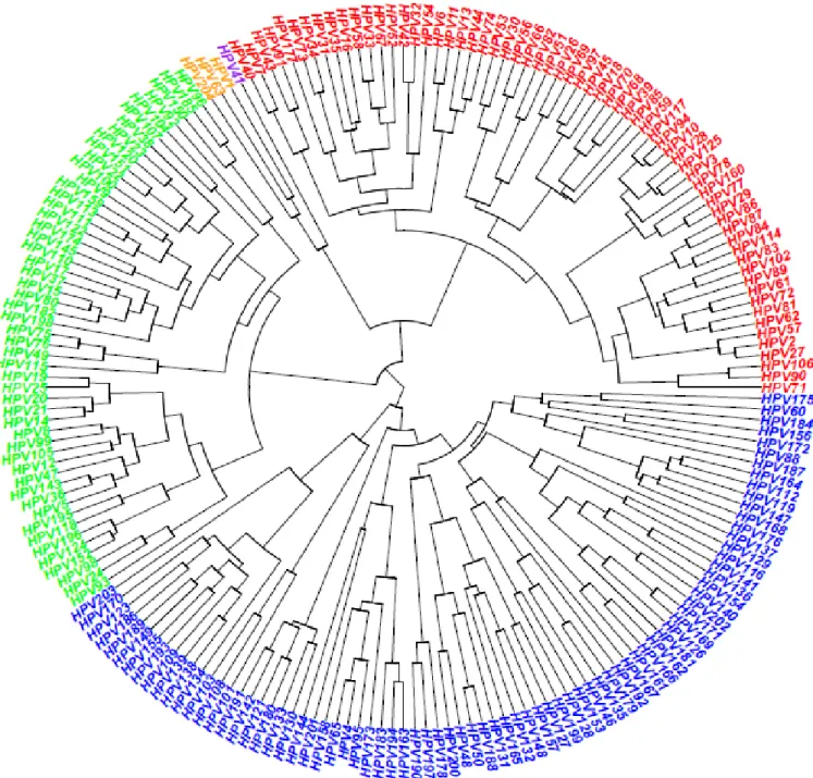
Figure 3: Phylogenetic tree of 204 HPV types. Alpha, beta, gamma,mu and nu papillomaviruses are presented in red, green, blue, orange and purple colors, respectively. The phylogenetic tree is based on the L1 part of the genome.
9
Genus Species First HPV type Other HPV types Date
Alpha Alpha-1 HPV32 42 1986-1987 Alpha-2 HPV3 10, 28, 29, 77, 78, 94, 117, 125, 160 1984-2009 Alpha-3 HPV61 62, 72, 81, 83, 84, 86, 87, 89, 102, 114 1989-2008 Alpha-4 HPV2 27, 57 1984-1989 Alpha-5 HPV26 51, 69, 82 1985-1997 Alpha-6 HPV30 53, 56, 66 1981-1987 Alpha-7 HPV18 39, 45, 59, 68, 70, 85, 97 1981-2004 Alpha-8 HPV7 40, 43, 91 1984-2001 Alpha-9 HPV16 31, 33, 35, 52, 58, 67 1984-1989 Alpha-10 HPV6 11, 13, 44, 74 1984-1993 Alpha-11 HPV34 73, 177 1985-2013 Alpha-13 HPV54 1987 Alpha-14 HPV71 90, 106 1991-2004 Beta Beta-1 HPV5 8, 12, 14, 19, 20, 21, 24, 25, 36, 47, 93, 98, 99, 105, 118, 124, 143, 152, 195, 196 1984 - 2014 Beta-2 HPV9 15, 17, 22, 23, 37, 38, 80, 100, 104, 107, 110, 111, 113, 120, 122, 145, 151, 159, 175, 182, 198 1984-2014 Beta-3 HPV49 75, 76, 115 1987-2008 Beta-4 HPV92 2001 Beta-5 HPV96 150, 185 2002-2013 Gamma Gamma-1 HPV4 65, 95, 95, 158, 173, 205 1984-2015 Gamma-2 HPV48 200 1987-2014 Gamma-3 HPV50 188 19872013 Gamma-4 HPV60 1989 Gamma-5 HPV88 2001 Gamma-6 HPV101 103, 108 2004-2006 Gamma-7 HPV109 123, 134, 138, 139, 149, 155, 170, 186, 189, 193 2007-2014 Gamma-8 HPV112 119, 147, 164, 168, 176 2007-2013 Gamma-9 HPV116 129 2009 Gamma-10 HPV121 130, 133, 142, 180, 191 2009-2013 Gamma-11 HPV126 136, 140, 141, 154, 169, 171, 181, 202 2010-2014 Gamma-12 HPV127 132, 148, 157, 165, 199 2009-2014 Gamma-13 HPV128 153 2009-2011 Gamma-14 HPV131 2009 Gamma-15 HPV135 146, 179, 192 2009-2013 Gamma-16 HPV137 2009 Gamma-17 HPV144 2010 Gamma-18 HPV156 2011 Gamma-19 HPV161 162, 166 2012 Gamma-20 HPV163 183, 194 2012-2014 Gamma-21 HPV167 2012 Gamma-22a HPV172 2012 Gamma-23a HPV175 2013 Gamma-24a HPV178 190, 197 2013-2014
10
Genus Species First HPV type Other HPV types Date
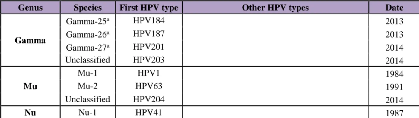
Gamma Gamma-25a HPV184 2013 Gamma-26a HPV187 2013 Gamma-27a HPV201 2014 Unclassified HPV203 2014 Mu Mu-1 HPV1 1984 Mu-2 HPV63 1991 Unclassified HPV204 2014 Nu Nu-1 HPV41 1987
Table 2: Established HPV types, stratified by species and genera. Date refers to the period when HPV types were officially assigned with an established number by the International HPV Reference Center. a: Species assignments are tentative and not official, but recommended to the
papilloma virus working group of ICTV. Modified table from www.hpvcenter.se, accessed on 2016-01-15.
1.3.1.2 HPV in Skin Cancer
The papillomavirus life cycle is tightly linked to the differentiation process of the infected epithelium. Papillomaviruses initially infect basal epithelial cells, which constitute the only cell layer in an epithelium that actively divides. The mechanisms by which HPV induces neoplastic transformation are probably various, and in fact, in vitro models demonstrate only a weak potential. It (neoplastic transformation) is attributed in a large part to the actions of the HPV E6 and E7 oncogenes [34-36].
These oncoproteins inactivate tumor suppressor genes that operate at key cell cycle checkpoints. E6 interferes with p53, leading to genomic instability and blocking of apoptosis, allowing cells with damaged DNA to replicate rather than self-destruct while E7 inactivates retinoblastoma signaling, leading to induction of DNA synthesis in keratinocytes that would otherwise be terminally differentiated and non-replicating [37].
Interestingly, SCCs derived from mice with a deletion of the retinoblastoma protein or the
p53 gene only in skin, exhibit similar molecular signatures to that of HPV-induced tumors, suggesting a role of HPV in the carcinogenesis of SCC [38]. Thus, when E6 and E7 act synergistically, not only do they promote inhibition of apoptosis and dysregulation
11 of the cell cycle leading to abnormal cell growth, but also induce cellular genomic instability contributing to carcinogenesis. However, the effect by itself is not enough to transform cells [39].
UV exposure is an important cofactor in HPV carcinogenesis. It may be then, that the contribution of HPV infection to cancer is via the anti-apoptotic effect in UV-damaged keratinocytes, which would have otherwise progressed to senescence and disintegration. This inhibition of apoptosis probably results in persistent viral infection and hence the accumulation of further DNA mutations, putatively leading to immortalized cells. Unrepaired DNA damage has been observed in UVB-irradiated cells expressing the E6 protein, and inactivation of the retinoblastoma protein with HPV 16 E7 has resulted in significant inhibition of the ability to recover mRNA synthesis and increased levels of apoptosis following UV radiation [40, 41].
An association between HPV and NMSC has been found among patients with epidermodysplasia verruciformis (EV), a rare hereditary immunosuppressive disease [42]. EV patients develop skin lesions in early infanthood and present eruptions of wart-like lesions which are refractory to conventional wart treatment and progress to SCC at sun-exposed sites of the skin [43]. The persistence of HPV infection in EV has been suggested to be due to the inability of the patient’s immune system to reject HPV-infected keratinocytes by a still unknown immunogenetic defect and is probably also influenced by environmental factors, particularly ultraviolet radiation [44]. The HPV types found in patients with EV are referred to as EV-HPV types, and include, among others, HPV types 5, 8, 9, 12, 14, 15, 17, and 19–25 [45, 46]. HPV 5 and 8 are the most prevalent types [47].
In contrast to cervical cancer where HPV genotypes 16 and 18 have been established as the most prevalent genotypes which cause this disease (70% of cancer cases) and in contrast to patients suffering from EV where HPV 5 and 8 are high-risk genotypes for skin cancer, the HPV types found in skin cancers of the general population have varied depending on which PCR-system was used [48, 49]. It is common to detect multiple genotypes in a single specimen [24, 50].
Metagenomic sequencing has revealed that >95% of the viral sequences present in NMSC samples belong to the Papillomaviridae family, mostly to the beta and gamma genera
12
[24] but so far, only one study, that is included in this thesis, has detected a particular genotype (HPV 197) with high frequency (37.4%) [30] .
Infections of HPV in skin are very common, but because of the diversity of HPV types, there does not appear to exist any single virus that is widely spread. Acquisition appears to occur already shortly after birth [51-54]. A broad spectrum of cutaneous HPV is commonly detected both on healthy skin [55, 56], in plucked eyebrow samples [57-59] as well as in different skin diseases such as SCC, BCC, actinic keratosis (AK) – a precursor lesion for SCC – and in KAs, in both immunocompetent and immunosuppressed patients [60-63].
It has to be highlighted that detection of an HPV type in skin tumors does not necessarily mean that an HPV infection has been detected, as it may merely be a viral contamination of the skin surface. Forslund et al. demonstrated that cleansing of the skin by simple tape stripping before sampling, strongly reduces the proportion of HPV positive samples [62]. Prevalence dropped from 69% in swabs from top of SCCs, BCCs and AK lesions, to 12% in the corresponding biopsies, after cleansing the skin surface.
When skin biopsies from NMSC only contain low viral loads (<1 copy/cell) [22, 64-66], it is debatable whether such low viral copy numbers are biologically relevant to tumor initiation and maintenance.
1.4 VIRAL DETECTION IN SKIN CANCER
When analyzing human skin specimens, one has to take into account that besides human DNA and RNA, human skin harbors various physiological populations of microorganisms, including commensal or symbiotic bacteria, fungi, parasites and viruses, overall known as the skin microbiota.
Sequencing studies reveal that viruses represent < 1% of the total genomic material in skin [31] and therefore, detection of any virus by NGS, formerly required performing some type of viral enrichment or amplification first. Viral enrichment can be achieved by
13 performing: low-speed centrifugation and/or filtration to remove bacterial and host cells, nuclease treatment to digest nucleic acids that are not protected with virions [67], separation of long chromosomal DNA from shorter DNA [31], high-speed gradient centrifugation [68] or targeted sequence capture [69-71]. Each of these procedures may bias against detection of some viruses and result in decreased assay sensitivity as a result of loss of viral nucleic acids. Novel protocols designed to overcome these problems look very promising. ViroCap for instance, is a viral targeted sequence capture panel, with multiple probes designed to capture most viral species that infect vertebrates (337 viral species). This test is also capable to capture those viruses that share up to 58% variation from the reference viruses used to select capture probes [72].
The publications included in this thesis analyze only DNA virus (no RNA) and thus, amplification techniques and detection methods will focus on DNA. RNA viral presence will be discussed in the section “Concluding remarks and future perspectives”.
1.4.1 Amplification techniques
1.4.1.1 PCR using “general” or “degenerated” primers
The relative ease and economic accessibility of the PCR technique made it one of the most widely used techniques in clinical diagnostics. Several general primer PCR systems targeting the L1 gene (FAP, CUT, PGMY, MGP) [33, 48, 73, 74] can amplify a broad range of HPV types. However, efficiency of any PCR based amplification depends on the number, position, and stability of mismatches between the primers and the template. The sequence to be amplified must be previously known and thus, amplification is biased to detect only sequences of high similarity to the primers used. HPV types with low similarity to the primer sequences will remain undetected.
As an example, Forslund et al., designed PCR FAP primers from two relatively conserved regions of the L1 open reading frame taking into consideration most known genome sequences, from HPV 1 to HPV 80 [49]. FAP primers could detect 65/75 (86.7%)
14
different HPV types but were not able to amplify HPV types 1, 2, 35, 41, 44, 55, 63, 66, 71 or 74. The failure in detecting HPV1, 41 and 63 is of particular importance as these types are the only genotypes that form the genera mu and nu (recently, HPV 204 was also classified as a mu papillomavirus (hpvcenter.se)). Other putative novel genotypes phylogenetically close to these HPV types (belonging to these genera) might also remain undetected.
Currently, there are 205 different HPV types recognized. Attempts to degenerate FAP primers more, in order to amplify a broader number of HPV types, as well as multiplexing specific HPV primers in the PCR reaction, might improve HPV detection, but will at the same time reduce specificity in the reaction (e.g. human DNA binding).
1.4.1.2 Unbiased approach: Whole genome amplification
By performing whole genome amplification (WGA), all the DNA present in a sample will be amplified without requiring previous knowledge of the DNA sequence. There are different methods that have been developed for high-fidelity WGA.
Initial PCR-based WGA techniques were based on the use of degenerate/semi-degenerate primers [75] and primer extension PCR [76]. These techniques used Taq polymerase and consequently, limited the amplicon fragment length to 3 kb and introduced errors in the sequence due to the lack of the enzyme´s 3´- 5´ exonuclease activity. Furthermore, they exhibited incomplete genome coverage and amplification bias [75-77] and therefore, were substituted by non-PCR based methods.
Multiple displacement amplification (MDA) is today a gold standard method for non-PCR based amplification techniques. It was first developed for rolling circle amplification [78] where the reaction starts by annealing random hexamer primers to the DNA template and DNA synthesis is carried out at constant temperature (Figure 4).
The DNA polymerase used in MDA originates from bacteriophage phi29 [79, 80] and has a 3´- 5´ proofreading activity, resulting in a low intrinsic error rate and about 1000-fold
15 less accumulation of mutations compared to PCR techniques using Taq DNA polymerase [81, 82]. Moreover, Phi29 possesses a strong strand displacement activity, being able to solve secondary structures as hair pin loops, thereby preventing slipping, stopping and dissociation of the polymerase during amplification. Average product length can be greater than 10 kb [83].
Figure 4: Overview of the whole genome amplification with focus on double stranded DNA (A). Random hexamer primers bind to single stranded DNA and amplification starts (B, C). When the polymerase reaches a downstream primer, the strand is displaced and new primers can anneal to the displaced product (D). The end product is double stranded repeated copies of the DNA in the sample (E). Figure adapted from Rector et al., A sequence-independent strategy for detection and cloning of circular DNA virus genomes using multiply primed rolling-circle amplification, in Journal of Virology, 2004; 78:4993-8, with permission from American Society for Microbiology.
MDA provides an effective and easy means of amplifying minimal quantities of DNA and is the least biased WGA method reported [83, 84]. However, there are also biases associated with this technology. Chimera formation, preferential amplification of circular single stranded DNA (ssDNA) and non-uniform amplification of linear genomes have been documented [85, 86].
The predominant mechanism for chimera formation is now elucidated [87]. More than 85% of chimeras are due to inverted sequences occurring when strand displacement takes place in the reaction. 3´-termini can be displaced and might reanneal at randomly occurring complementary segments on nearby 5´-strands, resulting in the joining of two
16
sequences in inverted orientation with an intervening deletion. Bioinformatic analysis might be able to find these sequences and remove them for further analysis.
MDA has not shown the ability to accurately estimate the amount of viral populations present [88], most probably due to the preferential amplification of particular genomic regions during initial MDA priming events [89, 90]. Several investigators have proposed that pooling several independent MDA reactions run on a single sample of template DNA minimizes representational bias in shotgun metagenome sequence libraries [91-96]. However, this assumption has not been thoroughly tested [97].
There is no reported method that overcomes all amplification bias in MDA products. Hence, the effective use of MDA in any application depends on the user´s needs. E.g.: if the user is only interested in ssDNA sequences, skipping the denaturation step in the amplification will completely bias the ssDNA amplification, as primers will not be able to bind to non-denatured dsDNA [98].
Even though MDA was traditionally used to amplify circular DNA [96], it can be used to amplify linear DNA [83, 100]. In Paper III, we quantified the amount of amplification of both human DNA and HPV DNA by adding 20 copies/μL of HPV 16 plasmid to samples of human placental DNA at 1 ng/μL. We amplified these samples in the same manner as for the clinical samples and quantified the amounts using real-time PCR for beta-globin and for HPV 16, respectively. Human DNA was found to be amplified 26-fold, whereas HPV 16 DNA was amplified 679-fold. Thus, although the WGA will have made it easier to detect the circular HPV genomes, it is unlikely that we would have missed linear and/or large dsDNA viruses unless they were present in only small amounts.
1.4.2 Detection techniques
Detection of amplified products is usually performed by hybridization of amplicons to type-specific probes coupled to fluorescent beads [101, 102] or by product sequencing [20, 21, 48, 103].
17
1.4.2.1 Hybridization to type-specific probes
Detection of amplicons using probes requires prior knowledge of the DNA sequence query in order to design specific probes. There is a large variety of molecular assays based on hybridization used for detection of different microorganisms [104, 105]. Some of the hybridization methods for HPV DNA typing include the Hybrid Capture II test (Digene Corporation, Gaithersburg, MD), the LINEAR ARRAY® HPV genotyping test (Roche Molecular Systems, Alameda, CA), the INNO LiPA® HPV genotyping test (Fujirebio, Gent, Belgium), and the Cervista® HPV (Hologic Inc., Marlborough, MA).
Accurate and internationally comparable DNA detection and typing methodology is an essential component both for research and for diagnosis. The World Health Organization (WHO) started a WHO Global HPV Laboratory Network (LabNet) in 2006 to support the world-wide development and implementation of HPV vaccines through improved laboratory standardization and quality assurance of HPV testing and typing methods (Technical Report on the Global HPV LabNet 2013 HPV DNA Genotyping Proficiency Panel). The results show a yearly increase in proficiency (sensitivity and specificity) of HPV typing assays when routinely used in laboratories worldwide [106, 107].
Despite a high sensitivity and specificity of these methods, their major limitation is the potential for error in HPV typing because of probe hybridization when cross-reactivity of one probe to several target groups occurs (false positivity). Furthermore, sequences with low similarity to the probes or sequences that present small variations and point mutations might not be detected (false negativity). Therefore, these methods are not valid when searching for novel viruses and/or sequences.
A recent study developed a comprehensive viral targeted sequence capture panel using hybridization probes, that were able to assess all viruses known to infect vertebrate cells (excluding human endogenous retroviruses), as well as to detect divergent viruses [72]. The main advantages of this design are: complete viral genomes targeting, detection of viruses that show up to 58% variation from the reference virus used to select capture probes and, the possibility to isolate the capture sequences in order to sequence them afterwards [72].
18
1.4.2.2 High throughput sequencing
With the establishment of the high throughput sequencing (HTS) technology (also called next generation sequencing (NGS) or deep sequencing), there is no need to test specimens for predefined microorganisms by PCR. Sequencing the DNA will determine which organisms are present by analyzing the sequence data. Furthermore, the entire sequence is obtained and this enables, both discovering novel viruses as well as finding small variations and point mutations within the previously known viruses.
1.5 HIGH THROUGHPUT SEQUENCING INSTRUMENTS
During the past decade, there has been a dramatic evolution of next generation sequencing technologies: 454 GS (Roche), SOLiD (ABI), Ion Torrent (Life Technologies) and Genome Analyzer System (Illumina).
These recent technological advances have revolutionized the study of genomics and molecular biology. NGS allows us to sequence DNA and RNA much more quickly and cheaply than the previously used Sanger sequencing. Using capillary electrophoresis-based Sanger sequencing, the Human Genome Project took over 10 years and costed nearly $3 billion. Nowadays, sequencing a human genome can be done in a variety of bench-top NGS instruments in a couple of days and at very limited costs.
In this thesis, we have used 2 different sequencing technologies. We started sequencing with 454 GS Junior (Roche) but after about a year, quicker, cheaper and deeper platforms came on the market such as the Genome Analyzer System from Illumina (MiSeq, HiSeq and NextSeq platforms). A comparison of some of the platforms´ specifications used in this thesis is shown in Table 3.
19
Method Sanger-CE 454 GS Junior MiSeq HiSeq NextSeq Year 1980’s 2000’s 2010’s 2010’s 2014
Output <100 kb 35 Mb 1500 Mb 600 Gb 120 Gb
Read length 500 bp 400 bp 300x2 bp* 100x2 bp* 150x2 bp*
Run time 7 h 8 h 56 h 11 days 29 h
$ per Mb 2400 31 0.5 0.05 0,45
Table 3: Next generation sequencing platforms and their specifications. *Pair-end sequencing.
1.5.1 454 GS technology (Roche)
The 454 Life Sciences (454; Branford, CT, USA; now Roche, Basel) sequencing platform (the 454 Sequencer) was the first next-generation technology to reach the market. It was first commercially introduced in 2004. It dramatically increased the volume of sequencing conducted by research groups and expanded the range of problems that can be addressed by the direct readouts of DNA sequence.
It was the first technology to sequence and assemble bacterial genomes de novo [108] and the first non-Sanger technology to sequence an individual human [109]. Other notable studies conducted by 454 included work as diverse as uncovering the potential cause of the disappearance of the honeybee [110], revealing the complexity of rearrangements between individual human genomes [111], providing new approaches to understand infectious diseases [112] - such as the mechanism of resistance to the drug R207910 in Mycobacterium tuberculosis [113] - and sequencing the first million base pairs of a Neanderthal [114-116].
The GS Junior (454) technology is based on emulsion-based amplification and pyrosequencing. Its workflow is comprised of three main steps: generation of a single-stranded template DNA library, emulsion-based clonal amplification of the library, and pyrosequencing (Figure 5).
20
1.5.1.1 Generation of a template DNA library
The DNA is isolated and fragmented by nebulization. DNA fragments are polished and end repaired to create 3’Adenine ends to allow TA ligation of multiplex identifiers (MIDs) adaptors. These adaptors are very important, as they both allow sample quantification, and permit the user to multiplex sequencing, as each DNA fragment can be ligated to a different MID.
Once the adaptor is ligated, a size selection step is performed to get rid of adaptors that were not ligated to the DNA fragments and samples are purified. Before continuing to the next step, it is important to assess the library quality and quantity in order to obtain the optimal number of molecules of library DNA for emulsion PCR.
1.5.1.2 Emulsion-based clonal amplification of the library
Pools of DNA libraries with different MIDs are combined with capture beads and then amplified by emulsion PCR. The beads are captured by droplets of a PCR reaction mixture in oil emulsion, and PCR amplification occurs in each droplet. This results in each bead carrying ten million copies of a unique DNA template.
The emulsion is then broken, the bead-attached DNAs are denatured and the beads are deposited into wells of a fibre-optic slide, the PicoTiterPlateTM, containing hundreds of thousands of wells, wide and deep enough to contain only one bead.
1.5.1.3 Pyrosequencing
Pyrosequencing is a method of DNA sequencing based on the "sequencing by synthesis" principle. It relies on the detection of pyrophosphate release on nucleotide incorporation. The desired DNA sequence can be determined by light emitted upon incorporation of the
21 next complementary nucleotide. Only one out of four of the possible A/T/C/G nucleotides are added and available at a time, thus only one letter can be incorporated on the single stranded template (which is the sequence to be determined) and light is emitted (once per nucleotide incorporation). The previous nucleotide letter is degraded before the next nucleotide letter is added for synthesis. The intensity of the light determines if there is more than one of these nucleotides in a row. This process is repeated with each of the four letters until the DNA sequence of the single stranded template is determined.
Figure 5: Schematic overview of high-throughput sequencing using 454 GS (Roche). Image reprinted from Mardis et al., The impact of next-generation sequencing technology on genetics, in Trends in Genetics, 2008; 24:133-41, with permission from Elsevier.
1.5.2 Genome Analyzer System technology (Illumina)
The Genome Analyzer System technology was first developed by Solexa Inc., which launched the first Genome Analyzer in 2006. This platform enabled scientists to sequence up to 1 Gb of data in a single run. In early 2007, Solexa was bought by Illumina Inc., and it now supports a broad range of applications. Agrigenomics, cancer, forensics, complex
22
diseases, drug development, and microbial genomics as well as reproductive and genetic health are the most common fields where this platform has been used. With the added depth of sequencing, it enables: the identification of low-abundance genomes or those exhibiting modest expression differences between samples, discovering all types of genetic variations (SNPs, rearrangements, copy number variants, insertions, and deletions) [117-119], characterization of new bacterial isolates [120-122] and/or new variants that cause diseases, profiling DNA methylation status across the entire genome [123-125], defining somatic variations in cancer [124], and characterizing complex RNA populations for new genes and transcript structures [126, 127], among other utilities.
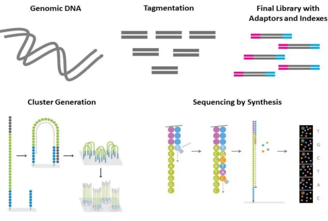
Illumina's sequencing technology is based on cluster generation and sequencing by synthesis, tracking the addition of labeled nucleotides as the DNA template is copied in a massively parallel fashion. Its workflow follows three main steps: generation of a single-stranded template DNA library, cluster generation, and sequencing by synthesis (Figure 6).
1.5.2.1 Generation of a template DNA library
The DNA sample is prepared into a “sequencing library” by using either an enzymatic fragmentation followed by adding unique adapter sequences (forward and reverse oligomucleotides) to the sample, or an engineered transposome that simultaneously fragments and tags ("tagment") input DNA in the process.
After tagmentation (or fragmentation and adapters ligation), a limited-cycle PCR reaction uses these adapter sequences to amplify DNA fragments. This PCR reaction also adds index sequences on both ends of the DNA fragments, enabling dual-indexed sequencing of pooled libraries on any Illumina Sequencing System.
23
1.5.2.2 Cluster generation
The sequencing libraries are denatured and attached to a lawn of single stranded oligonucleotides immobilized on a flow cell surface. These olignonucleotides correspond to the complementary sequences of the adapters ligated during the library preparation step. Subsequently, each end of every library molecule matches one of the two primers on the glass surface.
Once the sequencing templates are attached, amplification in a bridge-fashion occurs. The free/distal end of the DNA template loops over to hybridize the complementary surface primer (oligonucleotide). A DNA polymerase copies the templates from the hybridized oligonucleotides forming dsDNA bridges, which are denatured and result in two single strands that serve as a new template, and will then loop over and hybridize again.
Priming will continue as the distal end of a ligated fragment bends over to a complementary oligo on the surface of the flow cell. Repeated denaturation and extension will result in millions of surface-bound colonies, each of them containing approximately one million copies of each template (the cluster).
1.5.2.3 Sequencing by synthesis
The Illumina sequencing method uses fluorescently-tagged dNTPs containing a terminator which blocks further polymerization. During each sequencing cycle, all 4 labelled nucleotides are added to compete for addition (thus, minimizing incorporation bias) to the DNA strand. Only one nucleotide can be incorporated based on the sequence of the template.
The included base behaves as a terminator for the polymerization. After each cycle, the fluorescent dye is imaged to identify the nucleotide and the terminator is cleaved to allow the incorporation of the next base. This process is repeated until the DNA sequence of the single stranded template is determined.
24
Figure 6: Schematic overview of high-throughput sequencing using Illumina technology.
1.6 HIGH THROUGHPUT SEQUENCING DATA ANALYSIS
As even just one sequencing run provides enormous amounts of data, analysis and interpretation are challenging.
Sequencing analysis usually starts by performing quality checking [128] (Figure 7). Quality checking is an important and effective measure for determining the quality of sample libraries, and it also serves to indicate whether the sequencing succeeded or failed. Bases are checked according to their Phred quality scores [128].Phred quality scores are logarithmically related to the base-calling error probabilities. For example, a Phred quality score of 10 corresponds to a base calling accuracy of 90% (10 errors per 100bp), while a quality score of 20 equals to a base calling accuracy of 99% (1 error per 100bp) [128].
25 Specific quality filtering conditions can be adapted for different downstream analyses [129].
To obtain a dataset that contains reads of interest, e.g. the virus-related reads for viral metagenomics, sequences that are not a target of the investigation (e.g. human and bacterial reads) need to be filtered out on the bioinformatics level (Figure 7). This will further speed up the downstream analysis and decrease the risk of mis- assemblies[129].
NGS sequences from human samples subjected to WGA typically contain more than 60 percent human-related sequences. Human- and bacteria-related reads are the most commonly obtained reads, followed by sequences classified as “other” and “unknown” [31, 130] (Table 4). Viral reads usually represent less than 0.5% of sequencing data and enrichment for viral particles by ultracentrifugation has not been shown to be helpful in the analysis of biopsies or skin swabs [130].
Negative control samples (PCR grade water) might also contain bacterial sequences and sequences classified as “other” and “unknown”[130]. Water controls have so far been found to be uniformly negative for viral sequences [130].
These background sequences might be present due to the background reactivity of the Phi29 polymerase reaction [131] or represent environmental contamination and therefore, it is imperative that all metagenomic sequencing projects include sequencing of negative control samples [130].
Once human and bacterial sequences are filtered out, data normalization is performed in order to decrease sample variation and discard redundant data such as duplicated reads (Figure 7). NGS technologies can produce duplicated reads due to errors in PCR amplification and/or sequencing [132, 133] and these reads might introduce an overestimation of the species abundance. Duplicated reads may also include natural duplicates that by chance originate from the same genomic position [132, 133]. Highly abundant species have a higher chance of natural duplicates [133] and their removal might introduce bias towards underestimation of abundances [132].
26
Raw NGS Sequence Data
Check Base Calling Accuracy
(Phred quality scores – manual cut off)
Filter out Human and Bacterial Genomes
(manual cut off)
Sequence Data Normalization
Assembly
(manual cut off)
Assembly Validation
(manual cut off)
Taxonomic Classification Taxonomic Classification
Similarity Based Methods Non-Similarity Based Methods
Genotype Abundances, Community Diversity and Structure Estimation
Results
Figure 7: Bioinformatics pipeline to analyze high-throughput sequencing data for viral metagenomics.
27
FFPE Biopsies Biopsies Skin swabs Water
Sequencing platform GSFLX GSFLX GSFLX PGM 400 GSFLX Human 37,3 99,8 69,1 76,3 2,8 Bacteria 21,3 0,1 24,2 18,3 52,2 Virus 0,2 0 0,3 0,3 0 Other 10,2 0 2,2 1 15,5 Unknown 30,9 0 4,2 4,1 29,5
Table 4: Typical taxonomic assignment of NGS reads (%). Summary of results in previous studies, using different types of biospecimens, pre-treatments and NGS platforms.FFPE: Formalin Fixed Paraffin Embedded. Adapted from Bzhalava et al., Unbiased approach for virus detection in skin lesions, in PloS One, 2013; 8:e65953, with permission from the Creative Commons Attribution License.
Sequence datasets are usually normalized using a digital normalization algorithm (http://ged.msu.edu/papers/2012-diginorm), which substantially reduces data size and computational resources for de novo assembly. NGS technologies produce billions of short reads from random locations in the genome by oversampling it and assembling those reads is the next step performed in the bioinformatics analysis (Figure 7).
Assembly algorithms, in a process called de novo assembly, reconstruct original genomes which are present in the sample by merging short genomic fragments into longer contiguous sequences (“contigs”). There are two main types of de novo assembly programs: Overlap/Layout/Consensus assemblers, most widely applied to the longer reads and de Bruijn Graph assemblers, applied to the shorter reads. To validate assembly results, several assembly algorithms might be used, as well as re-mapping all singletons reads to assembled contigs [31, 134].
The possibility always exists that assembly algorithms may construct erroneous “chimeric” sequences by assembling sequences from different organisms or species. This problem may be particularly relevant for viral metagenomics where the biospecimens may contain a multitude of related viral sequences. For HPVs, we developed an algorithm to identify possible “chimeric” HPV sequences [135]. It is based on the assumption that an HPV genome should have similar degree of identity to the most closely related HPV type over its entire genome. Thus, HPV related sequences that have different degrees of
28
similarity over their length to the most closely related HPV sequence in GenBank are considered as possible chimeras (i.e. it is assumed that they contain parts of different HPVs). The algorithm checks these chimeras by dividing the sequence into three equal segments. If at least one of the segments has less than 90% similarity, another segment has more than 90% similarity and the difference between these segments is more than 5% (e.g. if segment 1 is 88% similar and segment 2 is 94% similar) the sequence is considered as “possibly chimeric”. This approach has been extremely valuable when analyzing HPV genotypes, however, it cannot be used for viruses that frequently rearrange parts of genomes with each other (e.g. Anelloviruses) and other algorithms must be developed [130].
Taxonomic classification of metagenomic reads can be performed by similarity and/or non-similarity-based methods. One of the most famous similarity-based taxonomic classifications is performed by NCBI BLAST, where sequences are compared to known genomes. However, a large part of the sequencing reads from de novo sequencing projects are classified as unknown [31, 130]due to incompleteness of public sequence databases or drawbacks of NGS technologies such as short read lengths and sequencing errors. Therefore, more sensitive algorithms, such as BLASTx and tBLASTx searches are conducted against protein databases after the BLASTn search on the nucleotide level. An example of this, would be to subject assembled contigs to taxonomic classification by comparing them against GenBank nucleotide database using paracel blast (www.strikingdevelopment.com) BLASTn to classify them as a) previously known sequences, b) related to previously known sequences, or c) unrelated to previously known sequences.
One of the biggest challenges for bioinformatics analysis is the taxonomic classification of NGS data as many of the sequences, especially those belonging to viruses, have no homologs in the public databases or these homologs are highly divergent [136]. In BLAST searches, sequences might have multiple matches and to classify these sequences, several methods have been developed. One of the most frequently used is called MEGA [137]. This method finds the ‘Lowest Common Ancestor’ node of all matching sequences in the phylogenetic tree, reducing the risk of false positive matches. However, MEGA might produce false negative results by discarding sequences that do not satisfy user-defined cut-offs.
29 In metagenomic samples, genome size is related to the number of reads and thus, MEGAN is suboptimal for quantitative metagenomic analyses. Nevertheless, other tools have been developed to address this issue. The GAAS (Genome relative Abundance and Average Size) tool [138] iteratively weighs each reference genome for all matching reads and normalizes the number of reads to the length of their genomes. GRAMMy (Genome Relative Abundance estimates based on Mixture Model theory) are another useful tool that models read assignment ambiguities, genome size biases and read distributions along the genomes on a unified probabilistic framework [139]. Both tools estimate similarities from the alignment qualities of the reads to the reference genomes but not from the reference genomes directly. Thus, they are very valuable for divergent genomes, but they are suboptimal in case there are highly similar genomes in the reference databases. The Genome Abundance Similarity Correction (GASiC) considers reference genome similarities to correct the observed abundances estimated via read alignments [140].
1.7 SIGNIFICANCE OF THE STUDY
The development of sequencing technologies that perform high throughput sequencing together with the possibility of performing an unbiased approach, allows for the accurate identification of all microorganisms that are present in large numbers of biological specimens.
The experiments carried out in this thesis have analyzed viral DNA present in skin tumors and the findings have contributed significantly to the knowledge of HPV genotypes. Until 2004, only 106 HPV types had been completely cloned, sequenced and given an official number at the International HPV Reference Center. However, during the last 10 years, 99 new types have been recognized (http://www.hpvcenter.se, accessed on 2016-01-15) and the number of putative novel HPV types is continuously growing. Our group has detected a total of 360 putative novel HPV types (called SE types) so far, all of them belonging to the genera alpha, beta and gamma (Figure 8) and one of them, HPV 197 (SE46) was found to be the most common genotype present in skin tumors.
30
The resulting “microbiological sequence atlas” obtained in this thesis can be used for large-scale molecular epidemiological analysis on whether any particular infection is regularly present in any specific form of skin cancer. Creating a solid and comprehensive basis for advancing knowledge in this area is important. Furthermore, if extensive sequencing efforts fail to find any infection present in cancer tissue, this would point to other hypotheses (e.g., a role of immunosurveillance in the recognition and elimination of precancerous lesions, regardless of cancer etiology, and in cancer cell gene expression) [15]. If properly carried out, therefore, also negative findings for viral sequences can provide useful clues about the origins of human skin cancer [16] .