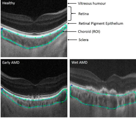
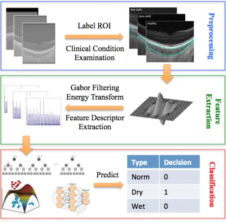
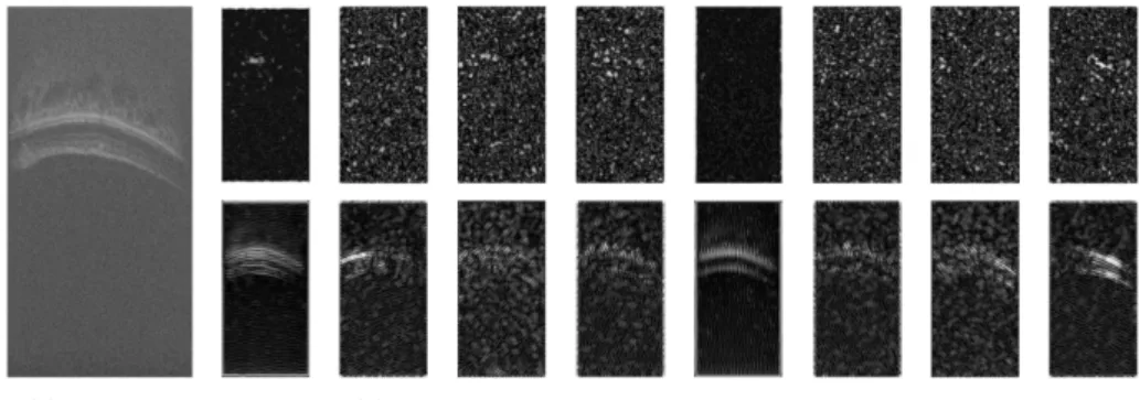
Age-related macular degeneration detection and stage classification using choroidal OCT images
9
0
0
Full text
Figure

Related documents