T H E ROLE OF E P I N E P H R I N E I N T H E R E A C T I O N S P R O D U C E D BY T H E E N D O T O X I N S OF G R A M - N E G A T I V E B A C T E R I A I. HEMORRHAGIC NECROSIS PRODUCED BY EPINEPHRINE IN THE SKIN OF
ENDOTOXIN-TREATED RABBITS* BY LEWIS THOMAS, M.D.
(From the Department of Pathology, New York University-Bellevue Medixal Center, New York)
PLATES 65 T O 67
(Received for publication, August 24, 1956)
In a series of studies beginning in 1941, Delaunay, Boquet, and their asso- ciates (1-3) described the reactions of peripheral arterioles which are char- acteristic of the early stages of the systemic response to the endotoxins of gram negative bacteria.
Within 15 minutes after an intravenous injection of endotoxin in rabbits, these authors observed an intense, prolonged arteriolar constriction, interrupted periodically by brief waves of vasodilation. In animals given large, shock-producing doses, the phase of arteriolar constriction was followed terminally by one of extreme arteriolar dilation with slowing and then stasis of capillary blood flow. The initial reaction of vasoconstriction was prevented by pretreatment with N-(2-chloroethyl)dibenzyl- amine (dibenamine). On the basis of these observations, it was suggested that the systemic effects of endotoxin were associated with a generalized sympathomimetic response analogous to stimulation by epinephrine.
There are other reasons for suspecting that epinephrine, or a substance resembling epinephrine, may be implicated in the reaction to endotoxin (4). Hyperglycemia, sometimes followed by hypoglycemia, is regularly produced by both endotoxin and epinephrine and, according to Boquet and Izard, (5) can be prevented by dihydro- ergotamine. Similarly, depletion of liver glycogen and elevated blood levels of lactic and pyruvic acid occur in response to both agents.
Additional points of resemblance between the actions of epinephrine and endotoxin include leucocytosis (although the leucopenia which occurs during the 1st hour after endotoxin is not produced by epinephrine), interference with diapedesis of leucocytes into inflamed tissues (1, 6), the production of petechial hemorrhages and focal necrosis in the walls of the stomach and upper gastrointestinal tract, mesenteric lymph nodes, spleen and liver (1, 7), and fibrinoid necrosis of the walls of the coronary arteries * This work was done under sponsorship of the Commission on Acute Respiratory Diseases, Armed Forces Epidemiological Board, and supported in part by the Office of the Surgeon General, Department of the Army and by grants from the National Institutes of Health, Public Health Service (H-2022), and the American Heart Association.
866 EPINEPHRINE AND ENDOTOXIN
(8-10). Each of these effects has been observed in experimental animals after admin- istration of both endotoxin and epinephrine (4).
I t might be postulated, for operational purposes, that the systemic effect of endotoxin is either to increase the reactivity of terminal arterioles to epineph- rine, or to increase the a m o u n t of epinephrine released b y the adrenal medulla or liberated in peripheral tissues. The present paper x is concerned with two observations which appear to bear directly on the problem: (a) When rabbits are given an intravenous injection of endotoxin, a subsequent intradermal in- jection of epinephrine at a n y time during the next 4 hours produces extensive hemorrhagic necrosis in the skin. (b) Similar lesions are caused b y injection of a mixture of small quantities of endotoxin and epinephrine into the skin. On the basis of a s t u d y of these and related findings to be described below, it is suggested t h a t endotoxin has the property of altering the reactivity of terminal arterioles to epinephrine, and t h a t this property m a y be the basis of m a n y of the pathophysiologic effects of endotoxin.
Material and Methods
Hybrid albino rabbits of either sex were obtained from several breeders. All animals were 6 to 8 weeks old, and weighed between 1.0 and 1.5 kilos. They were maintained on a diet of Purina rabbit pellets and water.
The following preparations of endotoxin were used: (a) purified lipopolysacchar/de Eber- the~a lyphosa endotoxin prepared by the method of Landy and Johnson (13) and supplied by Doctor Maurice Landy, of the National Institutes of Health, Bethesda; (b) lipopolysac- charide endotoxins (14) derived from gscherichia coli and Salmonella equi, prepared by Doctor Otto Westphal, of the University of Freiburg, Germany; (c) Serratia marcescens endo- toxin (15), supplied by Doctor Murray Shear, of the National Institutes of Health, (d) Shigdla paradysenteriae endotoxin (16), supplied by Doctor Walther Goebel, of The Rockefeller Institute, (e) E. coti endotoxin prepared by the method of Landy and Johnson in the Difco Laboratories, Detroit, and (f), crude meningococeal endotoxin, as for the Shwartzman reaction (17), prepared by a method described previously (18).
Epinephrine was obtained from several commercial sources as a 1:1000 dilution in sealed ampoules. Dilutions of the material were made as indicated in pyrogen-free, sterile, physiologi- cal saline.
Other reagents included norepinephrine, cortisone, nitrogen mustard, heparin, ephedrine, pitressin, proferrin (colloidal sacchaxate of iron oxide), dibenzyline, and chlorpromazine. The use of these will be described in the text which follows.
F.XPERIMENTAL
The
Effect of
Intradermal Epinephrine in Rabbits Given Endotoxin by Vein The capacity of epinephrine to produce hemorrhagic necrosis of the skin in animals receiving intravenous endotoxin is illustrated b y the following experi- m e n t : - -1 Some of the observations described in this paper were reported at meetings of the American Society for Clinical Investigation (11) and the Federation of American Societies for Experi- mental Biology (12).
L E W l S T H O M A S 867 Six rabbits were injected intravenously with 10 #g. of Westphal's E. coil llpopolysaccharide endotoxin. Within 5 minutes after the injection of endotoxin, each animal was given epineph- fine intradermally in each quadrant of the abdomen, in doses of 100 #g. (right upper), 50 gg. (right lower), 10/~g. (left upper), and 5 #g. (left lower). Each dose of epinephrine was contained in a 0.2 co. volume of physiological saline. Six control animals, not given endotoxin, received the same injections of epinephrine.
Within 4 to 6 hours after the injections, each of the endotoxin-treated rab- bits showed scattered petechiae, and numerous engorged superficial vessels resembling telangiectases, in the right upper and lower skin sites, and extreme pallor in both quadrants on the left. B y 12 hours the lesions on the right had developed into confluent oval or circular areas of deep blue hemorrhagic necrosis, measuring approximately 4 X 5 cm. each. In the left quadrants there were smaller areas of bluish discoloration caused b y a distended mesh- work of superficial blood vessels in which were scattered small petechiae. None of the lesions showed any swelling or induration, nor any evidence of inflammation in the surrounding tissues.
At 24 hours the lesions in all four quadrants had become somewhat larger and more deeply hemorrhagic. Typical 24 hour epinephrine lesions are shown in Fig. 1. No further extension of the lesions occurred after 24 hours, and they gradually faded in color during the next 3 or 4 days.
Histologic sections of the lesions showed hemorrhages throughout all portions of the corium, and necrosis of hair follicle cells. Occasional small thrombi were seen in some of the veins. The capillaries and venules were greatly dilated and filled with blood, and the hemorrhages appeared to be derived from rupture of these vessels. Sections taken 24 hours after injection, with fully developed hemorrhage and necrosis, showed very little evidence of inflammatory cell infiltration; occasional collections of polymorphonuclear leucocytes were present around small veins, but the major portion of the involved tissues contained no leucocytes.
Six different preparations of endotoxin were tested to determine the effec- tive doses with which dermal necrosis was produced b y epinephrine; each animal received 100 #g. of epinephrine intradermally, immediately after the injection of endotoxin. With Westphal's S. equi and E. coli endotoxins, Goebel's
Sh. paradysenteriae endotoxin, L a n d y and Johnson's E. typhosa endotoxin, and the Difco E. coli endotoxin, an intravenous dose of 1 #g. produced epi- nephrine lesions in the majority of animals. With doses of 10/zg., more exten- sive lesions occurred in all rabbits with each preparation. Shear's S. marcescens
endotoxin gave negative results with a dose of 1 #g., but 10 #g. consistently produced epinephrine lesions. A summary of experiments showing the effec- tive doses of both epinephrine and endotoxin is presented in Table I.
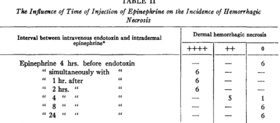
I n order to determine the influence of the timing of injections of endotoxin and epinephrine, the following experiment was performed:--
Six rabbits received an intravenous injection of 20 #g. of Difco E. toll endotoxin, and six different abdominal skin sites were injected with 100 #g. of epinephrine at various intervals of time. The results are shown in Table IL
8 6 8 E P I N E P I I R I N E A N D E N D O T O X I N
W h e n epinephrine was injected 4 hours before endotoxin, no lesions oc- curred, b u t when given simultaneously with, or 1, 2, or 4 hours after endo-
TABLE I
The Production of Hemorrhagic Necrosis of the Skin by E#inephrine in Rabbila Receiving Inlra~enous Endoloxin
Dermal hemorrhagic Dosages necrosis* N o . r rabbits Epine- + + + + Endotoxin pnrine $ + + 0 E. coli (D/fco) ~. coti (Westphal) ~. egui (Westphal) ~k. paradysenteriae (Goebel) Z marcescens (Shear) E. typhesa (Landy) ~eningococcal filtrate I /~g. ~ g . 50 10 6 6 - - i 20 10 6 6 - - - - 10 10 6 4 2 - - 5 10 6 2 4 I 1 10 6 - - 5 1 10 100 10 50 10 10 10 S 10 I00 1 100 10 100 6 1 100 6 50 100 6 10 100 6 20 100 10 10 100 6 0.02 ce. 10 12 0.002 cc. 10 12 6 - - - - 1 5 - - 4 - - 4 5 1 6 - - - - 4 2 10 - - - - 12 - - - - -- 10 2
* Figures indicate No. of rabbits with of epinephrine.
+ + + + indicates lesions 4 X 4 cm. in diameter; 0, no lesions.
dermal hemorrhagic necrosis 24 hours after injec- cm. or larger; + + indicates lesions 1 or 2
toxin, typical hemorrhagic lesions occurred in every case. W h e n epinephrine was n o t given u n t i l 8 hours or longer after endotoxin, the results were negative. Tests W i t h Intravenous Materials Other Than E n d o t o x i n . - - I n an a t t e m p t to learn whether the production of epinephrine lesions represented a specific property of endotoxins, the following materials were injected b y vein simul- taneously with a n i n t r a d e r m a l injection of 100 #g. of epinephrine: h u m a n a n d
L E W I S T H O M A S 869
horse serum (5 cc. each), crystalline ovalbumin (100 rag.), rabbit liver glycogen (100 rag.), colloidal saccharate of iron oxide ("proferrin"--I cc. per kilo), and crystalline ferritin (1 mg. per kilo). None of the animals receiving these injec- tions developed hemorrhage or necrosis at the site of epinephrine injection.
Stetson has recently isolated a material with biological properties similar to endotoxin in extracts of Group A hemolytic streptococci, and reports that intradermal epinephrine has produced hemorrhagic necrosis in rabbits receiv- ing this material by vein (19).
TABLE II
Tke Influence of Time of Injection of Epinephrine on the Incidence of Hemorrhagic Necrosis
Interval between intravenous endotoxin and intradermal epinephrine*
Epinephrine 4 hrs. before endotoxin " simultaneously with " " 1 hr. after " " 2 hrs. " " cc 4 t t ~ ,ct tt 8 t c t~ ~c " 24 . . . . "
Dermal hemorrhagic necrosis
+ + + + + + o - - - - 6 6 - - - - 6 - - - - 6 - - - - - - 5 1 - - - - 6 - - - - 6
* Each of 6 rabbits received an intravenous injection of 20/~g. of Difco E. coli endotoxin and an intradermal injection of 100 /~g. epinephrine at the indicated times. Size of lesions indicated as in preceding table.
Tests with Intradermal Norepinephrine and Other Vasoactive Materials.--
Groups of 4 rabbits were injected intravenously with 20/~g. of E. ¢oli endotoxin (Westphal), and simultaneously tested with intradermal injections of the following materials: norepineph- rine (100 #g.), ephedrine (100 #g.), pitressin (2 international units), histamine phosphate (100 #g.), serotonin (S-hydroxytryptamine) (0.1, 1, and S nag.), and ferritin (100 #g.). Each a g e n t w a s injected in a volume of 0.2 cc.
With norepinephrine, severe hemorrhagic lesions, identical with those caused by epinephrine, occurred in all animals. None of the other materials caused hemorrhage or necrosis.
Prevention of Epinephrine Lesions by Dibenzyline, Chlorpromazine, and Cortisone.--The capacity of two epinephrine antagonists, dibenzyline and chlorpromazine, to protect against the hemorrhagic skin lesions is illustrated by the following experiment:
Six rabbits were injected with 0.5 rag. dibenzyllne intravenously. 30 minutes later, each received 20 ~tg. of Westphal's E . eoli endotoxin by vein, and, simultaneously, 100 #g. of epi- n e p h r i n e intradermally. Six animals were given S gin. chlorpromazine intramuscularly, foI-
870 EPINEPHRINE AND ENDOTOXIN
lowed 30 minutes later by the same test doses of endotoxin and epinephrine. Six control animals received endotoxin and epinephrine alone.
The results are shown in Table III. None of the dibenzyfine or chlorproma- zine-treated rabbits developed hemorrhagic necrosis, while typical lesions occurred in all the untreated controls.
Similar experiments were performed with cortisone. Six rabbits received 10 rag. cortisone intramuscularly 1 hour before the injection of endotoxin and epinephrine. None of these rabbits showed inhibition of the epinephrine skin lesion. However, when cortisone was administered for 3 days before endotoxin and epinephrine, in a daily intramuscular dose of 10 rag., complete protection was observed in all animals (Table III).
Protection against the Lethal Effect of Endotoxin by Chlorpromazine and Dibenzyline.--It was shown previously that cortisone protects rabbits against
TABLE H I
The Effect of Various Agents on the Incidence of Endotoxin-Epinephrine Lesions Agent* Cortisone Chlorpromazine Dibenzyline Heparin Nitrogen mustard
Dermal hemorrhagic necrosls1:
Test group 0/6 o/6 o/6 6/6 6/6 Con~oh 6/6 5/6 4/4 6/6 6/6 * See text for methods of administration.
Numerator refers to No. of rabbits with skin lesions, denominator to No. in group. the lethal action of intravenously injected endotoxin (20). Abemathy and Halberg (21) have recently reported protection of mice against endotoxin by chlorpromazine. A similar effect of chlorpromazine and also dibenzyline was noted in the course of this study, as illustrated by the following experiment.
Two groups d ten mature rabbits, weighing approximately 4 kilos each, were treated with chlorpromazine (5 mg./kilo) and dibenzyline (0.5 reg./kilo), and 30 minutes later they received 50/zg. of Westphal's g. coli endotoxin by vein. A third group of ten untreated rabbits were given endotoxin as controls.
Nine of the ten controls died within the ensuing 24 hours, while none of the chlorpromazine and only two of the dibenzyline group died.
The possibility that the lethal action of endotoxin may be mediated through its effect on epinephrine reactivity will be dealt with in the discussion to follow.
Failure to Prevent Epinephrine Lesions with Heparin and Nitrogen Mustard.--
LEWIS THOMAS 871
man reaction is prevented by heparin (22) and nitrogen mustard (23). Both agents were found to have no effect on the lesions produced b y epinephrine and endotoxin. The results, summarized in Table I I I , are illustrated by the following experiments:--
Six rabbits received 20 mg. heparin intravenously, and 30 minutes later each was injected by vein with Dffco g.. coli endotoxin (20/zg.) and intradermal epinephrine (100 gig.). The administration of heparin was repeated 30 minutes later, and at hourly intervals for 4 sub- sequent doses. All six animals developed hemorrhagic skin lesions similar to, or slightly larger than, those in six untreated controls.
Nitrogen mustard was tested by the method employed by Stetson and Good (23). Six rabbits were injected with a dose of 1.75 rag. per kilo. 3 days later, when the polymorphonu- clear leucocyte count had fallen to less than 500 cells per c.mm. on each aafimal, intravenous endotoxin and intradermal epinephrine were administered as above, and six control rabbits were similarly injected. No differences in the size or severity of the skin lesions were demon- strable between the treated and control rabbits.
Resistance to Epinephrine Lesions in Rabbits with Induced "Tolerance" to Endotoxin.--It has been repeatedly demonstrated b y numerous investigators that the daily administration of small doses of endotoxin brings about a tem- porary, irnmunologically non-specific, state of tolerance to m a n y of the physi- ologic effects of endotoxin, including pyrogenicity, the production of prostra- tion and shock, and the local and generalized Shwartzman reactions (4). In order to determine whether such animals are also resistant to the epinephrine lesions produced with endotoxin, the following experiment was p e r f o r m e d : - Eight rabbits were injected intravenously with 1/~g. of Difco E. toll endotoxin each day for 4 days, then with 10/~g. for 4 more days. On the day following the last injection, each animal received 50 #g. of the same endotoxin by vein and, simultaneously, 100 gg. of epi- nephrine intradermally. On the same day, these doses of endotoxin and epinephrine were injected in eight control rabbits.
No skin lesions appeared in any of the animals given repeated injections of endotoxin, while extensive areas of hemorrhagic necrosis recurred in each of the controls, indicating that the phenomenon of induced tolerance applies to this type of lesion as it does to other effects of endotoxin.
The Role of Vasoconstriction in the Lesions Caused by Epinephrine.--It was assumed, early in the course of these studies, that the epinephrine lesion was probably the result of a great prolongation of the vasoconstrictor effect of epinephrine, due perhaps to a failure of the locally involved tissues to oxidize or otherwise inactivate the substance. Three observations cast doubt on this interpretation.
One was that the most central area of the epinephrine-injected skin site, where one would expect maximally effective concentrations of adrenalin, often seemed to be the least damaged portion of the lesion; frequently a small, circular island of intact skin persisted in the center of a large area of hemor-
872 E P I N E P H R I N E A N D E I ~ T O X I N
rhagic necrosis. The second observation was that the earliest visible change, as the lesion developed, was the appearance of engorged, greatly dilated super- ficial skin vessels, which antedated the occurrence of petechiae, suggesting the possibility that extreme vasodilation and local pooling of blood might be of more importance in the causation of necrosis than vasoconstriction itself. The third observation was that dermal necrosis could not be produced by the maintenance of constantly high levels of epinephrine in a single skin area in normal rabbits. In a group of eight rabbits, for example, which received re- peated intradermal injections of 200/~g. of epinephrine in the same skin site every hour for 8 hours, no lesions of hemorrhagic necrosis resulted.
The question as to the role of vasoconstriction in this lesion will be con- sidered further in the discussion to follow.
The Effect of Intradermal Injection of Mixtures of Endotoxin and Epinephrine
A possible explanation for the epinephrine lesions described in the previous section is that the dermal hemorrhage and necrosis may represent a direct action of endotoxin on vessels in the epinephrine-injected area, due to entrap- ment of circulating endotoxin in blood confined to this region by virtue of the local stasis caused by epinephrine. This explanation cannot be fitted with the observation that epinephrine can be injected as late as 4 hours after intra- venous endotoxin and still produce the lesion, in view of the substantial evi- dence that the major portion of intravenously injected endotoxin is cleared from the circulating blood within a few minutes after injection (24). One would not expect the level of circulating endotoxin at 1, 2, or 4 hours to be sufficient to produce local effects comparable to those seen when endotoxin and epinephrine are given simultaneously.
A second possibility is that endotoxin has its effect on some central nervous system area which governs peripheral reactivity to epinephrine. The present section presents evidence against this possibility, and in favor of the view that the effect of endotoxin is directly exerted on blood vessels, and on their reactivity to epinephrine. In brief, it has been found that a mixture of endo- toxin and epinephrine, injected locally into an area of skin, produces hemor- rhagic necrosis in the injected region. That this necrosis is mediated locally by the combined action of endotoxin and epinephrine, and is not due to a systemic effect of absorbed endotoxin, is demonstrated by the finding that separate skin sites, simultaneously injected with epinephrine alone, do not undergo hemorrhagic necrosis. The observations are illustrated by the follow- ing experiments:--
Mixtures of Dffco/L toll endotoxin and epinephrine were prepared so t h a t an intradermal injection of 0.2 ee. contained 10 #g. of epinephrine and either 50, 10, or 5 #g. of endotoxin. These were injected in six rabbits, into three quadrants of the abdominal skin, and 100/zg. of epinephrine alone were injected into the fourth quadrant.
LEWIS THOMAS 873
By the end of 12 hours, hemorrhage and necrosis had appeared in each of the sites receiving mixtures of epinephrine and endotoxin, while the control sites injected with epinephrine alone showed no lesions. The endotoxin-epi- nephrine lesions advanced somewhat in size and intensity until 24 hours, and remained stationary thereafter. T h e y resembled the previously described lesions produced b y intradermal epinephrine in animals injected with endo- toxin b y vein, except t h a t they were considerably smaller, rarely occupying areas larger than 3 X 3 cm. Also, a conspicuous zone of erythema, edema, and induration occurred around the area of hemorrhagic necrosis, unlike the uniformly flat appearance of the former lesions. Typical lesions are illustrated in Fig. 2. Similar results were obtained with each of the other preparations of endotoxin employed in the experiments described in the previous section. Norepinephrine combined with endotoxin produced lesions identical with those with epinephrine; no lesions occurred with serotonin, ephedrine or pitressin. To obtain information concerning the effective doses of epinephrine, a similar experiment was performed with a fixed quantity of endotoxin (10/~g.) mixed with 4 different doses of epinephrine (50, 10, 5, and 1 #g.), injected intradermally, in 0.2 cc. volumes, in each abdomb nal quadrant. Six rabbitswere used. The sitesinjected with endotoxin combined with 50, 10, or 5/~g. of epinephrine showed hemorrhagic lesions averaging 2 or 3 cm. in diameter in all rabbits, while the areas receiving 1 #g. developed small lesions, less than 1 cm. in diameter, in 2 of the 6 animals.
The histologic appearance of the lesions caused by endotoxin-epinephrine mixtures varied, depending on the age of the lesions. At 8 hours, early in the development of petechiae hem- orrhage, visible in the gross, the central portions were ~;mi!ar to the lesions produced with intradermal epinephrine and intravenous endotoxin, exhibiting marked dilation of capillaries and venules, diffuse hemorrhage, and necrosis of hair follicle cells, with little infiltration by inflammatory cells. By the end of 24 hours, the entire area contained numerous polymorpho- nuclear leucocytes, some of which formed masses within the lumen and around the walls of venules.
Prevention of Lesions by Dibenzyline, Chlorpromazine, and Cortisone.--As
was the case in the lesions produced b y intradermal epinephrine and intra- venous endotoxin, the administration of 0.5 rag. of dibenzyline or 5 rag. of chlorpromazine 30 minutes before injection of an endotoxin-epinephrine mix- ture completely prevented the development of hemorrhagic necrosis. Cortisone failed to protect when given as a single dose 1 hour before injection, but pre- treatment with cortisone for 3 days provided complete protection.
The Enhancing Effect of Nitrogen Mustard.--The
administration of nitrogen mustard not only failed to prevent the lesions, but consistently caused the development of much larger and more extensive areas of necrosis. These lesions continued to spread over the entire abdomen and eventually brought about sloughing of both skin and muscle in the abdominal wall. This observa- tion is illustrated b y the following experiment:--874 E P I N E P H R I N E A N D E N D O T O X I N
Six rabbits received 1.75 rag. of nitrogen mustard intravenously. 3 days later, when the polymorphonuclear leucocyte count was less than 600 cells per c.min, in all animals, a mixture of 10 #g. of Difco/L coli lipopolysaccharide endotoxin and 10 gg. of epinephrine was injected intradermaily on the right side of the abdomen. At the same time, similar doses of endotoxin alone, and epinephrine alone, were given in the left upper and lower quadrants respectively. At 12 hours, the right side of each rabbit showed a flat dead-white area of completely necrotic skin occupying an area of 3 X 3 cm.; surrounded by a narrow zone of subcutaneous hemorrhage (Fig. 3, a). By the end of 24 hours the lesions had spread to involve a larger area of skin on the right side (Fig.
3, b), and during the next day denudation occurred, exposing large areas of necrotic subcutaneous tissue and muscle. The skin sites on the left side, in- jected with endotoxin alone, or epinephrine alone, showed no visible lesions.
The Failure of Heparin to Protect against the Lesion.--Heparin was adminis- tered to 6 rabbits, as described in an earlier section, and caused no protection against the lesions. In several instances, hemorrhage was somewhat more intense than in untreated controls, but the size of the lesions was unchanged.
Induced "Tolerance" to the Lesions Produced by Mixtures of Epinephrine and Endotoxin.--Eight rabbits were given daily intravenous injections of Westphal's E. coli endotoxin, in doses of I t~g. for 4 days and 10 ~g. for 4 days. On the day after the last injection, an intradermal injection of 50 #g. of endotoxin mixed with 50 #g. of epinephrine was given in these rabbits and in 8 untreated controls. No lesions of hemorrhage or necrosis developed in any of the treated animals, while extensive lesions occurred in all of the controls.
The Enhancing Effect of Intravenous Endotoxin on the Hemorrhagic Necrosis Produced by Endotoxin-Epinephrine Mixtures.--The inflammatory component of the local skin lesions produced by mixtures of epinephrine and endotoxin was eliminated when an intravenous injection of endotoxin was given at the same time as an intradermal injection of the mixture, and the resulting lesions showed more extensive hemorrhagic necrosis. The point is illustrated by the following experiment : - -
Eight rabbits were injected intradermally with a mixture of 50 #g. of E. coli endotoxin, and 50 #g. of epinephrine. Immediately before this injection, 4 of the animals received 10 #g. of endotoxin by vein. On the following day, it was observed that each of the latter rabbits had developed large, fiat areas of hemorrhagic necrosis measuring 5 X 6 cm. in diameter, without any surrounding edema or erythema, in contrast to the much smaller areas of necrosis sur- rounded by swollen, indurated skin which occurred in the untreated rabbits. Histologic sections revealed an almost complete lack of infiltration by leucocytes in the group given intravenous endotoxin, while the lesions in the controls showed dense accumulations of polymorphonuclear leucocytes throughout the involved skin.
The results suggest that the presence of a local inflammatory reaction, in response to endotoxin, m a y limit to some extent the degree of necrosis caused by epinephrine. Delaunay (1) has demonstrated the capacity of intravenous endotoxin to interfere with the diapedesis of leucocytes, and it m a y be this
LEWIS T H O M A S 875 effect which allows greater progress of the lesions. This interpretation may also apply to the violent lesions observed in the animals with leucopenia fol- lowing nitrogen mustard. This matter will be reviewed in the discussion below.
DISCUSSION
The two basic observations encountered in this study, for which some ex- planation must be attempted at the outset, are the following:--(a) Epineph- rine (and norepinephrine) produce extensive lesions of hemorrhagic necrosis when injected into the skin of rabbits at any time during a period of 4 hours after an intravenous injection of endotoxin. The lesions are consistently pro- duced by intravenous endotoxin in doses as small as 1.0 #g., and by epineph- rine in doses as small as 5.0 ~tg. In control rabbits, untreated with endotoxin, repeated injections of 200/~g. of epinephrine into the same skin site, each hour for 8 doses, did not produce hemorrhage or necrosis of the skin. (b) Comparable skin lesions, of somewhat smaller size and accompanied by a vigorous inflam- matory reaction around the periphery, are produced by the intradermal injection of mixtures of epinephrine and endotoxin in similar doses.
Taken separately, each of these two observations might be accounted for on the basis of several different hypothetical mechanisms. The occurrence of dermal necrosis at the site of an epinephrine injection, following intravenous endotoxin, might be due to an effect of endotoxin on some central neural mechanism governing epinephrine reactivity, or to local anoxia in the presence of fever, or to the superimposition of localized, epinephrine-induced ischemia on the generalized arteriolar constriction caused by endotoxin. The second observation might be explained by local vasoconstriction and failure to absorb the injected materials, with consequent prolonged exposure of blood vessels to a necrotizing action of endotoxin.
But taken together--and the lesions resemble each other in so many respects that it seems reasonable to regard them as manifestations of the same mecha- n i s m - t h e explanation which best accounts for both observations is that endotox/n exerts an effect on blood vessels which alters their reactivity to epi- nephrine to such an extent that this hormone now becomes a potent necrotiz- ing agent. When endotoxin is injected by vein, the effect is demonstrable wherever epinephrine is locally placed, and when mixtures of endotoxin and epinephrine are injected into the skin the reaction is confined to the injected area.
One other explanation should perhaps be considered to account for both observa- tions by a single mechanism, and this is the possibility that in both cases the actual necrotizing agent is endotoxin rather than epinephrine. Thus, it might be held that the lesion caused by intravenous endotoxin and intradermal epinephrine is due to the trapping of drculating endotoxin in the area of vascular stasis created by epinephrine, and a similar situation is created by local injection of a mixture of the two substances. However, this would require the assumption that the levels of circulating endotoxin remain fairly constant for as long as 4 hours after intravenous injection, since epineph-
876 E P I N E P H R I N E A N D ENDOTOXIN
rine will produce necrosis if injected at any time during this period. On the contrary, the available evidence indicates that a major portion, if not all, of intravenously injected endotoxin is cleared from the circulating blood within a few minutes after injection (24), and it is difficult to conceive of a sufficient amount persisting in the blood for longer than 1 hour.
If, as seems the case, the lesions are due to a necrotizing action of epineph- rine on vessels altered by endotoxin, the mechanism by which hemorrhagic necrosis is brought about remains unclear. I t does not appear to be simply an exaggerated or prolonged vasoconstrictor response. Repeated injections of large amounts of epinephrine into the same skin site do not produce lesions in normal rabbits. Other vasoconstrictor agents, such as serotonin, pitressin, and ephedrine, do not cause lesions in endotoxin-treated animals. I t is perhaps important that the earliest visible change in the developing epinephrine lesion is the appearance of a widely dilated meshwork of small vessels, resembling telangiectases. Further evidence that an abnormal degree of vasodilatation, rather than constriction, may be involved in the response to epinephrine will be presented in the paper which follows.
Protection against both types of lesion was provided, as might be expected, by pretreatment with dibenzyline and chlorpromazine, both of which are known to act as antagonists of epinephrine. The protection observed with cortisone may be on the same basis, since this hormone has been shown to limit the reactivity of terminal arterioles to epinephrine (25). I t is of interest that all 3 agents not only prevent the epinephrine-endotoxin lesion but also protect against the lethal effect of intravenous endotoxin.
Although there is a gross similarity between the epinephrine lesion and the local Shwartzman reaction, the two phenomena seem to involve quite different mechanisms.
Histologically, the Shwartzman reaction is characterized by severe local inflam- mation and transient occlusion of small blood vessels by aggregates of platelets and leucocytes (26), while the epinephrine lesion occurs in the absence of leucocytic infiltration and does not exhibit intravascular thrombi. The Shwartzman reaction is prevented by heparin and nitrogen mustard, which do not protect against epinephrine- endotoxin necrosis, and is not inhibited by cortisone (27), dibenzyline or chlorpro- mazine (28), which prevent the epinephrine lesion. Stetson (29) has reported that provocation of the Shwartzman reaction can be brought about by infiltrating with epinephrine around the periphery of prepared skin sites; whether this effect was due to ischemia in an inflamed, vulnerable tissue, or represented potentiation of epineph- rine by endotoxin still present in the prepared tissue, remains to be determined.
The lack of participation by leucocytes in the epinephrine-endotoxin lesion, in contrast to the apparently essential role of these cells in the Shwartzman reaction (4), is emphasized by the experiments with nitrogen mustard. This agent not only fails to protect against the lesion, but, in the case of the local reaction to mixtures of epinephrine and endotoxin, brings about a remarkable
LEWIS THOMAS 877 enhancement of the extent and severity of necrosis. This result suggests that local accumulations of leucocytes may have some protective action against the spread of the lesion. Such an effect might also account for the difference between the size of the lesions produced by mixtures of epinephrine and endo- toxin and those in which endotoxin was given by vein. The latter procedure always produced large, flat lesions without an inflammatory exudate, while the mixtures caused smaller areas of necrosis which appeared to be limited by a surrounding zone of vigorous inflammation.
In this connection, the effect of intravenous endotoxin on the lesions pro- duced by local mixtures of both reactants may be of significance. Delaunay (1), Miles (6), and others have noted that the diapedesis of leucocytes into locally irritated tissues is prevented by an intravenous injection of endotoxin. Instead of the limited area of necrosis surrounded by a zone of erythema and edema, the reaction to a mixture of epinephrine and endotoxin is converted into a large, flat lesion of hemorrhagic necrosis by an intravenous injection of endotoxin. The end result is the same as though epinephrine alone had been injected into the skin. I t is possible that endotoxin has two opposing effects when mixed with epinephrine: to potentiate the necrotizing action of epineph- fine, and to limit the extent of necrosis by stimulating the local appearance of leucocytes. The role of leucocytes may be to oxidize or otherwise inactivate epinephrine.
The mechanisms which are responsible for activation or inactivation of tissue epinephrine and norepinephrine, and which may be presumed to regu- late the flow of blood through capillary nets, remain undefined.
It has been proposed that epinephrine is destroyed in living tissues by monoamine oxidase or catecholase enzymes (30), or by iron conveyed to the tissues by means of ferritin (31). It is also likely that the degree of reactivity' of arterioles to epinephrine is conditioned by the composition of electrolytes in the cells of the vessel wails; Tobian (32), for example, has reported an abrupt shift of potassium from arterial wails during epinephrine perfusion. Among the possible experimental approaches to the action of endotoxin, therefore, are its effects on epinephrine-inactivating enzymes or their inhibitors, or on the permeability of endothelial or smooth muscle cells to electrolytes. In addition, the availability and degree of activity of cholinergic mediators in the tissues may be important in influencing local responses to epinephrine.
The altered reactivity to locally injected epinephrine in endotoxin-treated rabbits suggests the hypothesis that other physiologic events which are brought about by endotoxin may be due to abnormal reactions to the epinephrine (or norepinephrine) which is produced or stored in the tissues of internal organs. Such an hypothesis might account for certain effects of endotoxin which resemble prolonged and exaggerated responses to epinephrine. Among these are the generalized reaction of peripheral artefiolar constriction and vasomotion, hyperglycemia, depletion of liver glycogen, and elevated blood
878 E P I N E P I ~ A N D EN-DOTOXII~
levels of lactate and pyruvate, all produced with regularity by both epineph- rine and endotoxin. A terminal shock-like state of arteriolar dilation and vascular collapse, similar to that which occurs after lethal doses of endotoxin, has been observed by Yard and Nickerson (33) in animals given large infusions of norepinephrine. Hemorrhages in the upper intestinal wall, mesenteric lymph nodes and gall bladder, which frequently occur after large doses of endotoxin, are also produced by parenteral injections of epinephrine (1, 7). The lesions of fibrinoid necrosis which occur in the rabbit coronary arteries during the generalized Shwartzman reaction (8, 9) resemble to a striking degree the arterial lesions encountered by Waters (10) in dogs subjected to epineph- rine intoxication.
The role of vascular spasm in the pathogenesis of renal cortical necrosis, the identifying lesion of the generalized Shwartzman reaction (17, 18) is prob- lematical. It has been shown that the lesion develops in rabbits after deposition of masses of fibrinoid material within the glomerular capillaries, and the origin of fibrinoid is believed to be fibrinogen which is precipitated intra- vascularly in combination with an acid polysaccharide (34, 35). As in the case of the local Shwartzman reaction, renal necrosis is prevented by heparin and nitrogen mustard, but not by cortisone (18, 27), dibenzyline or chlorpro- mazine (28). Although there is no direct evidence to implicate epinephrine in the pathogenesis of this lesion in the rabbit, Sheehan and others (36) have proposed arterial spasm as a major factor in the development of renal cortical necrosis in other species.
The observations reported here may have bearing on several other lines of investigation not immediately related to the problem of endotoxin. As exam- pies, the following: (a) Delayed type bacterial hypersensitivity reactions have several important points of resemblance to the reactions caused by endotoxin (29), and the possibility that epinephrine may participate in the tissue damage of hypersensitivity merits consideration. (b) The similarities between the vasomotor manifestations of traumatic shock and endotoxin shock have long been recognized, and Fine et al. (37) have suggested that the absorption of endotoxin from the gastrointestinal tract may be a causative mechanism in traumatic shock. Alterations in vascular reactivity to topical epinephrine have been observed to be similar in both types of shock, as is shown in the following paper (38). (c) Enhancement of bacterial infection has been shown to occur in experimental animals subjected to endotoxin (39, 40) and trau- matic (6, 37) shock. The possibility that epinephrine hyperreactivity may be involved in both circumstances is suggested by the work of Miles (41) on the potentiation of local infection by epinephrine.
In the paper which follows, further evidence is presented for a direct effect of endotoxin on the epinephrine reactivity of blood vessels, utilizing the vascular bed of the rat mesoappendix and the isolated perfused rabbit ear as experimental models.
LEWIS T~OMnS 879
SUMMARy AND CONCLUSIONS
Extensive lesions of dermal hemorrhagic necrosis occurred in rabbits when epinephrine (or norepinephrine) was injected into the skin within 4 hours after an intravenous injection of endotoxin. As little as 5 #g. of intradermal epinephrine, and 1 #g. of intravenous endotoxin, were sufficient to produce lesions.
Similar lesions, but smaller in size and surrounded by a zone of acute in- flammation, were produced by intradermal injection of a mixture of compa- rable amounts of endotoxin and epinephrine.
No lesions were produced by combinations of endotoxin with serotonin, pitressin, or ephedrine.
Both types of epinephrine-endotoxin lesion were prevented by pretreat- ment with cortisone, dibenzyline, and chlorpromazine. They were not pre- vented by heparin or nitrogen mustard. The lesions produced by intradermal mixtures of epinephrine and endotoxin were greatly enhanced in size and severity in animals treated with nitrogen mustard.
Both types of lesion were prevented in rabbits rendered "tolerant" b y repeated injections of sublethal amounts of endotoxin.
It is concluded that endotoxin has the property of altering the reactivity of blood vessels to epinephrine in such a way that this hormone becomes a potent necrotizing agent. The possibility that this effect may represent a basic mechanism in the various intoxicating actions of endotoxin, and certain im- plications of this hypothesis, are discussed.
BIBLIOGRAPHY
1. Delaunay, A., Boquet, P., Lebmn, J., Lehoult, Y., and Delaunay, M., J. physiol. (Paris), 1948, 40, 89.
2. Delaunay, A., Lebrun, J., and Cotereau, H., Ann. Imt. Pasteur, 1947, 73, 565. 3. Delaunay, A., Delaunay, M., and Lebrnn, J., Compt. rend. Acad. so. 1947, 224, 72. 4. Thomas, L. Ann. Rev. Physiol., 1954, 16, 467.
5. Boquet, P., and Izard, Y., Proc. Soc. l~xp. Biol. Med., 1950, 75, 254. 6. Miles A. A., and Niven, J. S. F., Brit. J. Exp. Pathol., 1950, 31, 73. 7. Penner, A., and Bernheim, A. I., J./~xp. Med. 1942, 76, 271.
8. Thomas, L., Denny, F. W., and Floyd, J., J. Exp. Med., 1953, 97, 751. 9. Brunson, J. G., Thomas, L., and Gamble, C. N., Am. J. Path., 1955, 31,555. 10. Waters, L. L., Symposium on Atherosclerosis, National Academy of Sciences,
National Research Council, Washington, D.C., 1956. 11. Thomas, L., and Zweifach, B. W., J. Clin. Inv., 1956, 35, 739. 12. Thomas, L., Fed. Proc. ,1955, 15, 518.
13. Landy, M., Johnson, A. G., Webster, M. E., and Sagin, J. F., J. Immunol., 1955, 74, 466.
14. Westphal, O., Liideritz, O., Eichenberger, E., and Keiderling, W., Z. Naturforsch., 1952, 9, 536.
15. Perrault, A., and Shear, M. J., Cancer Research, 1949, 9, 626.
880 E P I N E P H R I N E AND E N D O T O X I N
17. Shwartzman, G., The Phenomenon of Local Tissue Reactivity, New York, Paul B. Hoeber, Inc., 1937, 461.
18. Thomas, L., and Good, R. A., J. Exp. Meg., 1952, 96, 605. 19. Stetson, C. A., J. Exp. Meg. 1956, 104, 921.
20. Thomas, L., and Smith, R. T., Proc. Soc. Exp. Biol. and Meg., 1954, 86, 810. 21. Abernathy, R., and Halberg, F., J. Lab and Clin. Meg., 1955, 46, 790. 22. Good, R. A., and Thomas, L., J. Exp. Meg., 1953, 97, 871.
23. Stetson, C. A., and Good, R. A., J. Exp. Meg., 1951, 93, 49. 24. Beeson, P., Proc. Soc. Exp. Biol. and Meg., 1947, 04, 146.
25. Zweifach, B. W., Shorr, E., and Black, N. M., Ann. New York Acad. Sc., 1953, 56, 626.
26. Stetson, C. A., J. Exp. Meg., 1951, 93, 489.
27. Thomas, L., and Good, R. A., J. Exp. Meg., 1952, 95, 409. 28. Thomas, L., unpublished observations.
29. Stetson, C. A., J. Exp. Meg., 1955, 101,421.
30. V. Euler, C., Acta Physiol. Scan&, 1956, 38, suppl. 118, 39. 31. Shorr, E., Harvey Lectures, 1954-55, 112.
32. Tobian, L., and Fox, A., J. Clin. Inv., 1956, 35, 297. 33. Yard, A. C., and Nickerson, M., Fed. Proc., 1956, 15, 502.
34. Thomas, L., Brunson, J., and Smith, R. T., J. Exp. Meg., 1955, 102, 249. 35. Thomas, L., Smith, R. T., and yon Korff, R., J. Exp. Meg., 1955, 102, 263. 36. Sheehan, H. L., and Moore, H. C., Renal Cortical Necrosis, Springfield, Charles
Thomas Co., 1953.
37. Schweinberg, F. B., Frank, H. A., and Fine, J., Am. J. Physiol., 1954, 179, 532. 38. Zweifach, B. W., Nagler, A., and Thomas, L., J. Exp. Meg., 1956, 1.04, 881. 39. Thomas, L., in The Affect of ACTH and Cortisone upon Infection and Resistance,
(G. Shwartzman, editor), New York, Columbia University Press, 1953. 147. 40. Dubos, R. J., and Schaedier, R. W., J. Exp. Meg., 1956, 1.04, 53.
41. Miles, A. A., in Experimental Tuberculosis, Bacillus and Host, Ciba Foundation Symposium, Boston, Little Brown & Co., 1955, 87.
EXPLANATION OF PLATES PLATE 65
FIGS. 1 a to lc. The skin lesions produced by epinephrine in rabbits receiving endo- toxin by vein. In each photograph, the size of the lesion is indicated by the centimeter rule above the area.
FIG. 1 a. 100/~g. of epinephrine injected intradermally at the same time as an intra- venous injection of 20/~g. of Difco E. coli endotoxin. Photographed 24 hours after injection.
FIG. 1 b. 50/zg. of epinephrine injected 1 hour after 20 ~g. of Westphaf E. coli
endotoxin.
FIG. 1 c. A titration of intradermal epinephrine. This rabbit received 20/Jg. of Di/co E. co//. endotoxin by vein, and, at the same time, 4 skin sites were injected with epinephrine in the following amounts: 1. (lower left in photograph) 50 ~g.; 2. (lower fight) 20/~g.; 3. (upper left) 10/zg.; 4. (upper right) 5/~g. Photographed 24 hours later.
THE JOURNAL OF EXPERIMENTAL MEDICINE VOL. 104 PLATE 65
PLATE 66
FIGS. 2 a. and 2 b. The lesions produced by intradermal injection of a mixture of endotoxin and epinephrine. Photographed 24 hours after injection.
FIG. 2 a. 20 t~g. of Westphal S. equi endotoxin and 20 #g. of epinephrine. FIc. 2 b. 10 #g. of same endotoxin and 5/*g. of epinephrine.
THE JOURNAL O F EXPERIMENTAL MEDICINE ¥OL, 104 PLATE 66
PLATE 67
FIGS. 3. a and 3 b. The effect of nitrogen mustard on the lesions produced by mix- tures of endotoxin and epinephrine. The rabbits received an intradermal injection of 20/~g. of Difco E. coIi endotoxin and 20 ~g. of epinephrine, 3 days after nitrogen mus- tard in a dose of 1.75 rag. per kilo.
FIG. 3 a. Skin lesion photographed 12 hours after the intradermal injection. FIG. 3 b. A lesion photographed 24 hours after injection.
THE JOURNAL OF EXPERIMENTAL MEDICINE VOL, 104 PLATE 67