For Peer Review
Multiple stimulus- and task-dependent asynchronous hierarchies (STDH) within the visual brain’s parallel processing systems
Semir Zeki
Wellcome Laboratory of Neurobiology, University College London Abstract
Results from a variety of sources lead ineluctably to a re-appraisal of the hierarchical and parallel processing strategies used by the brain to construct an image of the visual world. Contrary to common supposition, there are at least three “feed-forward” anatomical hierarchies that reach the primary visual cortex (V1) and the visual areas outside it, in parallel. These anatomical hierarchies do not conform to the temporal order with which visual signals reach and activate V1 and the specialized visual areas. Furthermore, neither the anatomical hierarchies nor the temporal order predict the perceptual hierarchies. The latter shows that we see (and become aware of) different visual attributes at different times, with colour leading form (orientation) and visual motion, even though it is signals from fast moving stimuli that are the earliest to reach the visual cortex (of area V5). Parallel processing, on the other hand, is much more ubiquitous than commonly supposed, but is subject to a barely noticed but fundamental aspect of brain operations, namely that different parallel systems operate asynchronously with respect to each other and reach perceptual end-points at different times.
This re-assessment leads to the conclusion that the visual brain is constituted of multiple, asynchronously operating, task- and stimulus–dependent parallel hierarchies even within systems sub-serving a single attribute such as form or visual motion; which of these parallel anatomical hierarchies has temporal and perceptual precedence at any given moment is stimulus- and task-related and critically dependent upon the visual brain’s ability to undertake multiple operations asynchronously.
For Peer Review
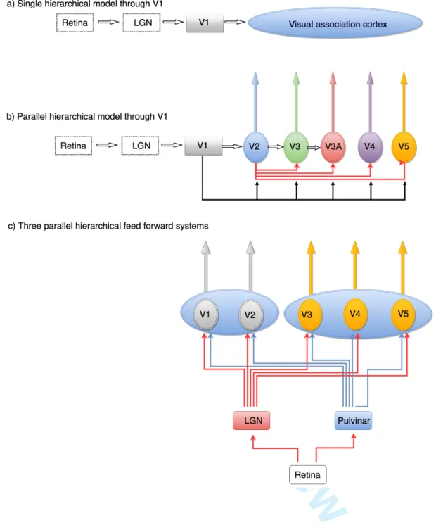
Introduction:Although we are removed by over a century of intensive work from early theories of how the visual brain is organized, these early theories nevertheless linger on, sometimes forcefully, in our present day theorising about the visual brain. Of these, none has been more persistent than the view that V1 is the sole “entering place of the visual radiation into the organ of psyche” (Flechsig, 1905), a view reflected today in the belief that V1 is the sole source of all processing related to colour and form(Marr, 1982)(Bruce & Young, 1986)(Lerner et al., 2001)(Riesenhuber & Poggio, 2002)(Kourtzi et al., 2003)(Grill-Spector & Malach, 2004)(Sasaki, 2007)(Nandy et al., 2013)(Wilson & Wilkinson, 2015), inter alia. Early theories conceived of V1 as the “visuo-sensory” cortex or the “cortical retina”(Henschen, 1893), the one with which we “see”, while the cortex surrounding it came to be known as “visual association” or “visuo-psychic” cortex, “constituted for the final elaboration and interpretation of [visual] sensations” (Campbell, 1905). Significantly, it was supposed that activity in both V1 and the “visuo-psychic” cortex had a conscious correlate, in different ways: V1 was thought to mediate the conscious perception of visual stimuli, a process which Heinrich Lissauer designated as apperception, “…the highest degree of perception, in which the consciousness accepts the sensory impressions with maximal intensity”. Next followed, in visuo-psychic cortex, the process “of connecting other conceptions (ideas) with the content of the perceptions” to give perceptions their meaning (Lissauer, 1890a) (Figure 1a).
Such, then, was the formulation that led to a dual, hierarchical, concept of how the brain processes visual signals and builds a visual image(Zeki, 1993).
Dominant until the 1950s, this hierarchical view was (implicitly) refined by Hubel and Wiesel who showed an hierarchy of complexities in the physiological responses of cells within V1 and between V1 and visual areas of the prestriate cortex(Hubel & Wiesel, 1962; 1965; 1969), an hierarchy that was consistent with the postulated anatomical (hierarchical) progression of visual signals within visual cortex. It led them to suppose that each station of the visual pathway processes the same information but at a more complex level than the antecedent one. Hence, instead of a dual hierarchical process, theirs was a multi-level hierarchical view of increasingly complex responses along a single hierarchical chain. As before, it considered V1 to be
For Peer Review
the source of all processing related to form; was based exclusively on the properties of orientation selective (OS) cells, considered to be critical for form perception. Their new hierarchical view did not take into account two other cardinal attributes of vision, namely colour and visual motion, which earlier theories had considered though only to dismiss the notion that they may have special representations in the brain, outside of V1(Zeki, 1993).
Yet in light of further anatomical and physiological experiments, and taking other visual attributes besides form into account, the exclusive hierarchical model turned out to be too simplistic. The ”visual association” cortex, which Campbell(1905), with Delphic wisdom, had thought of as consisting of “one or more areas”, turned out to contain multiple visual areas, in both macaque (Cragg, 1969;Zeki, 1969, 1971a) and owl monkey (Allman & Kaas, 1971, 1974, 1975,1976), the evidence for the former being based on anatomical connections and for the latter derived from evoked potential studies. The anatomical evidence showed that each of the visual areas outside V1 receives an independent output, in parallel, from V1. Hence the cortical output from V1 is not organized along a single hierarchical chain; there are instead multiple parallel outputs from it to different prestriate visual areas(Zeki, 1976) (Figure 1b). A plausible interpretation of this was that different prestriate areas, being recipient of different signals from V1, undertake different tasks in parallel, not the same task at a more complex level, leading to the view that there is a functional specialization in the visual brain, with different areas processing different visual attributes more or less independently (Zeki, 1978a; 1978b). This has indeed been shown in a wide variety of physiological and imaging experiments(Zeki, 1978c; Livingstone & Hubel, 1984; Hubel & Livingstone, 1987;(Corbetta et al., 1991Zeki et al., 1991;Allison et al., 1994; Merigan et al., 1997; Morita et al., 2004;Cavina-Pratesi
et al., 2010 inter alia.
Hence, when other visual attributes besides form, based on the responses of OS cells, came to be considered, it became obvious that parallelism and functional specialization are major strategies for processing visual signals (Zeki, 1976, 1978a, 1978b). Parallel processing has since played a prominent role in theorising about the visual brain(Marr, 1982; Ballard et al., 1983; Grossberg, 1991). Yet even when considered as acting alongside, or in combination with, an hierarchical processing
For Peer Review
system, it also turns out to be in need of revision, or at least a better integration into the concept of hierarchies, especially since it, too, considered V1 to be the “sole” entering place of visual signals into the rest of the brain and has not taken into account the asynchrony of parallel operations.
My aim here is to show that the term “hierarchical” processing is meaningless unless specified with respect to task and stimulus and that the term “parallel” must not be equated with “simultaneous”, as is commonly implicitly supposed, since different parallel systems act asynchronously with respect to one another. Here, I propose an alternative model of the strategy used by the brain to build an image of the visual world. This new formulation does not pit hierarchical strategies against parallel ones. Rather, it posits that there are multiple hierarchical processes operating in parallel but asynchronously with respect to each other, the temporal precedence of one hierarchical system over another, both physiologically and perceptually, being task and stimulus dependent. This model thus combines the hierarchical and parallel strategies but incorporates two further critical strategies: one is a task- and stimulus-dependent strategy while the other is the related strategy of asynchronous processing. In this essay, I restrict myself to discussing the significance of the “feed-forward” inputs and the role they play in brain strategies for building an image of the visual world. I do not discuss, except in a cursory way, the role of return inputs to an area or of lateral connections within areas. These are of undoubted importance and have been very widely discussed but do not constitute the focus of this article.
Figure 1.
Definition of terms: I give a brief definitions of terms used here; a detailed rationale for their use can be found elsewhere (Zeki & Bartels, 1999).
For Peer Review
Node: an area of the visual brain or a collection of cells within it that are specialized for a visual attribute. An example of the former is area V5, specialized for visual motion, and an example of the latter are the thick, thin and interstripes of V2 or the blobs and the interblobs of V1, which have functionally distinct groupings of cells. An area such as V5 may have sub-populations of cells dealing with fast and slow motion; these would constitute functionally distinct nodes within V5.
End-point: denotes neural activity at a node that requires no further processing to acquire a conscious correlate although the results of the processing may be relayed to further areas. This definition is tied to the “acquisition of a conscious correlate” (see below). There are many end-points in the visual brain, activity at which needs no further processing. The route to each “end-point” is hierarchical but relatively autonomous. Consequently there are many relatively independent hierarchies.
Activity that acquires a conscious correlate: When activity reaches an “end-point” and requires no further processing, it may acquire a conscious correlate. Indeed, it would be computationally wasteful for a node or an area to process signals with the sole aim of relaying the results of the processing to the next stage in the hierarchy. It is more reasonable to suppose that something of what has been processed at a given node can acquire a conscious correlate (Zeki & Bartels, 1999) and hence reach a
perceptual end-point.
Asynchronous processing refers to differences in processing times between different nodes, subserving different visual attributes (for example colour and motion) or between different subdivisions within a node (e.g. fast and slow motion within V5). The differences may be due to the asynchronous arrival of signals, their asynchronous processing or their asynchronous outputs – or all three.
Problems with the hierarchical and parallel models
A problem shared by hierarchical and parallel models is the common assumption that, in the primate visual brain, all visual signals arrive and are processed in V1 first and that the different (parallel) systems starting in V1 undertake their tasks simultaneously. In fact, visual areas outside V1 dealing with the cardinal attributes of form, colour and motion all receive “feed-forward” visual signals that by-pass
For Peer Review
V1(Cragg, 1969;Benevento & Rezak, 1976;Benevento & Yoshida, 1981;(Beckers & Zeki, 1995;ffytche et al., 1995;Sincich et al., 2004;Baldwin et al., 2012;Leh et al., 2008; Schmiedt et al., 2014;Gaglianese et al., 2015). Furthermore, different, hierarchically organized, parallel systems operate asynchronously with respect to each other (Moutoussis & Zeki, 1997a; Viviani & Aymoz, 2001; Arnold et al., 2001; Gauch & Kerzel, 2008; Aymoz & Viviani, 2004; Linares & López-Moliner, 2006; Self, 2014; Barbur, Wolf, et al., 1998), with the consequence that activity in different parallel systems reach perceptual end-points at different times, leading to different visual attributes being perceived at different times, not simultaneously (see below). Hence the parallel systems are only spatially (anatomically) parallel; in terms of the reception, processing and outputting of signals, they operate asynchronously with respect to each other. This leads to a perceptual hierarchy that is not predictable from the anatomical hierarchies. Moreover, V1 is neither necessarily the sole or earliest recipient of all visual signals from the thalamus (Beckers & Zeki, 1995; ffytche et al., 1995; Shigihara & Zeki, 2013; 2014a, 2014b; Shigihara et al., 2015). There are, therefore, at least three different hierarchies – anatomical, physiological and perceptual. These are in different directions and the direction of one is not predictable from that of the other two. Taken together, the three hierarchies create an unaddressed problem of considerable interest, namely how the results of activities in different, hierarchically organized but asynchronously operating parallel systems are integrated (or bound) to give us our unitary experience of the visual world.
Figure 2:
Entry through V1, “tiers” of visual areas and “feed-forward” models: After the discovery of parallel processing channels serving different visual attributes (Zeki, 1976; 1978a), Hubel and Wiesel’s exclusively hierarchical model was significantly modified to include a parallel strategy (Livingstone & Hubel, 1988). But a general hierarchical model applicable to each of the different parallel processing systems also
For Peer Review
received strong support from anatomical and physiological experiments showing that each can be functionally characterised as consisting of an hierarchical chain. Anatomically, this was already evident in the sequence of connections between visual areas, starting from V1 and extending to prestriate areas V2, V3, V4 and V5 (Cragg, 1969; Zeki, 1969, 1971a) (Figure 1b). It was also evident in the laminar pattern of connections between reciprocally connected areas (Rockland & Pandya, 1979)(Felleman & Van Essen, 1991)(Markov et al., 2014); this showed that the input to a cortical area from “lower” areas occupies a different cortical layer than the input to it from higher areas, thus allowing a systematic classification of cortical areas into “lower” or “higher” with respect to each other.
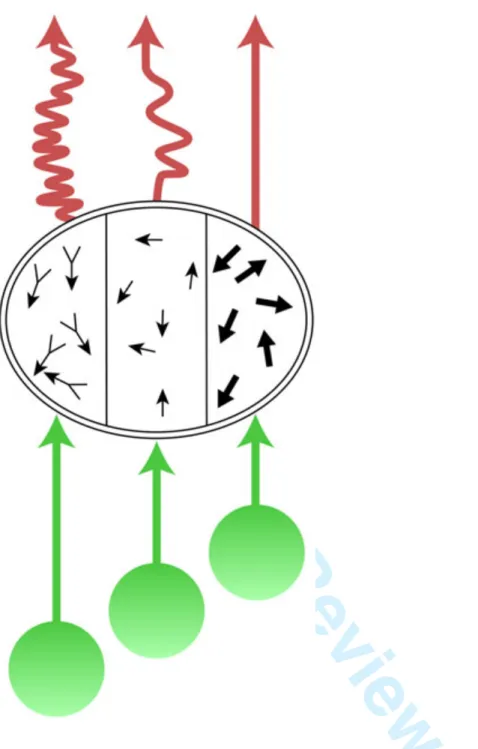
Physiologically, an hierarchical organization became manifest in the gradual enlargement of receptive fields beyond V1 (Zeki, 1978b) and the apparent increasing complexity of the response properties of cells in a specialized hierarchical chain. The best confirmation of a functional hierarchy within one of the parallel processing systems came from studies of the visual motion system(Movshon et al., 1985) (Figure 2). The cortical motion pathway extends from V1 to V5, directly and through V2(Zeki, 1971b), thus constituting a sort of double parallel hierarchical chain(Zeki, 1971a)(Shipp & Zeki, 1989a) (Figure 1b). Because of a convergent input from V1 to V5, the cells of the latter have larger receptive fields than their counterparts in V1(Zeki, 1971b). The V5-projecting cells of V1 are more concerned with the components of the moving stimulus, which may not always correspond to the true, overall direction of its motion (Rust et al., 2006), while those in V5 are more concerned with the overall direction of motion of the entire stimulus, regardless of the direction of motion of its component parts (Dubner & Zeki, 1971;Zeki, 1974); Movshon et al., 1985); (Watson et al.)Simoncelli & Heeger, 1998; (Rust et al., 2006) (Figure 2). A similar, essentially hierarchical, picture emerges from the colour system extending from V1 to V4, both directly and through V2 (Zeki, 1971a). Cells in V4 register the hue of stimuli (Zeki, 1980; Bartels & Zeki, 2000; Kusunoki et al., 2006; Stoughton & Conway, 2008; Brouwer & Heeger, 2009; Brouwer & Heeger, 2013) a process to which V1 may contribute weakly (Wachtler et al., 2003). By contrast, cells in V1 and V2 are less specific with respect to colour, often registering the wavelength composition (components) of the light reflected from a surface and changes in that composition(Zeki, 1983a, 1983b; Moutoussis & Zeki, 2002); they are thus capable of
For Peer Review
responding to a surface of any colour provided it reflects a sufficient amount of light of their preferred wavelength
Paradoxically, the evidence for a functional hierarchical progression with V1 as its source is less compelling in the form system, where it is most frequently, indeed universally, invoked; it is derived mainly from the observation that cells in V1, V2, V3 and V3A respond to oriented lines or boundaries (Zeki, 1978b; Gaska et al., 1988; Larsson et al., 2006, 2010; Tong et al., 2012), those in V4 to oriented lines (Zeki, 1983c; Desimone & Schein, 1987) but also to curvatures and convexities (Dumoulin & Hess, 2007; Pasupathy & Connor, 1999; Müller et al., 2009) while visual areas in the fusiform gyrus respond to more complex forms such as faces, houses and objects (Kanwisher et al., 1997; Haxby et al., 2001; Lerner et al., 2001), thus constituting stimuli of increasing perceptual, and therefore hierarchical, complexity, or so it is generally supposed (Figure 3). How the output of OS cells in V1 is modified physiologically to respond specifically to curvatures and convexities, although explored computationally (Cadieu et al., (2007); Rodríguez-Sánchez & Tsotsos, 2012), has not been demonstrated physiologically; nor is it clear how the responses of OS cells are combined physiologically to produce cells that are specifically responsive to faces and objects. As with the motion and colour systems, computational models have also not factored in the contribution that the direct input from the LGN and the pulvinar make to elaborating the properties of cells in visual areas that are critical for form perception, the only general point of agreement being that the processing of form signals, whether of simple objects and forms such as triangles or squares or more complex ones such as faces or objects, begins exclusively in V1, through its OS cells (Bruce & Young, 1986; Riesenhuber & Poggio, 1999, 2002; Dumoulin & Hess, 2007); Dumoulin et al., 2014); Ostwald et al., 2008); Wilson & Wilkinson, 2015; Haxby et al., 2000), inter alia.
“Tiers” of visual areas
The supposition that entry through V1 constitutes the sole “feed-forward” cortical entry to the rest of the visual brain, as well as the hierarchical progression in physiological properties through prestriate areas of different specialization, has led to two widely held beliefs: one is the conceptual classification of visual areas into three “tiers” or levels - “low-level”, “mid-level” and “high-level”, V1 and V2 constituting
For Peer Review
examples of the first, areas V3, V4 and V5 of the second, and areas critical for face and object perception of the third(Sergent et al., 1992)(Kanwisher et al., 1997)(Epstein & Kanwisher, 1998)(Kanwisher et al., 1997)(Grill-Spector et al., 2001)(Riesenhuber & Poggio, 2000), inter alia. There are compelling reasons, rooted in anatomical connections and the hierarchical progression in physiological responses discussed above, for this classification. But there are equally compelling reasons, discussed below, for supposing that tiers may be conceptually reversible, in the sense that an area occupying a given tier when one set of stimuli or of task conditions is considered may occupy another when a different set of conditions is applied; that (for example) visual areas may not be easily categorized into the same tiers when one considers the latency of arrival of signals in different visual areas or the perceptual precedence that one attribute (such as colour) may have over another (such as directional motion).
The other belief posits the necessity for signals to be processed by V1 to generate a conscious visual experience, either through the “feed-forward” output from it to the specialized visual areas(Weiskrantz, 1986) or through a return input to it from the prestriate areas (Engel et al., 1999;Lamme & Roelfsema, 2000) or both (Hochstein & Ahissar, 2002; Tong, 2003; Koivisto et al., 2010). These theories require to be re-considered in light of more recent evidence because the assumption that V1 is the sole source of visual information in the cortex, for all three cardinal visual systems – those of motion, colour and form - is no longer tenable. This of course does not imply that the return inputs to V1 do not play a significant role in visual processing or in conscious visual perception but only that they are not essential for producing a conscious correlate, however crude (for reviews, see (Zeki & ffytche, 1998; ffytche & Zeki, 2011b); (Overgaard et al., 2008; Overgaard & Mogensen, 2014)
For Peer Review
In the discussion that follows, I group V1 and V2 together as distributor areas, and separate them from other visual areas of prestriate cortex, to which I do not assign levels or hierarchies, for reasons which will become evident (Figure 1c). In spite of differences between V1 and V2, in terms of their cyto- and myelo-architectures and in the disposition of anatomical and functional compartments within them, both contain anatomically (metabolically) identifiable compartments largely dedicated to different attributes of vision and projecting selectively to different specialized areas of the prestriate cortex (Livingstone & Hubel, 1984; Horton, 1984; Livingstone & Hubel, 1988; Shipp & Zeki, 1985; DeYoe & Van Essen, 1988; Hubel & Livingstone, 1987; Shipp & Zeki, 1989a; Shipp & Zeki, 1989b). I acknowledge that there are those who do not believe in such a segregation or who believe in only a moderate segregation(Lennie, 1998; Leventhal et al., 1995; Gegenfurtner, 2003). I do not agree with them, but the issue is not important in this context because there is universal agreement that the three cardinal attributes of form, colour and motion are represented in both areas.
V1 and V2, thus have a special status. This is partly because all visual attributes are represented in both; hence damage to either area usually (but not always) leads to total blindness corresponding in extent and position to the part of the visual field that is represented in the affected brain area (a scotoma or a hemianopia). This has been well known for V1, ever since the topography of the projections from retina to cortex was charted by Henschen (1893), Inouye (1905) - see (Glickstein & Whitteridge, 1987) and Holmes (1918). It is rather less well known that damage to V2 leads to similar results (Horton & Hoyt, 1991). By contrast, damage to the more specialized visual areas leads to visual deficits that correspond more to their specializations, as in achromatopsia, akinetopsia and prosopagnosia which are, nevertheless debilitating (for general reviews, see Meadows, 1974; Zeki, 1990; 1991; Sergent & Signoret, 1992).
For Peer Review
A. The&three&anatomical&“feed&forward”&hierarchical&systems&of&the&visual&brain:! At! least! three! hierarchically! organized! “feed?forward”! systems,! not! one! as! is! commonly!emphasized,!reach!the!prestriate!visual!areas;!one!passes!!through!V1! and!the!other!two!by?pass!it!to!reach!them!directly!from!the!LGN!and!pulvinar! (Yukie! &! Iwai,! 1981)(Fries,! 1981)(Cragg,! 1969)(Benevento! &! Rezak,! 1976)(Sincich!et# al.,! 2004)(Leh!et# al.,! 2008)! (Figure& 1c& and& Figure& 4).! ! The! “feed?forward”! system! that! passes! through! V1! consists! of! two! major! subdivisions,! relayed! through! the! parvocellular! (P)! and! the! magnocellular! (M)! layers! of! the! LGN;! these! reach! V1! separately! but! are! intermixed! to! varying! extents!within!it(Lachica!et#al.,!1992)(Vidyasagar!et#al.,!2002)(Nassi!&!Callaway,! 2009).! The! “V1?bypassing”! input,! by! contrast,! comes! mainly! from! the! intercalated,! koniocellular! (K),! layers! of! the! LGN! (Hendry! &! Reid,! 2000);! it! constitutes!a!direct!input!that!is!uncontaminated!or!much!less!contaminated!by! the!M!and!P!inputs,!which!is!not!to!say!that!there!is!no!intermixing!between!M,!P! and!K!signals!in!individual!visual!areas.!A!hierarchical!system!may!therefore!be! routed! through! V1! or! may! reach! the! prestriate! visual! areas! directly;! all! three! systems! are! “feed?forward”! hierarchical! ones,! in! the! sense! that! they! involve! sequential!anatomical!steps,!starting!at!the!retina!and!extending!to!the!cortical! visual! areas!(see Figures 1c, 3). ! While! the! relationship! of! these! three! feed? forward! to! each! other! remains! to! be! clarified,! there! is! enough! evidence! to! suggest!that!“feed-forward” connection directly from the LGN to prestriate areas is able to sustain a crude but conscious visual experience even in the absence of V1 (Zeki & ffytche, 1998; ffytche & Zeki, 2011; Brent et al., 1994b; Morland, Jones, Finlay, Deyzac, Lê, et al., 1999) while the precise role of the “feed-forward” input from the pulvinar remains to be determined (Schmid et al., 2010). !
!
With!the!exception!of!the!fast!visual!motion!input!to!V5!(Rodman!et#al.,!1989);! Ceccaldi!et# al.,! 1992;! Barbur!et# al.,! 1993;! Beckers! &! Zeki,! 1995;! Benson!et# al.,! 1998;! Zeki! &! ffytche,! 1998;! Morland,! Jones,! Finlay,! Deyzac,! Lê,!et# al.,! 1999;! Stoerig!&!Barth,!2001;!Overgaard,!2011;!Ajina!et#al.,!2015),!the!“V1?bypassing”! inputs!to!the!prestriate!visual!areas!have!been!!neglected;!they!have!not!played!a! prominent! role! in! computational! models! of! the! brain,! assuming! them! to! have! played!one!at!all,!and!have!not!!figured!in!work!that!questions!the!predominance!
For Peer Review
of!hierarchies(Hegdé!&!Felleman,!2007);!yet!all!the!evidence!suggests!that!they! constitute! functionally! significant! inputs,! capable! of! triggering! activity! in! specialized! visual! areas! that! has! a! conscious! correlate,! even! if! only! crude,! without!the!participation!of!V1!(see!below).!!
!
Figure 4.
B. The Physiological (temporal) hierarchies:
Past single cell studies suggested that V1 is always active before the prestriate visual areas(Nowak et al., 1995; Schmolesky et al., 1998b; Luck, 2014). The earliest mean latencies were reported to be in V1, with considerable overlap in the mean latency of responses from so-called “mid-level” visual areas (Nowak et al., 1995; Schmolesky et al., 1998a; Bullier, 2003) suggesting that they become active in parallel temporally. Mean latencies were also reported to occur significantly later in “high level” areas, such as those critical for face and house perception, compared to “low-“ or “mid-level” ones (Hadjikhani et al., 2009). Once again, these results were consistent, or so it appeared, with early views on hierarchical progression of visual processing and with V1 as the sole “entering place” of visual signals into the rest of the visual brain. In spite of methodological differences, the results of evoked potential studies using electro-encephalography (EEG) and magneto-encephalography (MEG) seemed also to reflect the general hierarchical organization of the visual brain (Luck, 2014), with V1 as the first staging-post. Here, too, the mean latency of activity in prestriate cortex appeared to follow that in V1, although some of the reported mean latencies in prestriate cortex, at between 160-220 ms after stimulus onset(Drasdo, N., Edwards, L., Thompson, 1993)(Probst et al., 1993), are surprisingly long, given latencies of less than 50 ms for the earliest responses from prestriate cortex obtained from single cell recordings (Chen et al., 2007). The (temporal) hierarchical conclusions drawn from these studies have no doubt been at least partially responsible for perpetuating
For Peer Review
the concept of exclusive hierarchies, especially in the form system, with the cortical source for all visual processing lying exclusively in V1.
But this consensus regarding a temporal hierarchy in the arrival of signals in different parts of the visual brain, apparently mirroring the anatomical hierarchy of the LGN-V1-prestriate pathway, no longer obtains when one considers the latencies of the
earliest responses (Figure 5); these can be earlier in visual areas of prestriate cortex than the mean latencies from V1. In fact, earliest response latencies have been revised downwards as methods and analytical tools have improved, with several studies giving early latencies, for both striate and prestriate cortex, at less than 50 ms (Whittaker & Siegfried, 1983; (Maunsell & Gibson, 1992; Nowak et al., 1995) and in one as early as 20 ms for V1 (Kraut et al., 1985). Signals from fast moving visual stimuli arrive in V5 before V1 (Beckers & Zeki, 1995)(ffytche et al., 1995b)(Gaglianese et al., 2015), while V1, V2 and V3 respond at the same early time window of 25-45 ms (Shigihara & Zeki, 2013) to form stimuli of varying degrees of perceptual complexity constructed from lines; as well, responses to chromatic stimuli can be detected in visual cortex (including prestriate cortex) much earlier than previously supposed, within the 25-45 ms time window (Shigihara et al, 2015, submitted). Finally, more complex forms such as houses and faces which, in Gestalt parlance, are “other than the sum of the parts”, also activate V1 and the relevant specialized visual areas within the same early time window (Shigihara & Zeki, 2014b), while the earliest responses from the fusiform face area (FFA) have been found to occur as early as 50 ms, some 20 ms before the mean latency of responses from V1 (Seeck et al., 1997)(Shigihara & Zeki, 2014b).
In sum, the (temporal) hierarchy obtained from considering the earliest response times gives a temporal progression that is different from what one might have expected from the classical anatomical hierarchical progression through V1 or from mean latencies; now signals are found to arrive, be processed in, and output from, specialized visual areas first, or simultaneously with V1 (ffytche et al., 1995b)(Schoenfeld et al., 2002)(Shigihara & Zeki, 2013)(Shigihara & Zeki, 2014b) (Shigihara et al., 2015, submitted). Such a temporal order or progression could not have been predicted from the classical “feed-forward” visual pathways through V1; it was not even predicted after the anatomical demonstration of “V1-bypassing”
For Peer Review
pathways to the specialized visual areas. Why earlier EEG and MEG studies did not detect these early responses, or emphasize them when detected, is not clear. It may have been due to the use of flash or reversing checker-board stimuli, not tailored to the specialization of the visual areas (but see Shigihara et al., 2015, submitted); it may have been that early responses, which are relatively weak (in the 50 fT range and indicative of activity in 10,000-50,000 cells (Murakami & Okada, 2006)) compared to the more robust mean responses, were not looked for or discarded or ignored (Tobimatsu & Celesia, 2006); it may have been due to the absence of analytical techniques which now enable their detection or it may have been due to an over-reliance on the classical anatomy of visual connections from the LGN exclusively to V1 and thence to the areas of prestriate cortex. Probably all factors played a role. It is critical to emphasize that the meticulously described sequential temporal hierarchy briefly reviewed above, with V1 showing the earliest mean latency, is not in any sense compromised by the demonstration that a different temporal sequence governs the earliest activation of areas, nor do I question these early results on mean latencies or their significance. But the difference between the two makes it interesting to consider the relationship between early latencies and mean latencies, the latter probably indicative of a more robust build-up of the activity profile in areas. Indeed the combination of the two sets of results – mean and early latencies - merely serves to emphasize that, unless one specifies the context, the term hierarchy becomes confusing.
C. The Perceptual hierarchies: A prediction from the “feed-forward” anatomical progression of cortical connections exclusively from V1 to prestriate visual areas, as well as from the temporal order of cortical activation judged from the mean latency picture derived from recording from V1 and from the so-called “mid-level” visual areas, might be that the three attributes of colour, motion and form are processed first in V1 and then simultaneously in the so-called “mid-level” visual areas, and that they are all perceived at the same time; a prediction from the earlier arrival of signals in V5 than in V1 might be that fast motion is perceived first, before colour and form. Neither prediction is necessarily correct. Psychophysical experiments undertaken to determine the relative time taken to perceive different visual attributes show that, though they are processed in parallel, they are not necessarily processed with the
For Peer Review
same speed or reach a perceptual end-point at the same time, thus indicating that different attributes are processed at different speeds, i.e. asynchronously. Over brief time windows, we see (and become aware of) different visual attributes at different times, with colour leading motion by about 80 ms and form (orientation) by about 40 ms (Moutoussis & Zeki, 1997b)(Viviani & Aymoz, 2001)(Arnold et al., 2001)(Clifford et al., 2003)(Linares & López-Moliner, 2006)(Self, 2014)(Žaric et al., 2015). In terms of overall brain activity, this perceptual asynchrony is also shown by the different activity time courses in different visual areas, in response to viewing the same dynamic, complex natural scenes in action movies(Bartels & Zeki, 2004). Perceptual asynchronies have been attributed to differences in processing times (Moutoussis & Zeki, 1997b)(Arnold & Clifford, 2002), a supposition for which there is some evidence(Lo & Zeki, 2014a)(Žaric et al., 2015). It seems that, in the brain, the fastest system does not “wait” for the slowest one to complete its operations, significantly different from even the most modern computers, whose operations are synchronous(Sutherland & Ebergen, 2002) although efforts are being made to inject a strong asynchronous component into computers (Moradi & Indiveri, 2014). The consequence is that, over brief time windows, the brain does not bind what occurs veridically but instead binds what it has processed and, because it processes colour before motion, mis-binds these two attributes in veridical terms(Moutoussis & Zeki, 1997b), at least over brief time-windows.
Perceptual asynchrony implies a relative independence of the processing systems, in the sense that to perceive a colour in a combined colour-motion task, for example, the brain evidently does not wait for the motion system to complete its processing(Moutoussis & Zeki, 1997b). This autonomy speaks in favour of the absence of a cortical end-point to which both the colour and motion systems must “report” before the correct combination of a colour-motion task is perceived. It suggests instead that different systems have different processing end-points, which acquire conscious correlates at different times (Zeki, 2003).
The consequences of perceptual asynchrony (Zeki, 2015a) and the attendant mis-binding over brief time windows, together with the correct mis-binding of attributes over periods in excess of 500 ms, are of importance in addressing the unresolved question of binding: of how, ultimately, the brain brings together what it has processed
For Peer Review
asynchronously in its different parallel processing systems to give a coherent picture of the visual world, in which all the attributes take their correct time and place. It is possible that this process may not occur solely through direct physiological interaction between visual areas, as is commonly supposed, but through the intervention of post-perceptual areas (Rangelov & Zeki, 2014).
D. Asynchronous operations:
The perceptual asynchrony described above probably has its roots in the unequal speeds with which processings are brought to a perceptual endpoint (as defined above) (Arnold & Clifford, 2002)(Lo & Zeki, 2014b). It is the most tangible manifestation of the visual brain’s asynchronous operations. In fact, it is almost certain that each station along each of the parallel processing pathways undertakes multiple operations asynchronously, unless one posits that a station or node will only commence its operations once it receives all the inputs destined to it, which seems unlikely. As an example, the shortest latency signals to reach V5 are the ones coming from fast moving stimuli (> 22o sec-1); they by-pass V1 and reach V5 with latencies of about 32 ms V1(Beckers & Zeki, 1995)(ffytche et al., 1995b). But V5 also receives signals from slowly moving stimuli (< 5o sec-1) that are relayed from V1 (and from V2 as well), and reach V5 about 60 ms after stimulus onset. And while it takes about 60 ms for pattern motion cells to build up their selective profile, component motion cells, significantly, start their responses 6 ms before them(Smith et al., 2005). This makes it likely that V5 either undertakes, or is able to undertake, several operations simultaneously but asynchronously, through the parallel hierarchical systems reaching it from the thalamus and from the cortex, which deliver their signals asynchronously (Figure 6). Hence, the hierarchical system reaching V5 without passing through V1 operates in parallel but asynchronously with the hierarchical one reaching it through V1 (and V2) (Figure 5). This becomes more compelling in light of the demonstration that activity in a group of cells in V5, signalling one direction of motion at a given speed, can evidently acquire a conscious correlate before activity of another group of cells, signalling a different direction of motion at the same speed (Lo & Zeki, 2014a). This suggests that even when signals belonging to the same attribute reach the same area or node synchronously, they may nevertheless be processed asynchronously.
For Peer Review
Activity in one node of one system can also be asynchronous with respect to activity at a given node of a separate system. For example, activity of the motion system based on V5 is asynchronous with respect to that of the colour system based on V4, evident in the fact that we perceive colour before directional motion(Moutoussis & Zeki, 1997b) and in the fact that we pair colours presented in different parts of the visual field asynchronously, compared to our pairing of directional motion or of colour and directional motion, similarly presented (Bartels & Zeki, 2006). Evidently, the hierarchical systems reaching V4 and V5 directly from the thalamus and through V1 (and V2) operate at different speeds (Figure 5). Hence, taking but two visual areas into account, signals belonging to the same visual attribute (fast and slow motion) or to two different attributes (motion and colour) may reach their respective nodes within an hierarchical system before or after they reach a node or nodes in other hierarchical systems (signals from fast moving stimuli reach V5 before those from slowly moving stimuli reach it, and before colour signals reach V4); and activity in one hierarchical system may reach a perceptual end-point before another (we perceive colour before we perceive motion). Overall, the precedence which one hierarchical system has over others is task and stimulus dependent, leading us to the ultimate hierarchy, which is a stimulus- and task-dependent one (for example, signals from fast and slow moving stimuli reaching V5). Indeed, the principle of dynamic parallelism, originally used only for the motion system(ffytche et al., 1995b) to describe the fact that the parallel inputs into V5 reach it asynchronously, depending upon whether they are relaying signals from fast or slow moving stimuli, may be reasonably enlarged to embrace all the operations of the visual brain, since what system has temporal and perceptual precedence during early time frames is, apparently, dynamically regulated by stimulus and task. Hence, hierarchies themselves are in dynamic, stimulus- and task-dependent relationship to each other.
For Peer Review
Disjunctive hierarchies: There is therefore a disjunctive relationship between the different hierarchies – anatomical, temporal and perceptual. This is at least partially explicable by the fact that each visual area undertakes several processes, which it accomplishes at different speeds, that is to say asynchronously(Bartels & Zeki, 2005)(Zeki, 2015a) (Arnold & Clifford, 2002)(Lo & Zeki, 2014a) (Figures 5 & 6). Unless one posits that an area “waits” until all signals reach it before commencing its processing, or that it “waits” until all processings are completed before outputs from it are initiated, one must assume that the processing within an area and outputting from it starts whenever signals from any source reach it and the processing of these signals within it reaches an end-point and acquires a conscious correlate (Figure 6). Even if two areas start processing signals reaching them at the same time, or if one area undertakes two processes simultaneously, it does not necessarily follow that the separate processings will start or terminate simultaneously (Zeki, 2015a). Hence two processes which start at the same or at different times, in the same or in two different areas, may reach end-points simultaneously or may take different times to reach completion, in the sense defined above.
The above gives a general account of why current concepts of the overall organization of the visual brain must be enlarged to include the three parallel “feed-forward” systems, the principle of asynchronous processing and to accommodate the relationship that the three different hierarchical classifications have to each other; such a classification raises the question of what constant relationship, if any, the different hierarchies have with respect to each other. I try to address this question after reviewing briefly the disjunctive hierarchies operating in the form, colour and motion systems, to show that each conforms to the same general pattern outlined above in terms of the intersection of disjunctive hierarchies and the parallel but asynchronous operations that they undertake.
For Peer Review
Figure 6:Parallel hierarchical inputs to three processing systems: form, motion and colour A. The visual form system:
Stimuli of differing perceptual complexity, such as lines, angles and rhombuses constitute intuitively a hierarchy in terms of form and, indeed, a physiological hierarchical progression mirroring this perceptual hierarchy seemed plausible from earlier results, which showed that some cells in V2 respond to angles rather than straight lines, which is more characteristic of V1(Hegdé & Van Essen, 2000)(Ito & Komatsu, 2004). Such a progression is also evident in the mean latencies with which different visual areas are activated (see above). Yet stimuli constituted from the same lines activate V1 and the prestriate visual areas (V2 and V3) within the same time frame(Shigihara & Zeki, 2013)(Shigihara & Zeki, 2014a) and more recent physiological results show no evident progression in complexity of response to shapes between areas such as V1, V2 and V4(Hegdé & Van Essen, 2007)(Hegdé & Felleman, 2007). As well, responses to shape from “high” level areas such as the parahippocampal place area cannot be predicted from the responses of cells in V1(Nasr et al., 2014), suggesting that inputs that by-pass V1 may contribute to their responses(Shigihara & Zeki, 2014b). Indeed, face and house stimuli constituted from the same straight lines activate V1 and the relevant specialized visual areas within the same time frame(Shigihara & Zeki, 2014b). In summary there is no single hierarchical order – whether temporal or anatomical – that is dominant within the form system.
The above studies are mirrored by physiological ones, which show that OS cells in V2 and V3 maintain their orientation selectivity when disconnected from V1(Schmid et al., 2009), presumably through the direct “V1-bypasing” inputs to them, which is not to say that the OS cells of V1 do not contribute to the elaboration of the properties of OS cells in prestriate cortex(Anzai et al., 2007)(El-Shamayleh et al., 2013). Moreover, subjects blinded by lesions to V1 can still discriminate oriented lines and shapes(Trevethan et al., 2007), though whether they have a crude conscious experience of them, as they do with colour and fast moving stimuli, is not clear. Taken together, these results imply that the OS cells of V1 cannot be the sole source
For Peer Review
of input to the form system in the brain, as is commonly assumed, a conclusion which finds support in the results of backward masking experiments; these show that lines are relatively ineffective in masking rhombuses whereas rhombuses are effective in masking lines (Lo and Zeki 2014). This is the precise opposite of what one might expect from the exclusive hierarchical doctrine, which supposes that rhombuses are constructed from oriented lines.
It has been argued that there are at least three form systems in the brain – a static one, a dynamic one and one linked to colour (Grossberg, 1991) (Zeki, 1993). This may yet turn out to have been an under-estimate. OS cells, a likely source for form construction, are widely distributed in different visual areas of the brain, including areas V2, V3, V3A, V3B(Zeki, 1978b)(Mannion et al., 2009)(Larsson et al., 2010), all of which have been linked to form perception(Zeki et al., 2003). Although early results suggested that the tuning properties of OS cells in this group of visual areas are not markedly different, more recent ones show differences in, for example, contrast thresholds for optimal activation (Zeki, 1978b) (Gaska et al., 1988)(Gegenfurtner et al., 1997) (Mannion et al., 2009). In fact, OS cells may also code for other variables such as size, phase and position (Goris et al., 2015). Given these similarities and differences, it is unlikely that OS cells serve the same purpose and may in fact have different roles in the different form systems; the three different feed-forward inputs to V1 and the specialized visual areas may contribute in different ways to the diversity of OS cell responses. In contrast to these areas, the OS cells of V4 have broader tuning curves and are associated with chromatic responses (Zeki, 1983d)(Bushnell & Pasupathy, 2012). It is generally supposed, though with no convincing direct physiological evidence, that the response of V4 to curved forms and convexities (Dumoulin & Hess, 2007)(Pasupathy & Connor, 1999)(Müller et al., 2009) has its origins uniquely in the OS cells in V1. But it is possible that the “V1-bypassing” input to V4 may also play a role, in addition to the input from V1.
Clinical studies show that perception of real objects may remain intact when line drawings of these objects are impaired (Hiraoka et al., 2009) and, conversely, that the perception of objects may be impaired without impairment of the perception of the individual elements (lines) that they are generally considered to be constituted from (Riddoch et al., 2008); agnosias for static forms need not be accompanied by an
For Peer Review
agnosia for them when in motion (Botez & Serbănescu, 1967) and shape from shading and that from edges may be separately represented(Humphrey et al., 1996). Finally, a dissociation between orientation and shape perception has been demonstrated within subdivisions of LO(Silson et al., 2013). All this attests to the possibility that we may be mistaken of thinking of a single or even multiple form systems whose source(s) lie exclusively in the OS cells of V1.
B. The motion system: dynamic parallelism
As discussed above, the shortest latency signals to reach V5 are the ones coming from fast moving, high contrast. In fact, many single cell studies show that fast motion is especially prominently registered in V5(Maunsell & van Essen, 1983)(Newsome et al., 1986)(Liu & Newsome, 2005)(Rodman & Albright, 1987)(Zeki, 2015b). But V5 also receives signals from slow-moving stimuli, reaching it through V1(ffytche et al., 1995b). This makes it likely that V5 either undertakes, or is able to undertake, several operations simultaneously but asynchronously through the parallel hierarchical systems reaching it from the thalamus and from the cortex (Figure 6). Hence, the hierarchical system reaching V5 without passing through V1 operates in parallel but asynchronously with the hierarchical system reaching it through V1 (and V2) (Figure 5). This becomes more compelling in light of the demonstration that activity in a group of cells in V5, signalling one direction of motion, can evidently acquire a conscious correlate before activity of another group of cells, signalling a different direction of motion(Lo & Zeki, 2014a).
The asynchronous routing of motion signals to V5, depending on the speed of the moving stimulus, implies a stimulus dependent dynamic parallelism (ffytche et al., 1995) that constitutes a reverse (temporal) hierarchy from the one predicted by the classical sequential cortical anatomical input originating in V1. Hence, as with the form system, the source of motion signals in V5 cannot be thought of as being exclusively the motion selective cells of V1, especially since in the absence of V1, the receptive field size of cells in V5 are maintained, as is the overall topography of V5 and the directional selectivity of its cells (Rodman et al., 1989)(Poppel et al., 1973). Indeed, the pulvinar may be implicated in higher order motion processing, including the processing of pattern motion(Casanova et al., 2001)(Villeneuve et al., 2012) but it is likely that the property of directional selectivity itself is not conferred on V5 by the
For Peer Review
pulvinar(Berman & Wurtz, 2011). Significantly, studies of humans blinded by lesions in V1 show that this V1 by-passing input is sufficient to elicit a crude but conscious experience of directional visual motion (the Riddoch Syndrome) (Zeki & ffytche, 1998)(ffytche & Zeki, 2011). This naturally calls into question theories that have posited a mandatory involvement of V1, either through feed-forward or return input processing, for visual activity to acquire a conscious correlate (Weiskrantz, 1986) (Lamme & Roelfsema, 2000)(Hochstein & Ahissar, 2002).
C. The colour system Just as with the form and motion systems, the colour one based on V4 receives a direct input from V1 and V2 as well as a “V1-bypassing” input from both the LGN and the pulvinar (Yukie & Iwai, 1981)(Fries, 1981)(Benevento & Yoshida, 1981)(Benevento & Rezak, 1976)(Cragg, 1969)(Leh et al., 2008)(Schmiedt
et al., 2014). V4 responds to colour stimuli within the same early time window and possibly earlier than V1 (Shigihara et al., 2015, submitted) and, again just like V5, the input is apparently sufficient to enable a crude but conscious experience of colour(Morland, Jones, Finlay, Deyzac, Le, et al., 1999), though the stimuli must be large(Brent et al., 1994a). Although the colour system has not been studied as extensively as the motion system in this regard, this evidence is consistent in showing that a mandatory passage of signals through V1 or a return input to it, are not necessary for eliciting a conscious visual experience.
It is interesting to consider the judgment of Henschen on the possibility of a colour centre outside V1 which he, along with Gordon Holmes, was vociferous and successful in dismissing as improbable (see Zeki 1993). Henschen believed that, if this were true, then “with the calcarine cortex [V1] destroyed and the cortex of that other gyrus [where we now know V4 to be located] intact, a patient would have to be absolutely blind and yet be able to see colours, which makes no sense”(Henschen, 1910). Really, in light of the evidence available to him at that time, this indeed made no sense; neither did the report that patients blinded by lesions in V1 are able, sometimes, to perceive directional motion consciously (Riddoch, 1917); Holmes (1918) dismissed such findings by writing assertively that “…occipital lesions do not produce true dissociations of function with intact retinal sensibility”, thereby ignoring one of his own cases of the sparing of motion after V1 lesions, in a patient, who was “…generally conscious only of the movement of the white test object” (Holmes,
For Peer Review
1918). Had either known about the direct projection from the LGN to the prestriate cortex, they may well have tempered their views and general hostility to any separate representation of colour and motion outside of V1.
Hence there are parallel inputs to the prestriate visual cortex that use V1 on the one hand and other pathways that by-pass it on the other, for all three of the cardinal visual attributes – form, colour and motion; in at least two of these, the direct “V1-bypassing” input is potent enough to elicit a conscious, if crude, experience of the relevant attribute.
A re-assessment
The general principle that can be derived from all these studies is (a) that there are multiple hierarchies in the visual brain – anatomical, temporal and perceptual – which are in different directions; and (b) that these hierarchies operate in parallel but asynchronously. Collectively, the above facts invite a re-assessment of views that have been, and continue to be, very basic to our thinking about how the visual brain operates. Among these are:
1. The assumption that V1 is the exclusive source of visual signals to the rest of the visual brain. This assumption, especially prominent in thinking about the colour and form systems, is no longer tenable when applied to them, just as it has not tenable for the motion system for some time; nor is the assumption that the OS cells of V1 are the exclusive source of signals for generating forms in the brain. While it is evident that OS cells must make a significant contribution to the responses of cells in areas outside V1, the survival of OS cells in areas such as V2 and V3 and the same early time frames with which lines activate visual areas implicated in form perception raise the question of whether OS cells are not, as well, partially independently constructed in different visual areas.
2. The concept of tiers of areas: the concept of “low-level”, “mid-level” and “high-level” tier areas made sense when considered against the background of an exclusive overall hierarchical strategy for processing visual signals derived from early anatomical studies of the classical visual pathway through V1, of the perceptual complexities of visual stimuli, or of exclusive hierarchical strategies within each of the parallel processing systems. They still make sense today, but
For Peer Review
only if the context is specified and qualified. In temporal terms, how can V5 be a “mid-tier-“ area when it receives fast motion signals before V1 and processes these to allow a conscious, if crude, experience of visual motion even in the absence of V1? How can V3 be a “mid-level” visual area when it is activated within the same early time frame as V1? Cortical areas critical for shape and face perception are indeed “high-tier” visual areas in perceptual terms, but not when the latency of early visual signals are considered; to consider them “higher-tier” areas when the early latencies at which they are activated are the same as V1 encourages the supposition that the construction of complex forms such as faces have their source exclusively in the orientation selective cells of V1, as also commonly assumed; but the direct V1-bypassing inputs to them may also play a significant role. To speak of tiers of areas thus makes sense only if the stimulus, the latency of arrival of signals in the cortex from it and the task are specified; an area can be “low-level” “mid-level” or “high-level” depending on them depending on the context.
3. The assumption of a perceptual hierarchy in form complexity: The notion that forms such as rhombuses are relatively simple compared to more complex forms such as faces and houses, though intuitively appealing, needs re-examination. It is perhaps more reasonable to consider them as different forms, as in Gestalt psychology which considers the “whole to be other than the sum of the parts”1. Apparently simple forms such as hexagons and triangles are in fact
!!!!!!!!!!!!!!!!!!!!!!!!!!!!!!!!!!!!!!!!!!!!!!!!!!!!!!!! 1!The!Gestalt!position!is!usually!rendered!as!“the!whole!is!more!than!the!sum!of! ! ! “Prinzipiell!identisch!ist!in!den!beiden!Thesen!–!und!darauf!soll!es!hier! ankommen!–!das!Und*Summenhafte:!der!Aufbau!aus!Stücken,!die,!das!eine!und! das!andere!und!ein!drittes…zunächst,!primär,!alles!Weitere!fundierend!gegeben! sind;!in!Und?Verbindung;!im!Auch?Dasein;!gegeneinander!inhaltlich!prinzipiell! beliebig!und!ohne!Ingerenz,!es!sei!den!eine!solche,!die!stückhaft!von!“unter!her”! –!wieder!von!Stücken!her!–!gemeint!ist;!entstehen!darüber!höhre!Gebilde.! Verbindungen,!Komplexe,!so!bauen!sich!diese!sekundär,!von!unten!her,!auf!der! Und?Summe!der!Stücke!auf!(wobei!etwa!wieder!sachlich!beliebig!hinzutretende! Funktionen,!Akte,!Verhaltungsweisen!der!Aufmerksamkeit!usw.!Eine!Rolle! spielen).! ! ! Was!zusammengefügt!erscheint,!im!Zugleich,!im!Nebeneinander,!im! Nacheinander,!is!prinzipiell!beliebig;!für!das!Zusammensein!is!der!“Inhalt”!oder! das!Zueinander!von!Inhalten!eigentlich!irrelevant.!Keine!sachlichen!Momente!
For Peer Review
mathematically complex structures and could be generated independently of other complex forms such as scenes, faces or objects. Nor need complex forms such as faces be considered as being simply built up from the OS cells of V1, which is not the same as saying that V1 does not contribute significantly to elaborating the properties of areas which are critical for face and object perception.
4. The mandatory involvement of V1 in the generation of conscious visual experiences. The notion that passage of signals through V1 or the return inputs to it from prestriate visual areas is mandatory for eliciting a conscious visual experience must also be revisited. There is now compelling evidence to show that V5 can act autonomously of V1 to mediate a conscious, if crude, experience of fast visual directional motion(Barbur et al., 1993) (Zeki & ffytche, 1998)(Sahraie et al., 2013)(Overgaard et al., 2008)(Overgaard & Mogensen, 2014) and less extensive evidence to suggest that V4 can, similarly, mediate a conscious if crude experience of colour in the absence of V1(Brent et al., 1994b)(Barbur, Sahraie, et al., 1998)(Morland, Jones, Finlay, Deyzac, Lê, et al., 1999). This is not to suggest that either area acts completely independently nor is it to question the well-documented fact that return inputs to V1 may be necessary for detailed vision, but only to question the view that the healthy functioning of V1 and that the passage of signals to and from it are mandatory for eliciting a conscious experience of the relevant visual attribute.
The organizing principle of hierarchies: multiple parallel stimulus- and task-dependent asynchronous hierarchies
The above account appears to make of the visual brain a hopelessly intricate organ. But it encourages us to speculate on a possible organizing principle, or at least some !!!!!!!!!!!!!!!!!!!!!!!!!!!!!!!!!!!!!!!!!!!!!!!!!!!!!!!!!!!!!!!!!!!!!!!!!!!!!!!!!!!!!!!!!!!!!!!!!!!!!!!!!!!!!!!!!!!!!!!!!!!!!!!!!!!!!!!!!!!!!!!!!!!!!!!!!!!!!!! sind!für!die!Zusammengefügtheit!bedingend,!sondern!inhaltsfremde,! “sachaussere”!Faktoren,!wie!z.!B.!das!Oft?zusammengewesensein,!das!simultane! Beachten!usw.”!! ! If!he!had!expressed!it!in!clearer!language,!his!statement!would!no!doubt!have! had!wider!circulation!today.! ! I!am!grateful!to!Michael!Herzog!for!pointing!this!out!to!me.!
For Peer Review
constant relationship between the different, disjunctive, hierarachies. One principle, I suggest, is that of stimulus- and task-dependent hierarchy (STDH), which operate asynchronously and do not necessarily use only V1 as the entry point to the rest of the visual brain, although they may do so. The concept of STDH is strongly linked to a much-ignored characteristic of the visual brain, namely its asynchronous operations. Between them, these two characteristics resolve conceptually the difficulties of having different hierarchical systems which are in different directions and hence apparently in opposition to each other.
Early theories of brain strategies for visual perception did not take into account the time factor; later ones assumed that the anatomical hierarchical progression is reflected in the hierarchical progression in latencies of activation (see above). But the demonstration that early response time windows do not conform to the classical temporal hierarchical picture, coupled to the demonstration that different visual areas, or different nodes within a visual area, undertake their operations asynchronously with respect to each other and reach perceptual end-points at different times(Zeki, 2015a), raises the question of asynchronous operations and their significance, which is superimposed upon the parallel and hierarchical strategies. Different nodes within a given hierarchical pathway do not receive signals in a hierarchical order consistent with the hierarchy revealed by classical anatomy through V1; instead the sequence of activation and the output from stations or nodes within a visual processing system are determined by stimuli and task, as in the example of fast motion discussed above. Anatomical hierarchies, which may be considered to form some sort of organizing principle, must therefore be integrated into a grander organizing principle, which is that of stimulus- and task-dependent hierarchies; these apparently determine not only the sequence of activation of areas but the perceptual precedence as well. That the precedence in arrival of signals and the perceptual precedence of one hierarchical system over others is dependent on both stimulus and task is suggested by experiments which show that: (a) pairing stimuli across attributes, for example the pairing of colour with motion or with orientation, depends upon the task subjects are asked to undertake. Thus, the perceptual asynchrony which is evident when subjects have to determine the colour and the direction of motion of a moving stimulus is not evident when the task is to determine solely that a change in the colour and a change in the direction of the stimulus have occurred, without specifying the colour or the
For Peer Review
direction of motion(Bedell et al., 2003) (Clifford et al., 2003) while the introduction of an exogenous attentional cue reduces or abolishes the colour-motion asynchrony (Holcombe & Cavanagh, 2008) (Wu et al., 2004)(Suzuki et al., 2013); (b) in pairing within attributes – for example when subjects are asked to pair right-left with up down motion of stimuli moving at the same speed, no perceptual asynchrony is evident (Zeki & Moutoussis, 1997) but pairing up down motion with up-right motion results in a perceptual precedence of up-right over up-down(Lo & Zeki, 2014a). Further evidence of the dependency of the perceptual hierarchies on the stimulus is provided by (c)experiments which show that adding an irrelevant transparency may improve and accelerate the pairing of colour and motion (Moradi & Shimojo, 2004). Hence one hierarchy is determined by the need for several steps to process a signal to a perceptual end-point while another hierarchy may need only one or two steps of cortical processing. Within such a context, the apparently disjunctive hierarchies begin to make better sense. Hence, the organizing principle that determines the relationship of one hierarchical system to another is stimulus and task based, the only constant feature in an ever-changing and apparently otherwise unpredictable relationship between different hierarchies.
This asynchronous and relatively independent processing is applicable to all the systems constituting the visual brain. Beyond demonstrating a perceptual asynchrony and hence giving insights into brain strategies, the results imply that there must also be a temporal hierarchy in binding different visual, which has in fact been demonstrated to a certain extent(Bartels & Zeki, 2006); indeed, if activity at each node of a processing system can potentially acquire a conscious correlate, it follows that binding and integration must be multistage processes, since activity at any station within any processing system can be bound to activity at any station of another processing system(Bartels & Zeki, 1998); this naturally introduces another hierarchy, a binding hierarchy whose temporal time course is also not predictable from either the anatomical or the physiological hierarchies but can be predicted from the principle of stimulus- task-dependent hierarchies. As well, if processing by different stations is asynchronous, it follows that the outputs of the different processing systems must also be asynchronous and therefore temporally hierarchical(Rangelov & Zeki, 2014) (Figure 6). Moreover, since the outputs are asynchronous, it can be conjectured that
For Peer Review
the return inputs to the visual areas from the reciprocal connections that they have with the areas that they project to (Felleman & Van Essen, 1991) may also be asynchronous, although this has yet to be demonstrated. It is therefore not at all certain, from what has been said above, that these reciprocal connections, which may and probably operate asynchronously (given the asynchronous output from areas), will predict the hierarchical status of an area in the same way that the laminar arrangements do.
This leads us ineluctably to the conclusion that the operations of the visual brain, and probably the brain at large, are massively asynchronous and parallel. This makes the visual brain’s parallel hierarchical systems much more ubiquitous than previously supposed and gives the term “hierarchical organization” much wider significance. The hierarchies, it turns out, are very widespread but reversible in terms of temporal status because they are governed by stimulus and task which, in turn, determine the asynchronous status of one hierarchical system with respect to that of another or of others.
In conclusion, enough evidence has been accumulated over the past quarter of a century to merit a significant re-evaluation of the strategies used by the visual brain to construct a picture of the visual world.
Acknowledgements
The work of this Laboratory is supported by the Wellcome Trust, London. I thank Stewart Shipp and Dominic ffytche for their helpful comments and suggestions on earlier versions of this manuscript.
ConflictofInterest
None to declare.