Tom, AML survivor
Acute Myeloid Leukemia
A Message from Louis J. DeGennaro, PhD
President and CEO of The Leukemia & Lymphoma SocietyThe Leukemia & Lymphoma Society (LLS) is committed to bringing you the most up-to-date blood cancer information. We know how important it is for you to have an accurate understanding of your diagnosis, treatment and support options. With this knowledge, you can work with members of your oncology team to move forward with the hope of remission and recovery. Our vision is that one day the great majority of people who have been diagnosed with Acute Myeloid Leukemia (AML) will be cured or will be able to manage their disease with a good quality of life. We hope that the information in this booklet will help you along your journey.
LLS is the world’s largest voluntary health organization dedicated to funding blood cancer research, education and patient services. Since the first funding in 1954, LLS has invested more than $814 million in research specifically targeting blood cancers. We will continue to invest in research for cures and in programs and services that improve the quality of life of people who have AML and their families.
We wish you well.
Louis J. DeGennaro, PhD
President and Chief Executive Officer The Leukemia & Lymphoma Society
Table of Contents
2 Introduction 2 Here to Help
5 Leukemia
6 Acute Myeloid Leukemia
9 Diagnosis
12 Treatment
29 Research and Clinical Trials 31 Normal Blood and Marrow 33 Medical Terms
51 More Information
Acknowledgement
The Leukemia & Lymphoma Society gratefully acknowledges, for their critical review and important contributions to the material presented in this publication,
Judith Karp, M.D.
Professor of Oncology and Medicine Director, Leukemia Program
Division of Hematologic Malignancies
The Sidney Kimmel Comprehensive Cancer Center at Johns Hopkins Hospital Division of Hematologic Malignancies
Baltimore, MD
This publication is designed to provide accurate and authoritative information about the subject matter covered. It is distributed as a public service by LLS, with the understanding that LLS is not engaged in rendering medical or other professional services.
Introduction
This booklet provides information about acute myeloid leukemia (AML) for patients and their families. Brief descriptions of normal blood and marrow and definitions of medical terms are included at the end of the booklet to help readers better understand the information about AML.
AML may be called by other names, including acute myelogenous leukemia, acute myelocytic leukemia, acute myeloblastic leukemia and acute granulocytic leukemia. About 12,950 new cases of AML were expected to be diagnosed in the United States in 2011. As of January 2008 an estimated 30,993 people were living with (or were in remission from) AML. Although AML can occur at any age, adults aged 60 years and older are more likely to develop the disease than younger people.1
Advances in AML testing and treatment are resulting in improved remission and cure rates, but much work remains to be done. For example, the vitamin A derivative all-trans retinoic acid (ATRA) has greatly improved survival rates for patients with acute promyelocytic leukemia (APL), a subtype of AML. A number of new therapies are under study in clinical trials.
1Howlader N, Noone AM, Krapcho M, Neyman N, Aminou R, Waldron W, Altekruse SF, Kosary CL, Ruhl J,
Tatalovich Z, Cho H, Mariotto A, Eisner MP, Lewis DR, Chen HS, Feuer EJ, Cronin KA, Edwards BK (eds). SEER Cancer Statistics Review, 1975-2008, National Cancer Institute. Bethesda, MD, www.seer.cancer.gov/ csr/1975_2008/, based on November 2010 SEER data submission, posted to the SEER website, 2011.
Here to Help
This booklet will help you talk to your doctor about the tests and treatment you need. We encourage you to take the lead in asking questions and discussing your fears and concerns. These actions will give members of your healthcare team the opportunity to answer your questions, extend emotional support and provide any needed referrals.
A diagnosis of AML is often a shock to the patient, family members and friends. Denial, depression, hopelessness and fear are some of the reactions people may have. Keep in mind that
{
{ Many people are better able to cope once they begin treatment and can look forward to recovery.
{
{ The outlook for people with AML is continuing to improve. New approaches to therapy are being studied in clinical trials for patients of all ages and at every stage of treatment.
LLS Has Ways to Help.Treatment for AML will affect your daily life, at least for a time. You may have questions about your treatment and want to have friends, family members or caregivers help you get information.
Making treatment choices, paying for medical care, communicating with healthcare providers, family members and friends—these are some of the stressors that go along with a cancer diagnosis. LLS offers free information and patient services for individuals and families touched by blood cancers.
Speak to an Information Specialist. Information Specialists are master’s level
oncology professionals. They provide accurate up-to-date disease and treatment information and are available to speak with callers Monday through Friday, 9 a.m. to 6 p.m. ET at (800) 955-4572. You can email infocenter@LLS.org or chat live with a Specialist at www.LLS.org.
Language Services. Free language services are available when you speak with an
Information Specialist. Let your doctor know if you want a professional healthcare interpreter who speaks your native language or uses sign language to be present during your visit. Many times, this is a free service.
Información en Español. LLS has a number of resources available in Spanish for
patients, caregivers and healthcare professionals. You can read and download these resources online at www.LLS.org/espanol or order printed copies by mail or phone.
Other Helpful Organizations. Our website, www.LLS.org/resourcedirectory,
offers an extensive list of resources for patients and families about financial assistance, counseling, transportation, summer camps and other needs.
Chapter Programs and Services. LLS chapter offices around the United States
and Canada offer support and education. Your chapter can arrange for peer-to-peer support through the Patti Robinson Kaufmann First Connection Program.
The Patient Financial Aid program offers a limited amount of financial aid for qualified patients. Find your local chapter by calling (800) 955-4572 or by visiting www.LLS.org/chapterfind.
Clinical Trials. Our Information Specialists help patients work with their doctors
to find out about specific clinical trials. Information Specialists conduct clinical-trial searches for patients, family members and healthcare professionals. You can also use TrialCheck®, an online clinical-trial search service supported by LLS that offers patients and caregivers immediate access to listings of blood cancer clinical trials. Please visit www.LLS.org/clinicaltrials.
Free Materials. LLS publishes many free education and support materials for
patients and healthcare professionals. PDF files can be read online or downloaded. Free print versions can be ordered. Visit www.LLS.org/resourcecenter.
Telephone/Web Education Programs. LLS provides a number of free, live
telephone and web education programs presented by experts for patients, caregivers and healthcare professionals. For more information, visit www.LLS.org/programs.
Suggestions From Other People Living With Cancer {
{ Get information about choosing a cancer specialist or treatment center.
{
{ Find out about financial matters: What does your insurance cover?
What financial assistance is available to you?
{
{ Learn about the most current tests and treatments for your type of AML.
{
{ Keep all appointments with the doctor and talk openly about your fears or
concerns or any side effects you experience.
{
{ Talk with family and friends about how you feel and how they can help.
{
{ Contact your doctor if you have fatigue, fever, pain or sleep problems so
that any issues can be addressed early on.
{
{ Get medical advice if you have experienced changes in mood, feelings
of sadness or depression.
The Trish Greene Back to School Program for Children With Cancer.
This program is designed to increase communication among parents, children, adolescents, young adults, healthcare professionals and school personnel.
Informative materials, videos and a wealth of literature are available through LLS chapters to help ensure a smooth transition back to school. For more information, please visit www.LLS.org/backtoschool. For practical guidance on how to support your child, yourself and other family members, see the free LLS booklet Coping
With Childhood Leukemia and Lymphoma.
Reach Out. You and your loved ones can reach out for support in several ways. For example:
{
{ LLS offers online Blood Cancer Discussion Boards as well as online chats at www.LLS.org/getinfo.
{
{ Local or Internet support groups and blogs can provide forums for support. {
{ Patients with cancer often become acquainted with one another, and these friendships provide support.
Depression. Treatment for depression has proven benefits for people living
with cancer. Depression is an illness that should be treated even when a person is undergoing AML treatment. Seek medical advice if your mood does not improve over time—for example, if you feel depressed every day for a 2-week period. Contact LLS or ask your healthcare team for guidance and referrals to other sources of help, such as counseling services or community programs. For more information you can contact the National Institute of Mental Health (NIMH) at www.nimh.nih.gov and enter “depression” in the search box at the top of the web page, or call the NIMH toll-free at (866) 615-6464.
We’d Like to Hear From You. We hope this booklet helps you. Please tell us
what you think at www.LLS.org/publicationfeedback. Click on “LLS Disease & Treatment Publications—Survey for Patients, Family and Friends.”
Leukemia
Leukemia is a cancer of the marrow and blood. The four major types of leukemia are acute myeloid leukemia, chronic myeloid leukemia, acute lymphoblastic leukemia and chronic lymphocytic leukemia.
Acute leukemias are rapidly progressing diseases that affect cells that are not fully developed. These cells cannot carry out their normal functions. Chronic leukemias usually progress more slowly, and patients have greater numbers of mature cells. In general, these more mature cells can carry out some of their normal functions (see Normal Blood and Marrow on page 31).
With myeloid leukemia, a cancerous change begins in a marrow cell that normally forms certain blood cells—that is, red cells, some types of white cells and platelets. With lymphocytic (lymphoblastic) leukemia, the cancerous change begins in a marrow cell that normally forms lymphocytes (another type of white cell). The four main types of leukemia are further classified into subtypes. Knowing the subtype of your disease is important because the treatment approach may be based on the subtype (see AML Subtypes on page 11).
More general information about leukemia is given in the free LLS publication
Acute Myeloid Leukemia
How AML Develops. AML results from acquired changes in the DNA (genetic material) of a developing marrow cell. Once the marrow cell becomes a leukemic cell, it multiplies into 11 billion or more cells. These cells, called “leukemic blasts,” do not function normally. However, they grow and survive better than normal cells. The presence of the leukemic blasts blocks the production of normal cells. As a result, when AML is diagnosed, the number of healthy blood cells (red cells, white cells and platelets) is usually lower than normal.
The medical term for
I
IsLow red cell count
I
AnemiaLow platelet count
I
Thrombocytopenia(“thrombocyte” is another word for platelet)
Low neutrophil count
I
Neutropenia (a neutrophil is a type of whitecell)Causes and Risk Factors.Most patients diagnosed with AML have no clear-cut triggering event.
Repeated exposure to the chemical benzene can be a factor in AML development. Benzene damages the DNA of normal marrow cells. According to the Agency for Toxic Substances and Disease Registry, despite the fact that petroleum products contribute to the majority of benzene in the atmosphere, half of the total national personal exposure to benzene comes from cigarette smoke. Benzene is also found in certain industrial settings; however, the strict regulation of its use has decreased benzene exposure in the workplace.
A small but increasing percentage of AML cases arise following treatment with chemotherapy (especially with alkylating agents or topoisomerase II inhibitors) or radiation therapy for other cancers, such as lymphoma, myeloma and breast cancer. But only a small proportion of people exposed to chemotherapy, radiation therapy and/or benzene develop AML. A theory about why AML develops in some people is that they have inherited genes that limit their ability to detoxify the causative agents. Genetic disorders, such as Fanconi’s anemia, Shwachman syndrome, Diamond-Blackfan syndrome and Down syndrome, are associated with an increased risk of AML. Very rarely, an unexpectedly high number of cases of AML may be diagnosed within the same family. Clusters of AML in unrelated people within a community are uncommon. AML is not contagious.
AML may develop from the progression of other blood cancers, including polycythemia vera, primary myelofibrosis, essential thrombocythemia and myelodysplastic syndromes (MDS).
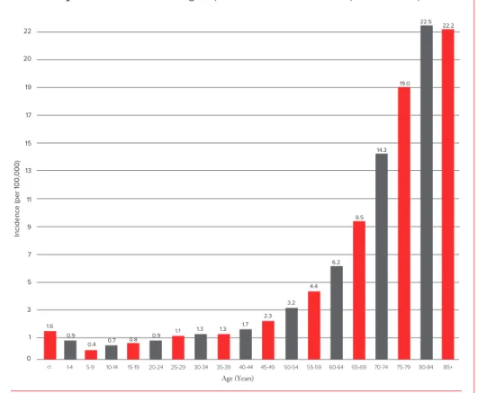
Incidence. AML is the most common acute leukemia affecting adults. Older people are more likely to develop AML than younger adults or children. However, AML is the most common type of leukemia diagnosed during infancy. About 15 to 20 percent of cases of acute childhood leukemia and 80 percent of cases of acute adult leukemia are AML.
The risk for developing AML increases about 10-fold from ages 30 to 34 years (about 1 case per 100,000 people) to ages 65 to 69 years (about 10 cases per 100,000 people). For people over 70, the incidence rate continues to increase, peaking between the ages of 80 and 84 (see Figure 1).
Acute Myeloid Leukemia:Age-Specific Incidence Rates (2004-2008)
Incidence (per 100,000) Age (Years) <1 1-4 5-9 10-14 15-19 20-24 25-29 30-34 35-39 40-44 45-49 50-54 55-59 60-64 65-69 70-74 75-79 80-84 85+ 11 13 15 17 19 20 22 9 7 5 3 1 0 0.4 0.8 1.1 1.3 1.3 2.3 3.2 4.4 6.2 9.5 14.3 22.5 19.0 22.2 0.9 0.9 1.6 0.7 1.7
Figure 1. I The horizontal axis shows 5-year age intervals. The vertical axis shows the frequency of new cases of AML per 100,000 people in a given age-group. Source: SEER Cancer Statistics Review, 1975-2008, National Cancer Institute. Bethesda, MD, www.seer.cancer.gov/csr/1975_2008/, based on November 2010 SEER data submission, posted to the SEER website, 2011.
Signs and Symptoms. A person with signs or symptoms that suggest the possibility of leukemia is usually referred to a specialist. This may be a hematologist or an oncologist. The doctor will order additional tests to make a diagnosis (see page 9). The signs and symptoms of AML are associated with a number of other, less serious diseases.
It is common for people with AML to feel a loss of well-being because of the underproduction of normal bone marrow cells. The person may tire more easily and have shortness of breath during normal physical activities.
People with AML may also have {
{ A pale complexion from anemia
{
{ Signs of bleeding caused by a very low platelet count, including
{ Black-and-blue marks or bruises occurring for no reason or because of a
minor injury
{ The appearance of pinhead-sized red spots on the skin, called “petechiae” { Prolonged bleeding from minor cuts
{
{ Mild fever {
{ Swollen gums
{
{ Frequent minor infections, such as perianal sores {
{ Loss of appetite and weight loss {
{ Discomfort in bones or joints {
{ Enlarged spleen {
{ Enlarged liver.
Bleeding. A low platelet count predisposes patients to bleeding. Bleeding in the
brain or lung is serious and can be fatal. However, such bleeding is usually preceded by minor bleeding, such as nose bleeds, blood in the urine or bruises (see Disease
and Treatment Side Effects on page 23).
Infection. Severe infection can occur at the time of diagnosis but becomes more
common and often more serious during treatment, when the bone marrow is completely suppressed. If the neutrophil count becomes or remains low because of AML or its treatment, serious infection almost invariably occurs and is a leading cause of death from AML (see Disease and Treatment Side Effects on page 23).
Myeloid Sarcoma. Rarely, a collection of AML cells, called a “myeloid sarcoma,”
forms outside the marrow. A myeloid sarcoma may occur in almost any part of the body. Other signs of AML may not appear in the blood and marrow until weeks or months after the initial myeloid sarcoma diagnosis. A myeloid sarcoma diagnosis is equivalent to a diagnosis of AML and is treated with chemotherapy rather than local therapy. Treatment may also include allogeneic or autologous stem cell transplant. Other names for a myeloid sarcoma are “chloroma,” “granulocytic sarcoma,” “myeloblastoma” or “monocytoma.”
Diagnosis
An accurate diagnosis of the type of leukemia is important. The exact diagnosis helps the doctor to
{
{ Estimate how the disease will progress {
{ Determine the appropriate treatment.
Talk to your doctor about
The diagnostic tests that are being done What the results mean
Getting copies of the test results.
Some of these tests may be repeated during and after therapy to measure the effects of treatment.
Blood and Bone Marrow Tests. Blood and bone marrow tests are used to
diagnose AML and the AML subtype. A change in the number and appearance of blood cells helps to make the diagnosis. AML cells look similar to normal immature white cells. However, their development is incomplete (see Figure 2).
Normal Marrow Cells and AML Blast Cells
Panel B
Panel A
Figure 2. I Panel A shows normal marrow cells seen through a microscope. The darker shapes are the nuclei of the cells. Some of the nuclei are circular and some are horseshoe shaped, reflecting the different developmental stages and the different types of cells. Panel B shows AML blast cells seen through a microscope. These cells are “arrested” in an early stage of development. The AML cells in panel B all have a similar appearance, in contrast to the varied appearance of the normal cells in panel A.
Blood and Marrow Samples. To do the tests, blood samples are generally taken
from a vein in the patient’s arm. Samples of marrow cells are obtained by bone marrow aspiration and biopsy (see page 36). The cells from the blood and marrow samples are examined under a microscope.
Most patients with AML have
I
Blood tests usedLower-than-expected
I
CBC – Blood cell counts are determined by ared cell and platelet counts blood test called a “complete blood count (CBC).
Too many immature white cells
I
Peripheral Blood Smear – A test called aand too few mature white cells “peripheral white cells blood smear” (an examination of the stained [dyed] blood cells with a microscope) usually shows the presence of leukemic blast cells (myeloblasts). These immature cells do not function like normal, mature white blood cells.
Confirmation of Diagnosis. In addition to looking at the number and appearance of the cells in the blood samples, your doctor will also order other tests to
{
{ Confirm the diagnosis {
{ Identify the AML subtype {
{ Develop a treatment plan.
Your doctor will work with a hematopathologist to confirm the diagnosis. A hematopathologist is a specialist who studies blood cell diseases by looking at samples of blood and marrow cells and other tissues. The diagnosis of AML is confirmed by identifying
{
{ Leukemic blast cells in bone marrow samples {
{ The percentage of blast cells. Blasts are normally 1 to 5 percent of marrow cells. Having at least 20 percent blasts is generally required for a diagnosis of AML. But AML can also be diagnosed if the blasts have a chromosome change that occurs in a specific type of AML, even if the blast percentage is less than 20 percent. {
{ Specific chemical activity in blast cells {
{ Characteristic markers (antigens) on the surface of blast cells, such as CD13 or CD33 (CD is an abbreviation for “cluster designation”).
{
{ Cells based on the types of markers (antigens) on the cell surface, a process called “immunophenotyping.” “Flow cytometry” is the name of one test that may be used to do immunophenotyping.
Other Tests. “Karyotyping” and “cytogenetic analysis” are processes used to
identify certain changes in chromosomes and genes. A laboratory test called “polymerase chain reaction (PCR)” may be done, in which cells in a sample of blood or marrow are studied to look for certain changes in the structure or function of genes, such as FLT3 and NPM1.
AML Subtypes. Most people who are diagnosed with AML have one of the eight AML subtypes shown in Table 1. This table is based on the French, American, British (FAB) classification system that is used by many doctors. Treatment is similar for most of these subtypes, with the exception of M3—acute promyelocytic leukemia (APL). Treatment for APL is described on page 20.
Cell Subtype
I
DescriptionMyeloblastic
I
M0 – minimally differentiated AMLMyeloblastic,
I
M1 – myeloblasts are the dominant leukemic cells inwith minimal maturation the marrow at the time of diagnosis.
Myeloblastic,
I
M2 – many myeloblasts, but some cells arewith maturation developing toward fully formed blood cells.
Promyelocytic
I
M3 – leukemic cells have a translocation betweenchromosomes 15 and 17.
Myelomonocytic
I
M4 – leukemic cells often have a translocation oran inversion of chromosome 16.
Monocytic
I
M5 – leukemic cells have features of developingmonocytes (white cells).
Erythroleukemic
I
M6 – leukemic cells have features of developingred cells.
Megakaryocytic
I
M7 – leukemic cells have features ofdeveloping platelets.
1Based on the FAB classification.
Table 1. I AML cells may have features of red cells, platelets or white cells (monocytes, eosinophils or, rarely, basophils or mast cells) in addition to myeloblasts or promyelocytes. When one cell line is dominant, the disease may be referred to as acute erythroid leukemia, acute megakaryocytic leukemia, acute monocytic leukemia and so forth.
The World Health Organization (WHO) classification system for AML is used by some doctors. It divides AML into several broad groups based on expected outcomes. The WHO classifications for AML include
{
{ AML with recurrent genetic abnormalities {
{ AML with myelodysplasia-related changes {
{ Therapy-related AML
{
{ AML not otherwise specified {
{ AML with a translocation between chromosomes 8 and 21
{
{ AML with a translocation or inversion in chromosome 16 {
{ AML with changes in chromosome 11
{
{ Acute promyelocytic leukemia (APL, M3), which usually has a translocation between chromosomes 15 and 17.
Treatment
A diagnosis of AML is associated with a wide range of outcomes.
Treatment Planning. A number of factors affect the choice and outcome of treatment, including
{
{ Your AML subtype
{
{ The results of cytogenetic analysis {
{ Whether you have received chemotherapy in the past to treat another type of cancer {
{ Whether you have had myelodysplastic syndrome (MDS) or another blood cancer {
{ Whether the AML is in your central nervous system {
{ Whether your AML has not responded to treatment or has relapsed {
{ The presence of systemic infection at diagnosis {
{ Your age and general health.
Changes to Chromosomes and Genes. A bone marrow examination of the
cytogenetic pattern and the status of the molecular markers, for example FLT3 and
NPM1, is important. Certain changes to the chromosomes and genes can provide
important information for treatment planning. Normal human cells contain 23 pairs of chromosomes (22 numbered pairs and either XX for female or XY for male). About 60 percent of people with AML have abnormal chromosomes (number and/or structure). In some cases of AML, the cells have chromosome changes that can be seen under a microscope. Not all chromosome changes can be seen under a microscope. Other laboratory tests may be used to detect chromosome changes. Common AML chromosome changes include trisomy 8, trisomy 21, monosomy 7, monosomy 21 and loss of an X or Y chromosome. Genetic changes may occur in patients with normal chromosomes, so it is important for your doctor to do a molecular analysis (see Table 2 on page 13).
Risk Group
I
Chromosomes1 (Cytogenetic Analysis)I
Genes (Molecular Analysis)Most favorable
I
8;21 translocation (M2 subtype)I
RUNX1-RUNX1T115;17 translocation (M3 subtype, APL)
I
PML-RARa (APL)16;16 translocation or inversion 16
I
CBF-ßMYH11(M4 subtype)
No chromosome changes
I
NPM1 or CEBPAmutation, without FLT3-ITD
Intermediate
I
No chromosome changes9;11 translocation
I
MLLT3-MLLOther nondefined chromosome
changes (fewer than 3 changes)
Trisomy 8 2, 3
Least favorable
I
Deletion of all or part of chromosomes 5 and 7b6;9 translocation
I
DEK-NUP214Inversion 3 or 3;3 translocation
I
RPN1-EVI1v;11q23 translocation
I
MLL-rearrangedMonosomy 5, del(5q), monosomy 7
3 or more chromosome changes without
one of the recurring translocations or inversions
No chromosome changes
I
FLT3-ITD with or withoutNPM1 mutation ERG and BAALC overexpression
1 Cytogenetic changes are sometimes abbreviated. For example: { A translocation may be written as t(8;21)
{ An inversion may be written as inv(16) { A deletion may be written as del(7) or -7
{ The letter “v” is an abbreviation used to indicate a variable chromosome. For example, an 11q23 translocation
sometimes involves genes other than MLL.
See pages 33 to 50 for definitions of terms.
2 Gene association not defined.
3 Trisomy 8 is equally distributed among risk subgroups and does not affect risk in the absence of genetic changes.
Impact of other genes such as IDH1, IDH2 and WT1 is under continued study.
Table 2. Some AML Risk Factors
Certain chromosome and gene abnormalities, alone or in combination, may affect a patient’s response to treatment. Researchers continue to work on developing better treatments for all patients.
High White Cell Count. About 5 percent of AML patients develop signs or
symptoms attributable to a very high blood blast cell count. A white cell count greater than 100,000 at the time of diagnosis is associated with unfavorable risk.
Fast Facts About AML Treatment {
{ For some patients, AML is curable with current therapies.
{
{ A person who has AML should (or must) be evaluated and treated by a
hematologist or an oncologist.
{
{ It is essential to seek treatment in a center where doctors are experienced in
the care of patients with acute leukemia.
{
{ Many patients with AML, particularly those with high white blood cell
counts, need treatment as soon as possible after diagnosis. The approach for treating each patient is based on an individual’s subtype, risk factors and treatment goals.
{
{ Achieving a remission is important because it is associated with prolonging
survival. The initial goal of treatment is usually to bring about a remission, in which
{
{ There is no evidence of leukemic blast cells in the blood or marrow.
{
{Normal blood cell production is restored and blood cell counts return to
normal levels.
{
{ Variations on standard approaches to treatment are undergoing intensive
study throughout the world. A patient may receive a different number of drugs, a different sequence of drugs, or drugs different from those described in this booklet, and still be receiving appropriate and effective treatment.
{
{ In most patients, intensive chemotherapy is required to achieve complete
remission. At least two drugs are combined to treat patients initially.
{
{ More treatment is needed once a remission is achieved to help prevent
a relapse.
{
{ Postremission treatment may consist of chemotherapy, stem cell
transplantation or low-dose maintenance chemotherapy.
{
{ If relapse occurs, treatment options may include different chemotherapy
regimens, allogeneic stem cell transplantation or other investigational therapies.
{
{ For older AML patients, age alone is not a contraindication to treatment.
Fit patients in their 70s and 80s can enter remission.
{
{ Patients who have the M3 subtype, acute promyelocytic leukemia (APL),
are treated with all-trans retinoic acid, arsenic trioxide and an anthracycline
Your treatment options and the results you can expect from treatment. It is important to be informed about the results you might expect with standard therapy and to discuss the possibility of participating in a clinical trial.
Talk to your doctor about
Chemotherapy. The initial phase of chemotherapy is called “induction therapy.” Induction may involve the simultaneous use of multiple drugs or a planned sequence of treatments. For most AML subtypes, patients are treated with an anthracycline, such as daunorubicin, doxorubicin or idarubicin, combined with cytarabine (also called “cytosine arabinoside” or “ara-C”). Other drugs may be added or substituted for higher-risk, refractory or relapsed patients. Autologous or allogeneic stem cell transplantation may be added to the treatment plan for patients with relapsed AML or patients at high risk of relapse after chemotherapy (see page 18).
The anthracycline and cytarabine act in different ways to stop AML cell growth and lead to AML cell death. The anthracycline is usually given in the first 3 days of treatment. Cytarabine is started at the same time but is given for 7 to 10 days of treatment. This treatment is also called “7 plus 3.” Both drugs are dissolved in fluids and given to the patient via an indwelling catheter (central line) or port. While “7 plus 3” is considered to be a standard, there are several clinical trials looking at ways to improve both the rate and duration of remission by adding specific molecularly-targeted drugs, increasing the doses of cytarabine and/or anthracyclines, or using a new drug that combines the cytarabine and anthracycline in a very specific ratio and delivers them together in an encapsulated form.
The central line is placed surgically in a vein in the upper chest. The catheter is tunneled under the skin of the chest so that it stays firmly in place. The external end of the port can be used to administer medications, fluids or blood products, or to withdraw blood samples for cell counts and chemical tests. See the free LLS booklet
Understanding Drug Therapy and Managing Side Effects for additional information
about drug administration.
Typically, the severity of the disease and the side effects of this initial therapy result in an initial hospital stay of 4 to 6 weeks. Some patients who live with a caregiver and near the medical facility may be safely discharged sooner. This depends on the policies of the treatment center and the status of the patient.
The goal of induction therapy is to rid the blood and marrow of visible leukemic blast cells. Generally, if blast cells are still evident after the first course of induction chemotherapy, a second course of the same chemotherapy is given. Table 3, on page 16, lists some of the standard drugs used to treat AML patients, as well as some of the drugs under study in AML clinical trials. Please check www.LLS.org or call our Information Specialists at (800) 955-4572 for updates to this information.
Table 3. Some Drugs Used to Treat Acute Myeloid Leukemia
Most antileukemic drugs interact with the cell’s genetic material (the DNA).
Anthracyclines (Antitumor Antibiotics) { { daunorubicin (Cerubidine®) { { doxorubicin (Adriamycin®) { { idarubicin (Idamycin®) { { mitoxantrone (Novantrone®) Antimetabolites {
{ cladribine (2-CdA; Leustatin®)
{
{ clofarabine (Clolar®)
{
{ cytarabine (cytosine arabinoside, ara-C; Cytosar-U®)
{ { fludarabine (Fludara®) { { hydroxyurea (Hydrea®) { { methotrexate { { 6-mercaptopurine (Purinethol®) {
{ 6-thioguanine (Thioguanine Tabloid®)
Topoisomerase Inhibitors {
{ etoposide (VP-16; VePesid®, Etopophos®)
{
{ topotecan (Hycamtin®)
DNA Damaging (Alkylating) Agents { { cyclophosphamide (Cytoxan®) { { carboplatin (Paraplatin®) { { temozolomide (Temodar®) Cell-Maturing Agents {
{ all-trans retinoic acid (ATRA, tretinoin; Vesanoid®)
{
{ arsenic trioxide (Trisenox®)
Hypomethylating Agents {
{ azacitidine (Vidaza®)
{
{ decitabine (Dacogen®)
Table 3. I This table lists some of the standard drugs and some of the drugs under study in clinical trials to treat AML patients. Various approaches to AML treatment are undergoing study in clinical trials. A patient may be treated with drugs that are not listed in this table and still be receiving appropriate and effective treatment. For a description of standard chemotherapy combinations, see page 15. It is essential to seek treatment in a center
Postremission Therapy. Normal blood cell production will return in many patients several weeks after initial treatment is completed. Blood cell counts
gradually approach normal, well-being returns and any remaining AML cells cannot be detected in blood or marrow. This is called a “remission.” A small number of residual AML cells will not interfere with normal blood cell development, but the number of cells has the potential to grow and cause a relapse of the AML. Postremission therapy, also called “consolidation therapy,” is needed to kill remaining AML cells and prevent relapse. Some of the main factors that influence the approach used include
{
{ Patient age {
{ Ability to tolerate intensive treatment {
{ Cytogenetic and molecular characteristics of the AML cells {
{ Availability of an HLA-matched related or unrelated stem cell donor.
AML postremission treatment consists of additional intensive chemotherapy after remission has been achieved, with or without autologous or allogeneic stem cell transplantation. Patients are hospitalized for postremission therapy. The length of stay varies depending on the treatment and other factors.
Patients who do not have a transplant generally are given four cycles of
chemotherapy. If chemotherapy alone is used, the best results occur if intensive treatment is applied. Intensive chemotherapy can be given with high dosages of cytarabine or other drugs.
Some patients may benefit from intensive chemotherapy alone followed by one of three types of stem cell transplantation:
{
{ Autologous
{
{ Allogeneic {
{ Reduced-intensity allogeneic (under study in clinical trials).
The question of which patients are likely to benefit from transplantation after their first complete remission is under study in clinical trials. Studies show that allogeneic stem cell transplantation may benefit poor- and intermediate-risk patients who are younger than 60 and have a sibling match. There does not seem to be any clear advantage for patients considered favorable or chemo-sensitive. Autologous transplant is being used in some centers as an alternative to multiple cycles of chemotherapy. Treating with a reduced-intensity transplant has shown some benefit for healthier older patients, up to age 75.
Clinical trials are examining several different approaches: modulating the activity of the immune system (e.g. vaccines or cytokines) or giving new drugs that differ from standard chemotherapy (e.g. tipifarnib [Zarnestra®], sorafenib [Nexavar®],
azacitidine [Vidaza®], lenalidomide [Revlimid®]). Various forms of less intensive maintenance treatment such as the role of hypomethylating agents or azacitidine and decitabine, after completion of postremission chemotherapy, are also under study in clinical trials.
Autologous Stem Cell Transplantation. Autologous transplantation is relatively
safe for many patients, including older patients. For some AML patients who do not have an HLA-matched stem cell donor, therapy can be further intensified with very-high-dose chemotherapy followed by an autologous transplant. This procedure uses the patient’s own stem cells to restore blood cell production after intensive chemotherapy.
Talk to your doctor about
The potential benefits and risks of this procedure.
For more information, see page 34 and the free LLS publication Blood and Marrow
Stem Cell Transplantation.
Allogeneic Stem Cell Transplantation. Allogeneic stem cell transplantation is
used to treat certain AML patients. It is a curative treatment option for some AML patients in first remission.
The upper age limit for transplantation varies by treatment center; many centers use age 60 or 65 years for allogeneic transplantation and 70 years for reduced-intensity allogeneic transplantation.
Patients in these age ranges who are in remission and have an HLA-matched stem cell donor may be candidates for this procedure. Umbilical cord blood, like bone marrow and peripheral blood, is a rich source of stem cells for transplantation. It is an alternative source for donor stem cells if an appropriate sibling or unrelated donor is not available.
Allogeneic transplantation is associated with a higher rate of side effects and mortality than autologous transplant. However, it may be considered for patients with worse-risk AML, based on cytogenetic and molecular test results. The decision to perform an allogeneic transplant also depends on the age of the patient and the patient’s (or his or her family’s) understanding of the potential benefits and risks. As one example, a younger patient with cytogenetic and molecular findings that are associated with a high probability of relapse would be a candidate for allogeneic stem cell transplantation early in treatment if he or she had a stem cell donor. Alternative therapies include intensive consolidation chemotherapy, reduced-intensity transplantation or autologous transplantation.
Reduced-Intensity Stem Cell Transplantation. Reduced-intensity allogeneic
ill to have an allogeneic stem cell transplant (based upon other medical conditions or general health status), if a suitable donor is available. The conditioning therapy used for a reduced-intensity transplant is of lower intensity than that for a standard stem cell transplant; it does not completely inactivate the patient’s immune system or treat the AML as intensively.
Reduced-intensity allogeneic stem cell transplantation is based on two considerations:
{
{ Much-improved immunosuppressive therapy prevents the patient from rejecting the donor’s stem cells, even though the patient’s immune system has not been fully suppressed by the lower-intensity conditioning therapy
{
{ The anticipated attack of the donor’s immune cells successfully suppresses the patient’s leukemia cells. This attack is referred to as "graft-versus-tumor effect" (graft-versus-leukemia effect or GVL). Over time, if the transplant is successful, the donor’s stem cells replace the patient’s immune cells. The engrafted donor immune cells recognize minor tissue antigens on the patient’s leukemia cells and continue to suppress their growth.
The risks and benefits of this treatment have not yet been clearly established. As is the case with allogeneic stem cell transplantation, the risk of graft-versus-host disease (GVHD) is an important consideration and a potentially disabling side effect.
Talk to your
doctor about Whether a reduced-intensity transplant is a potential option for you.
See the free LLS publications Blood and Marrow Stem Cell Transplantation and
Cord Blood Stem Cell Transplantation for comprehensive information about
allogeneic stem cell transplantation.
Central Nervous System (CNS) AML. CNS disease occurs in approximately 1 in 50 cases at the time of diagnosis. Preventive therapy is usually not indicated for CNS AML, but examination of the spinal fluid after remission should be considered for patients with
{
{ Monocytic subtypes
{
{ Masses of AML cells outside the marrow {
{ Inversion 16 and 8;21 translocation {
{ CD7- and CD56-positive (neural-cell adhesion molecule) immunophenotypes
{
{ Very high blood blast-cell counts at diagnosis.
Refractory Leukemia and Relapsed Leukemia. Most patients achieve an initial remission. However, some patients have residual leukemic cells in their marrow even
after intensive treatment. This is referred to as “refractory leukemia.” There are other patients who have a return of leukemia cells in the marrow and a decrease in normal blood cells after achieving a remission. This is referred to as “relapsed leukemia.” With refractory leukemia, approaches such as using drugs not used in the first course of treatment may be taken in an effort to induce remission. Stem cell transplantation may be used when remission is achieved, which may result in a more durable remission. In patients who relapse, the duration of the remission, the patient’s age and the cytogenetic findings in the leukemia cells influence the approach to therapy. Drugs similar to those administered initially, different drugs or stem cell transplantation may be used to treat the leukemia.
The Information Specialists at LLS offer guidance on how patients can work with their doctors to find out if a specific clinical trial is an appropriate treatment option. Information Specialists conduct clinical-trial searches for patients, family members and healthcare professionals. You can use the LLS-supported online tool TrialCheck® at www.LLS.org/clinicaltrials, a clinical-trial search service that offers patients and caregivers immediate access to listings of blood cancer clinical trials.
Transplantation in Relapsed Patients. Some form of allogeneic transplantation
may be recommended for patients in early first relapse or second remission. For patients who lack a sibling donor, matched-unrelated donor transplants can be effective, although this is a high-risk procedure. Patients with AML who relapse after allogeneic stem cell transplantation may have a long-term remission if they have a second transplant. Donor leukocyte infusion is sometimes used to treat patients with AML relapse after transplant. This therapy is most effective in early relapses and in the absence of extensive chronic graft-versus-host disease (GVHD).
Talk to your
doctor about Therapies under study in clinical trials if you have refractory or relapsed AML.
Several drugs and drug combinations that can be used to treat AML are being studied in clinical trials. For more information about specific clinical trials for relapsed and refractory leukemia, go to www.LLS.org/clinicaltrials or contact our Information Specialists.
Acute Promyelocytic Leukemia (APL) Treatment. APL is the M3 subtype of AML (see Table 1 on page 11). Patients with APL are among the most frequently cured. APL treatment differs from the other AML treatments described in this booklet. With APL, the cells that accumulate in the marrow can be identified as
promyelocytes, the step in blood cell formation that comes after the development of myeloblasts. These cells also have a specific chromosome abnormality involving chromosome 15, usually in conjunction with chromosome 17.
All-trans retinoic acid (ATRA), a vitamin A derivative, is a standard component of induction therapy for APL. ATRA is also known as tretinoin (Vesanoid®). Retinoic acid is capable of inducing the leukemic promyelocytes to develop into mature cells (neutrophils). It causes a marked decrease in the concentration of leukemic blast cells in the marrow, and a remission frequently follows.
Used alone, ATRA can induce a short-term remission in at least 80 percent of patients. Treatment with ATRA must be followed by or given with chemotherapy in order for the remission to be long-lasting. ATRA often minimizes the side effects of chemotherapy because blood cell counts may be improved and the number of leukemic cells may be decreased at the time that chemotherapy is started.
The remission rate of APL patients treated with ATRA and an anthracycline, such as idarubicin, is about 70 to 80 percent. Nevertheless, problems with hemorrhage during the initial phases of treatment, resistance to treatment and relapse occur in a proportion of patients, as they do in some patients with other types of AML. Therefore, long-term follow-up of patients in remission is required to identify those who are cured and those who may require further therapy.
For APL patients with a white cell count of 10,000/μL or greater at diagnosis, cytarabine may be added to induction or consolidation regimens.
The ideal duration of maintenance therapy is also being investigated. Currently, it consists of 2 years of 6-mercaptopurine (6-MP), methotrexate, and ATRA.
A small number of APL patients have persistent minimal residual disease (MRD) at the end of consolidation therapy. These patients may benefit from arsenic trioxide (Trisenox®), followed by allogeneic stem cell transplantation, if an HLA-matched donor is available.
Patients who do not have a donor, or cannot have an allogeneic stem cell transplant for other reasons, may be candidates for an autologous stem cell transplantation. Arsenic trioxide is approved to treat APL patients who have relapsed or are resistant to treatment with chemotherapy and ATRA.
See page 30 for an example of a treatment under study in clinical trials.
Acute Monocytic Leukemia Treatment. In some types of leukemia, including the subtype of monocytic leukemia (M5; see Table 1 on page 11), the leukemic blast cells sometimes invade the lining of the spinal cord or brain. This does not usually occur with other types of acute myeloid leukemia. When the lining of the spinal cord or brain is involved, chemotherapy is injected into the spinal fluid. A lumbar puncture (also known as a “spinal tap”) is a commonly used medical procedure, performed under local anesthesia or with heavy sedation. During a lumbar puncture, a needle is placed into the spinal canal and the spinal fluid is removed and examined for leukemia cells. The extracted fluid volume is then replaced with fluid containing appropriate drugs, usually cytarabine or methotrexate.
AML Treatment in Older Adults. Acute myeloid leukemia occurs more frequently with advancing age. At least half of patients are older than 65 years of age when the disease is diagnosed. Today there are curative options available for some older patients, including those who may have other significant health issues. For AML patients older than 60 years, patient performance status, other health issues and AML risk features are all considered in developing a treatment plan. Age alone is not a contraindication to treatment, and fit patients in their 70s and 80s can enter remission. Standardized measures of strength and reaction time are used to determine physiological age, which is a better indicator of tolerance for therapy. However, older patients may have a poorer response to therapy because
{
{ The leukemic cells of older AML patients have a higher occurrence of unfavorable cytogenetic and molecular abnormalities.
{
{ Older patients may have other medical problems (called “comorbidities”), including heart, lung or kidney disease or diabetes mellitus. The doctor may have to select less toxic AML drugs or decrease the dosage and frequency of treatment.
It is important to know that even in otherwise healthy patients aged 75 years or older, the principal cause of treatment failure is not toxicity, but failure of the treatment to eliminate the AML cells.
Treatment for older adults can be tailored to decreased tolerance if needed. Azacitidine (Vidaza®) and decitabine (Dacogen®) are low-intensity treatment options. Vidaza and Dacogen are approved to treat patients with certain types of myelodysplastic syndromes (MDS) and are being studied in clinical trials for the treatment of patients with AML. There are diverse clinical trials looking at novel drugs and combinations for the treatment of AML in the elderly. Examples include tipifarnib (Zarnestra®), CPX-351, bortezomib (Velcade®), lenalidomide (Revlimid®), clofarabine (Clolar®) and the combination of azacitidine (Vidaza®) or decitabine (Dacogen®) with other “gene-expression modifying” agents (entinostat, vorinostat [Zolinza®], valproic acid [Depakene®; Stavzor®]).
Occasionally, very elderly patients refuse treatment or are so ill from unrelated illnesses that treatment may be unreasonable.
Talk to your doctor about
Whether treatment in a clinical trial is right for you.
AML Treatment in Children. Most children who are diagnosed with leukemia have acute lymphoblastic (lymphocytic) leukemia. Acute myeloid leukemia accounts for about 15 to 20 percent of cases of acute childhood leukemia.
Children who have AML are treated with an induction therapy similar to that for adults with AML: cytarabine and drugs such as doxorubicin or daunomycin, or a third drug, such as mitoxantrone. This treatment is followed by a complex multidrug program that results in about an 80 percent remission rate and a nearly 50 percent 5-year, free remission rate. Slightly more than half of the children in relapse-free remission are considered cured. Infants are usually treated with the same therapy. Children less than 2 years of age who have AML have a decreased rate of remission and cure. In addition, the AML subtype acute monocytic leukemia (see page 11 and page 21) and a very-high-blast-count leukemia called “hyperleukocytic leukemia” are variants of AML that are much more difficult to treat, with lower remission and cure rates than the average results noted above.
Allogeneic stem cell transplantation (see page 18) may be used to treat children who have
{
{ Worse risk, based on cytogenetic and molecular test results {
{ Primary induction failure {
{ Relapse after intensive multidrug therapy.
Clinical Trials for Childhood AML. AML is one of the most challenging
childhood cancers to treat. Multi-institution clinical trials are under way to determine the best treatments for worse-risk patients. The expected outcomes for children who have AML with cytogenetic or molecular abnormalities may be different from those for adults who have the same abnormalities.
Chemotherapy has been used in different combinations and dosages over the past several decades, leading to improved childhood AML cure rates, but more research is needed to further improve cure rates and decrease the side effects and long-term and late effects of chemotherapy.
Researchers have identified cell targets that appear to be the key to treatment with the new generation of chemotherapy agents. These new targeted agents are being studied in conjunction with chemotherapy to examine their impact upon cure rates and their effect on toxic complications associated with traditional chemotherapy. Researchers are also studying risk factors and treatments for AML chemotherapy complications, especially infections, to make AML therapy safer for children. See the free LLS booklet Learning & Living with Cancer: Advocating for your child’s
educational needs for information about planning for the child’s entry or return to
school following diagnosis and treatment.
Disease and Treatment Side Effects. Most AML side effects are temporary and subside once the body adjusts to therapy or when therapy is completed. During the course of therapy and after therapy is completed, healthy new cells begin to grow and develop. Severe side effects are treated on an inpatient basis.
Low Blood Cell Counts. AML decreases the production of normal blood cells.
In addition, chemotherapy is toxic to both normal blood cells and AML cells. The normal blood cells are eliminated from the marrow along with AML cells. For the patient, this results in a severe deficiency in the
{
{ Red cells (anemia) {
{ Platelets (thrombocytopenia) {
{ White cells called “neutrophils” and “monocytes” (neutropenia and monocytopenia). Transfusion of red cells and platelets is almost always needed for a period of
several weeks during treatment. After that, the blood cell counts usually return toward normal.
Infection. During treatment for AML, the deficiency of neutrophils and
monocytes (types of white cells) can lead to infection from bacteria and fungi normally present in the environment, on the skin and in the nose, mouth or colon. The risk of infection may be increased because chemotherapy damages the lining of the mouth and intestines, making it easier for bacteria to enter the blood. When the white cell count is low and infection risk is increased, antibiotics are given to prevent or treat infection. Transfusion is not generally used for patients with a low neutrophil count, but can be used in patients with high fever, infection that is unresponsive to antibiotics, blood fungal infections or septic shock.
Growth factors may be given to the patient to stimulate the marrow to make new white cells. The growth factors used most frequently are G-CSF (granulocyte colony-stimulating factor; filgrastim [Neupogen®] and pegfilgrastim [Neulasta®]) and GM-CSF (granulocyte-macrophage colony-stimulating factor; sargramostim [Leukine®]). These agents are used in children only in special circumstances. Because the patient has an increased risk of developing an infection, the medical staff and family and friends need to practice frequent and vigorous hand washing and take other precautions to avoid exposing patients to bacteria, viruses and other infection-causing agents. Caregivers for patients with central lines or ports need to be meticulous in the cleaning of catheters.
Patients at home should not delay in seeking medical attention if any signs of infection develop. A rise in temperature to 101°F or higher, or the onset of chills, may be the only sign of infection in a patient with a very low white cell count. Other signs of infection may include persistent coughing; tenderness at a site prone to infection, such as the area surrounding the anus or the facial sinuses; sore throat; pain on urination; or frequent loose stools.
Other Side Effects. Chemotherapy affects tissues that normally have a high rate
of cell turnover. Thus, the lining of the mouth, the lining of the intestines, the skin and the hair follicles may be affected. Common side effects may include
{
{ Mouth ulcers
{
{ Diarrhea
{
{ Temporary hair loss {
{ Rashes
{
{ Nausea and vomiting
{
{ Fatigue.
Some AML patients may build up uric acid in their blood as a result of a very high white cell count. The use of chemotherapy may also increase uric acid, which is a chemical in the cell. Uric acid enters the blood and is excreted in the urine. If many cells are killed simultaneously by therapy, the amount of uric acid in the urine can be so high that kidney stones can form. This may seriously interfere with the flow of urine. Drugs such as allopurinol (Zyloprim®) or rasburicase (Elitek®) can be given to minimize the buildup of uric acid in the blood.
There are drugs and other supportive therapies to prevent or manage many side effects. For more information see the free LLS publications Blood Transfusion,
Cancer-Related Fatigue Facts and Understanding Drug Therapy and Managing Side Effects.
Sometimes, a drug or a drug combination causes effects that continue for a period of time after treatment ends. Some effects may be long-lasting (see Long-Term
Effects of Treatment on page 26). Talk to your
doctor about
Possible side effects and follow-up care.
Follow-up Care. Some of the tests that were done to diagnose AML may be repeated to
{
{ Follow the effects of treatment {
{ Make decisions about whether to continue, intensify, change or stop treatment. After treatment, patients who are in remission and have completed postremission therapy continue to be examined regularly by their doctors. Careful periodic
assessment of the patient’s health, blood cell counts and, if indicated, marrow is required. As time progresses, the length of time between assessments may grow, but assessments should continue indefinitely.
Long-Term Effects of Treatment. Children and young adults who have been
treated for AML may be at increased risk for heart damage, other cancers and neurologic or cognitive problems. Patients should be seen by a primary care physician for general health examinations at least once a year. They should also be examined regularly by an oncologist.
It is important to know about the potential for long-term effects of treatment so that any problems can be identified early and managed. Treatment for individuals who have AML sometimes causes effects that continue after treatment ends (long-term effects) or develop much later in life (late effects). Various factors can influence the risk of developing long-term or late effects, including
{
{ Type and duration of treatment {
{ Age at the time of treatment {
{ Gender and overall health.
Most AML patients are treated with an anthracycline, such as daunorubicin. Anthracyclines have been associated with increased risk for heart muscle injury or chronic heart failure. Heart disease may not become apparent until many years after therapy ends.
Stem cell transplantation is used to treat some patients with AML. It has been associated with long-term or late effects, including infertility, thyroid dysfunction, chronic fatigue and risk for developing a second cancer (lymphoma; melanoma of the skin; or cancer of the tongue and salivary glands, central nervous system, bone, soft tissue and thyroid gland). The number of patients who develop secondary cancers is small.
These and other possible long-term and late effects can be managed. For more information see the free LLS fact sheets Long-Term and Late Effects of Treatment for
Childhood Leukemia or Lymphoma and Long-Term and Late Effects of Treatment in Adults.
Talk to your doctor about
Treatment Outcomes. Patients with AML have a difficult disease to cure. However, a few decades ago almost no adults with AML were cured. Today, advances in AML treatment have resulted in improved remission and cure rates.
Terms for AML Treatment Outcomes
Active disease
I
AML is still present during treatment or aftertreatment (refractory) or AML has come back after treatment (relapsed).
A patient with AML that has relapsed has more than 5 percent blast cells present in the marrow.
Minimal residual disease
I
No AML cells are detected in bone marrowusing standard tests, such as looking at cells under a microscope. But more sensitive tests, such as flow cytometry, or very sensitive tests, such as polymerase chain reaction (PCR), detect remaining AML cells in the marrow.
Complete molecular remission
I
No evidence of AML cells in the marrow whenusing very sensitive tests such as PCR.
Remission
I
No evidence of disease after treatment,(complete based on remission) {
{ Less than 5 percent blast cells in
the marrow {
{ Blood cell counts within normal limits
{
{ No signs or symptoms of the disease.
Sensitive molecular techniques permit the identification of small amounts of cells (minimal residual disease [MRD]) that cannot be detected by standard tests of the patient’s blood and marrow. This approach can be used if the leukemia cells have a detectable molecular abnormality. This feature can permit more sensitive follow-up of patients who are in remission and can help determine whether additional treatment is necessary. It is worth noting that, after treatment, a finding that 1 to 5 percent of the white cells in a patient’s marrow are blast cells is not an indication of MRD. This percentage of blast cells may be found in persons who do not have leukemia. Age is one of the main determinants of AML cure rate. Children with the disease have a cure rate just below 50 percent. Younger adults and patients with certain cytogenetic patterns and with certain subtypes, such as APL, have a greater possibility of cure. Allogeneic stem cell transplantation can cure some patients.
Relative survival compares the survival rate of a person diagnosed with a disease to that of a person without the disease. Based on data posted to the SEER website in April 2011, the overall AML 5-year relative survival rates for 2001-2007, by age at diagnosis, are as follows:
{
{ Patients diagnosed with AML before age 65 have a 5-year relative survival rate of 39.6 percent
{
{ Children less than 15 years of age have a total averaged 5-year relative survival rate of 60.9 percent
{
{ Patients diagnosed at age 65 and older have an overall 5-year relative survival rate of 5.2 percent.
Figure 3 shows additional 5-year relative survival by age data. Note that these numbers do not take into account differences by gender, race and subtype of AML; the patient’s risk based on cytogenetic and molecular test results; or the most recent advances in therapy and supportive care.
Acute Myeloid Leukemia:5-Year Survival Rates (2001-2007)
AML 5-year survival rates
Age (Years) <45 45-54 55-64 65-74 >75 50% 40% 30% 20% 10% 0 53.9 35.4 21.4 10.1 1.8
Figure 3. I Source: SEER Cancer Statistics Review, National Cancer Institute. 2011.
For more information about survivorship, including follow-up care, contact our Information Specialists at LLS.
Research and Clinical Trials
The proportion of patients with AML who enter remission, stay in remission for years or are cured has increased during the last 30 years. However, AML is still one of the most difficult cancers to treat. The challenge remains to develop treatments that cure patients of all ages and with all subtypes of AML. LLS invests research funds in both basic and applied-research programs to improve the cure rate for AML patients.
Fast Facts About Clinical Trials {
{ Studies of new treatments in clinical trials are conducted under rigorous
guidelines to help doctors find out if new cancer treatments are safe and effective or better than the standard treatment.
{
{ Patients in cancer clinical trials usually receive either the study treatment
or the best standard treatment.
{
{ Clinical trials take place throughout the United States and Canada and
around the world.
{
{ Many of today’s standard treatments for cancer are based on earlier
clinical trials.
{
{ Taking part in a clinical trial may be the best treatment choice for some
AML patients.
{
{ There are some clinical trials for patients at every stage of treatment and for
patients in remission.
{
{ Our Information Specialists at LLS offer guidance on how patients
can work with their doctors to find out about specific clinical trials. This service can be accessed by calling (800) 955-4572 or visiting www.LLS.org/clinicaltrials.
{
{ To learn more about clinical trials, read the free LLS booklet
Understanding Clinical Trials for Blood Cancers and visit www.LLS.org.
Clinical Trials. Every new drug or treatment regimen goes through a series of studies called “clinical trials” before it becomes part of standard therapy. Clinical trials are carefully designed and rigorously reviewed by expert clinicians and
researchers to ensure as much safety and scientific accuracy as possible. Participation in a carefully conducted clinical trial may be the “best available” therapy.
LLS Information Specialists, at (800) 955-4572, can offer guidance on how patients can work with their doctors to determine if a specific clinical trial is an appropriate treatment option. Information Specialists will conduct individualized
clinical-trial searches for patients, family members and healthcare professionals. This service is also available at www.LLS.org/clinicaltrials
Research Approaches. There are clinical trials for newly diagnosed patients and patients with relapsed or refractory disease. A number of approaches are under study in clinical trials for the treatment of patients with AML, as follows:
{
{ A concept called “epigenetics” is based on the idea that certain genes become silenced (or turned off), which contributes to causing or maintaining cancer. Drugs that can reverse the silencing process are being studied in clinical trials, either alone or in combination with other drugs.
{
{One process that leads to gene silencing is called “methylation,” and there
are two drugs that inhibit the process: azacitidine (Vidaza®) and decitabine (Dacogen®).
{
{Another mechanism of gene silencing is called “histone deacetylase
inhibition.” Histone deacetylases attack silenced genes differently than methylation. Histone deacetylase inhibitors under study in clinical trials include valproic acid, suberoylanilide hydroxamic acid (SAHA) and entinostat. These drugs are being studied in combination with Vidaza or Dacogen.
{
{ There are novel drugs that kill cells by triggering new pathways that cause cell death and thereby overcome resistance. The novel drugs may be combined with standard AML drugs such as ara-C and daunorubicin. Some novel drug examples are clofarabine (Clolar®), which is approved to treat acute lymphoblastic leukemia; vosaroxin (which is being studied in combination with cytarabine for relapsed/refractory AML); tipifarnib (Zarnestra®); flavopiridol; interleukin-2 (IL-2) with histamine dihydrochloride (Ceplene®); and a class of drugs called “antisense molecules.” New drugs that target FLT-3-ITD include midostaurin, sorafenib (Nexavar®) and AC220. These drugs are being combined with other chemotherapy drugs. CPX-351 is a treatment currently being studied in newly diagnosed older adults and in relapsed/refractory adults.
{
{ Another concept called “differentiation therapy” involves studying the use of all-trans retinoic acid (ATRA), which is approved to treat APL, and some types of histone deacetylase inhibitor drugs to promote the growth and differentiation of immature leukemic blast cells.
{
{ Donor lymphocyte infusion after transplantation, vaccine therapy and other immunotherapies.
We encourage you to contact our Information Specialists and visit www.LLS.org for more information about specific treatments under study in clinical trials.
Normal Blood and Marrow
Blood is composed of plasma and cells suspended in plasma. Plasma is largely made up of water in which many chemicals are dissolved. These chemicals include
{
{ Proteins
{
{Albumin, the most common protein in blood
{
{Blood-clotting proteins, made by the liver {
{Erythropoietin, a protein made by the kidneys that stimulates red cell production {
{Immunoglobulins, antibodies made by plasma cells in response to infections
including those we develop from our vaccinations (such as poliovirus antibodies, which are made by normal plasma cells in the bone marrow) {
{ Hormones (such as thyroid hormone and cortisol) {
{ Minerals (such as iron and magnesium) {
{ Vitamins (such as folate and vitamin B 12) {
{ Electrolytes (such as calcium, potassium and sodium).
The cells suspended in plasma include red cells, platelets and white cells (neutrophils, monocytes, eosinophils, basophils and lymphocytes).
{
{ The red cells make up a little less than half the volume of the blood. They are filled with hemoglobin, the protein that picks up oxygen in the lungs and delivers it to the cells all around the body; hemoglobin then picks up carbon dioxide from the body’s cells and delivers it back to the lungs, where it is removed when we exhale.
{
{ The platelets are small cells (one-tenth the size of red cells) that help stop bleeding at the site of an injury in the body. For example, when a person has a cut, the vessels that carry blood are torn open. Platelets stick to the torn surface of the vessel, clump together and plug up the bleeding site with the help of blood-clotting proteins such as fibrin and electrolytes such as calcium. Later, a firm clot forms. The vessel wall then heals at the site of the clot and returns to its normal state.
{
{ The neutrophils and monocytes are white cells. They are called “phagocytes” (eating cells) because they can ingest bacteria or fungi and kill them. Unlike the red cells and platelets, the monocytes can leave the blood and enter the tissue, where they can attack the invading organisms and help combat infection. Eosinophils and basophils are types of white cells that respond to allergens or parasites.
{
{ Most lymphocytes, another type of white cell, are found in the lymph nodes, the spleen and the lymphatic channels, but some enter the blood. There are three major types of lymphocytes: T lymphocytes (T cells), B lymphocytes (B cells) and natural killer (NK) cells. Each of these cells is a key part of the immune system.