Ultrasound in the management of thoracic disease
Daniel A. Lichtenstein, MD
T
he lung has, step by step, found a place in the field of critical care and emergency ultrasound. The slow develop-ment of this discipline is not truly ex-plained, as the techniques themselves are quite simple. Similarly, the concept of using ultrasound as a clinical tool for the intensivist (with or without examining the lung) has also been surprisingly long to develop. Since 1989, the author has used an ultrasound machine comprising 1982 technology (built much before the recent explosion of technology that favors miniaturization) and has had the plea-sure to discover a tool permitting an ac-curate whole-body imaging approach to the critically ill. Although it is thought that the delay that occurred between 1982 and today will remain unexplained, this “sleepy giant” is now awake.Traditionally, although other complex tests and devices exist, the practicing in-tensivist has most commonly assessed lung function using physical examination and auscultation (these simple tools be-ing available since 1810) (1), radiography
(available since 1895) (2), or with com-puted tomography (CT) (available since 1972) (3). However, the flaws of these familiar tools are increasingly acknowl-edged. Auscultation’s low accuracy in the critically ill has recently been highlighted (4), and bedside radiography (typically obtained supine) has even greater limita-tions (4 –10). Even CT, which has con-tributed to saving countless lives, has some major drawbacks that may not be initially apparent. This is further dis-cussed in part 2 (page S253). Basically, the most unstable patients are the very patients who do not fully benefit from a CT scan. Ultrasound is a tool with at-tributes that have only recently begun to be appreciated by the greater medical com-munity, in distinction to its use for cardiac concerns (11). Previously, respected sources considered that the place for ultra-sound in assessing the lung was limited (12). Despite scientific evidence proving otherwise, this opinion has persisted (13). Only recently has this begun to change.
The scientific principles of ultrasound largely arise from the work of Langevin (1915), with additional contributions from Curie and Einstein early in the early 20th century. Utilizing this technology for medical purposes was proposed in 1946 (14) and has since been developed for more than half a century by other pioneers (Wild and Howry, 1951, and Henry and Griffith, 1974). Since these pioneers, ultrasound has become an in-dispensable and cost-effective medical
tool. Since the sentinel studies like the one by Joyner et al. (15) studying pleural effusion, the utility of thoracic ultra-sound was largely limited to this single diagnosis. Recent reviews of the state-of-the-art of lung investigations devoted lit-tle if any space to ultrasound (16, 17). Working in the team of François Jardin, who had pioneered the use of echocardi-ography with his ADR-4000, using a dou-ble working knowledge in intensive care and general ultrasound, and having to cope with critical situations in the heat of the nights, we had a privileged place to appreciate and develop hidden potentials of ultrasound. Although appreciating the countless advantages of applying general ultrasound for managing the critically ill at the bedside (18), we noted that there were many conditions for which lung ul-trasound proved immensely helpful. We thus undertook the challenging task to prove that the lung should be considered as much of a legitimate target as any other organ with respect to the use of bedside ultrasound.
The aim of this article will be to review and detail the scientific basis that sufficed to disprove the previously incorrect dogma surrounding the field of lung ul-trasound. This work relies on the analysis of the artifacts that air, in the tissues, pleural spaces, or within the lung itself, produces. Thus, the very substance that was previously thought to make lung ul-trasound impossible actually forms the basis of this science. These basic physical From Service de Réanimation Médicale, Hôpital
Ambroise-Paré, Faculté Paris-Ouest, Boulogne, France.
The author has not disclosed any potential con-flicts of interest.
For information regarding this article, E-mail: [email protected]
Copyright © 2007 by the Society of Critical Care Medicine and Lippincott Williams & Wilkins
DOI: 10.1097/01.CCM.0000260674.60761.85
Using simple and standardized semiology, the lung appears accessible to ultrasound, despite previous opinions otherwise. Lung ultrasound allows the intensivist to quickly answer to a majority of critical situations. Not only pleural effusion but also pneumothorax, alveolar consolidation, and interstitial syndrome will have accurate ultrasound equivalents, the recognition of which practically guides management. Combined with venous, cardiac, and abdominal examination, ultrasound investigation of this vital organ provides a transparent overview of the critically ill, a kind of stethoscope for a visual medicine. It is believed that by using this tool, the intensivist may more confidently manage
acute dyspnea and make emergency therapeutic decisions based on reproducible data. Further benefits include reduced require-ments for computed tomographic scans, therefore decreasing delay, irradiation, cost, and above all, discomfort to the patient. Thus, ultrasound of the lung can also be added to the classic armamentarium as a clinical tool for emergency use. (Crit Care Med 2007; 35[Suppl.]:S250–S261)
KEYWORDS: chest ultrasonography; lung; ultrasound diagnosis;
respiratory failure; intensive care unit; pneumothorax; alveolar consolidation; pleural effusion; pulmonary edema; chronic ob-structive pulmonary disease; interstitial syndrome
properties are described in part 1. Part 2 describes the comprehensive range of acute respiratory disorders amenable to diagnosis with ultrasound. In part 3, the daily applications of clinical lung ultra-sound are illustrated.
PART 1: ANALYSIS TECHNIQUE, REQUIRED MATERIAL, AND NORMAL PATTERN
Seven Principles of Lung Ultrasound. The concept of lung ultrasound can be based on seven principles (19).
1) A simple, unsophisticated ultrasound machine is perfectly adequate. 2) The thorax is an anatomic area where
air and water are intimately mixed. From these interactions arise the ar-tifacts. In addition, air and water have opposite gravitational dynamics (air rises, water descends). It is thus cru-cial to define “dependent disorders” that are water-rich, such as pleural effusions and alveolar consolidation, and “nondependent disorders” that are air-rich, such as pneumothorax or the interstitial syndrome. One may then refer to a sky– earth axis. 3) All lung patterns arise from the
pleu-ral line.
4) Lung ultrasound is largely based on the analysis of artifacts.
5) Lung patterns are largely dynamic. A ret-rospective analysis of static images does not provide for an adequate analysis. 6) The majority of acute lung disorders
abut the lung surface, thus explaining the wide-ranging feasibility of lung ul-trasound.
7) As the lung surface is extensive (about 1500 cm2), constituting the most
vo-luminous organ, precise areas should be defined, as is the norm for the abdominal examination. One may ask where to put the probe. The answer is simple: at the same places as the stethoscope.
Choice of the Ultrasound Unit: A Crit-ical Step.For performing both lung and whole-body ultrasound, we think that simplicity can be favored. The required image resolution has been fully satisfac-tory since 1991. We wrote our first text-book in 1992 using only ADR-4000 fig-ures, which were already sufficient to illustrate the developing field of lung ul-trasound by using a technology from 1982. We think the gray-scale analogic
resolution available since 1991 (that we currently use) is more than adequate to perform abdominal, cardiac, venous, and craniofacial (optic nerve) applications. We rarely find this quality in digital sys-tems. Our unit has ideal dimensions for hospital use (30 ⫻38 cm footprint), be-ing easily portable from bed to bed and from floor to floor. A bigger unit would be a hindrance, but even smaller units could become paradoxically more cum-bersome than our basic model once they become affixed to a cart. Without such a cart, although it may be at risk for theft, but more importantly, the actual ultra-sound unit might be placed on the pa-tient’s bed, constituting an infection con-trol hazard. Further questions are where to put contact product, disinfectants, and interventional materials? It was maybe unnecessary to await for the current de-velopment of ultraminiature units to pro-vide a whole-body assessment of the crit-ically ill. The ultrasound unit we use has exactly, in the updated versions, the same internal properties (notably image reso-lution) as in its original 1991 version. We think that the various Doppler functions are not truly required to assess the lungs or the venous system (20, 21) or for an adequate hemodynamic assessment. Us-ing a sUs-ingle probe without Doppler capa-bilities simplifies a whole-body diagnostic approach for the head, heart, and venous diseases and their evaluation (22). We thus think that a Doppler evaluation should be incorporated only if the sim-pler examination does not answer the clinical question. The unnecessary reli-ance on Doppler capability produces drawbacks, such as decreased spatial res-olution, and increases the complexity of the unit and the approach, increases costs, is maybe not fully harmless (23– 25), and may add physical volume to the
bedside unit. Our preferred microconvex 5-MHz probe permits a full evaluation of the abdomen, small hard-to-access parts (such as the apex of the lungs), compres-sion of the subclavian vein or adductor area for the femoral veins, and allows all manner of interventional procedures (Fig. 1). We also avoid linear probes (be-cause human beings are not linear), deep structures are not explored, and com-pression maneuvers of deep veins are hard to achieve using linear probes.
The need to avoid cross-contamina-tion of the critically ill and to limit nos-ocomial infections is rarely adequately discussed in current practice, yet respect-ing basic principles and practices is vital to prevent needless morbidity and mor-tality. The use of more than one probe in the same examination should raise con-cerns regarding the ability to maintain a sterile technique. Harsh disinfectants should be avoided because they can grad-ually damage the probe head. The flat keyboard we use can be cleaned, whereas keyboards full of prominent buttons can-not—a point of prime importance in the intensive care unit (ICU). Our device can also be immediately switched on, which is not the case of most digital units. Fi-nally, the coupling gel, an unpleasant part of ultrasound since its advent, can be avoided with a new non-gel coupler soon available. This will greatly enhance the comfort of ultrasound for both physicians and patients.
Ultrasound Examination of the Nor-mal Lung. Lung ultrasound is a recent field of study, and a rigorous approach is required to produce consistent results. To obtain the best from the examination, the operator should simply follow the seven principles sequentially.
A brief review of the normal pattern is useful. As air rises and water descends, Figure 1.Stage 1.Left, the probe is gently applied on the anterior chest wall in a supine patient (at the earth level). This defines stage 1. Note the microconvex shape of the probe—a basic requirement. Stage 2 includes zone 1 and zone 2.Right, extension of the examination to stage 3.
the position of the patient should be spec-ified. What is dependent in one position is no longer dependent in another. One should define a gravitational, earth–sky axis and specify the area where the probe is applied. The thorax should be scanned directly, avoiding the traditional abdom-inal route, which can lead to erroneous diagnoses. Exclusive longitudinal scans are desirable, and thus, we think that a linear probe will be a hindrance for this purpose. The operator must have access to superficial and deep areas with only one probe. Here again, a linear probe will be a hindrance.
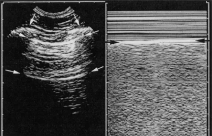
As the lung is the most voluminous organ of the body, a careful and method-ical examination comprising three basic steps is desirable. First the thorax has to be located (in the craniocaudal axis), then the lung surface located, and then zones have to be defined. The thorax is distin-guished from the abdomen by locating the diaphragm, which is a basic land-mark. Once the probe is applied to the thorax, lung sonography will largely con-sist of the analysis of artifacts because only artifacts appear on the screen (Fig. 2). However, the upper and lower ribs can already be identified, casting a frank pos-terior shadow. Between two ribs and typ-ically 0.5 cm deeper (in the adult), a roughly horizontal, hyperechoic line pro-duced by the pleural interface is visible. The pleural line indicates the parietopul-monary interface (i.e., the lung surface). The ribs and the pleural line outline a characteristic pattern, the bat sign (Fig. 2). The bat sign, visible only in
longitu-dinal scan, should be recognized first in any lung examination and considered a mandatory first sign to acquire. Like a G key in a musical partition, it is a perma-nent landmark of the lung surface.
Precise areas of interest will be defined using clinical landmarks. The anterior and posterior axillary lines are practical landmarks that delineate anterior, lateral, and posterior areas. Each of these areas can be divided into smaller areas. These anatomic areas are considered in our ap-proach to lung sonography, which incor-porates four clinical stages of investiga-tion. Stage 1 is defined by examining the anterior chest wall in a supine patient (zone 1) at the earth level (i.e., our daily conditions of work, under the gravity rules) and is immediately informative re-garding pneumothorax, interstitial syn-drome, or atelectasis such as can result from right mainstem intubation. Stage 1 primedefines this same examination per-formed when the patient is half-supine (as small pneumothoraces move toward the apex). This does not regard trauma-tized patients. In stage 2, the lateral chest wall (zone 2) is added to the anterior zone, until the bed physically prevents further lateral placement of the probe. Stage 2 gives information on substantial pleural effusions, substantial alveolar consolidations, and phrenic nerve func-tion. A stage 3 examination is performed by slightly moving the ipsilateral shoul-der of the supine patient to position the probe as posterior as possible without moving the back (in the case of trauma) and to gain a few centimeters of
sono-graphic exploration of the posterior lung fields (zone 3). As the probe is required to “point to the sky” to perform this exam-ination, small probes are mandatory. Small pleural effusions (beginning hemo-thorax for instance) and small alveolar consolidations, not detected by stage 1 and 2 examinations, may be thus de-tected. In a stage 4 (exhaustive analysis in nontrauma patient), the patient is posi-tioned laterally, or seated, to study fully the posterior chest wall. In addition, the apex is investigated. To optimally compare the ca-pabilities of lung ultrasound with CT, full stage 4 examinations should be the re-quired standard, yet in most cases, stages 1, 2, or 3 answer the clinical questions.
At the pleural line, two important dy-namic and static signs can be described. First is the dynamic normal sign of lung sliding. This is the basic sign of normal-ity. Lung sliding is a kind of dynamic twinkling movement visible at the pleural line and synchronized with respiration. It corresponds to the displacement of the lung along the craniocaudal axis. For ob-jectifying and documenting lung sliding, M mode yields a simple pattern, the sea-shore sign (Fig. 2). With these simple signs, the use of Doppler is not required. Much could be written about lung slid-ing. Basically, the 2.5-MHz probes equip-ping many echocardiographic-Doppler units usually have insufficient image res-olution. Modern units also have dynamic noise filters or persistence filters. These filters, designed to provide a flattering image, can render lung sliding hard or impossible to detect and must be by-passed. Lung sliding is a relative move-ment alongside the superficial chest tissues, which are motionless. The ampli-tude of lung sliding is maximal at the bases. Very discrete lung sliding should carefully be sought, as any degree of lung sliding has the same meaning (all-or-nothing rule). Further, lung sliding can be detected even with mechanical venti-lation, morbid obesity, advanced age, or lung emphysema (even with giant bul-lae). It should be noted, however, that in a dyspneic patient, muscular contrac-tions can make lung-sliding analysis dif-ficult, unless the seashore sign is sought. The second sign is the normal static sign. Air artifacts normally arise from the pleural line. In general, two diametrically opposed types can be described: either horizontal or vertical. Several clinically relevant kinds of artifacts exist, and a practical alphabetic classification is re-quired to avoid long descriptions (19). Figure 2.Normal lung pattern.Left, longitudinal scan of an intercostal space. Only artifacts (ribs and
air) are visible. However, between two ribs (vertical arrows), strictly half a centimeter below in the adult, the pleural line is located (upper horizontal arrows). Upper rib, pleural line, and lower rib outline the bat sign. The horizontal lines (lower horizontal arrows) that arise from the pleural line have clinical applications (the A lines).Right, seashore sign (M mode). A flagrant difference in pattern appears on either side of the pleural line (arrows). The motionless superficial layers generate horizontal lines—the waves. The deep artifacts follow the lung sliding, hence the sandy pattern.
The basic normal sign is a horizontal repetition of the pleural line recurring at regular intervals, called ultrasound A-line sign (Fig. 2). The B line and some other artifacts will be further described in the section on pathologic conditions. Other artifacts (C, I, J, N, O, S lines . . .) and other subtle signs will not be further de-tailed here.
The normal lung pattern combines lung sliding with a predominance or to-tality of A lines. In a ventilated patient without respiratory concerns, the cupolas are usually located one or two spaces below the mamillary line. They move to-ward the abdomen at inspiration, with an amplitude of around 10 –15 mm.
PART 2: ULTRASOUND SEMIOLOGY AND CLINICAL APPLICATIONS OF THE MAIN ACUTE LUNG DISORDERS
According to the second principle of lung ultrasound, the image and artifact patterns produced are a function of the air/fluid ratios. Pleural effusion contains pure liquid. Alveolar consolidation con-tains mainly liquid and very little air. Interstitial syndrome contains mainly air and little liquid. Pneumothorax contains pure air.
Pleural Effusion
Fluid pleural effusion is a disorder containing exclusively fluid and no air. Although detecting this entity with ultra-sound was imagined in 1946 (14) and assessed in 1967 (15), this simple appli-cation is not fully exploited in all institu-tions.
Maybe this application was not exten-sively exploited because radiologists have easy access to CT. Whereas pleural effu-sion can be obvious in echogenic pa-tients, it needs standardized diagnostic criteria in others. Using some not well-known signs, ultrasound accuracy proves nearly as efficient as that of CT (26). Both tests have better accuracy than the su-pine chest radiograph (4).
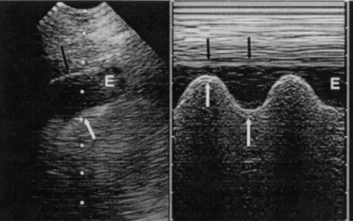
Signs.Customarily, a pleural effusion is detected during abdominal examina-tions, using a subcostal approach. We do not use this traditional access. We find it safer to analyze the pleura directly through the intercostal spaces with a short probe. The effusion should first be sought in a stage 2 examination (i.e., lat-erally in a supine patient) at bed level. Substantial effusions are immediately vis-ible. If no effusion is visible, and if more information is required, the examiner may proceed to a stage 3 (posterior chest) examination to detect minimal effusions. The classic anechoic pattern is not a per-fect criterion, although it can be nondi-agnostic in critical cases. Apart from the obvious diagnostic criteria of a dependent fluid image located above the diaphragm, it is possible to add two more subtle signs, one static and one dynamic, that will greatly help in the difficult cases (Fig. 3). One static sign is the sharp sign. A pleural effusion is limited by four reg-ular borders forming the shape of a sharp. These borders consist of the pleu-ral line, from where it arises, the upper and lower shadows of the ribs, and the deep border, which is always regular. This border is assumed to be the visceral pleura and was called the lung line. The
dynamic sign is the sinusoid sign. It shows the respiratory variation of the in-terpleural distance with inspiratory de-crease (Fig. 4). The sinusoid sign indi-cates the centrifugal shifting of the lung toward the wall during inspiration. As the lung moves toward a “core–surface” axis, the pattern, on M mode, is a sinusoid. The sharp and sinusoid signs confirm the presence of pleural effusion with a spec-ificity of 97% when the gold standard used is withdrawal of pleural fluid (26).
With CT as a gold standard, sensitivity and specificity of ultrasound are 93% (4). Minimal effusions can be detected using ultrasound, provided the probe is applied over the adequate area of the chest. Ex-tremely small effusions are not detected using CT, raising the problem of the per-tinent gold standard. An aerated lung lobe will float over the effusion. A consol-idated lobe will swim within the effusion (the jellyfish sign).
Clinical Applications. The rapid bed-side diagnosis of pleural effusions has ob-vious diagnostic and therapeutic implica-tions for the critically ill. Mattison et al. (27) described a prevalence of 62% in medical ICUs, with 41% of effusions be-ing present at admission. Ultrasound is superior to radiography in all respects. Ultrasound will detect the effusion, eval-uate its volume, provide information on its nature, and indicate the appropriate area for a thoracentesis, with better ac-curacy than radiography. Bedside radiog-raphy rarely detects small effusions and can also miss effusions of up to 525 mL (28). It can also prompt false-positive di-agnoses. Ultrasound is acknowledged as the method of choice to detect an
effu-Figure 3.Pleural effusion in an intercostal ap-proach. Note one basic static sign, the sharp sign, as the effusion (E) is outlined by four regular borders: pleural line, shadow of ribs, visceral pleura. Note that the lung at this area is not consolidated, as air artifacts are visible.
Figure 4.Pleural effusion, a basic dynamic sign, the sinusoid sign. The image provided to theleft(as in Fig. 3) is not specific to pleural effusion, can be difficult to see in poorly echoic patients, and can be echoic. In addition, it does not provide any information about its viscosity. The image atright(M mode) highlights the sinusoid sign, a sign specific to liquid pleural effusion, and indicates a low viscosity.E, expiration.
sion in a supine patient (29). In our ob-servations, one third of ultrasound-visible and easy-to-puncture effusions in ventilated patients remained occult to su-pine bedside radiography (26). Bedside radiography does not provide reliable in-formation on the volume of an effusion. We have no special ultrasound technique for measuring the exact volume either, estimating one effusion as between 500 and 1000 mL and another as between 15 and 30 mL. We think these approxima-tions are sufficient in clinical practice. Other approaches are available (30). However, ultrasound provides informa-tion about the nature of the pleural effu-sion, data that we do not expect from radiography. The main causes of pleural effusions in the ICU are heart failure (35%), atelectasis (23%), pneumonia (11%), and empyema (1% of cases) (27). Theoretically, a transudate is anechoic, an exudate echoic. A liquid with mobile particles (plankton sign) or septa is sug-gestive of exudate, hemothorax, or puru-lent pleurisy and should be aspirated and formally analyzed (see Fig. 1 on p. S263). When faced with an anechoic effusion, we believe that it is prudent to perform ul-trasound-assisted thoracentesis whenever knowledge of the nature of the effusion might improve the prognosis. This very simple procedure may make long discus-sions of differential diagnoses irrelevant. Diagnostic or therapeutic thoracente-sis is not often routinely performed on a critically ill, ventilated patient because of concerns regarding the risks. We think that with practice, care, and ultrasound guidance, thoracentesis can become rou-tine in this situation. With ultrasound detection, even radio-occult effusions in ventilated patients may be safely aspi-rated. The principle is based on the visual approach rules (26). Precise and repro-ducible criteria are mandatory. Briefly, one must check for an inspiratory en-largement of the interpleural space of
ⱖ15 mm, with effusion visible at the ad-jacent upper and lower intercostal spaces. The patient can remain in the supine position in half of the cases. One checks for the absence of respiratory interposi-tion of a vital organ (lung, heart, liver, spleen). The sinusoid sign has the at-tribute of clearly indicating low viscosity of the pleural fluid, in other words, the possibility of using a fine needle to min-imize procedural trauma. Thoracentesis should be done immediately after ultra-sound localization, with the patient re-maining in the same position. A clinical
landmark made in the radiology depart-ment and followed by an aspiration once the patient is back in the ICU seems in-adequate. Skinfolds can displace the cu-taneous landmark. All these precautions are easy to follow, and in our experience, the success rate in ventilated patients is 97% (26). Using these criteria is like driv-ing a car: with open eyes and an attentive brain behind, the risk of an accident is low, whereas the converse is true as well. Complications such as a pneumothorax vary between rarely (31) and nil (26). Typically,⬍10 secs are needed to obtain a liquid sample in ⬎88% of cases. With-drawal of pleural fluid should improve the ventilatory mechanics (32) and assist weaning from the ventilator, among other benefits.
Alveolar Consolidation
Alveolar consolidation contains mainly fluid and little air. This daily con-cern in the ICU is not always accurately detected by bedside radiography. Auscul-tation is sometimes superior to radiogra-phy (4). These limitations may necessi-tate use of a CT scan. However, 98.5% of cases of alveolar consolidation abut the pleura (33), a mandatory condition for its ultrasound detection. Whereas the loca-tion of pleural effusion, pneumothorax, or interstitial syndrome are rather stan-dardized, the location of an alveolar con-solidation varies with pathogenesis. Alve-olar consolidation is usually dependent, thus being demonstrated by a stage 3 examination, often lateral, thus being demonstrated with a stage 2 examination, and sometimes anterior, being detectable with a stage 1 examination. Those cases amenable to expedient diagnosis with the simple stage 1 examination roughly cor-respond to the middle and upper lobes. It should be noted that a subcostal ap-proach often yields ghost artifacts of the liver or spleen (mirror artifacts through the diaphragm). This is why, among other reasons, we do not use this route. Detecting alveolar consolidation is not a new application for ultrasound (14, 34). However, despite these previous descrip-tions, ultrasound has been seldom used for this purpose in general.
Signs.Using basic but rigorous termi-nology to define alveolar consolidation, we found a sensitivity of 90% and a spec-ificity of 98% using ultrasound corrobo-rated by CT as the gold standard (33). Apart from some obvious criteria (image located in the thorax, that is, above the
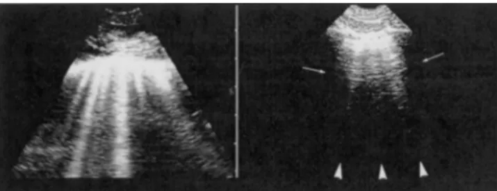
diaphragm, image arising from the pleu-ral line or from an associated pleupleu-ral ef-fusion, tissue-like pattern, reminiscent of the liver), two specific criteria of interest can be defined (Fig. 5). Analogous to the critical criteria for pleural effusion, there is both an important static and dynamic criterion. The static criterion states that an alveolar consolidation usually has ir-regular deep boundaries. The superficial boundary is the pleural line or the deep boundary of a pleural effusion, if present. The deep boundary is irregular, as in con-nection with the aerated lung, a pattern therefore different from the lung line. Only when the whole lobe is involved will the deep boundary be regular. The dy-namic criterion requires an absence of any dynamic sinusoidal component, thus excluding pleural effusion as a cause. In the case of alveolar consolidation, cranio-caudal inspiratory movement is present or even impaired (in the most severe cases), but no inspiratory centrifugal shift (i.e., from the bottom to the top of the screen) should occur in the core– surface axis. This is mandatory for distin-guishing alveolar consolidation from pleural effusion, which are potentially as-sociated but distinct entities and diag-noses.
Many subtle findings can be described using ultrasound. The volume can be as-sessed. Abscesses or necrotizing areas within the consolidation can be detected (see Fig. 2 on p. S263). Hyperechoic punctiform or linear images are possibly present and indicate air bronchograms (34). These air bronchograms can be mo-tionless or have an intrinsic inspiratory centrifugal movement, called dynamic air bronchogram, as opposed as static air bronchograms. The dynamic air bron-chogram allows distinction between non-Figure 5. Massive alveolar consolidation of the lower lobe. Note the air bronchograms (dynamic in dynamic acquisition). Note the homogeneous pattern, indicative of noncomplicated pneumonia (compare with Fig. 2 on p. S263).
retractile (pneumonia) and retractile (at-electasis) consolidations with 100% specificity for diagnosing nonretractile ones (35, 36). The absence of satellite lung rockets is suggestive of aspiration pneumonia. Lung sliding is frequently abolished.
Late-stage atelectasis yields alveolar consolidation with static air bron-chograms, shift of neighboring organs, pinching of intercostal spaces, and aboli-tion of lung sliding. The lung pulse is a sign available early after single-lung in-tubation, when the lung is still aerated, as in the seconds immediately after intuba-tion. The lung pulse is a vibration visible at the pleural line, in rhythm with the heart beat, clearly visible because lung sliding is abolished, and objectified with M mode. The heart vibrations are usually dominated by the lung expansion. With marked atelectasis, abolition of lung slid-ing allows the heart beat to be observed more readily. The lung pulse had a sen-sitivity of 90% for the diagnosis of one-lung intubation in one study (37).
Why Use Ultrasound?The value of ul-trasound follows from the inadequacy of radiography (4 –10). Radiography gives a rough summation of consolidation, pleu-ral effusion, and abscesses, whereas ultra-sound accurately distinguishes each dis-order. Ultrasound can have diagnostic (immediate diagnosis of pneumonia in a patient with fever, pain, and normal ra-diograph, for instance), monitoring (pro-gression of acute respiratory distress syn-drome, indication for prone positioning, positive end-expiratory pressure setting), or even therapeutic effect (see
“Point-of-Care Ultrasound: Infection Control in the Intensive Care Unit” in this supplement).
Interstitial Syndrome
Despite being described back in 1994 (38) and confirmed since 1997 (39), this is an area of bedside diagnosis that will be new to many practicing clinicians. Using this approach provides information that is not provided on a bedside chest radio-graph and that has no auscultatory equiv-alent when using the stethoscope. Inter-stitial syndrome seen in the critically ill is mostly due to thickening of interlobular septa, which generate Kerley lines, and ground-glass areas, which are visible on CT scans. The major causes are cardio-genic pulmonary edema and infectious processes. In the interstitial syndrome, predominant air components are mingled with a minimal amount of fluid. The ul-trasound study of an aerated organ, with diagnoses based exclusively on the anal-ysis of artifacts, requires the examiner to think in an abstract manner. We will see how to diagnose interstitial syndrome and, above all, why.
Signs of the Interstitial Syndrome. The sign is a vertical artifact (a comet-tail artifact) with special features. It arises from the pleural line, is a well-defined and laser-like beam, is dominant (it erases the A lines), spreads up without fading to the edge of the screen, and is synchronous with lung sliding. This arti-fact, as described, has been calledB line. Several B lines visible in a single view are reminiscent of a rocket at liftoff and have been termed “lung rockets,” or B⫹lines (Fig. 6). Diffuse lung rockets dissemi-nated all over the anterolateral wall de-fine diffuse interstitial syndrome. The test is defined as negative when such B lines are absent, isolated, or exclusively
confined to the last intercostal space above the diaphragm, a variant observed in 27% of healthy subjects (39). Diffuse lung rockets have a sensitivity and spec-ificity of 93% for the diagnosis of inter-stitial syndrome when compared with ra-diography, and the concordance is complete when the gold standard is CT (39). A separation of the artifacts of about 7 mm indicates thickening of the inter-lobular septa (B7 lines), whereas a sepa-ration of 3⫾1 mm (B3 lines) is corre-lated with ground-glass lesions (39). One or two B lines visible in a single view are dubbed b lines (lower case) and seem to have no pathologic meaning.
The B line must critically be distin-guished from two other artifacts: the E and the Z lines (Fig. 7). E lines (E for subcutaneous emphysema) are long but do not arise from the pleural line. The Z lines arise from the pleural line like the B lines, but four features allow easy distinc-tion. They are ill-defined, quickly vanish, are independent from lung sliding, and do not erase the A lines. Z lines are very frequent, visible in 80% of patients (40). They should be considered as a parasite artifact devoid of clinical meaning (40). This generates an important basic rule. Lung artifacts have the characteristic fea-ture that A lines and B lines cannot be visible at the same location. Lung arti-facts areeitherA lines orB lines.
What is the structure detected by ultra-sound? The B lines are generated by ele-ments with a high acoustic impedance gra-dient from the surrounding structures, such as fluid surrounded by air (water is an excellent transmitter, whereas air im-pedes ultrasound). The detected elements are smaller than the resolution of ultra-sound. They are present at and all over the lung surface. They are separated from each other byⱕ7 mm. They are present Figure 6. Interstitial syndrome. These vertical
comet-tail artifacts have the specific peculiarities of strictly arising from the pleural line, being well-defined and laser-like, moving with the lung sliding, spreading to the edge of the screen with-out fading, and erasing normal A lines. This pat-tern defines B lines. Several B lines in a single view define lung rockets. Diffuse lung rockets indicate interstitial syndrome. This patient has cardiogenic pulmonary edema.
Figure 7.Some artifacts: E and Z lines.Left, these well-defined comet tails descend to the edge of the screen. However, the bat sign is absent (as with Fig. 6). This pattern cannot be due to B lines. The patient has subcutaneous emphysema with extensive collections of gas between anatomic struc-tures—a condition generating E lines.Right, the ill-defined comet-tail artifacts (three visible here,
arrowheads) arise from the pleural line but do not erase the physiologic A lines (arrows) and quickly vanish without reaching the edge of the screen. These are Z lines.
in pulmonary edema, but labile, resolving on its treatment. All these features (and some others) are characteristic of thick-ened subpleural interlobular septa, which perfectly fulfill this description. CT cor-relation has proven that B lines corre-spond to thickened interlobular septa. A normal septum has a width of 300 m and cannot be seen using ultrasound. The thickened septum has a width of 700m, a size that remains under the power of ultrasound but allows generation of the artifact. Ultrasound B lines are thus an ultrasound equivalent of the familiar Ker-ley B lines (41). The superficial septa alone can be detected using ultrasound. They are indicative of the deeper septal thickening. Acute interstitial syndrome is generally diffuse, especially from cardio-genic cause. This explains why the diag-nosis is immediate, the moment the probe is applied to the chest wall.
Why Use Ultrasound?Initially, the in-tensivist may question the relevance of detecting interstitial syndrome (using ul-trasound or any other method). Devoid of stethacoustic or radiologic signs allowing diagnosis, the intensivist has likely be-come accustomed to practicing without this information. This does not discount the fact that this application of ultra-sound and this information may have an immediate effect on the critically ill. The recognition of diffuse interstitial syndrome in emergency situations is virtually equiv-alent to diagnosing acute pulmonary edema (cardiogenic or permeability re-lated). Detecting B lines rules out pneu-mothorax (42). In a dyspneic patient, de-tecting lung rockets allows immediate differentiation between cardiogenic pul-monary edema and exacerbation of chronic obstructive pulmonary disease. Only a few seconds are required, and a permanent digital record may be ob-tained of the examination; something that is impossible with simple ausculta-tion. The sensitivity of the ultrasound detection of pulmonary edema in this set-ting is 100% and specificity is 92% (43). Lung rockets are unusual in pulmonary embolism. Their absence is found with a 92% sensitivity (44). Other uses such as distinction between lesional and cardio-genic pulmonary edema, morphologic analysis of acute respiratory distress syn-drome, qualitative assessment of the oc-clusion pressure, and measuring lung fluid or lung compliance are under inves-tigation.
Pneumothorax
Pneumothoraces contain pure air and no fluid. Can ultrasound detect air (a classic foe to ultrasound) within an air-containing area? Numerous studies have now conclusively proven the answer to be yes, provided one more step is made to-ward abstraction and provided that arti-facts are accepted as providing clinical information. This indication for immedi-ate bedside diagnosis has a marked ad-vantage in both an accuracy and timeli-ness to that of radiography, especially in the supine patient. After adequate train-ing, any intensivist will be able to rule out pneumothorax in a few seconds and will need⬍1 min to rule it in.
Pneumothoraces remain common in the critically ill, from initial traumatic injuries, iatrogenic procedures, or ac-quired from illness or from barotrauma. They may quickly be life threatening (45). Bedside radiography misses a large per-centage of cases (9, 46 – 48), even tension pneumothoraces (49); thus, this situation often requires CT for confirmation, time permitting. Bedside radiographs, even when they show the pneumothorax, are a poor indicator of its volume. However, CT cannot be routinely used for this indica-tion either. The excessive use of CT will lead to over-irradiation and increased costs and will subject patients to the risks of medical transport, whereas the serious consequences may occur if pneumotho-rax is overlooked. We believe that using ultrasound is an extremely simple way to resolve this quandary.
Signs. Pneumothorax semiology may appear abstract, as it refers exclusively to
artifact analysis. It may also appear com-plex, as several signs have to be investi-gated. However, after minimal training in acknowledged centers, the signs are per-fectly reproducible. Pneumothorax is a “nondependent” semiotic. It should be sought first at the anterior and lower area, as 98% of significant pneumothora-ces are at least anterior and inferior in supine patients (50). This easy-to-investi-gate location is fortuitous. Many signs are available, three covering the majority of situations. All our studies have been per-formed with CT as a gold standard.
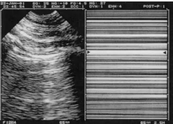
Lung sliding should be sought in area 1. Absent lung sliding is a basic and ini-tial step for the diagnosis, which was ac-tually first described in horses (51). Lung sliding allows pneumothorax to be confi-dently discounted, in a matter of seconds, because the negative predictive value is 100% for the diagnosis of pneumothorax (52). With a pneumothorax, a striking absence of motion arising from the pleu-ral line is observed instead of the familiar lung sliding. Sensitivity is 100% (when nonfeasible cases are not considered). The abolition of lung sliding can be ob-jectified in M mode, which gives a char-acteristic pattern, the stratosphere sign (Fig. 8). One can use Doppler, but it is not essential. One can also use a linear probe, but our microconvex probe, com-bined with our black-and-white technol-ogy, consistently permits a full investiga-tion of this area (and of the deep lung and whole body).
The role of ultrasound for detecting abolition of lung sliding is increasingly being described (53, 54). However, absent
Figure 8. Pneumothorax and stratosphere sign. The complete abolition of lung sliding can be objectified using M mode (right). Exclusively horizontal lines are displayed, indicating complete absence of dynamics at the level of, and below, the pleural line (arrowheads), a pattern called the stratosphere sign. Note the absence of a B line atleft.
lung sliding is extremely frequent in crit-ically ill patients. Specificity, which is 91% in a general population (52), falls to 78% when patients are all critically ill (55) and occurs in up to 60% when acute respiratory distress syndrome patients are studied. In dyspneic patients seen in the emergency room, abolished lung slid-ing has a positive predictive value of only 27% for the diagnosis of pneumothorax, (unpublished data). Thus, absent lung sliding does not mean pneumothorax. Countless other situations yield abolished lung sliding: jet or high-frequency venti-lation, massive atelectasis (including one-lung intubation), acute pleural sym-physis (inflammatory adherences), severe fibrosis, phrenic nerve palsy, cardiopul-monary arrest, simple apnea, and inap-propriate operator technique (abusive use of persistence filter, inappropriate probe, etc.). Paradoxically, lung sliding is most often abolished precisely in the patient who both is at maximal risk for pneumo-thorax and who will not tolerate it phys-iologically. We should reiterate that ab-sent lung sliding is not specific to pneumothorax. By combining the assess-ment of lung sliding with other signs, however, the effectiveness of ultrasound is improved.
From the pleural line in stage 1 arise exclusively horizontal artifacts, A lines (Fig. 8). No B line is visible, a pattern called the A-line sign. It should be noted, however, that Z lines are very frequently visible. We believe that the use of linear probes usually prevents correct recogni-tion and distincrecogni-tion of B, Z, and A lines, a serious concern. In fact, this distinction relies on deep analysis, which linear probes usually do not achieve, whereas the probe we use adequately studies su-perficial and deep areas. The A-line sign, which is 100% sensitive for the diagnosis of complete pneumothorax, is in no case specific. Specificity is 60% (42). What matters is that the slightest B line allows prompt ruling out of pneumothorax (42). As we saw that B lines arise from the lung alone, this finding is logical. This is pre-cious information in numerous cases in which lung sliding is absent.
The lung point is a specific sign that allows pneumothorax to be confirmed and that confidently indicates those pa-tients who will benefit from chest tube placement in an extreme emergency. When a profile suggestive of pneumotho-rax (A lines with absent lung sliding) is detected on stage 1, the probe gradually moves to the lateral areas, until it finds a
fleeting, sudden inspiratory visualization, of either lung sliding or B lines, in an area where abolished lung sliding and exclusive A lines were previously re-corded. This is an all-or-nothing law, cor-responding to whether the lung is in con-tact with the chest wall (Fig. 9). The specificity of the lung point is 100%. Its overall sensitivity is 66% and falls with major pneumothoraces with complete lung retraction (55). Interestingly, sensi-tivity for occult pneumothorax is high: 79% of pneumothoraces not visible on bedside radiographs are definitely diag-nosed using ultrasound (40). Once again, ultrasound seems to be more accurate than bedside radiography.
The lung point is a critical sign be-cause it confirms that the abolition of lung sliding is real and not due to tech-nical defaults. In addition, the lung point provides indication about the pneumo-thorax volume and evolution if not treated. A lateral lung point was corre-lated with a 90% need for drainage vs. 8% with anterior lung point (40). Briefly, an anterior lung point indicates moderate pneumothorax (generally radio-occult), whereas a very posterior or absent lung point characterizes massive pneumotho-races with complete retraction.
Some Applications.The clinical appli-cations are multiple. Ultrasound has proven superior to radiography (55). Ul-trasound can thus complete or replace radiography and decrease use of CT. The recognition of pneumothorax in an emer-gency is the main application. This has long been feasible in prehospital
medi-cine (56). We believe that in extreme emergencies, ultrasound will replace ra-diography. Drainage, previously done blindly in most unstable patients, can be done confidently using visual approach. The other basic application is ruling out pneumothorax in a few seconds when managing acute dyspnea or cardiac ar-rest, after any chest procedure (subcla-vian catheterism, thoracentesis), or even routinely in a ventilated patient. For such applications, a bulky device can do more harm than good. An elegant application is the possibility of monitoring a pneumo-thorax based on ultrasound alone. All in all, the need for repeated radiographic studies, which thicken the medical file, increase the hospital budget, and contin-uously irradiate the patient, will be de-creased.
Changes in the volume of a small and especially occult pneumothorax can be monitored if the intensivist chooses to manage the patient conservatively but not insert a tube. This logic can be pushed to its extreme when radiographs are wholly un-desirable (pregnancy, children). Concerns about irradiation, especially in pediatric pa-tients, are being increasingly discussed (57–59). Thus, it is questionable whether using CT scanning to follow conservatively managed pneumothoraces offers a good balance between therapeutic benefit and ir-radiation (60).
Airway Control
The dynamic real-time nature of lung ultrasound allows the immediate diagno-Figure 9.Lung point. On theright(time-motion), a sudden change is visible at the precise location where the collapsed lung, subject to a slight increase in volume during inspiration, reaches the wall. The “sandy” pattern generated by lung sliding instantaneously replaces a pattern formed by horizontal lines (arrow).
sis of complete atelectasis, such as occurs immediately after one-lung intubation. The detection of absent lung sliding and the sole presence of a lung pulse has 90% sensitivity for the diagnosis of immediate complete atelectasis after one-lung intu-bation (37). From the diagnosis of one-lung intubation, absence of one-lung pulse allows check radiography to be postponed after intubation. The ability of ultrasound to help such procedures is beginning to be appreciated (61). Other ultrasound signs have also been described that may assist in safely managing the airway. When the patient is correctly intubated, both cupolas of the diaphragm should have the same amplitude. When right mainstem intubation occurs, the left cu-pola remains motionless, whereas the right one has an abnormally increased amplitude, often⬎15–20 mm with usual tidal volumes. The position of the endo-tracheal tube within the upper trachea gives a characteristic pattern using subtle to-and-fro movements of the tube (⬍1 mm to avoid mucosal damage). Visual perception is sufficient (and M mode can objectify it, yielding a variant of the sea-shore sign). Doppler is also not required for these diagnostic adjuncts. Ultrasound is also useful for guiding a percutaneous (or surgical) tracheostomy by precisely defining the anatomic structures that should be avoided (62).
Other Applications
Multiple other disorders can be de-tected using ultrasound, providing a scope for this modality limited only by the imagination of the clinician. The am-plitude of lung sliding is informative re-garding correct lung compliance, pres-ence of pleural symphysis, or as seen, massive atelectasis. Markedly diminished lung compliance is frequent in acute re-spiratory distress syndrome or massive pneumonia, and ultrasound yields a dy-namic pattern that no other test can de-tect, namely, abolition of lung sliding. The analysis of the interstitial syndrome, areas of ground-glass, consolidated lung areas, and pleural effusions will help in distinguishing cardiogenic from perme-ability-related pulmonary edema. In pul-monary embolism, a normal pattern (“A” profile) in a dyspneic patient is expected. This is the equivalent of the traditionally normal chest radiograph. In a patient with acute respiratory distress and no history of asthma or chronic obstructive pulmonary disease, this is immediately
suggestive of a pulmonary embolism. We have found a sensitivity of 92% for de-tecting the A profile as opposed to the B profile, and this accuracy increases to 100% if only the B3 profile is considered (44). Some authors have further de-scribed pulmonary infarction (63), a sign that we rarely observe, perhaps because the patients seen by the intensivist have severe pulmonary embolism, a setting in which pulmonary infarction has little time to develop. Lung abscess is also ac-cessible to ultrasound (see “Point-of-Care Ultrasound: Infection Control in the In-tensive Care Unit” in this supplement).
Countless disorders can also be found in the mediastinum. An experienced user can often avoid immediate referrals to invasive or time-consuming techniques. Aortic aneurysm or dissection of the tho-racic aorta can often be detected using our probe that has a small footprint. Both simple and complex conditions such as mediastinitis, tracheal stenosis, or accu-mulation of secretions above an ET tube are nicely documented in many cases (22). Bedside ultrasound can be concep-tualized as comprised of both diagnostic and interventional attributes. For diagno-sis, several signs such as the swirl sign, plankton sign, and lung pulse improve ultrasound accuracy in the diagnosis of conditions like pneumothorax and pleu-ral effusion. Interventional ultrasound plays a major part in managing condi-tions such as pneumothorax, lung ab-scess, and pleural effusions The dia-phragm is also amenable to study by ultrasound. The location, amplitude and direction of movement, and degree of in-spiratory thickening are all easily as-sessed by ultrasound, and ultrasound alone. Even a ruptured diaphragm will be better documented on ultrasound than on CT.
PART 3: CLINICAL
CONSIDERATIONS ARISING FROM THE USE OF LUNG ULTRASOUND
Interesting applications are accessible by combining the potentials described above. To respect the word count, we will see the role of a simple black-and-white unit when compared with radiography and CT, we will investigate a dyspneic patient, define who is interested (which patients, which operators?), and appreciate strong and weak points of the method.
Lung Ultrasound: Answer to the Tra-ditional Quandary of Radiography or CT
in the ICU.CT is often requested by cli-nicians due to the poor accuracy of bed-side radiography (10). Although CT re-mains an invaluable diagnostic tool in critical care medicine, it is currently time-consuming and requires patient transport. Scientific analysis of the po-tential of lung ultrasound shows that it has an intermediate role between CT scanning and radiography. Ultrasound in general has a near 90 –100% accuracy, depending on the application (4, 40). Al-though CT has major advantages of pro-viding detailed, relatively easy to inter-pret images, a good regional overview, and that its use has saved many lives (17), it does have significant drawbacks (22). Because the main acute chest disorders can be assessed using ultrasound, the question arises as to whether it is re-quired to transport such critically ill pa-tients to a CT scan. Apart from the need for transportation, the time spent (typi-cally 1 hr, all in all, even if the image acquisition is done in 10 secs), and the drawbacks of iodine injection, one partic-ular disadvantage should be borne in mind: the irradiation. One CT scan cre-ates as much irradiation as ⱖ100 chest radiographs, an increasing concern in young women and children (57–59). The high cost of CT is also not an insignifi-cant issue; in daily practice, how many patients on Earth have access to this method? Finally, we think that the ultra-sound detection of occult pregnancy should be routine and can take a few seconds. Positive findings may further in-fluence decision making regarding fur-ther ultrasound studies and radiographic imaging.
In the future, bedside radiography may become redundant; its indications should gradually decrease. Overall, the slight inferiority of ultrasound com-pared with CT in some applications can be balanced with others for which ul-trasound clearly seems to be superior. This regards spatial resolution, which al-lows detection of septations within pleu-ral effusion (which are never seen in a CT scan) or necrotizing areas in consolida-tions (64). Also, CT is unable to detect all real-time dynamic signs such as lung sliding, dynamic air bronchogram, phrenic dynamics, among others. By mingling the points of slight inferiority with those of slight superiority, one can envisage ultrasound as a credible alternative to chest CT.
Approach to a Dyspneic Patient. As pulmonary edema, chronic obstructive
pulmonary disease, pneumothorax, pul-monary embolism, and pneumonia yield particular patterns, these signs can be invaluable at the bedside of a dyspneic patient. Currently, when investigating acute dyspnea with the usual tools, incor-rect initial diagnoses are frequent (65). The idea of performing lung ultrasound here may seem peculiar. However, if an ultrasound unit is readily available, the ultrasound data will complete or correct both the clinical and radiologic findings that are often nondiagnostic or even mis-leading. In emergencies, ultrasound data obtained using a simple device without Doppler have enabled the physician to give a correct etiological diagnosis in 85% of cases, whereas the traditional ap-proach (clinical examination comple-mented by laboratory tests and chest ra-diography) was accurate in only 51% of cases (65). Timeliness may be life saving, however, and any delay that waiting for a ultrasound machine creates cannot be ac-cepted. Therefore, the intensivist should have mastered the ultrasound profiles of simple lung, two-dimensional cardiac, and venous pathology (22). The best ex-ample is the challenge of chronic ob-structive pulmonary disease vs. pulmo-nary edema.
Obviously, traditional information can and should be combined with the ultra-sound findings to take the best of each. Physical examination and thorough re-view of the radiographic studies (if formed) remain necessary if time per-mits. We believe, however, that with increased experience, the intensivist will come to increasingly rely on ultrasound in the sickest patients.
Some reminders will be useful to ap-preciate the place of lung ultrasound.
A Field to Be Defined: For Whom? Although the critically ill patient in an ICU will be the first to benefit from lung ultrasound, these benefits also extend to the emergency room (66), trauma room, and even the prehospital environment. Lung ultrasound has proven its feasibility in remote areas (56). We believe that once these benefits become apparent, pa-tients in cardiology, pneumology, pedia-try of course, anesthesiology, chest sur-gery, and even internal or family medicine will benefit from these tech-niques.
A Field to Be Defined: By Whom? Lung ultrasound is an opportunity for those who practice intensive care medi-cine. As no discipline has legitimately claimed, studied, or adopted this field, it
will rightly belong to those who first ded-icate themselves and prove benefit to their patients. Intensivists are in close physical contact with the patient, and ap-propriate training should enable us to master the technique (67). Clearly, on-site availability of an ultrasound device within the ICU will simplify patient man-agement. We also think that although medicolegal issues must be considered in many practices, when the patient’s life is in danger, medicolegal issues should be relegated to a secondary concern.
Lung Ultrasound: A Space for Simplic-ity.An issue that likely slowed the recog-nition and acceptance of general ultra-sound is the perception that this is a difficult exercise. As regards lung ultra-sound, paradoxically, we think this is a marked misconception. Appreciating lung sliding or lung rockets has an ex-tremely short learning curve (4, 33, 67). B lines and the stratosphere sign are es-sentially the most simple signs one can imagine in ultrasound (or even in medi-cine). This focus on simplicity can also be applied to the equipment required. The unsophisticated equipment we describe adequately covers whole-body applica-tions. Doppler functions are unnecessary. A cardiac analysis in a dyspneic patient can be reduced to assessing left ventricle contractility alone (or to no analysis in some cases), a major advantage for phy-sicians unfamiliar with this discipline. Lung ultrasound feasibility is 98% in our observations (68).
Versatility: An Access to the Neighbor-ing Organs. Although the focus of this article, assessing the lung is only a single first step in assessing the critically ill (22). Combining cardiac and lung ultra-sound results in the thorax being consid-ered as a whole. Unexpected diagnoses will be made in the abdominal (pneumo-peritoneum, mesenteric infarction, etc), cephalic (maxillary sinusitis, intracranial hypertension), and venous areas. With the same system, interventional ultra-sound can be liberally performed (22).
The hemodynamic control of an un-stable patient is a classic in the ICU. Many tools (too many?) are available. Some ap-plications (under submission) of lung ul-trasound will help the physician in im-mediate decisions in this field.
Harmlessness, Cost Savings. Irradia-tion is an increasing concern in the ra-diologic literature. Its deleterious side ef-fects in the child and the young woman are now acknowledged. Lung ultrasound is an elegant way to circumvent this
is-sue. The indication for radiographs and CT scans should progressively decrease. This decrease should yield cost savings. Costs should decrease both due to re-duced immediate complications (such as pneumothorax due to thoracentesis) and from remote ones (such as neoplasia as a consequence of irradiation). In addition, it has been shown that onesingle appli-cation allows reimbursement of the unit we describe in⬍3 yrs (37).
Limitations of Lung Ultrasound. A comprehensive understanding of the lim-itations of ultrasound is required to make ultrasound the safe and high-precision tool it is. Prudent operators will promptly recognize a limitation and rely on the traditional diagnostic tools that sufficed in the past. Hindrances to ultrasound can be organic or artificial. Organic obstacles can be innate (a poorly echoic patient) or acquired (subcutaneous emphysema, pleural calcifications, obese body habi-tus). Artificial obstacles, mostly dressings and tubes, can be limited by smart policy and generally dealt with by the attending clinician who is not afraid to redress a wound after the ultrasound assessment. Intraparenchymal lesions (pneumatocele, deep abscess, rare cases of central consol-idations) will escape surface ultrasound. Paradoxically, obesity is not a major hin-drance to lung ultrasound. Operator skill is a familiar limitation. Insufficient train-ing will result in avoidable pitfalls. Non-longitudinal scans, incorrect or loosely held probes, use of the subcostal route (which can create ghost artifacts mimick-ing effusions or consolidations), disre-gard of the sky– earth axis in terms of searching for pleural effusion in a non-dependent area, abusive use of the dy-namic noise filter, and incorrect loca-tion of the diaphragm will all result in errors that should be decreased with adequate training.
Each application has its limitations. Pleural effusion, if loculated, will not yield the sinusoid sign, and then the di-agnostic criteria for liquid will depend on the operator’s judgment. The dark ultra-sound lung is a rare pattern in which none of the numerous discriminative signs is available. It usually corresponds to a white radiologic lung and is most often due to massive pleural effusion. Here, CT can be useful. B lines alone do not discriminate between acute and chronic interstitial syndrome. Subphrenic fat can mimic alveolar consolidation if care was not taken to locate the cupola. Lastly, confusing B, Z, and E lines can occur. In
cases of subcutaneous emphysema, which is not always associated to pneumothorax, experienced users will sometimes identify conserved lung sliding (22). Subcutaneous emphysema can generate E lines, a com-mon pitfall for the beginner who risks con-fusion with B lines. Posterior pneumotho-races will escape anterior analysis but are accompanied by suggestive anterior pat-terns (22). In dyspneic patients, lung slid-ing is sometimes hard to detect because of muscular contractions, but an answer is generally forthcoming with careful study. Major dyspnea with intense muscular effort is rare in spontaneous pneumothorax and in that occurring in a ventilated patient. A lung point is not always present, but clearly, a patient who displays chest pain, dyspnea, tympanism, A lines, and absent lung sliding after an invasive procedure is likely the victim of a pneumothorax.
Training in Lung Ultrasound: Issue or Strong Point?Clinical medicine can only be mastered by dedicating years to careful medical study. Likewise, critical care ul-trasound, which is a discipline unto itself, cannot be learned in a few hours. We think the future of lung ultrasound train-ing should be the responsibility of the university, which should involve students as early as possible for maximal societal benefit. The practicing intensivist who desires to save lives more easily will see that training in lung and emergency ul-trasound is paradoxically easier to learn than standard general ultrasound (4, 33, 67). There is nothing in common be-tween the recognition of a B line (a few minutes of training) and a fetal malfor-mation. In practice, the best way for mas-tering lung ultrasound is to spend a pe-riod as resident or fellow in the ICUs regularly practicing lung ultrasound.
The aim of our training center is to give to the operator the keys for using ultrasound alone for diagnosis and for performing therapeutic actions. With practice, life-saving drainage of compres-sive pleurisy or pneumothorax or correct management of an acute dyspnea should become routine.
CONCLUSIONS
Lung ultrasound constitutes a visual medicine and provides a transparent ap-proach to the acutely ill, guiding man-agement and care. Although the use of this modality has been largely neglected by the critical care community, its value to patients is being increasingly demon-strated. Provided minor limitations are
accepted, lung ultrasound seems to have only advantages; noninvasive, immedi-ately implemented, highly feasible, easy to execute, and versatile (from bedside to aircraft, from head to feet), it provides diagnoses with an accuracy superior to that of radiography and is time saving in a dyspneic patient. Substantial cost sav-ings are possible, global irradiation de-creases, and patients’ comfort increases. Everyone wins, there is no loser. Scien-tific considerations aside, we like to highlight again this basic advantage: sim-plicity. Sometimes answering with dis-concerting ease questions for which only sophisticated approaches were hitherto indicated, ultrasound elegantly simplifies daily problems encountered in extreme emergencies (69). Symbolizing for some the stethoscope of tomorrow, ultrasound is actually a genuine stethoscope of today if we consider the etymology: scopein (to observe) and stethos (the chest wall).
ACKNOWLEDGMENTS
I thank, one more time, but never enough, Enrico Storti and Luca Neri for their outstanding achievement in this small revolution and also for a priceless gift, their friendship; and Alan Sustic, who will also find a devoted place in this section.
REFERENCES
1. Laënnec RTH: Traité de l’auscultation médi-ate, ou traité du diagnostic des maladies des poumons et du coeur. New York, Hafner, 1962; Paris, JA Brosson and JS Claudé, 1819 2. Williams FH: The Roentgen Rays in Medicine
and Surgery. New York, Macmillan, 1901 3. Hounsfield GN: Computerized transverse
ax-ial scanning.Br J Radiol1973; 46:1016 –1022 4. Lichtenstein D, Goldstein I, Mourgeon E, et al: Comparative diagnostic performances of auscultation, chest radiography and lung ul-trasonography in acute respiratory distress syndrome.Anesthesiology2004; 100:9 –15 5. Greenbaum DM, Marschall KE: The value of
routine daily chest X-rays in intubated pa-tients in the medical intensive care unit.Crit Care Med1982; 10:29 –30
6. Janower ML, Jennas-Nocera Z, Mukai J: Util-ity and efficacy of portable chest radiographs.
AJR Am J Roentgenol1984; 142:265–267 7. Peruzzi W, Garner W, Bools J, et al: Portable
chest roentgenography and CT in critically ill patients.Chest1988; 93:722–726
8. Wiener MD, Garay SM, Leitman BS, et al: Imaging of the intensive care unit patient.
Clin Chest Med1991; 12:169 –198
9. Tocino IM, Miller MH, Fairfax WR: Distribu-tion of pneumothorax in the supine and
semi-recumbent critically ill adult.AJR Am J Roentgenol1985; 144:901–905
10. Ivatury RR, Sugerman HJ: Chest radiograph or computed tomography in the intensive care unit?Crit Care Med2000; 28:1033–1039 11. Jardin F, Dubourg O: L’exploration échocar-diographique en médecine d’urgence. Paris, Masson, 1986, pp 3–154
12. Friedman PJ: Diagnostic procedures in respi-ratory diseases.In:Harrison’s Principles of Internal Medicine. 12th Edition. New York, McGraw-Hill, 1992, p 1043
13. Weinberger SE, Drazen JM: Diagnostic pro-cedures in respiratory diseases. In: Harri-son’s Principles of Internal Medicine. 16th Edition. New York, McGraw-Hill, 2005, pp 1505–1508
14. Dénier A: Les ultrasons, leur application au diagnostic.Presse Méd1946; 22:307–308 15. Joyner CR, Herman RJ, Reid JM: Reflected
ultrasound in the detection and localisation of pleural effusion.JAMA1967; 200:399 – 402 16. Desai SR, Hansel DM: Lung imaging in the adult respiratory distress syndrome: Current practice and new insights. Intensive Care Med1997; 23:7–15
17. Wyncoll DL, Evans TW: Acute respiratory distress syndrome. Lancet 1999; 354: 497–501
18. Lichtenstein D, Axler O: Intensive use of gen-eral ultrasound in the intensive care unit: A prospective study of 150 consecutive pa-tients.Intensive Care Med1993; 19:353–355 19. Lichtenstein D: Lung ultrasound in the crit-ically ill.In:Yearbook of Intensive Care and Emergency Medicine. Heidelberg, Springer, 2004, pp 625– 644
20. Cronan JJ: Venous thromboembolic disease: The role of ultrasound, state of the art. Ra-diology1993; 186:619 – 630
21. Lichtenstein D, Jardin F: Diagnosis of inter-nal jugular vein thrombosis.Intensive Care Medicine1997; 23:1188 –1189
22. Lichtenstein D: General Ultrasound in the Critically Ill. Third Edition. New York, Springer-Verlag, 2005, pp 1–200
23. Taylor KJW: A prudent approach to Doppler ultrasonography. Radiology 1987; 165: 283–284
24. Miller DL: Update on safety of diagnostic ultrasonography.J Clin Ultrasound1991; 19: 531–540
25. Guidelines of the British Medical Ultrasound Society, 2000
26. Lichtenstein D, Hulot JS, Rabiller A, et al: Feasibility and safety of ultrasound-aided thoracentesis in mechanically ventilated pa-tients.Intensive Care Med1999; 25:955–958 27. Mattison LE, Coppage L, Alderman DF, et al: Pleural effusions in the medical ICU: Preva-lence, causes and clinical implications.Chest
1997; 111:1018 –1023
28. Collins JD, Burwell D, Furmanski S, et al: Minimal detectable pleural effusions. Radiol-ogy1972; 105:51–53
29. Doust B, Baum JK, Maklad NF, et al: Ultra-sonic evaluation of pleural opacities. Radiol-ogy1975; 114:135–140