Movement disorders Review
Biomarkers for Dementia and Mild Cognitive Impairment in Parkinson’s Disease
Manuel Delgado-Alvarado1,2, Belén Gago1,2*, Irene Navalpotro-Gomez1,2, Haritz Jiménez-Urbieta1,2, María C. Rodriguez-Oroz1,2,3,4,5,6
1
Biodonostia Health Research Institute, San Sebastián, Spain.
2
Centro de Investigación Biomédica en Red sobre Enfermedades Neurodegenerativas (CIBERNED), Madrid, Spain.
3
Neurology Department, University Hospital Donostia, San Sebastián, Spain.
4
Ikerbasque (Basque Foundation for Science), Bilbao, Spain.
5
Basque Center on Cognition, Brain and Language (BCBL), San Sebastián, Spain.
6
Physiology Department, Medical School University of Navarra, Pamplona, Spain.
*Current address: Universidad de Málaga, Instituto de Investigación Biomédica, Facultad de Medicina, Málaga, Spain.
Corresponding author:
Maria C Rodriguez-Oroz Tel.: +34943006258. Fax: +34943005250; E-mail address: maria.rodriguezoroz@biodonostia.org
Article content: 62 pages; 5287 words; 298 references; 3 tables; 2 images; 2 supplementary tables
Running Title: Biomarkers and cognition in Parkinson’s disease
2
Financial Disclosure/Conflict of Interest: the authors have no conflict of interests to declare
Funding Sources: Institute of Health Carlos III (ISCIII), grants PI08/1539 and PI14/00763; Government of the Basque Country, grants 2011111074 and SAIO12-PE12BN012; and CIBERNED. M.D.-A. is funded by a Basque Country Ph.D. Studentship and Jesús de Gangoiti Barrera Foundation grant
Abstract
Cognitive decline is one of the most frequent and disabling non-motor features of Parkinson´s disease. Around 30% of patients with Parkinson’s disease experience mild cognitive impairment, a well-established risk factor for the development of dementia. However, mild cognitive impairment in patients with Parkinson’s disease is a
heterogeneous entity that involves different types and extents of cognitive deficits. Since it is not currently known which type of mild cognitive impairment confers a higher risk of progression to dementia, it would be useful to define biomarkers that could identify these patients to better study disease progression and possible interventions. In this sense, the identification among patients with Parkinson’s disease and mild cognitive impairment of biomarkers associated with dementia would allow the early detection of this process. This review summarizes studies from the last 25 years that have assessed potential biomarkers of dementia and mild cognitive impairment in Parkinson’s disease patients. Despite the potential importance, no biomarker has as yet been validated. However, features such as low levels of epidermal and insulin-like growth factors or uric acid in plasma/serum and of Aß in CSF, reduction of cerebral cholinergic innervation and metabolism measured by
PET mainly in posterior areas, and hippocampal atrophy in MRI might be indicative of dementia or of subgroups of patients with distinct subtypes of cognitive deficits with a distinct risk of dementia. Therefore, longitudinal studies combining the existing techniques and new approaches will be needed to identify patients at higher risk of dementia.
INTRODUCTION
It is only in recent decades that cognitive impairment has become recognized as a relevant clinical manifestation of PD, the prevalence of dementia reaching 80% in long-term patients.1, 2 Mild cognitive impairment (MCI) is also highly prevalent in PD (PD-MCI: mean 26.7%; range 18.9%–38.2%)3 and it is known to be a risk factor for dementia (PDD).3-7 PD-MCI is defined as cognitive decline that is not normal for the age and educational level of the patient but that is not associated with impaired functional activity.8 However, PD-MCI is a heterogeneous entity that covers several forms of cognitive impairment in function of the number and type of cognitive domains affected.8 It is not currently known which types of PD-MCI confer a higher risk of progression to dementia. In this sense, useful biomarkers are needed that can predict future outcome or that are useful to longitudinally track the underlying disease pathology in an objective way. Thus, there is increasing interest in the search for biomarkers that could aid in the identification of such PD-MCI patients. The assessment of biomarkers already associated with PDD in PD-MCI patients is one interesting approach to define subtypes of MCI that share biological features with dementia. In this review, we summarize the data currently available regarding the biological markers of dementia and MCI in PD.
4
LITERATURE SEARCHING STRATEGY
The literature in Medline (PubMed) from 1990 to July 2015 was reviewed using the free search terms Parkinson’s disease AND (dementia OR mild cognitive impairment), combined with the following terms/sets of terms: cerebrospinal fluid; blood OR plasma; genes OR DNA OR polymorphism; magnetic resonance imaging; PET; SPECT;
electroencephalogram; magnetoencephalography; evoked potentials. The search was limited to articles in English and the reference lists were searched for additional
publications. Since the concept of PD-MCI was introduced only a few years ago and the diagnostic criteria (MDS Task Force)8 were only recently adopted, some studies only distinguished between PDD and non-demented PD patients (PDND), while in others cognitively normal PD patients (PDCN) and PD-MCI were considered. Notably, several studies did not specify whether PD patients were PDCN or PDND, referring to them just as PD. In this review we will maintain the nomenclature used in the original papers. In addition to the studies in which a diagnosis of PDD or PD-MCI was indicated, those studying correlations between biomarkers and cognitive performance have been
considered. Studies in which cognitive diagnosis was only based on subjective medical assessment, without any formal neuropsychological evaluation, case reports and case series were excluded.
CEREBROSPINAL FLUID
The presence of Lewy bodies (LB), amyloid plaques and neurofibrillary tangles in the neocortex and limbic system is associated with dementia9-13 and MCI14, 15 in PD. Hence, the levels of amyloid-ß (Aß), tau protein and α-synuclein have been studied in the
cerebrospinal fluid (CSF) of PD patients (Table 1). In most studies, there was less Aß in PDD than in healthy controls16-20 and PDND16, 18, 19 patients, and lower levels of Aß were associated with progression to dementia in PDND patients21 and a deterioration in
attention,22 executive function,22, 23 memory22, 23 and global cognition.24 By contrast, the data for total (t-tau) and phosphorylated tau (p-tau) are less consistent, with increased16, 18,
25, 26
or unchanged levels17, 27-30 in PDD patients. In PD-MCI patients, there was less17 or similar31, 32 Aß to that in PDCN patients, while t-tau was higher32 or no different,17, 31 and p-tau was comparable in both.17, 31, 32 Interestingly, in PDND patients, low levels of Aß24,
25, 33-35
and a low Aß 1-42/total tau24, 34 ratio were associated with impairment in several cognitive domains or tests: attention and working memory,34 executive function,35
memory,33, 35 and phonemic21 and semantic fluency.34 Although the total α-synuclein was similar in PDD and PDND patients or controls in initial studies,26, 36 technically more advanced analyses show that PDD patients have more oligomeric forms of α-synuclein,18, 37
and a higher total α-synuclein concentration was associated with a faster decline in cognitive performance in de novo patients.38 However, most studies fail to find any association between total or oligomeric α-synuclein and cognition in PDND patients.24, 35, 39
6
Table 1. Summary of the studies that evaluated CSF amyloid ß1-42 (Aß1-42), total tau (t-tau), phosphorylated tau (p-tau), total synuclein (t-syn), and oligomeric α-synuclein (o-α-syn) as potential biomarkers for PDD or PD-MCI.
STUDIED BIOMARKERS PATIENTS COGNITIVE EVALUATION /
DIAGNOSTIC CRITERIA MAIN RESULTS/FINDINGS Aß1-42 T-tau P-tau T-α-syn O-α-syn PDND PDCN PDD PD- MCI Control PDD Jansen et al., 199827 + + 67 48 41 MMSE / PDD if< 26 No differences Parnetti et al., 200828
+ + + 20 8 20 MMSE / PDD by McKeith et al.,
1996 Maetzler et al., 201141 + + 21 10 39 MMSE / PDD by DSM-IV Maetzler et al., 201230
+ + 77 26 72 MMSE / PDD by DSM-IV and
MDS Task Force
Wennström et al., 201336
+ 38 22 52 MMSE / PDD by MDS Task Force
Mollenhauer et al., 200616
+ + 23 73 41 MMSE / PDD if< 25 PDD vs. PDND and C: ↓ Aß1-42
PDD vs. C: ↑ t-tau Maetzler et
al., 200920
+ 14 12 MMSE / PDD by DSM-IV PDD vs. PDND: ↓ Aß42
Compta et al., 200925
+ + 20 20 30 MMSE, attention and working
memory, executive, memory, language, visuospatial / PDD by DSM-IV-R and MDS Task Force
PDD vs. PDND and C: ↑ t-tau
PDND: ↓ Aß1-42 positively correlated with phonemic fluency
Compta et al., 201119
+ + + 19 19 9 MMSE / PDD by DSM-IV-R and
MDS Task Force
PDD vs. PDND and C: ↓ Aß1-42
↑ t-tau and p-tau in a subgroup of PDND and PDD with the rs242557 A-allele of MAPT and with levels of Aß1-42 < 500 pg/mL
Hall et al., 201226
+ + + + 90 33 107 MMSE / PDD by MDS Task Force PDD vs. PDND: ↑ p-tau
No differences in Aß1-42 or t-α-syn Vranová et al.,
201429
+ + 27 14 24 MMSE, attention and working
memory, executive, memory / PDD by MDS Task Force
PDD vs. PDND: ↑ t-tau/Aß1-42 index No differences in Aß1-42 or t-tau
Hansson et al., 201437
+ + 30 98 MMSE / PDD by MDS Task Force PDD vs. C: ↑ o-α-syn
Compta et al., 201518
+ + + + 21 20 13 MMSE / PDD by MDS Task Force PDD vs. PDND and C: ↓ Aß1-42 and ↑ t-tau
PDD vs. C: ↑ o-α-syn PD-MCI Beyer et al., 201331 + Aß38, Aß40
+ + 73 18 MMSE, attention and working
memory, executive, memory, visuospatial / PD-MCI if performance < 1.5 SDs below predicted level in ≥ 1 cognitive domains No differences Montine et al., 201017 + + + 41 11 58 150 CDR / PDD by MDS Task Force; PD-MCI by CDR=0.5 PDD vs. C: ↓ Aß1-42 PD-MCI vs. C: ↓ Aß1-42 No differences in t-tau and p-tau Yu et al.,
201432
+ + + 26 36 31 MMSE, MOCA / PDD and PD-MCI
by MDS Task Force
PD-MCI vs. PDCN and C: ↑ t-tau
PD-MCI: MOCA negatively correlated with t-tau
PDND* Alves et al., 201033 + Aß38, Aß40
+ + 109 36 MMSE, attention and working
memory, executive, memory, visuospatial / PDD by MDS Task Force
Positive correlation between Aß42, Aß38 and Aß40 and memory
Siderowf et al., 201022 §
+ + + 45 DRS-2, memory, attention and
working memory, initiation-perseveration, construction, and conceptualization / PDD if DRS-2 < 124
↓ Aß1-42 associated with decline in attention, conceptualization, memory and
8
Abbreviations. Subjects: PDND, Parkinson’s disease non-demented; PDD, Parkinson’s disease with dementia; PD-MCI, Parkinson’s disease with mild cognitive impairment; PDCN, Parkinson’s disease cognitively normal; C, control. Cognitive assessment: MMSE, Mini Mental State Examination; MOCA, Montreal Cognitive Leverenz et
al., 201134
+ + 22 MMSE, attention and working
memory, memory, semantic fluency, executive, processing speed / PDD by consensus panel based on CDR
Positive correlation between Aß1-42, attention and working memory
Positive correlation between Aß42/t-tau and working memory, attention and semantic fluency
Compta et al., 201321 §
+ 27 MMSE, executive, memory,
language, visuospatial / PDD by MDS Task Force
↓ Aß1-42 in dementia-converters
Positive correlation between Aß1-42 and lower phonemic fluency
Stewart et al., 201438 §
+ 304 MMSE, attention and working
memory, memory, visuospatial / PDD if MMSE < 23
↑ t-α-syn at baseline predicts faster cognitive decline
Parnetti et al., 201424 §
+ + + + + 44 25 MMSE, MOCA Aß1-42 negative correlation with decline in MMSE
and MOCA
Aß1-42 and t-tau negative correlation with decline in MMSE
Liu et al., 201523 §
+ + + 403 MMSE, attention and working
memory, executive, memory, visuospatial
No association with cognitive function at baseline T-tau and p-tau/Aß1-42 predicted decline in memory and executive function
Buddhala et al., 201539
+ + + + 77 30 CRD, Attention and working
memory, executive, memory, language, visuospatial / PDD by CRD
No association with cognition
Stav et al., 201535
+ + + + 31 34 Attention and working memory,
executive, visuospatial
Positive correlation between Aß1-42 and memory and response inhibition
Backstrom et al., 201547 §
+ + + + 99 Attention and working memory,
executive, memory, visuospatial / PDD and PD-MCI by modified MDS Task Force
Assessment; DRS-2, Mattis Dementia Rating Scale (version 2); MDS, Movement Disorders Society; CDR, Clinical Dementia Rating; DSM, Diagnostic and Statistical Manual of Mental Disorders.
* In these studies PDD patients were excluded according to the criteria shown. No distinction between PDCN and PD-MCI was considered in PDND patients
10 Proteins involved in inflammatory processes, oxidative stress and neuronal viability have also been investigated (Supplementary table 1). More C-reactive protein (CRP) was found in PDD patients than in PDND and controls,40 and interleukin-6 (IL-6) and IL-1β were more elevated in PD-MCI than in PDCN patients or controls.32 PD-MCI patients also had less interferon-γ (IFN-γ) and tumor necrosis factor α (TNF-α), and higher levels of nitric oxide and hydroxyl radical than controls.32 In addition, some of these proteins were associated with global cognition in PDND40 and PD-MCI patients.32 Uric acid (UA),41 a scavenger of free radicals, and cystatin C,42 which has anti-amyloidogenic properties, were also reduced in PDD and dementia with Lewy bodies (DLB) patients. Other proteins, such as brain derived neurotrophic factor (BDNF), eotaxin, ferritin, hypocretin and transthyretin32, 34, 40, 43, 44 did not differ between PDD and PDND or controls. Finally, recent proteomic analyses reveal that some proteins involved in signaling pathways, axonal guiding or protein folding45, 46 were differentially expressed in PDD and PDND patients. Interestingly, a profile characterized by low Aß1-42, high neurofilament light chain protein and high heart fatty acid-binding protein was associated with progression to PDD, with a relatively high diagnostic accuracy.47
Despite some variability, reduced Aß in PDD patients and in PDND who progressed to dementia appears to be relatively consistent.16-24 Along with the heterogeneity in Aß concentrations in PD-MCI17, 31, 32 and the fact that low Aß correlates with specific cognitive deficits in PDND patients,24, 25, 33-35 this suggests that Aß protein might
represent a useful biomarker to identify specific types of PD-MCI that might be at higher risk of suffering dementia (patients with concomitant Alzheimer’s disease [AD] or AD pathological changes). Although based on few studies, an increase in α-synuclein oligomers should also be considered.18, 26, 36-38 Differences in proteins related to
inflammation,32, 40 oxidative stress41 and cell survival are also promising.45, 46 Proteomic analysis might also be considered to validate and expand current data in future studies.
12
Supplementary table 1. Summary of studies assessing CSF potential biomarkers for PDD and PD-MCI other than neuropathologic proteins and studies assessing potential biomarkers in plasma/serum and urine.
BIOMARKER PATHWAY /
MECHANISM PATIENTS
COGNITIVE EVALUATION /
DIAGNOSTIC CRITERIA MAIN FINDINGS / RESULTS CSF
PDD
Kuiper et al., 199443
Ferritin Iron carrier PDND (n=72)
PDD (n=15) C (n=20) PDD by DSM-III-R No differences Compta et al., 200944 Hypocretin Neuropeptid PDND (n=21) PDD (n=20) C (n=22)
MMSE / PDD by DSM-IV-R and MDS Task Force No differences Maetzler et al., 201042 Cystatin C Antiamyloidogenic PDND (n=52) PDD (n=24) C (n=36) MMSE / PDD by DSM-IV PDD vs. C: ↓ Maetzler et al., 201141
Uric acid Oxidative stress PDND (n=55) PDD (n=20) C (n=76)
MMSE / PDD by DSM-IV PDD/DLB vs. PDND: ↓
Maetzler et al., 201230
Transthyretin Thyroxine and retinol carrier
PDND (n=77) PDD (n=26) C (n=72)
MMSE / PDD by DSM-IV and MDS Task Force
No differences
Jesse et al., 201245 Serpin A1 Serine protease inhibitor
PDND (n=24) PDD (n=21) C (n=24)
MMSE, attention and working
memory, executive, memory, language, visuospatial / PDD by DSM-IV
Differentially sialylated isoforms of Serpin A1 identified PDD vs. PDND with 100 % sensitivity and 58 % specificity
Lehnert et al., 201246
Eotaxin, Netrin G1, non-receptor Tyrosine-kinase type 13
Miscellaneous PD (n=12) PDD (n=12) C (n=12)
MMSE, executive, visuospatial / PDD by DSM-IV
PDD vs. C: ↑ Netrin G1 and non-receptor Tyrosine-kinase type 13
Hall et al., 201226 Neurofilament light chain Neuronal structure PDND (n=90) PDD (n =33) C (n=107)
Lindqvist et al., 201340 CRP, IL-6, TNF- α, IP10, MIP1β, MCP1 Inflammation/immune response PDND (n=71) PDD (n=16) C (n=33)
MMSE / PDD by MDS Task Force PDD vs. PDND and C: ↑ CRP
PDND: IL-6 negative correlation with MMSE.
Wennström et al., 201336
Neurosin Serin protease PDND (n=38)
PDD (n=22) C (n=52)
MMSE / PDD by MDS Task Force No differences
PD-MCI
Yu et al., 201432 IL-1β, IL-6, TNF- α, INF-γ, PGE2 OH, H2O2, NO Inflammation/immune response Oxidative stress PDCN (n=26) PD-MCI (n=36) C (n=31)
MMSE, MOCA / PD-MCI by MDS Task Force
PD-MCI vs. C: ↑ IL-1β and IL-6, ↓TNF-α and INF-γ. ↑ OH and NO. No differences in H2O2
PD-MCI vs. PDCN: ↑ IL-6. No differences in PGE2 PD-MCI: IL-6 negative correlation with MOCA.
PDND
Leverenz et al., 201134
Brain Derived Neurotrophic Factor
Neurotrophic factors PDND (n=22) MMSE, attention and working memory, memory, semantic fluency, executive, processing speed / PDD by consensus panel based on CDR
Positive correlation with attention and working memory
Backstrom et al., 201547
Neurofilament light chain protein, heart fatty acid-binding protein Neuronal structure / Lipid metabolism PDND (n=99) Longitudinal (9 years)
Attention and working memory, executive, memory, visuospatial / PDD and PD-MCI by MDS Task Force
↑ neurofilament light chain protein and heart fatty acid-binding protein associated with progression to PDD
PLASMA/SERUM PDD / PDND
Bialecka et al., 201248
Homocysteine (P) Cardiovascular risk PDND (n=153) PDD (n=64) C (n=254)
MMSE, attention and working
memory, executive, memory, language / PDD by MDS Task Force
PDD vs. PDND: ↑
Song et al., 201349 Homocysteine (P) Cardiovascular risk PDND (n=33) PDD (n=28) C (n=48) MMSE, CDR / PDD by DSM-IVR PDD vs. PDND: ↑ Chen-Plotkin et al., 201164 Epidermal Growth Factor (P) Neurotrophic factors PDND (n=70) Longitudinal (21 months)
DRS-2 / PDD if ≤ 5 Positive correlation with cognitive status at baseline and ↓ associated with greater risk of cognitive decline at follow-up
14 Song et al., 201356 High-sensitivity
C-reactive protein (S) Inflammation/immune response PDND (n=72) PDD (n=45) C (n=84)
MMSE, CDR / PDD by DSM-IV-R No differences
Peterson et al., 201354
Vitamin D (P) Neurotrophic factors PDND (n=225) PDD (n=61)
MMSE, MOCA, DRS, attention and working memory, executive, memory, visuospatial / PDD by DSM-IV
No differences.
PDND+PDD: positive correlation with memory and semantic fluency
González-Aramburu et al., 201455
Uric acid (S) Oxidative stress PDND (n=271) PDD (n=72)
MMSE, attention and working memory, executive, memory, visuospatial / PDD by MDS Task Force No differences PD-MCI/ PDCN/PDD Mielke et al., 201373 Ceramide and monohexosylceramide (P) Lipid metabolism PDCN (n=26) PD-MCI (n=14) PDD (n=12) C (n=5)
MMSE, attention and working
memory, executive / PD-MCI and PDD by MDS Task Force
PDD+PD-MCI vs. PDCN: ↑ ceramide C16:0, C18:0, C20:0, C22:0, and C24:0 and monohexosylceramide C16:0, C20:0, and C24:0 species
Rodriguez-Oroz et al., 201451
Homocysteine (P) Cardiovascular risk PDCN (n=37) PD-MCI (n=22) PDD (n=30) C (n=30)
MMSE, attention and working memory, executive, memory, language / MCI by Petersen et al., 1999; PDD by DSM-IV
No differences
Li et al., 201570 Phospholipids (P) Lipid metabolism PDCN (n=44) PD-MCI (n=41) C (n=75)
MOCA / PD-MCI if < 26 PD-MCI vs. PDND and C: ↑
PDND or PD
O'Suilleabhain et al., 200450
Homocysteine (P) Cardiovascular risk PDND (n=97) MMSE, attention and working
memory, executive, memory, language, visuospatial
↑ associated with worse performance in visuospatial and executive functions
Hassin-Baer et al., 200652
Homocysteine (P) Cardiovascular risk PD (n=72) MMSE, FAB, attention and working memory, executive, memory, language, visuospatial
No association with cognitive performance
200953 C (n=50)
Annanmaki et al. 200857*
Uric acid (P) Oxidative stress PD (n=40) MMSE, attention and working
memory, executive, memory, language, visuospatial
Positive correlation with lower performance in several cognitive tests
Moccia et al., 201458
Uric acid (S) Oxidative stress PD (n=80) MMSE, attention and working memory, memory
↓ associated with impairment of attention/memory Moccia et al.,
201459
Uric acid (S) Oxidative stress PD (n=69) Longitudinal (2 years)
MMSE, attention and working memory, memory
↓ associated with worsening in attention/memory
Connolly et al., 200872** 8,12-isoprostaneF2α-VI (P) Lipid metabolism PD (n=36) C (n=30)
MMSE, executive, memory No association with cognitive performance
Sclazo et al., 201061
IL-6 (S) Inflammation/immune response
PDND (n=44) MMSE Negative correlation with MMSE
Menza et al., 201060
IL-6, TNF- α (P) Inflammation/immune response
PDND (n=52) MMSE, attention and working
memory, executive, memory, language
Both markers: negative correlation with global cognition
Hassin-Baer et al., 201152
CRP (P) Inflammation/immune
response
PD (n=73) MMSE, FAB, attention and working memory, executive, memory, language, visuospatial
No association with cognitive performance
Pellecchia et al., 201363 Epidermal Growth Factor (S) Neurotrophic factors PD (n=65) Longitudinal (2 years)
MMSE, attention and working memory, executive, memory, visuospatial
Positive correlation with performance in semantic fluency and color naming task of Stroop at follow up
Pelleccia et al., 201465 Insulin-like growth factor-1 (S) Neurotrophic factors PD (n=65) Longitudinal (2 years)
MMSE, attention and working memory, executive, memory, visuospatial
↓ associated with faster decline in memory and executive function
Ma et al., 201566 Insulin-like growth factor-1 (P)
Neurotrophic factors PD (n=100) C (n=76)
16
Abbreviations. Biomarkers: (P), plasma; (S), serum;CRP, C reactive protein; IL-1β, Interleukin-1β; IL-6, Interleukin-6; TNF-α, Tumor necrosis factor alpha; IFN-γ, Interferon gamma; PGE2, Prostaglandin E2; IP10, Interferon gamma-induced 10; MIP1β, Macrophage inflammatory 1; MCP1, Monocyte chemotactic
protein-1; Hcy, Homocystein. Subjects: PDD, Parkinson’s disease with dementia; PD-MCI, Parkinson’s disease with mild cognitive impairment; PDND, Parkinson’s disease non-demented; PDCN, Parkinson’s disease cognitively normal; C, control. Cognitive assessment: MMSE, Mini Mental State Examination; MOCA, Montreal Cognitive Assessment; MDS, Movement Disorders Society; FAB, Frontal Assessment Battery; DSM, Diagnostic and Statistical Manual of Mental Disorders
*Low urine uric acid levels associated with worse performance on information, similarities, BD, picture completion, DS-B, rule shift cards, 10-CRT, statement verification, cognitive processing vigilance.
OTHER BIOLOGICAL FLUIDS: PLASMA/SERUM AND URINE
Apart from CSF, other biological fluids represent an attractive source of biomarkers due to the ease of obtaining samples (Supplementary table 1). Regarding plasmatic
homocysteine, while some studies associated higher levels with dementia and worse cognitive outcome,48-50 others failed to find any relationship with PD-MCI, dementia or neuropsychological performance.51-53
Plasma or serum levels of proteins involved in inflammation (CRP), oxidative stress (UA) or neuroprotection (vitamin D, transthyretin) were no different in PDD and PDND
patients.30, 54-56 However, in PDND patients, low UA concentrations was associated with a worse outcome in global cognition,57, 58 attention, and memory;59 high vitamin D levels with better semantic fluency and memory;54 and high concentrations of IL-6,60, 61 TNF-α,60
and IFN γ-induced protein 1062 with lower cognitive scores. Importantly, low levels of epidermal growth factor (EGF)63, 64 and insulin-like growth factor (ILGF)65 have certain predictive value for the development of dementia and cognitive decline, and ILGF positively correlates with global cognition66 and executive function.65 Interestingly, after cognitive rehabilitation plasma BDNF levels increased in PD-MCI patients.67
In addition to proteins, lipids have also been evaluated as abnormal lipid peroxidation may play a role in the pathogenesis of PD and other neurodegenerative diseases.68, 69 Whereas plasma levels of phospholipids were higher in PD-MCI than in PDCN patients,70 prostaglandin isomers derived from free radical peroxidation of polyunsaturated fatty acids71 (i.e. F2-isoprostanes) did not differ between PDD and PDND patients, nor were they associated with the severity of cognitive impairment.72 Lipids involved in the metabolism of glucosylceramide (a GBA substrate) have also been investigated, and interestingly, in the absence of mutations in GBA the levels of some ceramide species were higher in PD-MCI or PDD than in PDCN patients.73
18 Regarding urine, only UA has been studied to date and in keeping with findings in
plasma, low UA levels are associated with poor neuropsychological performance.57 In summary, the fact that cognitive outcomes are associate with neurotrophic factors and markers of inflammation, and that a few longitudinal studies in small cohorts with early PD show that EGF,63, 64 ILGF65 and UA59 may predict cognitive decline, suggest that these proteins could be useful as biomarkers of dementia in PD. The differences in some lipids between groups of PD patients with distinct cognitive states may also be of
potential value.70, 73 These findings are in keeping with recent data linking neurodegeneration and aging with disturbances in lipid metabolism74, 75 and neuroinflammation.76
GENETIC BACKGROUND
Genes are part of our inborn biological fingerprint and although they can be useful to predict outcome (i.e .risk of dementia) they are not useful to track disease course (i.e. cognitive evolution in a patient with PD). Thus, genetic factors are better considered as “predictive markers” rather than true biomarkers. Genes related to the aggregated proteins encountered in the brain of PDD patients9-13 (Table 2) have been extensively pursued. The ε4 allele of apolipoprotein E gene (APOE) is associated with increased amyloid plaque load77 and it was found to be more prevalent in PDD than in PDND patients,78-81 as well as being associated with lower performance in memory,82, 83 working memory, executive function and semantic fluency83 in PDND. However, such results were not evident elsewhere,31, 84-90 this discrepancy probably being due to the significant methodological variability among the studies (Table 2). Most of these cross-sectional studies have been pooled in one meta-analysis,91 suggesting an over-representation of APOE ε4 carriers
amongst PDD patients. In addition, although not uniformly,22, 89, 91 longitudinal studies show that APOE ε492-95 and APOE ε294, 95 are associated with more rapid cognitive decline and a risk of PDD. In relation to the tau gene (MAPT), despite the absence of uniform data,83, 88, 92 the H1 haplotype in PD patients was found to be associated with dementia96 and with a higher risk of progression to dementia97 and cognitive decline98 in the most extensive cross-sectional96 and longitudinal studies.97, 98 In addition, in PDND patients the H1/H1 genotype was associated with poor visual and memory outcomes,92, 99 although this finding was not replicated in a larger study.83 Duplications, triplications100,
101
and some mutations of the SNCA gene that encodes the α-synuclein protein102, 103 are associated with early onset dementia, although patients with idiopathic PDD and PDND did not show different polymorphisms of this gene.104 Interestingly, a recent multicenter study in a large cohort of PD patients, show that the APOE ε4 allele but not the MAPT
20
Table 2. Summary of genetic studies assessingAPOE, MAPT and GBA as potential biomarkers of PD-MCI and PDD.
AUTHOR PATIENTS COGNITIVE EVALUATION /
DIAGNOSTIC CRITERIA MAIN RESULTS / FINDINGS PDND PDD PD- MCI Cont rols APOE CROSS-SECTIONAL
Koller et al., 199584 61 52 DRS / PDD by NINCDS
No differences Parsian et al., 200285 250 34 96 MMSE / PDD by McKhann et al.,
1984
Camicioli et al., 200586 19 28 MMSE / PDD by DSM-IV
Jasinska-Myga et al., 200787
100 98 MMSE, attention and working
memory, executive, memory, language, visuospatial / PDD byICD-10 and DSM-IV Ezquerra et al., 200888 138 86 91 NA / PDD by MDS Task Force
Beyer et al., 201331 73* 18 MMSE, executive, memory, visuospatial / PD-MCI if cognitive performance < 1.5 SDs below predicted level
Feldman et al., 200679 49 38 NA / DSM-IV APOEε4 associated with PDD
Tröster et al., 200690 62 146 DRS, attention and working memory, executive, memory, language
Absence of APOEε4 associated with working memory impairment
Pankratz et al., 200680 274 50 PDD by MMSE with education-specific cutoff
APOEε4 associated with PDD Papapetropoulos et al.,
200781
33 39 PDD by American Psychiatric
Association 1987,1994 or MMSE < 24
APOEε4 associated with PDD
Blazquez et al., 200678 276 212 MMSE / PDD if < 24 APOEε4 associated with cognitive impairment in familial PD Mata et al., 201483 1079** MOCA, attention and working
memory, executive, memory, language, visuospatial
APOEε4 associated with ↓ memory, executive function, attention and language.
APOE
LONGITUDINAL
Harhangi et al., 200095 79 25 4673 PDD by DSM-III-R APOEε2 and APOE ε4 ↑ risk of PDD De Lau et al., 200594 139 MMSE / PDD by DSM-III-R APOEε2 and APOE ε4 ↑ risk of PDD Kurz et al., 200989 95 73 MMSE / PDD by DSM-IV APOE not associated with cognitive
performance at baseline or annual decline
Williams-Gray et al., 200991
101 MMSE, executive, memory,
language / PDD byMMSE ≤ 24 and DSM-IV
No differences
Siderowf et al., 201022 45 DRS-2 / PDD if < 124 APOEε4 not associated with cognitive decline
Abbreviations. NA,Not Available. Genes: APOE, Apolipoprotein E; MAPT, Microtubule Associated Protein Tau; GBA, Glucocerebrosidase. Subjects: PDND, Parkinson’s disease non-demented; PDD, Parkinson’s disease dementia; PD-MCI, Parkinson’s disease mild cognitive impairment; PDCN,
Parkinson’s disease cognitively normal; C, control. Cognitive assessment: DRS, Mattis Dementia Rating Scale; DRS-2, Mattis Dementia Rating Scale (version 2); NINCDS, National Institute of Neurological Disorders and Stroke; MMSE, Mini Mental State Examination; MOCA, Montreal Cognitive Assessment; DSM-III-R, Diagnostic and Statistical Manual of Mental Disorders, Revised Third Edition; DSM-IV, Diagnostic and Statistical Manual of Mental Disorders, Fourth Edition; ICD-10, International Statistical Classification of Diseases and Related Health Problems 10th Revision; MDS, Movement Disorders Society.
* PD cognitively normal
** PD (the cognitive status is not specified)
Morley et al., 201292 212 DRS-2 APOEε4 associated with cognitive
decline
MAPT
CROSS-SECTIONAL
Ezquerra et al., 200888 138 86 91 PDD by MDS Task Force No differences Mata et al., 201483 1079** MOCA, attention and working
memory, executive, memory, language, visuospatial
No association with cognitive performance
Setó-Salvia et al., 201196
2154 48 374 DRS / PDD by DSM IV-R PDD vs. C: ↑ frequency of H1. rs1467967-A allele and haplotype H2a (del-In9 variant) ↓ in PDD
MAPT
LONGITUDINAL
Goris et al., 200798 109** MMSE H1/H1 ↑ cognitive decline
Williams-Gray et al., 200997
126 MMSE, executive, memory,
language / PDD by MMSE ≤ 24 and DSM-IV
H1/H1 predictor of cognitive decline over 5.2 years
Morley et al., 201292 212** DRS-2, attention, memory, initiation-perseveration, construction, and conceptualization
H1/H1 associated with ↓ memory at baseline, but not with changes over time
GBA
CROSS-SECTIONAL
Alcalay et al. 2010107 699 MMSE, Self-report of cognitive impairment
Association with self-reported cognitive impairment
Setó-Salvia et al., 201196
225* 186 Clinical Dementia Rating Scale / PDD by DSM IV-R
Association with PDD
Alcalay et al., 2012106 72* MMSE, CDR, executive, memory, visuospatial
Association with ↓ memory and visuospatial function
GBA
LONGITUDINAL
Winder-Rhodes et al., 2013109
121 MMSE / PDD by DSM IV Associated with progression to PDD
Brockmann et al., 2015108
22 Recently, a higher prevalence of dementia,105, 106 MCI,106 and poor outcomes in global cognition,107 memory and visuospatial function106 was observed in PD patients carrying
GBA mutations. Moreover, GBA mutations are associated with greater cognitive decline108 and development of PDD109 in longitudinal studies, and with higher high LB burden.110 It has been postulated that the poorer lysosomal activity linked to GBA
mutations could reduce the turnover of α-synuclein through chaperone mediated autophagy, leading to LB formation.110
Genes related to defective neurotransmission relevant to cognition in PD have also been studied. In relation to dopamine metabolism, the Val158Met polymorphism of the catechol-o-methyl transferase (COMT) gene is associated with poor performance in executive function or attention92, 111-115 but apparently, not with dementia.97 No studies have assessed genes related to acetylcholine metabolism in PDD or PD-MCI patients, and no association between polymorphisms in the CHRNA4 gene (the nicotinic receptor subunit alpha-4) and cognition have been encountered in PDCN patients.116 Similarly, an association between genes related to the metabolism of homocysteine and cognition in PD has not been observed.48, 51 Conversely, polymorphisms in the IL-17A117 and BDNF genes
118119
have been associated with poorer global cognition119 and delayed recall.118
In summary, although there are contradictory results regarding the APOE ε4 allele and the H1 haplotype of the MAPT gene, methodological issues (i.e. differences in the size of the cohorts studied, the cognitive assessment and diagnostic accuracy for dementia and MCI, disease duration, the age of the patients, etc.) impair making comparisons between them. Meta-analysis of cross-sectional studies91 and longitudinal studies with larger cohorts92, 94,
95, 97
suggest that these genetic variants can be considered risk factors for dementia in PD. More recently, GBA mutations emerged as the strongest genetic predictive marker of
dementia.105, 106, 108, 109 These genetic variations may account for, or have a great impact on, the subtype of PD-MCI and the risk of dementia.
MAGNETIC RESONANCE IMAGING (MRI) Gray matter changes
Several cross-sectional studies have demonstrated higher brain atrophy in PDD and PD-MCI patients (more extensive in PDD) than in control subjects, PDCN or PDND,31, 120-153 particularly in the parietal, occipital, temporal and frontal lobes, yet also in the
hippocampus, amygdala, caudate, putamen, thalamus and substantia innominata (Figure 1). As expected, PDD patients had less gray matter (GM) volume than PD-MCI patients in several temporal and prefrontal areas,137 including the amygdala,123 and they had reduced cortical thickness in the anterior cingulate, entorhinal and orbitofrontal cortices, as well as in the parahippocampus, temporal pole, precuneus, and fusiform and lingual areas.121 Interestingly, several studies report correlations between different areas of GM loss and cognitive function,31, 121, 123, 127, 131, 134, 142, 146, 150, 154-162 although in terms of biomarkers of dementia and MCI, longitudinal studies are much more valuable. Thus, reduced cortical thickness in the right precentral and superior frontal gyri, as well as in the anterior cingulate cortex,21 and less GM volume in the prefrontal areas, insular cortex and caudate nucleus,163 along with hippocampal atrophy, were observed in PD patients who developed dementia during follow-up.164 Interestingly, hippocampal volume was also a major factor predicting the development of MCI in PD patients,164 and a sophisticated analysis using Bayesian network classifiers showed that PDD, PD-MCI and PDND patients were classified on this basis with high sensitivity and specificity.165 In this study, reduced GM in the left hippocampus and right entorhinal cortex, and enlargement of the lateral ventricles identified PDD patients, and brainstem and left hippocampus atrophy was seen in PD-MCI patients.165
24 In summary (Figure 1), although there are many studies in this field, the most valuable are those with larger cohorts 31, 122, 131, 138, 142, 152, 153, 158 and more advanced analytic
approaches121, 125, 148, 152, 157, 165, especially the longitudinal studies,21, 152, 163, 164
Accordingly, reduced cortical volume or thickness in several areas, and especially in the hippocampus,164 appears to be associated with progression to dementia and MCI. This is a promising avenue to be followed, whereby well-designed prospective studies using
modern analytical models might help to validate these findings or identify new patterns that could serve as potential biomarkers.
White matter microstructure: Diffusion Tensor Imaging
Reduced fractional anisotropy (FA) or increased mean diffusivity (MD) in diffusion tensor imaging (DTI) studies can indicate alterations in the microstructure of WM tracts. Both approaches show that dementia and MCI in PD are associated with extensive areas of modified WM microstructure (Figure 1).153, 166-172 Reduced FA is widespread in PDD compared with PDND patients or controls, compromising the main tracts (the superior and inferior longitudinal, inferior fronto-occipital and uncinate fasciculi, the cingulum, the anterior limb of the internal capsule, and the hippocampus).153, 166-170 In PD-MCI patients, the superior longitudinal, inferior fronto-occipital and uncinate fasciculi, as well as the cingulum, corpus callosum and corona radiata had a lower FA than in PDCN or control subjects.153, 166, 169, 171 Notably, while PDD patients had reduced GM volume and FA, PD-MCI subjects only showed FA abnormalities in the main WM tracts,153 suggesting that tract damage may precede GM atrophy. Regarding MD, results are analogous to those found in FA for both PD-MCI and PDD patients.166, 168, 172 On the other hand, both FA
and MD values have been correlated with cognitive outcomes,153, 161, 167-170, 172-175 possibly indicating a role in the detection of subtypes of cognitive failure associated with PD. Overall, the corpus callosum,153, 171 corona radiata,166, 171 and the inferior and superior longitudinal fasciculi are the areas most consistently altered in PDD and PD-MCI patients.153, 166, 171 The fact that in absence of GM changes, PD-MCI patients exhibited alterations in the main WM tracts153 suggests that DTI studies might help in the early diagnosis of cognitive decline. Longitudinal studies are not currently available and they will be needed to determine whether any of these changes may be considered as true biomarkers of MCI or dementia in PD.
Functional MRI: Cerebral blood flow
Functional MRI (fMRI) in resting state or during the execution of tasks indirectly measures neural activity and is used to study the regional activation of the brain and the association or dependency between two or more anatomic locations, termed functional connectivity. During the execution of working memory or executive function tasks, PD-MCI patients more weakly recruit the anterior cingulate cortex, caudate, medial and dorsolateral prefrontal cortex (DLPFC), and left precentral gyrus than PDCN patients.
176-178
However, similar findings were observed in PDCN patients when compared with controls179 suggesting that these changes are associated with executive dysfunction in PD.180, 181 Interestingly, genetic variants in PDND patients are associated with reduced recruitment of specific brain areas when executing certain tasks: the MAPT H1 haplotype in parietal115 and medial temporal99 regions when performing visuospatial and memory tests;99APOE ε4 allele in the temporo-parietal network in relation to memory encoding;115 and COMT met/met homozygosity of the Val158Met polymorphism in the prefrontal cortices, frontoparietal network and caudate nuclei when executing frontal tasks.111, 113, 115
26 In the resting state, reduced inter-regional correlations have been encountered in PDD (caudate nucleus-posterior cingulate cortex/precuneus182 and inferior occipital-right parahippocampal gyri172, 183) and in PD-MCI (long range connections)184, 185 patients. In addition, reduced connectivity in the frontoparietal network is associated with poor cognitive outcome in PD-MCI,186 and progressive loss of functional connectivity in posterior parts of the brain was associated with cognitive decline in the only longitudinal study available.187 One interesting approach is to study the default network that reflects the predominant activity at rest, which is dampened when switching to a cognitive task. In PDD patients, this network has weaker connectivity in the right inferior frontal gyrus188 and is less intensely deactivated than in controls when confronted with a complex visual task.183
Considering the data available (Figure 1), it can be speculated that there are two main functional networks in the resting state: one more anterior one that seems to be related to executive dysfunction; and another more posterior one that might herald the evolution to dementia.187 Activation studies indicate that fMRI might be helpful in the diagnosis of PD-MCI subtypes, which may also complement studies of genetic predictive markers in patients.
Proton magnetic resonance spectroscopy: metabolite spectra
Proton magnetic resonance spectroscopy (PMRS) allows certain metabolites to be quantified in vivo, reflecting the integrity of different elements in the brain, such as N-acetyl aspartate (NAA, neurons), choline compounds (Cho, cell membranes) and creatine (Cr, energy metabolism).189 PDD patients have less NAA190 and PD-MCI patients a lower NAA/Cr ratio191 than PDND patients in the occipital lobe, which correlates with poorer
visuospatial and working memory function.190 In addition, PDD191 and PD-MCI189 patients had lower respective NAA/Cr and Cho/Cr ratios in the cingulate cortex than controls. Moreover, levels of NAA were reduced in the DLPFC of PD-MCI patients and in the hippocampus of PDD patients,192 and they were positively correlated with frontal tasks and language function,192 respectively. A correlation between the NAA/Cr ratio in the anterior cingulate cortex with short-term memory,193 and with executive function and perception, 194 has also been observed. Finally, the inorganic phosphate/ATP ratio, a measure of oxidative metabolism, was negatively correlated with global cognition and language in PDND patients.195 Longitudinal studies are needed to decipher whether the biochemical alterations described might predict conversion to dementia or identify subtypes of PD-MCI.
In summary, despite the number of studies undertaken with MRI, no reliable biomarker of dementia has been identified. From the information provided by the different MRI
modalities in the few longitudinal studies available, the most consistent conclusion points to the fact that a lower hippocampal volume and dysfunction of the posterior-hippocampal network, witnessed either by fMRI or spectroscopy, might signal the eventual
development of dementia. Nevertheless, their value at the individual level has to be further explored.
PET AND SPECT IMAGING Dopaminergic denervation
Several studies have identified dopaminergic deficits in the striatum, anterior cingulate and midbrain in PDD patients, and in the striatum and the insula of PD-MCI patients compared with PDCN patients.176, 196-199 However, in all types of patients (PDCN,
PD-28 MCI and PDD) these deficits are associated with poor executive function, especially those in the striatum,176, 200-206 and less consistently with verbal and visual memory126 or global cognition.196 In addition, several studies failed to find an association between dopamine depletion and cognitive impairment.207-209 Therefore, the assessment of the dopaminergic system with these imaging techniques could serve as a biomarker of executive PD-MCI, an important entity with implications for functional performance. However, this does not seem to be sufficiently reliable to serve as a biomarker of dementia or MCI.
Cholinergic denervation
Pathological studies210 and pharmacological trials with acetylcholinesterase inhibitors211 indicate that the cholinergic disfunction is relevant in dementia in PD. Studies using different radiotracers (Supplementary Table 2), show that the cholinergic activity in PDD patients was weaker in the whole cortex,212-214 and in the occipital,213 precentral, parietal, temporal and posterior cingulate cortex215 than in PDND and healthy subjects.216, 217 Compared with controls, cholinergic deficits were evident in the midbrain, pons and cerebellum in one study of PD-MCI patients218 but not in another that included more patients.198 In addition, lower cortical cholinergic activity has been associated with worse global cognition219, 220 and language,221 and with working memory impairment in PDD and PDND patients.217
PET studies indicate that assessing the cholinergic state might be useful as a biomarker of dementia in PD. Despite the evidence available, the current accessibility of the
Supplementary table 2. Summary of PiB PET and acetylcholine-related PET and SPECT studies assessing PD-MCI or PDD and those reporting correlations with any cognitive measure in PD, PDND or PDCN.
AUTHOR AND YEAR PATIENTS COGNITIVE EVALUATION / DIAGNOSTIC CRITERIA MAIN RESULTS [11C]-PIB PET Amyloid load Maetzler et al., 2008225 PDD (n=10) C (n=6)
MMSE / PDD by DSM-IV 2/10 PDD ↑ uptake in cortical areas
Edison et al., 2008222 PDND (n=10) PDD (n=12) C (n=41) MMSE / PDD by MDS Task Force 2/12 PDD ↑ uptake Maetzler et al., 200920 PDND (n=14) PDD (n=12)
MMSE / PDD by DSM-IV 4/12 PDD ↑ uptake in the frontal, posterior cingulate, cuneus/precuneus, temporo-parietooccipital cortices and striatum
Burack et al., 2010226
PDD (n=3) MMSE, CDR / PDD by MDS Task Force
2/3 autopsy confirmed PDD ↑ uptake in orbitofrontal, prefrontal cortex, precuneus and temporal lobes
Foster et al., 2010229 PDCN (n=8) PD-MCI (n=9) PDD (n=15) C (n=9) CDR, attention and working memory, executive, memory, language, visuospatial / PDD by MDS Task Force; PD-MCI if CDR=0.5 No differences Jokinen et al., 2010253 PDND (n=8) PDD (n=11)
MMSE, attention and working memory, executive, memory / PDD by MDS Task Force
3/11 PDD and 2/24 C ↑ uptake in cortical areas
Gomperts et al., 2012228 PDCN (n=29) PD-MCI (n=14) PDD (n=12) C (n=85) MMSE, CDR, attention and working memory, executive, memory, language, visuospatial / PDD by MDS Task Force; PD-MCI if one abnormal aggregate cognitive domain score -1.5 SD No differences Petrou et al., 2012224 PDNC (n=5) PD-MCI (n=30) PDD (n=5)
MOCA, attention and working memory, executive, memory, visuospatial / PD-MCI based on domain Z-scores (not specified)
4/5 PDD and 2/30 PD-MCI ↑ cortical uptake. Negative correlation between cortical binding and global cognition Gomperts et al., 2013230 PDNC (n=35) PD-MCI (n=11) Longitudinal (2.5±1.4 years follow-up)
MMSE, attention and working memory, executive, memory, language, visuospatial /PD-MCI by Winblad et al., 2004
PD-MCI vs. PDCN: At baseline no differences Longitudinal course: Subjects progressing to a more severe cognitive diagnosis (n=14) ↑ baseline uptake
Campbell et al., 2013227 PDD (n=53) C (n=67) MMSE, CDR / PDD by MDS Task Force PDD vs. C: No differences
30 al., 2013223 C (n=17) by DSM-IV uptake and neuropsychological tests
Lucero et al., 2015231 PDND (n=130) PDD (n=15) MMSE, CDR / PDD defined by CDR
Uptake correlated with cognition in patients with <16 years of education [11C]-PMP PET Acetylcholinester ase activity Bohnen et al., 2003216 PDND (n=11) PDD (n=14)
MMSE / PDD by DSM-IV AChE activity ↓ in PDD 20%) and in PDND (-12.9%) vs. controls Bohnen et al., 2006217 PDND (n=13) PDD (n=11) C (n=14)
MMSE / PDD by DSM-IV PDD vs. C: ↓ cortical AChE activity
PDD+PDND: Cortical AChE activity correlated with attention. Shimada et al., 2009213 PDND (n=18) PDD (n=10) C (n=26)
MMSE / PDD by DSM-IV PDD vs. PDND↓ AChE activity in the inferior tem- poral, supramarginal, and posterior cingulate gyri.
Bohnen et al., 2012219
PD (n=101) C (n=29)
MMSE, attention and working memory, executive, memory, visuospatial / PDD by DSM-IV
↓ cortical AChE activity, associated with ↓ in verbal learning, executive function, and attention
Bohnen et al., 2015220
PDND (n=143)
MMSE, attention and working memory, executive, memory, visuospatial / Not reported
↓ cortical AChE activity in PDND with worse cognitive outcome 2-[18F]FA-85380 PET α4β2* nicotinic acetylcholine receptors Meyer et al., 2009218 PDCN (n=7) PD-MCI (n=8) C (n=9) MMSE, DemTecscale, attention and working memory, executive, memory, visuospatial / PD-MCI defined if
DemTecscale=8-13
PD-MCI vs. PDCN: ↓ midbrain, pons and cerebellum PD-MCI vs. C: ↓midbrain, pons, left parietal cortex, cerebellum [11C]-MP4A PET Acetylcholinester ase activity Hilker et al., 2005215 PDND (n=17) PDD (n=10)
MMSE, attention and working memory, executive, memory, language, visuospatial / PDD by DSM-IV
PDD vs. PDND: ↓ left inferior parietal lobe, left precentral gyrus and right posterior cingulate gyrus
Klein et al., 2010212 PDND (n=8) PDD (n=9) C (n=9) [123I]IBVM SPECT Presynaptic cholinergic terminal density Kuhl et al. 1996214 PDND (n=9) PDD (n=6) C (n=36) CDR / PDD by CDR 0.5 or clinical data
PDD vs. PDND: ↓ extensive cortical uptake
5-I-A-85380 SPECT α4β2* nicotinicacetylch olinereceptors Lorenz et al., 2014221
PD (n=25) Language Correlation between language performance and uptake in right superior parietal lobule, left thalamus, posterior subcortical region
Abbreviations. PET/SPECT technique: PiB, Pittsburgh compound B; PMP, Methyl-piperidin-4-yl propionate; FA-85380, 3-((S)-2-azetidinylmethoxy)pyridine; MP4A, Methylpiperidin-4-yl acetate; IBVM, Iodobenzovesamicol; A-85380, 5-iodo-3-[2(S)-azetidinylmethoxy]pyridine. Subjects: PDND, Parkinson’s disease non-demented; PDD, Parkinson’s disease dementia; PD-MCI,
Parkinson’s disease mild cognitive impairment; PDCN, Parkinson’s disease cognitively normal; C, control.Cognitive assessment: CDR, Clinical Dementia Rating of Hughes et al., 1982; MMSE, Mini Mental State Examination; MOCA, Montreal Cognitive Assessment; DSM-IV, Diagnostic and Statistical Manual of Mental Disorders, Fourth Edition; MDS, Movement Disorders Society.
Imaging amyloid
Fibrils of Aß can be assessed in vivo by [11C]-Pittsburgh compound B (PiB) PET imaging. From the few such studies undertaken in PD patients (Supplementary Table 2), 15-80 % of PDD cases20, 222-226 and only 2 out of 30 PD-MCI patients had elevated cortical PiB binding.224 Although no differences were evident between PDD and controls227-229 or between PD-MCI and PDCN patients in several studies,228, 229 higher PiB retention in PD-MCI patients predicted a greater risk of cognitive worsening.230 In addition, a robust correlation between global cognition and PiB binding was observed in PDCN, PD-MCI and PDD patients.224, 231 Therefore, while it can be argued that amyloid load might
contribute to the development of cognitive impairment in PD,10, 14, 232 the in vivo results of PiB-PET studies are rather variable,233 with low sensitivity and specificity in the diagnosis of dementia and MCI in PD patients. Nonetheless, the paucity of studies and the
heterogeneity in the cognitive deficits of PDD and PD-MCI patients could explain these inconsistent findings. As mentioned for other biomarkers related to Aß, PiB-PET might represent a useful biomarker for specific types of PDD with concurrent AD but not for all types of PDD (e.g. purer Lewy body type cases). Another factor to be considered is that PiB retention in PDD patients seems to be related to diffuse Aß plaques and not to mature plaques, which are not associated with AD but with pathological aging.226
32 Regional cerebral blood flow (rCBF) and glucose consumption (FDG uptake -
metabolism) can be measured by SPECT and PET respectively (Figure 2). In several studies PDD and PD-MCI patients exhibit areas of reduced rCBF234-249 and metabolism (more extensive in PDD),212, 250-258 such as in the frontal, parietal, temporal, occipital and cingulate cortex, the basal ganglia, thalamus and cerebellum. Interestingly, compared to PD-MCI, hypometabolism is more widespread in PDD patients, especially in the posterior cortical areas,251 and this heralds the progression to dementia in PD patients when
considered in conjunction with the hypometabolism in the posterior cingulate and caudate nucleus.259, 260 Moreover, impairment in specific cognitive domains is correlated with patterns of rCBF and hypometabolism in several studies.235, 251, 261-267. Furthermore, a recent study in a cohort of PDD patients with a multimodal PET approach show a correlation between hypometabolism, amyloid load and microglial activation ([11 C]-(R)PK11195 PET) in the anterior and posterior cingulate, striatum and frontal, temporal, parietal and occipital cortical regions.250
In summary, reduced rCBF and FDG uptake in the posterior cortical areas seems to be a useful biomarker of dementia in PD,251 in line with functional and spectroscopic MRI data.187, 190 In addition, considering the heterogeneity of PD-MCI, a multi-tracer approach might be worth studying to decipher biomarkers of MCI that could eventually develop into dementia.
NEUROPHYSIOLOGY
Electroencephalography (EEG) and magnetoencephalography
PDD patients have slow EEG activity,268-271 with an increase in power at the delta (1-4 Hz) and theta (4-8 Hz) frequencies.272-274 In addition, PD-MCI patients have increased
activity in the theta band in the frontal region275 and reduced alpha activity (8-10 Hz) in the right temporal region276 compared with PDCN patients. Moreover, PDND patients with a low background rhythm frequency had a higher ratio of progression to dementia277 or global cognitive decline,278 and low background EEG frequencies have also been associated with poor outcomes in global cognition,279, 280 attention, executive function, verbal fluency and long-term memory.281, 282
Although expensive and with restricted accessibility, another way to measure cortical neuron activity is magnetoencephalography, which provides a measure of rhythmic activity and of the synchronization of oscillatory activity over long distances with higher temporal resolution. In addition to a widespread increase in the relative power of the delta band, and a decreased power of the alpha and beta bands,283 PDD patients have reduced long distance intra-hemispheric bilateral fronto-temporal and inter-temporal
synchronization.284 As such, current evidence indicates a clear association between low frequency oscillatory activity and cognitive impairment. Thus, the use of novel analytical EEG approaches should be considered in the search for biomarkers of dementia in PD, particularly as they are cheap and easy to employ.
Event-related potentials
Event-related potentials (ERPs) involve cortical responses associated with sensory, motor or cognitive events,285 in which later components (after 100 ms) are thought to reflect cognitive processing. The most widely studied in PD is the P3 or P300 (elicited by visual or auditory stimuli), which appears to be delayed in PDD compared with controls286-289 or PDND patients.290, 291 Indeed, P300 correlates with many cognitive functions in PDND patients like memory, visual perception and executive functions.292-296 A recently developed transcranial
34 magnetic stimulation (TMS) method that is coupled to simultaneous peripheral nerve
stimulation at different intervals can produce short latency afferent inhibition (SAI), which is thought to provide a measure of cholinergic function. Several studies show that SAI is
dampened in patients with PDD297 and PD-MCI,298 while there is no difference in SAI between PDCN and control subjects. Due to the relevance of the cholinergic system in the cognitive deficits in PD, this is a promising technique to be considered in the search for biomarkers of PD-MCI with higher risk of dementia.
CONCLUSIONS AND FUTURE DIRECTIONS
An important factor to consider when evaluating the potential biomarkers studied is the fact that dementia and MCI in PD are heterogeneous, both from the clinical and the pathological perspective. In addition, different biomarkers may provide complementary information that could be combined to achieve better sensitivity and specificity in the detection of dementia, and MCI with a high risk of progressing to dementia (Table 3). The data currently available indicate that genetic information could aid the detection of PD patients at a high risk of dementia at the time of diagnosis, yet this sheds no light on the probability and the time lag in this cognitive decline. By contrast, other biomarkers aim to prematurely detect dementia during the evolution of the disease and they could be
especially useful to flag those PD-MCI patients at high risk of developing dementia in the short- to mid-term.
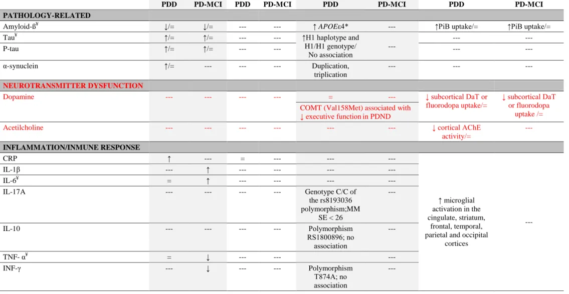
Table 3. Summary of the potential biomarkers of PDD and PD-MCI in function of the pathological processes implicated.
BIOMARKER CSF PLASMA GENES PET/SPECT IMAGING
PDD PD-MCI PDD PD-MCI PDD PD-MCI PDD PD-MCI
PATHOLOGY-RELATED
Amyloid-ߥ ↓/= ↓/= --- --- ↑ APOEε4* --- ↑PiB uptake/= ↑PiB uptake/=
Tau¥ ↑/= ↑/= --- --- ↑H1 haplotype and
H1/H1 genotype/ No association --- --- --- P-tau ↑/= ↑/= --- --- --- --- α-synuclein ↑/= --- --- --- Duplication, triplication --- --- --- NEUROTRANSMITTER DYSFUNCTION
Dopamine --- --- --- --- = --- ↓ subcortical DaT or
fluorodopa uptake/=
↓ subcortical DaT or fluorodopa
uptake /= COMT (Val158Met) associated with
↓ executive functionin PDND
Acetilcholine --- --- --- --- --- --- ↓ cortical AChE
activity/= --- INFLAMMATION/INMUNE RESPONSE CRP ↑ --- = --- --- --- ↑ microglial activation in the cingulate, striatum, frontal, temporal, parietal and occipital
cortices --- IL-1β --- ↑ --- --- --- --- IL-6¥ = ↑ --- --- --- --- IL-17A --- --- --- --- Genotype C/C of the rs8193036 polymorphism;MM SE < 26 --- IL-10 --- --- --- --- Polymorphism RS1800896; no association --- TNF- α¥ = ↓ --- --- --- INF-γ --- ↓ --- --- Polymorphism T874A; no association ---
36 PGE2 --- = --- --- --- --- IP10 = --- --- --- --- --- MIP1β = --- --- --- --- --- MCP1 = --- --- --- --- --- OXIDATIVE STRESS Uric acidδ, ¥ ↓ --- = --- --- --- --- --- NEUROTROPHIC/NEUROPROTECTION EGF¥ --- --- ↓ --- --- --- --- --- BDNF = --- --- --- PolymorphismVal1 96Met; no association --- --- --- Vitamin D --- --- = --- --- --- --- --- OTHER
Homocysteine¥ ↑/= = ↑/= = Variants in MTHFR, COMT and
other genes related to homocysteine metabolism; no association
--- ---
Abbreviations ”=”, No differences. Biomarkers: CRP, C reactive protein; IL-1β, Interleukin-1β; IL-6, Interleukin-6; L-17A, Interleukin 17A; IL-10, Interleukin 10; TNF-α, Tumor necrosis factor alpha; IFN-γ, Interferon gamma; PGE2, Prostaglandin E2; IP10, Interferon gamma-induced protein-10; MIP1β, Macrophage inflammatory protein-1;
MCP1, Monocyte chemotactic protein-1; EGF, Epidermal Growth Factor; BDNF, Brain Derived Neurotrophic Factor; PiB, Pittsburgh Compound B. Subjects: PDND, Parkinson’s disease non-demented; PDD, Parkinson’s disease with dementia; PD-MCI, Parkinson’s disease with mild cognitive impairment.
*APOE encodes for apolipoprotein E, and it is associated with the cerebral Amyloid-ß load
δLow urine uric acid levels associated with worse performance in several cognitive outcomes ¥
The data available allow us to conclude that genetic variants in COMT may account for differences in frontal dopaminergic function, they are typically associated with an executive subtype of PD-MCI with a low risk of dementia.92, 97, 111-115 By contrast, the APOE ε4 allele,92,
94, 95
the H1 haplotype of the MAPT gene97, 98 and mutations in the GBA gene109 all seem to be risk factors of dementia. However, they might be associated with different PD-MCI subtypes and probability of progression to dementia with slightly different cerebral pathology. Indeed, even in PDCN patients these mutations are associated with deficient activation in restricted brain territories when confronted with specific cognitive tests.99, 111, 113, 115 Thus, APOE ε4 might be associated with a more amnestic AD-like phenotype of PD-MCI, while MAPT and
GBA might be involved in more visuospatial patterns.
Regarding the proteins related to pathological cerebral inclusions, currently only low Aß levels in the CSF and PiB PET studies detecting Aß fibrils in the brain could have some potential to detect early dementia among PD-MCI patients. This could reflect a subgroup of patients with concurrent AD or AD pathological changes in whom the APOE ε4 allele might be also overrepresented. In this sense, these markers might not be useful for the purer LB cases of PDD, which could in turn be more related to GBA mutations.108-110 Oligomers of α-synuclein in the CSF are worth pursuing, both in sporadic PD but importantly, also in patients with GBA
mutations. In addition, the ratios of tau, Aß and α-synuclein could improve the predictive value of each protein independently, or help to differentiate between different forms of PD-MCI. New PET radiotracers to reliable mark in vivo tau and α-synuclein are needed to derive a complete picture of the clinical and pathological phenotypes of MCI subtypes, and how they progress towards dementia.
In addition, independently of the genetic fingerprint and pathological cerebral inclusions, it may be worth pursuing the study of lipids and of proteins related to metabolic processes (inflammation, oxidative stress, etc.) in CSF or plasma/serum, as well as neurotrophic factors
38 putatively involved in neurodegeneration such as EGF, ILGF, UA, neurofilament light chain protein and fatty acid binding-protein.47, 59, 63-65. Besides their diagnostic value, these fluid biomarkers may be particularly relevant in revealing dysfunctional biological processes that might be targets for pharmacological modulation (e.g. inflammation).
On the other hand, the cerebral cholinergic deficits detected by PET also seem to be a
promising biomarker of dementia212-215, 219, 220 but due to practical issues, alternative cheap and easy ways to evaluate cholinergic function should be considered, such as new
neurophysiological studies (TMS combined with ERP).297, 298
Pathological inclusions and neurotransmission deficits provoke neuronal dysfunction and death, which can be assessed by structural and functional imaging. Despite the fact that atrophy in structures like the hippocampus21, 163, 164 and reduced activity/metabolism in posterior cerebral areas187, 251, are associated with progression to dementia, no MRI modalities or FDG-PET are sufficiently reliable as to accurately predict progression to dementia at the single-patient level, as seen in CSF and peripheral fluid studies. Similarly to what has been discussed for other biomarkers, it could be that these findings are associated with subtypes of MCI (ie.
hippocampal atrophy with AD/amnestic type, and posterior dysfunction with visuospatial/LB type). The coupling of different PET studies, multimodal MRI and classic and modern
neurophysiological approaches (EEG and ERP), to more sophisticated analytical methods (i.e. neuronal networks, classifiers, etc.) represents a promising approach that might give rise to new tools to identify and stratify patients with distinct types of cognitive impairment and risk of dementia.
Multidisciplinary prospective studies in large cohorts of properly classified PD-MCI patients that assess the most promising biomarkers encountered in PDD patients, are now necessary to diagnose PD-MCI patients that are at high risk of progressing to dementia. In addition, studies
in early PD patients could also allow subtypes of PD with precocious development of dementia to be identified.
Acknowledgements
Author’s Roles
(1) Research Project: A, Conception; B, Organization; C, Execution. (2) Statistical Analysis: A, Design; B, Execution; C, Review and Critique
(3) Manuscript Preparation: A, Writing of the first draft; B, Review and Critique. M.D.-A.: 1A, 1C, 3A, 3B
B.G.: 1C, 3B I.N.-G.: 3B H.J.-U.: 3B
M.C.R.-O.: 1A, 1B, 1C, 3A, 3B
Financial Disclosures of all the authors (for the preceding 12 months): M.D.-A. holds a Basque Country Government Predoctoral Research Grant and has received a research award from Fundación Jesús de Gangoiti Barrera. B.G. has no disclosures. I.N.-G. receives support from CIBERNED. H.J.-U. holds a Basque Country Government Predoctoral Research Grant. M.C.R.-O. received honorarium for lectures, travel and accommodation to attend scientific meetings from UCB, and Boston Scientific, and she received financial support for her research from national and regional government bodies in Spain (Institute of Health Carlos III, Basque Country Government, Diputacion Foral Guipuzcoa, CIBERNED) and Europe.
40
References
1. Hely MA, Reid WG, Adena MA, Halliday GM, Morris JG. The Sydney multicenter study of Parkinson's disease: the inevitability of dementia at 20 years. Mov Disord 2008;23(6):837-844.
2. Aarsland D, Andersen K, Larsen JP, Lolk A, Kragh-Sorensen P. Prevalence and characteristics of dementia in Parkinson disease: an 8-year prospective study. Arch Neurol 2003;60(3):387-392.
3. Litvan I, Aarsland D, Adler CH, et al. MDS Task Force on mild cognitive impairment in Parkinson's disease: critical review of PD-MCI. Mov Disord 2011;26(10):1814-1824.
4. Janvin CC, Larsen JP, Aarsland D, Hugdahl K. Subtypes of mild cognitive impairment in Parkinson's disease: progression to dementia. Mov Disord 2006;21(9):1343-1349.
5. Gasca-Salas C, Estanga A, Clavero P, et al. Longitudinal Assessment of the Pattern of Cognitive Decline in Non-Demented Patients with Advanced Parkinson's
Disease. J Parkinsons Dis 2014.
6. Pedersen KF, Larsen JP, Tysnes OB, Alves G. Prognosis of mild cognitive impairment in early Parkinson disease: the Norwegian ParkWest study. JAMA Neurol 2013;70(5):580-586.
7. Broeders M, de Bie RM, Velseboer DC, Speelman JD, Muslimovic D, Schmand B. Evolution of mild cognitive impairment in Parkinson disease. Neurology
8. Litvan I, Goldman JG, Troster AI, et al. Diagnostic criteria for mild cognitive impairment in Parkinson's disease: Movement Disorder Society Task Force guidelines. Mov Disord 2012;27(3):349-356.
9. Aarsland D, Perry R, Brown A, Larsen JP, Ballard C. Neuropathology of dementia in Parkinson's disease: a prospective, community-based study. Ann Neurol
2005;58(5):773-776.
10. Sabbagh MN, Adler CH, Lahti TJ, et al. Parkinson disease with dementia:
comparing patients with and without Alzheimer pathology. Alzheimer Dis Assoc Disord 2009;23(3):295-297.
11. Braak H, Rub U, Jansen Steur EN, Del Tredici K, de Vos RA. Cognitive status correlates with neuropathologic stage in Parkinson disease. Neurology
2005;64(8):1404-1410.
12. Compta Y, Parkkinen L, O'Sullivan SS, et al. Lewy- and Alzheimer-type pathologies in Parkinson's disease dementia: which is more important? Brain 2011;134(Pt 5):1493-1505.
13. Halliday GM, Leverenz JB, Schneider JS, Adler CH. The neurobiological basis of cognitive impairment in Parkinson's disease. Mov Disord 2014;29(5):634-650. 14. Adler CH, Caviness JN, Sabbagh MN, et al. Heterogeneous neuropathological
findings in Parkinson's disease with mild cognitive impairment. Acta Neuropathol 2010;120(6):827-828.
15. Jellinger K. Heterogenous mechanisms of mild cognitive impairment in Parkinson's disease. J Neural Transm 2012;119(3):381-382.