University of Zurich Main Library Strickhofstrasse 39 CH-8057 Zurich www.zora.uzh.ch Year: 2015
Clear Cell Papillary Renal Cell Carcinoma and Renal
Angiomyoadenomatous Tumor: Two Variants of a Morphologic,
Immunohistochemical, and Genetic Distinct Entity of Renal Cell Carcinoma
Deml, Karl-Friedrich ; Schildhaus, Hans-Ulrich ; Compérat, Eva ; et al
Abstract: Clear cell papillary renal cell carcinoma (ccpRCC) and renal angiomyoadenomatous tumor (RAT) share morphologic similarities with clear cell (ccRCC) and papillary RCC (pRCC). It is a matter of controversy whether their morphologic, immunophenotypic, and molecular features allow the definition of a separate renal carcinoma entity. The aim of our project was to investigate specific renal immunohis-tochemical biomarkers involved in the hypoxia-inducible factor pathway and mutations in the VHL gene to clarify the relationship between ccpRCC and RAT. We investigated 28 ccpRCC and 9 RAT samples by immunohistochemistry using 25 markers. VHL gene mutations and allele losses were investigated by Sanger sequencing and fluorescence in situ hybridization. Clinical follow-up data were obtained for a subset of the patients. No tumor recurrence or tumor-related death was observed in any of the pa-tients. Immunohistochemistry and molecular analyses led to the reclassification of 3 tumors as ccRCC and TFE3 translocation carcinomas. The immunohistochemical profile of ccpRCC and RAT samples was very similar but not identical, differing from both ccRCC and pRCC. Especially, the parafibromin and hKIM-1 expression exhibited differences in ccpRCC/RAT compared with ccRCC and pRCC. Genetic analysis revealed VHL mutations in 2/27 (7%) and 1/7 (14%) ccpRCC and RAT samples, respectively. Fluorescence in situ hybridization analysis disclosed a 3p loss in 2/20 (10%) ccpRCC samples. ccpRCC and RAT have a specific morphologic and immunohistochemical profile, but they share similarities with the more aggressive renal tumors. On the basis of our results, we regard ccpRCC/RAT as a distinct entity of RCCs.
DOI: https://doi.org/10.1097/PAS.0000000000000456
Posted at the Zurich Open Repository and Archive, University of Zurich ZORA URL: https://doi.org/10.5167/uzh-112056
Journal Article Published Version Originally published at:
Deml, Karl-Friedrich; Schildhaus, Hans-Ulrich; Compérat, Eva; et al (2015). Clear Cell Papillary Renal Cell Carcinoma and Renal Angiomyoadenomatous Tumor: Two Variants of a Morphologic, Immunohisto-chemical, and Genetic Distinct Entity of Renal Cell Carcinoma. American Journal of Surgical Pathology, 39(7):889-901.
Clear Cell Papillary Renal Cell Carcinoma and Renal
Angiomyoadenomatous Tumor
Two Variants of a Morphologic, Immunohistochemical, and Genetic
Distinct Entity of Renal Cell Carcinoma
Karl-Friedrich Deml, MD,
*
Hans-Ulrich Schildhaus, MD,w
Eva Compe´rat, MD,
z
Adriana von Teichman, PhD,
*
Martina Storz, BA,
*
Peter Schraml, PhD,
*
Joseph V. Bonventre, MD, PhD,y
Falko Fend, MD,8
Barbara Fleige, MD,z
Andreas Nerlich, MD,#
Helmut E. Gabbert, MD,
**
Nikolaus Ga
ler, MD,ww
Rainer Grobholz, MD,
zz
Seife Hailemariam, MD,yy
Raoul Hinze, MD,88
Ruth Knu¨chel, MD,ww
Benoit Lhermitte, MD,zz
Gabriella Nesi, MD,## Thomas Ru¨diger, MD,
***
Guido Sauter, MD,www
and Holger Moch, MD
*
Abstract:Clear cell papillary renal cell carcinoma (ccpRCC) and renal angiomyoadenomatous tumor (RAT) share morphologic similarities with clear cell (ccRCC) and papillary RCC (pRCC). It is a matter of controversy whether their morphologic, im-munophenotypic, and molecular features allow the definition of a separate renal carcinoma entity. The aim of our project was to investigate specific renal immunohistochemical biomarkers in-volved in the hypoxia-inducible factor pathway and mutations in theVHLgene to clarify the relationship between ccpRCC and RAT. We investigated 28 ccpRCC and 9 RAT samples by im-munohistochemistry using 25 markers. VHL gene mutations
and allele losses were investigated by Sanger sequencing and fluorescence in situ hybridization. Clinical follow-up data were obtained for a subset of the patients. No tumor recurrence or tumor-related death was observed in any of the patients. Immunohistochemistry and molecular analyses led to the re-classification of 3 tumors as ccRCC and TFE3 translocation carcinomas. The immunohistochemical profile of ccpRCC and RAT samples was very similar but not identical, differing from both ccRCC and pRCC. Especially, the parafibromin and hKIM-1 expression exhibited differences in ccpRCC/RAT compared with ccRCC and pRCC. Genetic analysis revealed VHLmutations in 2/27 (7%) and 1/7 (14%) ccpRCC and RAT samples, respectively. Fluorescence in situ hybridization analysis disclosed a 3p loss in 2/20 (10%) ccpRCC samples. ccpRCC and RAT have a specific morphologic and immunohistochemical profile, but they share similarities with the more aggressive renal tumors. On the basis of our results, we regard ccpRCC/RAT as a distinct entity of RCCs.
Key Words: kidney, clear cell papillary renal cell cancer, cyto-keratin 7,VHL, renal angiomyoadenomatous tumor, clear cell renal cell carcinoma
(Am J Surg Pathol2015;39:889–901)
C
lear cell papillary renal cell carcinoma (ccpRCC) has been proposed as a new entity of renal cell cancer by the International Society of Urological Pathology to be included in the next World Health Organization Classi-fication of Renal Tumors.1 It was initially discovered in kidneys with end-stage renal disease in 2006.2 Since then>100 ccpRCC cases have been described, and the majority were found in normal functioning kidneys.3–8 They are
characterized by tumor cells with clear cytoplasm, linear arrangement of low-grade nuclei located apically distant from the basal membrane, and containing varying amounts of tubular, papillary, and cystic architecture. Strikingly, the ccpRCCs lack mitoses, atypia, pleomorphism, necrosis,
From the *Institute of Surgical Pathology, University Hospital Zurich, Zurich;zzCantonal Hospital Aarau;yyInstitute of Histological and Cytological Diagnostics, Aarau;zzUniversity Institute of Pathology, Centre Hospitalier Universitaire Vaudois (CHUV), Lausanne, Switzerland;wInstitute of Pathology, University Medicine Go¨ttingen (UMG), Go¨ttingen; 8Institute of Pathology, University Hospital Tu¨bingen, Tu¨bingen; zInstitute of Pathology, HELIOS Hospital Berlin-Buch, Berlin; #Institute of Pathology Bogenhausen, Sta¨dtisches Klinikum Mu¨nchen GmbH, Munich; **Institute of Pathology, Heinrich Heine University Hospital, Dusseldorf;ww In-stitute of Pathology, RWTH University, Aachen; 88Institute of Pathology, HELIOS Hospital Schwerin, Schwerin; ***Institute of Pathology, Sta¨dtisches Klinikum Karlsruhe gGmbH, Karlsruhe;
wwwInstitute of Pathology, University Medical Center Hamburg-Eppendorf, Hamburg, Germany;zService d’Anatomie et Cytologie Pathologiques 1, Groupe Hospitalier Pitie´-Salpeˆtrie`re, Paris, France;
yDepartment of Medicine, Renal Division, Brigham and Women’s Hospital, Boston, MA; and ##Institute of Pathology, University of Florence, Florence, Italy.
Conflicts of Interest and Source of Funding: Supported by the Swiss National Science Foundation (3238BO-103145) and the Zurich Cancer League to H.M. J.V.B. received grants from the NIH (R37DK39773, RO1DK072381). J.V.B. is a co-inventor on KIM-1 patents, which have been assigned to Partners Healthcare and li-censed by Partners Healthcare to a number of companies. For the remaining authors none were declared.
Correspondence: Karl-Friedrich Deml, MD, Institute of Surgical Path-ology, University Hospital Zurich, Schmelzbergstrasse 12, CH-8091 Zurich, Switzerland (e-mail: karl-friedrich.deml@usz.ch).
hyaline globules, foamy macrophages, and vascular in-vasion. Despite significant morphologic, immunohisto-chemical, and genetic similarities to clear cell RCC (ccRCC) and papillary RCC (pRCC), characteristic genetic differences include VHL gene mutations and 3p losses, found in ccRCC. Gain of the chromosomes 7 and 17 or loss of chromosome Y are absent or extremely rare in ccpRCC cases.4,9,10No disease-defining mutation has been identified to date.
The renal angiomyoadenomatous tumor (RAT) was first reported in the kidney of a 93-year-old man by Michal et al.11 Nine years later, the same group charac-terized 5 additional tumors.12 Verine13 pointed out that ccpRCCs are a major differential diagnosis of RAT and emphasized their morphologic, immunohistochemical, molecular, and clinical similarities. In the literature many terms have been used to probably describe the same entity, including ccRCC with prominent leiomyomatous pro-liferation and RCC with smooth muscle stroma.12,14–17
The epithelial component of ccpRCC and RAT is composed of cells with abundant clear cytoplasm, strong diffuse CK7 activity, and low-grade nuclei (Fuhrman grades 1 and 2). Because of their many similarities, several authors regard ccpRCC and RAT to be a variant of the same entity.6,9,13,17,18
The aims of our study were to clarify the relation-ship between ccpRCC and RAT and to identify markers to reliably distinguish ccpRCC and RAT from the bio-logically more aggressive renal neoplasms.
MATERIALS AND METHODS
Case Cohort
All tumors were consultation cases from H.M. and E.C. and were received from Austria, France, Germany, Italy, and Switzerland. Hematoxylin and eosin–stained slides were re-viewed for morphologic features of ccpRCC and RAT as previously described.3,5–7,11,12,19 Diagnostic features of ccpRCC include tumor cells with abundant clear cytoplasm, varying papillary, cystic, and tubular architecture, low-grade nuclei (Fuhrman grades 1 and 2) located apically distant from the basal membrane, and strong diffuse CK7 and CA-IX expression. For diagnosis of RAT, the following criteria were required: cells with clear cytoplasm, low-grade nuclei (Fuhrman grades 1 and 2) embedded in a smooth muscle stroma, and strong diffuse CK7 staining of the epithelial component. Tumors were staged according to the TNM sys-tem20 and graded according to Fuhrman et al.21 The mor-phologic characteristics were scored as previously described.7
Immunohistochemistry
A total of 25 antibodies were selected as (i) they are involved in theVHLsignaling pathway, (ii) they are known to be prognostic biomarkers of ccRCC, and (iii) they have been reported as markers of ccpRCCs and RATs in a small group of ccpRCCs described in recent USCAP meetings (2011 to 2014). Tissue microarray sections (2.5mm) were transferred to glass slides and treated using Ventana Benchmark XT, Bond-max (Leica Microsystems) automated systems, and manual
protocols. Tissue microarray construction was not possible in 5 of the ccpRCC cases because of the absence of tissue. The immunohistochemical staining product was described as nuclear, membranous, or cytoplasmic (Table 1). The im-munohistochemistry results were interpreted as 0 (negative), 1+ (weak staining), 2+ (moderate staining), and 3+ (strong staining). For statistical analysis, all 2+ and 3+ stainings were defined as positive and 0 and 1+ as negative. Antibodies and protocols are listed in Table 1.
Fluorescence In Situ Hybridization
Fluorescence in situ hybridization (FISH), performed to detectVHLallele losses, was carried out using the ZytoLight SPEC VHL/CEN 3 Dual-color Probe (ZytoVision, Bremerhaven, Germany). Tissue sections were cut from for-malin-fixed paraffin-embedded (FFPE) blocks, deparaffinized, and hybridized as previously described.22 Sixty
non-overlapping tumor nuclei from 3 different areas were ana-lyzed, and the number of VHL and CEN3 signals was recorded for each nucleus. The total number of VHL and CEN3 signals as well as the VHL/CEN3 ratio and the per-centage of tumor cells with <2 VHL signals were calculated. Tumors were considered VHL deleted if >50% of the tumor nuclei displayed <2 VHL signals.23In 2 cases TFE3 FISH
using SPEC TFE3 dual-color break-apart probe from Zyto-Vision was performed on whole sections as previously de-scribed by our group.24
VHL
Sequencing Analysis
Tumor areas displaying >80% tissue in the epi-thelial portion of the ccpRCC and RAT were marked on the hematoxylin and eosin slides. DNA from FFPE tu-mor tissue samples was obtained by punching 1 to 2 tissue cylinders (diameter 0.6 mm) from each sample. DNA was extracted from the tumor tissue samples according to the Maxwell 16 FFPE Plus DNA Purification protocol (Promega, Fitchburg) for automated DNA purification. DNA concentrations in the samples were measured using the Nanodrop (Thermo Fisher Scientific, Waltham, MA). Polymerase chain reaction (PCR) of the VHL gene was performed as previously described25using approximately
40 ng of DNA for each amplification. DNA sequencing was performed with the dideoxy chain-termination method using the BigDye Terminator v1.1 Cycle Se-quencing kit (Applied Biosystems, Foster City). The same forward and reverse primers were used for PCR and se-quencing analyses. Cycle sese-quencing products were ana-lyzed using the AbiPrism 3100 Genetic analyzer (Applied Biosystems). The obtained sequences were compared with the NCBI sequence AF010238 using NCBIs Blast 2 Se-quences. All VHL point mutations obtained were vali-dated by a second separate PCR and sequencing analysis.
RESULTS
Clinical and Pathologic Findings
The patients with ccpRCC ranged from 29 to 75 years of age (mean age 58 y) and those with RAT from 32 to 68 years of age (mean age 43.3 y) at the time of
nephrectomy. The male to female ratio was 1.5:1 in the ccpRCC group (17 men and 11 women) and 3.5:1 in the RAT group (6 men and 1 woman).
Clinical follow-up data were available for 78% (21/ 27) of the ccpRCC patients and 71% (5/7) of the RAT patients. Mean follow-up time was 29.7 months (range, 7 to 84 mo) for the ccpRCC patients and 32.3 months (range, 25 to 38 mo) for the RAT patients. There was no evidence of recurrence or disease-related death in any of the patients. None of the RAT (0/5) patients and 14% (3/ 22) of the ccpRCC patients had end-stage renal disease.
In the RAT group, the average diameter of the tumor was 3.1 cm (range, 1.8 to 5.0 cm) compared with 2.6 cm (range, 0.5 to 8 cm) in the ccpRCC group. Among the RAT patients, 67% (4/6) displayed pathologic stage pT1a and 33% (2/6) stage pT1b. Overall, 86% of the tumors (6/7) were Fuhrman nuclear grade 1, and 14% (1/7) were nuclear grade 2. In the ccpRCC cases, 77% (20/26) were stage pT1a, 19% (5/26) were pT1b, and 4% (1/26) were pT2a. Fuhrman nuclear grade 1 was found in 48% (13/27) and nuclear grade 2 in 52% (14/27) of the tumors. All the ccpRCCs and 6/7 RATs showed at least focal papillary architecture and branched ducts. In contrast to ccpRCC, secretory cells were completely absent in the RAT cases. Both showed variable amounts of cystic areas. All tumors were characterized by the absence of mitotic formations, foamy macrophages, calcifications, and vascular invasion.
Table 2 summarizes the clinicopathologic findings. Morphologic characteristics are shown in Table 3.
Immunohistochemical Findings
The immunohistochemical findings are detailed in Table 4. ccpRCC and RAT were strongly positive for CK7, CK19, CA-IX, GLUT-1, E-cadherin, vimentin,
b-catenin, parafibromin, PAX-2, PAX-8, p27, p53, and c-MET. Staining for GLUT-1 (P= 0.0572), CD70 (P= 0.1499), and p16 (P= 0.3702) differed slightly in the RAT samples compared with ccpRCCs, although differences did not show statistical significance. Following the recent results by Cui et al,26Aron et al,27 and Schwartz et al,28we tested parafibromin, hKIM-1,29and CD133 expression to distinguish
ccpRCC/RAT from ccRCC/pRCC. As shown in Table 5, the expression difference reached statistical significance (P< 0.0001). The biomarkers CD70,30 MET,31 and E-cadherin32 were able to distinguish between ccpRCC/RAT and ccRCC (P< 0.0001). Furthermore, the hKIM-1 and parafibromin were able to distinguish between ccpRCC/RAT and pRCC. All ccpRCC cases exhibited a characteristic CA-IX “cup-like” distribution, sparing the luminal border as it has been de-scribed in the literature before.6,33 In contrast, the RAT
tu-mors and the ccpRCC-like tumor with the VHL mutation showed a circumferential membranous staining pattern. Two RAT samples stained weakly positive for TFE3 and were, therefore, further analyzed by HMB45 and TFEB. Both stainings revealed a negative result. In addition, TFE3 FISH was performed (see below).
FISH Findings
Three deletions of the short arm of chromosome 3 were identified. All of them occurred in the ccpRCC cases (3/21, 14%), and no deletion was found in the RAT cases (0/7, 0%). The presence of the 3p deletions in the 1 ccRCC controls was correctly identified. In 9 of the cases FISH was not performed, as there was insufficient tissue after VHL mutation analysis and immunohistochemistry. TFE3 FISH was performed with the 2 above-men-tioned RAT-like cases that showed weak TFE3 ex-pression. One case showed the typical break-apart pattern
TABLE 1. Antibody Overview
Antibody Clone Species Vendor Dilution Staining Pattern
b-catenin 14/Beta-Catenin Mouse BD Biosciences 1:50 Membranous
Carbonic anhydrase IX — Rabbit Abcam Limited 1:6000 Membranous (partially cytoplasmatic) c-MET SP44 Rabbit Ventana Prediluted Membranous
CD10 SP67 Mouse Ventana Prediluted Membranous CD70 301731 Mouse R&D Systems 1:75 Membranous CD133 — Rabbit Abcam Limited 1:500 Membranous Cytokeratin 7 OV-TL 12/30 Mouse Dako A/S 1:100 Membranous Cytokeratin 19 RCK108 Mouse Abcam Limited 1:200 Membranous E-cadherin EP700Y Rabbit Cell Marque Lifescreen Ltd 1:200 Membranous Estrogen receptor SP1 Rabbit Labvision 1:50 Nuclear GATA3 L50-823 Mouse Biocare Medical 1:250 Nuclear GLUT-1 — Rabbit Millipore Corporation 1:1000 Membranous hKIM-1 — — Bonventre lab — Membranous and cytoplasmatic Melanosome HMB45 Mouse Dako A/S 1:50 Cytoplasmic
OCT3/4 N1NK Mouse Novocastra Laboratories Ltd 1:150 Nuclear p16 JC8 Mouse Santa Cruz Biotechnology Inc. 1:200 Nuclear p27 — Rabbit Santa Cruz Biotechnology Inc. 1:30 Nuclear
p53 DO-7 Mouse Dako A/S 1:80 Nuclear
Parafibromin 2H1 Mouse Santa Cruz Biotechnology Inc. 1:10 Nuclear PAX-2 — Rabbit Zymed Laboratories Inc. 1:100 Nuclear PAX-8 — Rabbit Protein Tech Group Inc 1:200 Nuclear Progesterone receptor 1E2 Rabbit Ventana Prediluted Nuclear TFE3 MRQ-37 Rabbit Cell Marque Lifescreen Ltd. Prediluted Nuclear TFEB — Goat Santa Cruz Biotechnology Inc. 1:100 Nuclear Vimentin Vim 3B4 Mouse Dako A/S 1:250 Cytoplasmatic
in >85% of the cells, whereas the second case was neg-ative. Both cases were reclassified as translocation carci-nomas because of immunohistochemical TFE3 positivity.
VHL
Gene Mutation Analysis
Three VHL mutations were detected in the ccpRCC group (3/27, 11%) in exon 2 (c.351G > C/p.Trp117Cys, c.461C > T/p.Pro154Leu, c.388G > C/p.Val130Leu) and 1 in the RAT group (1/7, 14%) in exon 1 (c.174_177delGCCG/ p.Pro59GlyfsX7). We identified 2 cases, harboring both a
VHLmutation and 3p loss. One case showed a 3p loss but no
VHLmutation, and 2 cases with aVHLmutation showed no 3p loss (Figs. 1–5).
DISCUSSION
In the present study we have sequenced the largest number of ccpRCC29and RAT7cases to date. We found
a VHL mutation rate of 11% in ccpRCC and 14% in RAT. Furthermore, we analyzed hypoxia-inducible factor
pathway–related proteins to compare these findings with recent findings.
ccpRCC and RAT are currently underrecognized. Recent studies have revealed that they are not rare7,34,35and
that among all RCCs the ccpRCCs have a prevalence rate between 1.2% and 4.1%, thus representing up to 4500 new cases of renal cancer in the United States annually.7,34,36 Awareness of its morphologic and immunohistochemical features is imperative for a correct classification. In a recent publication Gill et al35 underscored the necessity of re-classifying low-grade and low-stage ccRCC, as up to 7% of the cases are in fact ccpRCC.
Morphologically, ccpRCC and RAT share many features. Their epithelial component is composed of cells with clear cytoplasm and low-grade nuclei. Both tumors have various amounts of smooth muscle stroma, and their epithelial component is characterized by either cystic or papillary architecture. In our cohort the majority of the RAT samples had focal papillary features of the epithelial
TABLE 2. Clinicopathologic Findings
# Age Sex Laterality
Grade (Fuhrman)
Size (cm)
No.
Tumors Stage ESRD
Tumor-related Death Follow-up (mo) Tumor
Recurrence Metastasis Special Remarks
RAT
1 32 M Right 2 1.8 1 pT1a No NA NA No No 2 32 M Left 2 4.7 2 pT1b NA NA NA NA NA
3 34 M Right 2 1.8 2 pT1a NA No 35 NA NA IBD, pRCC type 1, kidney adenomas 4 45 F Right 2 NA 1 NA NA NA NA NA NA 5 56 M Left 2 2 1 pT1a No No 25 No No 6 68 M NA 1 3.5 1 pT1a NA NA NA NA NA 7 36 M Left 2 5 1 pT1b No No 19 No No CCPRCC 1 55 M NA 2 NA NA NA NA NA NA NA NA 2 58 M Right 2 0.9 1 pT1a No NO 39 No No 3 57 F Right 1 4 1 pT1b NA NO 25 NA NA 4 50 M NA 2 3 1 pT1a NA NA NA NA NA
5 38 M Left 2 2 1 pT1a Yes NA NA NA NA IgA nephritis 6 63 M Right 2 2 NA pT1a NA NA NA NA NA Large cell b-cell
lymphoma 7 62 M Left 2 3 2 pT1a No No 12 No No pRCC type 2 8 31 F Left 2 8 1 pT2a NA NA NA NA NA 9 55 M Right 1 4.5 1 pT1b No No 24 No No 10 68 F NA 1 3.8 pT1a NA Na NA NA NA 11 51 M Left 2 1.3 1 pT1a No No 11 No No 12 75 F Left 2 5.1 1 pT1b No No 7 No No 13 38 M Left 1 2.2 1 pT1a No No 8 No No 14 53 F Right 1 2 1 pT1a No No 84 No No 15 51 M Left 2 1 8 pT1a No No 71 No No 16 62 M Left 1 1.3 1 pT1a Yes No 67 No No 17 52 M Left 1 5 1 pT1b No No 60 No No 18 71 F Right 2 2.5 1 pT1a No No 39 No No 19 57 F Left 1 1.5 1 pT1a No No 37 No No 20 72 M NA 1 0.5 2 pT1a No No 29 No No 21 61 F Left 2 5 1 pT1b No No 22 No No 22 71 M Right 1 2 1 pT1a No No 21 No No 23 53 F Right 1 1 3 pT1a No No 17 No No 24 70 F Left 1 2.8 1 pT1a No No 15 No No 25 65 F NA 1 0.5 1 pT1a No No 14 No No 26 74 M Left 2 1.8 1 pT1a Yes No 12 No No 27 54 M Left 2 2 1 pT1a No No 10 No No
component, which are typically diffuse CK7 and CA-IX positive. The most relevant differential diagnoses include ccRCC that exhibit papillary features, pRCC exhibiting clear cell characteristics, and Xp11 translocation carci-noma. In our cohort, 2 cases initially classified as RAT had to be reclassified as Xp11 translocation carcinomas after immunohistochemical and TFE3 FISH analyses. The translocation carcinomas were identified by nuclear TFE3 protein expression. Only 1 case showed a positive TFE3 FISH result. It is controversial whether TFE3 positivity is sufficient to diagnose TFE3 translocation carcinoma,24,37but, from these 2 cases, we concluded that TFE3 translocation cancer falls within the differential diagnostic spectrum of ccpRCC/RAT. Another differ-ential diagnosis for the case with weak TFE3 staining and negative FISH is TFEB-associated RCC. Those tumors can overlap tremendously with the TFE3-rearranged RCC.38,39 To rule out this differential diagnosis, we
per-formed 2 additional immunohistochemical stainings (HMB45 and TFEB). Both stainings showed a negative result making that differential diagnosis unlikely.
One ccpRCC case was reclassified as ccRCC. That case exhibited typical ccpRCC morphology but was completely negative for CK7 and strongly positive for hKIM-1. This case also revealed a mutation in the VHL
gene and a 3p loss in the FISH analysis. These findings highlight the importance of molecular testing and should raise awareness of ccpRCC mimicking ccRCC.40
VHL gene mutations are the genetic hallmark of ccRCC. Initially, it was reported thatVHLalterations are absent in ccpRCC. However, 3 groups have recently identified VHL mutations in ccpRCC at frequencies varying from 15% to 27%.41–43In concordance with these studies, we also identified VHL gene alterations in ccpRCC, but the prevalence of VHL gene mutations is significantly lower than in ccRCC.44–46 The discrepancy
between the number of mutations found in our ccpRCC cases and that reported may be explained by the different detection methods used, including single-nucleotide polymorphism genotyping array and Sanger sequencing, and by the limited number of cases in previous studies. Alternatively, cases withVHLmutations could represent ccRCCs with morphology and immunoprofile closely mimicking that of clear ccpRCC and RAT tumor. Cur-rently, ccpRCCs are diagnosed on the basis of morphol-ogy and diffuse strong CK7 expression. The absence of VHL mutations/3p deletions is not diagnostic for ccpRCC. Therefore, we suggest diagnosis of tumors with diffuse CK7 expression combined with the typical mor-phology as ccpRCC. In previous studies, ccRCCs with a diffuse CK7 profile have had a completely different prognosis than ccRCCs without that CK expression pattern.47 These previous findings justify such an ap-proach.VHLinactivation leads to an HIF-dependent CA-IX and GLUT-1 upregulation. We only found few VHL TABLE 3. Morphologic Characteristics of RAT and ccpRCC
# Papillary Architecture Branched Ducts Secretory Cells % Cystic RAT 1 1f Yes No 15 2 0 No No 0 3 2f Yes No 65 4 1f Yes No 5 5 2f Yes No 0 6 1f Yes No 55 7 2f Yes No 15 ccpRCC 1 1 Yes Yes 85 2 2 Yes No 10 3 3 Yes Yes 10 4 3 Yes No 10 5 2 Yes No 15 6 3 Yes Yes 55 7 1 Yes Yes 0 8 3 Yes No 0 9 3 Yes No 0 10 2 Yes No 5 11 3 Yes No 0 12 3 Yes Yes 40 13 1 Yes Yes 15 14 1 Yes No 5 15 2 Yes No 20 16 2 Yes No 10 17 1 Yes Yes 15 18 3 Yes Yes 45 19 2 Yes No 30 20 2 Yes Yes 20 21 2 Yes No 5 22 1 Yes No 10 23 3 Yes Yes 55 24 1 Yes Yes 10 25 3 Yes No 35 26 2 Yes Yes 5 27 3 Yes Yes 30
Scored as previously described.7 F indicates focal.
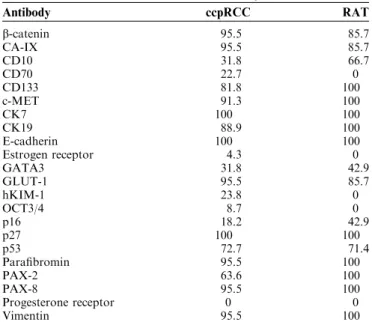
TABLE 4. Results of Immunohistochemistry
Antibody ccpRCC RAT b-catenin 95.5 85.7 CA-IX 95.5 85.7 CD10 31.8 66.7 CD70 22.7 0 CD133 81.8 100 c-MET 91.3 100 CK7 100 100 CK19 88.9 100 E-cadherin 100 100 Estrogen receptor 4.3 0 GATA3 31.8 42.9 GLUT-1 95.5 85.7 hKIM-1 23.8 0 OCT3/4 8.7 0 p16 18.2 42.9 p27 100 100 p53 72.7 71.4 Parafibromin 95.5 100 PAX-2 63.6 100 PAX-8 95.5 100 Progesterone receptor 0 0 Vimentin 95.5 100
mutations but in combination with CA-IX and GLUT-1 immunoreactivity in both ccpRCC and RAT. This clearly sets the ccpRCC and RAT apart from ccRCC, which shows VHL mutations in up to 80% of the cases.44,48 Therefore, we believe that the HIF pathway may be ac-tivated in aVHL-independent manner in most ccpRCCs and RATs, also hypothesized by Rohan et al.6
Recently, Lawrie et al49found various mutations in
ccpRCC by using next-generation sequencing, including a nonsynonymous T992I mutation in the MET proto-on-cogene. This gene was originally described as causing hereditary pRCC.50 Interestingly, Lawrie and colleagues detected noVHLmutation, but found overexpression in all 5 members of the miR-200 family. The miR-200 family plays an essential role in tumor suppression by inhibiting epithelial-mesenchymal transition.51 To support Lawrie and colleagues’ results, we also noted immunoreactivity for E-cadherin andb-catenin. These findings suggest that epithelial-to-mesenchymal transition may be incomplete or blocked in ccpRCC contributing to their indolent course.49
Other genetic alterations characteristic for pRCC include gain of chromosome 7 and loss of chromosome Y. However, in ccpRCC, gain of chromosome 7 has very rarely been reported,4,5,9,10and no loss of chromosome Y has been observed to date. Fisher et al52 found a unique
gene expression profile of ccpRCC when investigating 8 different genes, with only some expression levels com-parable to those observed in ccRCC and pRCC.
In our FISH analysis, we identified 3 chromosome 3p deletions in 20 ccpRCC and 7 RAT samples. All 3p deletions occurred in ccpRCC with a frequency of 14.3%, but none was detected in RAT. To date, only 4 cases with a 3p loss have been reported in ccpRCC.36,43Interestingly, the single case described by Martignoni et al43 con-currently harbored aVHLmutation like 2 of our 3 cases with a 3p loss. Shi et al36 also used FISH and observed monosomy of chromosome 3 in 3 cases in a series of 11 ccpRCCs all lacking mutations in theVHLgene. In 2009, Shannon et al14 published a study on 5 ccRCCs with
smooth muscle stroma and found loss of the entire chro-mosome 3 in 2 cases and a 3p loss in 1 case using FISH. In contrast, Martignoni et al17found no 3p loss in a series of 3 cases of ccRCC with smooth muscle stroma. Given these molecular findings, it has been suggested that RAT and ccRCC with smooth muscle stroma are interchangeable
terms.53 However, some of the cases of ccRCC with
smooth muscle stroma, particularly those that showed 3p loss, might represent ccRCCs with exuberant, infiltrative smooth muscle, whereas the others might in fact be RAT tumors, particularly the ones that do not show 3p loss.15 In addition, recent data show that some tumors with RAT morphology and immunophenotype share a common mutation in the TCEB1 gene, which inactivated the
VHLpathway and upregulated proteins along the hypoxia-inducible pathway.54 Twenty-five different antibodies were used to characterize ccpRCC and RAT. We were particularly interested in hypoxia-inducible factor path-way–related proteins and other antibodies, which were reportedly used in small series of ccpRCC cases in the 2011 and 2014 USCAP meetings. This gave us the opportunity to compare immunohistochemical findings in ccpRCC and RAT to clarify their interrelationship. Remarkably, there were no statistically significant differences in the staining properties in any of the antibodies in ccpRCC compared with RAT.
Parafibromin and hKIM-1 expression levels differed significantly between ccpRCC/RAT and ccRCC/pRCC. Cui et al26 recently demonstrated that parafibromin can be very helpful in differentiating ccpRCC from ccRCC and pRCC. In a study by Aron et al,27 the difference in the staining positivity rate of ccpRCC and ccRCC was even more striking compared with our study. In addition to parafibromin and hKIM-1 expression, CD70 also proved to be a useful marker in differentiating ccpRCC from ccRCC, as CD70 expression is rare in ccpRCC and very frequent in ccRCC. CD70 was used for immuno-histochemistry, because we have previously demonstrated that CD70 is a potential biomarker for ccRCC.30,55 The
importance of immunohistochemical stainings in the correct identification of true ccpRCC was also highlighted by Williamson et al.56 They studied 14 ccpRCC-like tumors, which could not be distinguished from ccpRCC morphologically, but which showed a high 3p deletion frequency (82%) and showed a different immunohisto-chemical profile, with negative or localized CK7 staining as the most striking feature. These characteristics also led to a reclassification of 1 of our tumors, primarily diag-nosed as ccpRCC.
Recently, Schwartz et al studied different stem cell markers in renal cancers. They reported a 90% positivity rate for OCT3/4 in a series of 10 ccpRCC samples.28This
TABLE 5. Different Expression Patterns of hKIM-1, Parafibromin, CD70, CD133, MET, and E-cadherin in ccPRCC, ccRCC, and pRCC (Own Data and Literature)
Antibody ccpRCC (n [%]) ccRCC (n [%]) Fisher Exact (P) pRCC (n [%]) Fisher Exact (P)
hKIM-1 6/22 (27.3) 54/73 (74)29 < 0.0001 28/30 (93)29 < 0.0001 Parafibromin 21/23 (91.3) 4/61 (7)26 < 0.0001 7/37 (19)26 < 0.0001 CD70 6/23 (26.1) 197/252 (78)30 < 0.0001 113/348 (32)30 0.6475 CD133 18/23 (78.3) 3/21 (14.3)31 < 0.0001 8/15 (53)31 0.16 MET 21/23 (91.3) 0/96 (0)32 < 0.0001 18/20 (90)32 1 E-cadherin 22/22 (100) 22/69 (31.9)33 < 0.0001 NA NA NA indicates not available.
finding is discrepant to our positivity rate of 8.7%, which may be due to the use of different antibodies or im-munohistochemical protocols. However, similarly to
Schwartz et al,28we also detected a high positivity rate of stem cell marker CD133 (81.8% and 100%, respectively) in ccpRCC. Interestingly, Schwartz and colleagues
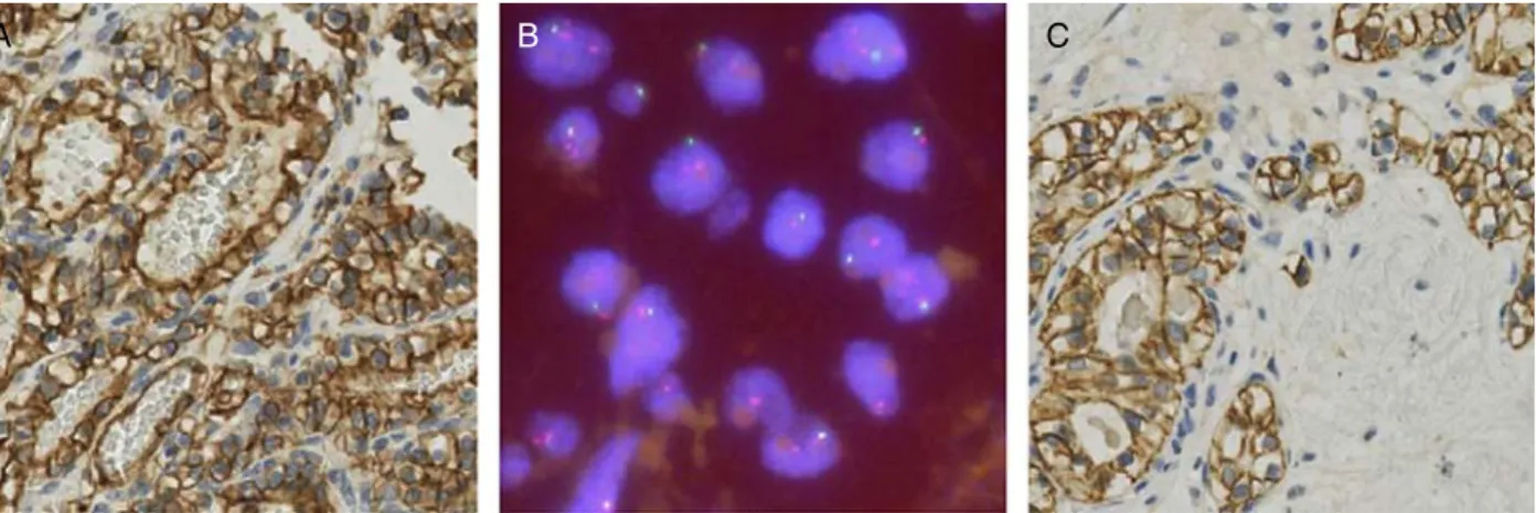
FIGURE 1. ccpRCC. Hematoxylin and eosin stain (A) and typical immunohistochemical profile with diffuse membranous CK7 positivity (B), membranous“cup-like”CA-IX positivity (C), nuclear parafibromin positivity (D), hKIM-1 negativity (E), and CD133 positivity (F).
reported a CD133 positivity rate of only 14% in ccRCC. It can therefore be concluded that CD133 is an additional tool to distinguish ccRCC from ccpRCC/RAT.
In concordance with Munari et al,57we found that
about one third of ccpRCC are positive for GATA3, a protein crucial for the regulation of Th2 development and
FIGURE 2. Hematoxylin and eosin morphology of a ccpRCC-like (A–C) and a RAT-like (D–F) case. Diagnostic features of ccpRCC include tumor cells with abundant clear cytoplasm, varying papillary, cystic and tubular architecture, and low-grade nuclei (Fuhrman grades 1 and 2) located apically distant from the basal membrane. The epithelial part of RAT tumors is composed of cells with clear cytoplasm and low-grade nuclei (Fuhrman grades 1 and 2) embedded in a smooth muscle stroma.
function. However, given that only a moderate staining intensity was seen in no more than 10% of the tumor cells, we do not consider OCT3/4 and GATA3 as diag-nostic tools to differentiate ccpRCC from ccRCC.
No previous studies have reported cancer-related death, vascular invasion, or metastasis in ccpRCC,4–7,53,58 suggesting that the disease follows an indolent course. Benign biological behavior was also observed in all
WT: Exon 2 Intron 1 WT: Exon 1 A D E B C
FIGURE 3. Molecular features of a ccpRCC-like (A) and a RAT-like (C) case both exhibiting a circumferential CA-IX staining pattern, harboring a VHL mutation (D: c.174_177delGCCG/p.Pro59GlyfsX7; E: c.351 G > C/p.Trp117Cys) and a 3p deletion detected by FISH (B). The mutation sites are denoted by an arrow. The boundaries between exon and intron are indicated. The upper base pair letter sequence shows the wild-type (WT) sequence (D and E). Tumor cells harbor only 1 VHL (green) signal and 2 CEN3 copies (orange) (B).
RAT cases.12,14,59 This is comparable to multilocular cystic RCC, which has an excellent prognosis with no disease recurrence after surgery.7,12,59 Specific molecular
alterations may account for the indolent course of mul-tilocular cystic RCC. Proposals have been put forward to rename multilocular cystic RCC as multilocular cystic
c.461C>T/ p.Pro154Leu Exon 2 Intron 2 A B C E D
FIGURE 4. ccpRCC look-alike showing classic ccpRCC morphology on the hematoxylin and eosin stain (A), with, however, a typical ccRCC immunohistochemical profile showing CK7 negativity (B), CA-IX positivity (C), hKIM-1 positivity (D), and proof of VHLmutation (E).
renal cell neoplasm of low malignant potential to under-score this specific biological behavior.1 Our group has
reported that the expression of p27, CA-IX, CK7, and CK19 is associated with a better prognosis in sporadic RCC.47,60 Interestingly, our ccpRCC/RAT cases stained strongly positive for all of these markers. Hence, the in-dolent clinical course of ccpRCC/RAT might in part be due to this specific signaling pathway. However, some of the low-grade ccRCCs included in our previous pub-lications may in fact be unrecognized ccpRCC.35,47
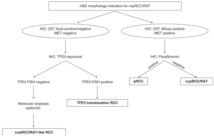
In summary, we have demonstrated that ccpRCC and RAT cannot be distinguished from one another by im-munohistochemistry and molecular analyses, and both follow a benign clinical course. We regard them as a spectrum of a distinct tumor entity. Precise diagnosis is crucial, as it has an excellent prognosis. Given the reliability of TFE3 immunohistochemistry, TFE3 FISH should be performed in cases with equivocal TFE3 immunohisto-chemistry.37 Taking into account the controversial relevance of theVHLmutation analysis in this differential diagnosis, directVHLsequencing is not helpful in separa-tion of ccRCC with prominent smooth muscle stroma from RAT. Our results suggest that a panel of antibodies against CK7, parafibromin, and MET is a helpful tool to differ-entiate most ccpRCCs/RATs from other renal tumors (Table 5). In some difficult casesVHLmutation testing and TFE3 FISH analysis are helpful tools to distinguish ccRCC and TFE3 translocation carcinoma from ccpRCC/RAT.
ACKNOWLEDGMENTS
The authors thank Susanne Dettwiler, Andre´ Fitsche, and Martina Storz for their outstanding technical assis-tance and Dorothee Pflueger for helping to interpret the TFE3 FISH data. We thank the sequencing service at the Institute of Surgical Pathology for performing numerous sequencing reactions.
REFERENCES
1. Srigley JR, Delahunt B, Eble JN, et al. The International Society of Urological Pathology (ISUP) Vancouver Classification of Renal Neoplasia.Am J Surg Pathol. 2013;37:1469–1489.
2. Tickoo SK, de Peralta-Venturina MN, Harik LR, et al. Spectrum of epithelial neoplasms in end-stage renal disease: an experience from 66 tumor-bearing kidneys with emphasis on histologic patterns distinct from those in sporadic adult renal neoplasia. Am J Surg Pathol. 2006;30:141–153.
3. Adam J, Couturier J, Molinie V, et al. Clear-cell papillary renal cell carcinoma: 24 cases of a distinct low-grade renal tumour and a comparative genomic hybridization array study of seven cases.
Histopathology. 2011;58:1064–1071.
4. Aydin H, Chen L, Cheng L, et al. Clear cell tubulopapillary renal cell carcinoma: a study of 36 distinctive low-grade epithelial tumors of the kidney.Am J Surg Pathol. 2010;34:1608–1621.
5. Gobbo S, Eble JN, Grignon DJ, et al. Clear cell papillary renal cell carcinoma: a distinct histopathologic and molecular genetic entity.
Am J Surg Pathol. 2008;32:1239–1245.
6. Rohan SM, Xiao Y, Liang Y, et al. Clear-cell papillary renal cell carcinoma: molecular and immunohistochemical analysis with emphasis on the von Hippel-Lindau gene and hypoxia-inducible factor pathway-related proteins.Mod Pathol. 2011;24:1207–1220.
H&E morphology indicative for ccpRCC/RAT
pRCC
IHC: Parafibromin
ccpRCC/RAT
IHC: TFE3 equivocal
TFE3 translocation RCC
TFE3 FISH positive
Molecular analyses (optional) TFE3 FISH negative
ccpRCC/RAT-like RCC
IHC: CK7 focal positive/negative MET negative
IHC: CK7 diffuse positive MET positive
7. Williamson SR, Eble JN, Cheng L, et al. Clear cell papillary renal cell carcinoma: differential diagnosis and extended immunohisto-chemical profile.Mod Pathol. 2013;26:697–708.
8. Herrera LP, Hirsch M, Comperat E, et al. Clear cell-papillary renal cell carcinoma (CP-RCC) not associated with end stage renal disease: clinicopathologic analysis of 50 tumors confirming a novel subtype of renal cell carcinoma (RCC) occurring in a sporadic setting.Mod Pathol. 2011;24(suppl):197A.
9. Wolfe A, Dobin SM, Grossmann P, et al. Clonal trisomies 7, 10 and 12, normal 3p and absence of VHL gene mutation in a clear cell tubulopapillary carcinoma of the kidney.Virchows Arch. 2011;459: 457–463.
10. Kuroda N, Shiotsu T, Kawada C, et al. Clear cell papillary renal cell carcinoma and clear cell renal cell carcinoma arising in acquired cystic disease of the kidney: an immunohistochemical and genetic study.Ann Diagn Pathol. 2011;15:282–285.
11. Michal M, Hes O, Havlicek F. Benign renal angiomyoadenomatous tumor: a previously unreported renal tumor. Ann Diagn Pathol. 2000;4:311–315.
12. Michal M, Hes O, Nemcova J, et al. Renal angiomyoadenomatous tumor: morphologic, immunohistochemical, and molecular genetic study of a distinct entity.Virchows Arch. 2009;454:89–99.
13. Verine J. Renal angiomyoadenomatous tumor: morphologic, immunohistochemical, and molecular genetic study of a distinct entity.Virchows Arch. 2009;454:479–480.
14. Shannon BA, Cohen RJ, Segal A, et al. Clear cell renal cell carcinoma with smooth muscle stroma. Hum Pathol. 2009;40: 425–429.
15. Kuhn E, De Anda J, Manoni S, et al. Renal cell carcinoma associated with prominent angioleiomyoma-like proliferation: re-port of 5 cases and review of the literature. Am J Surg Pathol. 2006;30:1372–1381.
16. Singh C, Kendi AT, Manivel JC, et al. Renal angiomyoadenoma-tous tumor.Ann Diagn Pathol. 2012;16:470–476.
17. Martignoni G, Brunelli M, Segala D, et al. Renal cell carcinoma with smooth muscle stroma lacks chromosome 3p and VHL alterations.Mod Pathol. 2014;27:765–74.
18. Behdad A, Monzon FA, Hes O, et al. Relationship between sporadic clear cell-papillary renal cell carcinoma (CP-RCC) and renal angiomyoadenomatous tumor (RAT) of the kidney: analysis by virtualkaryotyping, fluorescent in situ analysis and immunohistochemistry (IHC).Mod Pathol. 2011;24(suppl):179A. 19. Michal M, Hes O, Kuroda N, et al. Difference between RAT and
clear cell papillary renal cell carcinoma/clear renal cell carcinoma.
Virchows Arch. 2009;454:719.
20. Edge SB, Byrd DR, Compton CC, et al. AJCC Cancer Staging Manual. New York: Springer-Verlag; 2010.
21. Fuhrman SA, Lasky LC, Limas C. Prognostic significance of morphologic parameters in renal cell carcinoma.Am J Surg Pathol. 1982;6:655–663.
22. Schildhaus HU, Deml KF, Schmitz K, et al. Chromogenic in situ hybridization is a reliable assay for detection of ALK rearrange-ments in adenocarcinomas of the lung. Mod Pathol. 2013;26: 1468–1477.
23. Sanjmyatav J, Hauke S, Gajda M, et al. Establishment of a multicolour fluorescence in situ hybridisation-based assay for subtyping of renal cell tumours.Eur Urol. 2013;64:689–691. 24. Pflueger D, Sboner A, Storz M, et al. Identification of molecular
tumor markers in renal cell carcinomas with TFE3 protein expression by RNA sequencing.Neoplasia. 2013;15:1231–1240. 25. von Teichman A, Comperat E, Behnke S, et al. VHL mutations and
dysregulation of pVHL- and PTEN-controlled pathways in multi-locular cystic renal cell carcinoma.Mod Pathol. 2011;24:571–578. 26. Cui C, Lal P, Master S, et al. Expression of parafibromin in major
renal cell tumors.Eur J Histochem. 2012;56:e39.
27. Aron M, Zhang P, De Peralta-Venturina M, et al. Expression of novel markers human kidney injury molecule-1 (Hkim-1), S100A1 and napsin A in the differential diagnosis of renal cell carcinomas (RCC) with clear and papillary features.Mod Pathol. 2012;25(suppl): 190A.
28. Schwartz JD, Amin MB, Zhang PL. Immunohistochemical profile of stem/progenitor cell marker CD133 in variants of renal tumors.
Mod Pathol. 2012;25(suppl):240A.
29. Lin F, Zhang PL, Yang XJ, et al. Human kidney injury molecule-1 (hKIM-1): a useful immunohistochemical marker for diagnosing renal cell carcinoma and ovarian clear cell carcinoma.Am J Surg Pathol. 2007;31:371–381.
30. Ruf M, Mittmann C, Nowicka AM, et al. pVHL/HIF-regulated CD70 expression is associated with infiltration of CD27+ Lymphocytes and increased serum levels of soluble CD27 in clear cell renal cell carcinoma.Clin Cancer Res. 2015;21:889–98. 31. Choi JS, Kim MK, Seo JW, et al. MET expression in sporadic renal
cell carcinomas.J Korean Med Sci. 2006;21:672–677.
32. Cai J. Roles of transcriptional factor Snail and adhesion factor E-cadherin in clear cell renal cell carcinoma. Exp Ther Med. 2013; 6:1489–1493.
33. Tickoo SK, Reuter VE. Differential diagnosis of renal tumors with papillary architecture.Adv Anat Pathol. 2011;18:120–132.
34. Zhou H, Zheng S, Truong LD, et al. Clear cell papillary renal cell carcinoma is the fourth most common histologic type of renal cell carcinoma in 290 consecutive nephrectomies for renal cell carcino-ma.Hum Pathol. 2014;45:59–64.
35. Gill S, Kandel S, Xu B. Frequency of clear cell papillary renal cell carcinoma in cases of low grade clear cell renal cell carcinoma: a 12 year retrospective study from a single cancer center. Mod Pathol. 2013;26(suppl):212A.
36. Shi SS, Shen Q, Xia QY, et al. Clear cell papillary renal cell carcinoma: a clinicopathological study emphasizing ultrastructural features and cytogenetic heterogeneity. Int J Clin Exp Pathol. 2013;6:2936–2942.
37. Green WM, Yonescu R, Morsberger L, et al. Utilization of a TFE3 break-apart FISH assay in a renal tumor consultation service.Am J Surg Pathol. 2013;37:1150–1163.
38. Argani P, Yonescu R, Morsberger L, et al. Molecular confirmation of t(6;11)(p21;q12) renal cell carcinoma in archival paraffin-embedded material using a break-apart TFEB FISH assay expands its clinicopathologic spectrum. Am J Surg Pathol. 2012;36:1516–1526.
39. Smith NE, Illei PB, Allaf M, et al. t(6;11) renal cell carcinoma (RCC): expanded immunohistochemical profile emphasizing novel RCC markers and report of 10 new genetically confirmed cases.Am J Surg Pathol. 2014;38:604–614.
40. Petersson F, Grossmann P, Hora M, et al. Renal cell carcinoma with areas mimicking renal angiomyoadenomatous tumor/clear cell papillary renal cell carcinoma.Hum Pathol. 2013;44:1412–1420. 41. Xu W, Deng F-M, Melamed J, et al. Incidence and genetic
characteristics of clear cell tububopapillary renal cell carcinoma.
Mod Pathol. 2014;27(suppl):270A.
42. Behdad A, Hes O, Hirsch M, et al. Relationship between sporadic clear cell-papillary renal cell carcinoma (CP-RCC) and renal angiomyoadenomatous tumor (RAT) of the kidney: analysis by virtual-karyotyping, fluorescent in situ analysis and immunohistochemistry (IHC).Mod Pathol. 2011;24(suppl):179A. 43. Martignoni G, Segala D, Borze I, et al. VHL mutation, VHL
methylation, chromosome 3p and whole genomic status in clear cell papillary renal cell carcinoma.Mod Pathol. 2013;26(suppl):233A. 44. Rechsteiner MP, von Teichman A, Nowicka A, et al. VHL gene
mutations and their effects on hypoxia inducible factor HIFalpha: identification of potential driver and passenger mutations.Cancer Res. 2011;71:5500–5511.
45. Young AC, Craven RA, Cohen D, et al. Analysis of VHL gene alterations and their relationship to clinical parameters in sporadic conventional renal cell carcinoma. Clin Cancer Res. 2009;15: 7582–7592.
46. Halat S, Eble JN, Grignon DJ, et al. Multilocular cystic renal cell carcinoma is a subtype of clear cell renal cell carcinoma. Mod Pathol. 2010;23:931–936.
47. Mertz KD, Demichelis F, Sboner A, et al. Association of cytokeratin 7 and 19 expression with genomic stability and favorable prognosis in clear cell renal cell cancer. Int J Cancer. 2008;123: 569–576.
48. Nickerson ML, Jaeger E, Shi Y, et al. Improved identification of von Hippel-Lindau gene alterations in clear cell renal tumors.Clin Cancer Res. 2008;14:4726–4734.
49. Lawrie CH, Larrea E, Larrinaga G, et al. Targeted next-generation sequencing and non-coding RNA expression analysis of clear cell papillary renal cell carcinoma suggests distinct pathological mechanisms from other renal tumour subtypes.J Pathol. 2014;232: 32–42.
50. Schmidt L, Duh FM, Chen F, et al. Germline and somatic mutations in the tyrosine kinase domain of the MET proto-oncogene in papillary renal carcinomas.Nat Genet. 1997;16:68–73. 51. Korpal M, Lee ES, Hu G, et al. The miR-200 family inhibits
epithelial-mesenchymal transition and cancer cell migration by direct targeting of E-cadherin transcriptional repressors ZEB1 and ZEB2.J Biol Chem. 2008;283:14910–14914.
52. Fisher KE, Yin-Goen Q, Alexis D, et al. Gene expression profiling of clear cell papillary renal cell carcinoma: comparison with clear cell renal cell carcinoma and papillary renal cell carcinoma.Mod Pathol. 2014;27:222–230.
53. Alexiev BA, Drachenberg CB. Clear cell papillary renal cell carcinoma: incidence, morphological features, immunohistochem-ical profile, and biologic behavior: a single institution study.Pathol Res Pract. 2014;210:234–241.
54. Guo J, Tretiakova MS, Troxell ML, et al. Tuberous sclerosis-associated renal cell carcinoma: a clinicopathologic study of 57
separate carcinomas in 18 patients. Am J Surg Pathol. 2014;38: 1457–1467.
55. Boysen G, Bausch-Fluck D, Thoma CR, et al. Identification and functional characterization of pVHL-dependent cell surface proteins in renal cell carcinoma.Neoplasia. 2012;14:535–546.
56. Williamson SR, Zhang S, Eble JN, et al. Clear cell papillary renal cell carcinoma-like tumors in patients with von Hippel-Lindau disease are unrelated to sporadic clear cell papillary renal cell carcinoma.Am J Surg Pathol. 2013;37:1131–1139.
57. Munari E, Segala D, Gobbo S, et al. GATA3 expression in clear cell papillary renal cell carcinoma and renal cell carcinoma with prominent leiomyomatous proliferation is a further evidence of the relationship between these two entities.Mod Pathol. 2014;27(suppl): 250A.
58. Leroy X, Camparo P, Gnemmi V, et al. Clear cell papillary renal cell carcinoma is an indolent and low-grade neoplasm with over-expression of cyclin-D1.Histopathology. 2014;64:1032–1036. 59. Williamson SR, Halat S, Eble JN, et al. Multilocular cystic renal cell
carcinoma: similarities and differences in immunoprofile compared with clear cell renal cell carcinoma. Am J Surg Pathol. 2012;36: 1425–1433.
60. Dahinden C, Ingold B, Wild P, et al. Mining tissue microarray data to uncover combinations of biomarker expression patterns that improve intermediate staging and grading of clear cell renal cell cancer.Clin Cancer Res. 2010;16:88–98.