Nitrate
Reductase Complex
of Escherichia coli K-12:
Participation
of
Specific Formate
Dehydrogenase
and
Cytochrome
b,
Components
in
Nitrate Reduction
JOSE RUIZ-HERRERA' AND J. A. DEMOSS
Departmentof Biology, UniversityofCalifornia, San Diego, La Jolla, California92037
Receivedfor publication9 June 1969
Theparticipation of distinct formate dehydrogenases and cytochromecomponents
innitratereduction by Escherichiacoliwasstudied. The formate dehydrogenase ac-tivitypresentinextractsprepared fromnitrate-induced cellsofstrain HfrHwas
ac-tive with variouselectronacceptors,including methylene blue,phenazine methosul-fate, and benzyl viologen. Certainmutants which areunabletoreduce nitrate had loworundetectablelevels of formate dehydrogenase activity assayed with methylene
blue or phenazine methosulfate as electron acceptor. Of nine such mutants, five producedgaswhengrownanaerobically without nitrate and possessedabenzyl
vio-logen-linkedformate dehydrogenaseactivity, suggesting that distinct formate dehy-drogenases participate in the nitrate reductase and formic hydrogenlyase systems. Theother fourmutantsformed littlegaswhengrownanaerobically in the absence of nitrate andlacked the benzylviologen-linkedformatedehydrogenaseaswellasthe methylene blueorphenazinemethosulfate-linkedactivity. The cytochrome b1 present in nitrate-induced cells was distinguished by its spectral properties and its genetic
control fromthemajor cytochrome b1 componentsofaerobic cells andofcellsgrown
anaerobically in the absence of nitrate. Thenitrate-specific cytochrome
bk
wascom-pletely and rapidly reduced by1 mmformate butwasnotreduced by 1 mmreduced nicotinamide adenine dinucleotide; ascorbate reduced only partof thecytochrome b1 which was reduced by formate. When nitrate was added, the formate-reduced cytochrome b1 was oxidized with biphasickinetics, but the ascorbate-reduced cyto-chrome
bk
was oxidized withmonophasic kinetics. The inhibitory effects of n-heptylhydroxyquinoline-N-oxide onthe oxidation of cytochrome b1 by nitrate provided evidence that thenitrate-specific cytochrome is composed oftwocomponentswhich have different redoxpotentials butidentical spectral properties. We conclude from thesestudies that nitratereduction in E. coli is mediated by the sequential operation ofa specific formate dehydrogenase, two specific cytochrome b, components, and nitrate reductase.
Anumber of observations indicate thatnitrate reduction in Escherichia coli occurs mainly by a pathway involving formate dehydrogenase,
cyto-chrome
bl,
and nitrate reductase. Among the various substrates which are metabolized byE. coli, formate is the most effective electron donorfor thereduction of nitrate in cellsgrown anaerobically in the presence of nitrate (2, 23). Insuchcells, formatedehydrogenase,cytochrome b1, and nitrate reductase are induced to
rela-IPresent address: Departamento de Microbiologia, Escuela
NacionaldeCienciasBiologicas,I.P. N., CarpioyPlan deAyala, Mexico,D.F.
tively high levels
compared
to those in cells grownintheabsence ofnitrate(2,
17, 23).These three components are present in membranefractions (6) and have been
partially
purified
as a unit from cellextracts
(8).
Finally,
mutants of E. coli which are unable toproduce
nitrite from nitrate (NR7mutants) lack either formatedehydrogenase or nitrate reductase or combina-tionsoftheseactivities and cytochrome
b& (1,
16,17,22).
The proposal that formate dehydrogenase and cytochrome
bi
arespecific
components of thenitrate reduction system raises some important
NITRATE REDUCIASE COMPLEX OF E. COLI questions concerning the relationship of this
pathway with other pathways of electron trans-port in E. coli. Formate dehydrogenase must
also be a component of the hydrogenlyase sys-tem (15) as well as of formate oxidase. These multiple functions of formate dehydrogenase have beenpreviously recognized, andit hasbeen suggested that at least two distinct formate dehydrogenases, particulate and soluble, are formedbyE.coli (4, 5).
The participation of cytochrome
b,
in nitrate reductioncreates an even morecomplexsituation, since cytochrome b1 is one of the majorcyto-chromes present in both aerobic and anaerobic
cells of E. coli and therefore must function in
other electron transport systems (3, 10, 18). At present it is not known whether distinct cyto-chrome b1 components are involved in different electron pathways or to what degree such
path-waysinteract.
Todefine the components of the nitrate reduc-tase complex and their relationship to other metabolic pathways, westudied the biochemical
events involved in nitrate reduction in the wild-type E. coli and in a number of NR- mutants.
Evidence is presented that a specific formate dehydrogenase, two distinctcytochrome b1
com-ponents, and nitrate reductase are obligatory
components of a nitrate reductase complex in
E. coli.
MATERIALS AND METHODS
Thestrains of E. coli used in these studies, HfrH andAB2102,and the NR- mutantsderived fromthem weredescribedpreviously (17).Allstrainsweregrown on a complete medium containing a salt base (20), thiamine hydrochloride (5 ,g/mI), 0.4% nutrient -broth (Difco), and1% glucose (sterilized separately). Whenindicated, the mediumwassupplemented with 1%potassium nitrate and 0.5%sodiumformate.
For measuring activities, cultures were grown in flaskswithKletttube side arms at37C in ashaking waterbath. Foraerobic conditions, themedium was spargedvigorously with sterile air.Foranaerobic con-ditions, the mediumwasspargedwith asterile mixture of95%N2plus 5%CO2.Thecultures wereinoculated with a 2 to 5% inoculum froman overnightculture grown insimilar mediumandallowedtogrow tothe middle of the exponential phase. The cells were col-lected by centrifugation, washed twice with 0.05 M potassium phosphate buffer (pH 6.8 or 7.3), and resuspended inthe same buffer. Thissuspension was used as such (whole cells) or frozen overnight at
-15C before use (frozen-thawed cells). Cell-free extracts wereprepared bythree treatments of15 sec
each in a 10-kc Branson Sonifier, followed by
cen-trifugationat3,000 X g for 10minto removewhole cells.
Formate dehydrogenase was measured with dif-ferent electronacceptorsbyutilizingaradioassayora
spectrophotometric assay described previously
(17),
or by the following manometric procedure using aWarburgapparatus. In the main compartment were
placed0.2 ml ofcellfsorextract,10
;umoles
of electron acceptor, and0.05/M
potassium phosphate buffer (pH6.8) in a total volum -of 2.7 ml; 30 ,umoles of formate was placedin the side arm. Flasksand ma-nometers were flushed with N2 and equilibrated at37 C before tippingthe formate into the main
com-partment. Activity was expressed as microliters of CO2 evolved per minute per milligram of protein. No CO2 was produced in the absence of added electron acceptors.
In some experiments, benzyl viologen-specific formate dehydrogenasewas measuredby thefollowing
qualitative procedure.InThunberg tubes,140 umoles ofpotassium phosphate buffer (pH 6.8), 1.0
,smoles
of benzylviologen, 60;umoles
of sodium formate, and cells or extract were mixed in a final volume of 5.0ml. Thetubes were flushed with N2, and reduction of the benzyl viologen was followed by use of a Klett color-imeter with a 660-nm filter. The development ofcolor was not linear and, therefore, the results were ex-pressedqualitatively.Formichydrogenlyase was assayedmanometrically by following hydrogen evolution from formate (15). Gas production by growing cultures was assessedby observing the accumulation of gas in inverted vials submergedin tubes of the culture medium.
Nitrate reductase was assayed with formate or reduced methyl viologen as electron donors, as pre-viously described (17). With other electron donors the procedure utilized with formate was followed, sub-stituting the electron donor for formate.
*The absorption spectra of the cytochromes were determined with a single-beam recording spectro-photometer constructed and kindly made available by Warren Butler at the University of California, San Diego. Monochromatic light of 1.0-nm half band-width was provided by a Bausch & Lomb grating monochromator. The output of the photometer and a potentiometer geared to the wavelength drum pro-videdtheinputsignals to a Moseley 7000 AM X-Y recorder. For measurement of absolutereduced spec-tra atliquidnitrogentemperature, a 1.0-ml sample was placedin a metal-sided absorption cell,reducedwith solidsodium dithionite, mixedwith 500 mg of Al203 (1,umdiameter),frozen inliquid N2, and placed ina
Dewarflask containingN2in the cell compartment of theinstrument. Formeasurement of absolute reduced spectra at room temperature, the same general pro-cedure was followed, omitting the liquid N2. For determining the differential spectra, an attachment whichprovidedasplit beam wasutilized. In this case, identical3.0-ml sampleswere placed in glass absorp-tion cells (10-nm light path). The contents of one cuvette was oxidized by flushing with air for 30 sec, and the other was reduced with solid sodium di-thionite to obtain the reduced-minus-oxidized spec-trum.
Cytochrome levels were calculated as follows. A basal line was drawnbetween540and 570 nm, and the heightof the peak of the a-band wasmeasuredfrom this baseline. The optical density units were calcu-721
lated, and thecytochromelevelisexpressedasoptical
density unitspermilligramof protein. The kineticsof reduction and oxidation ofcytochrome b1 was
fol-lowed with an Aminco-Chance double-wavelength
spectrophotometer at either 35 C or room
tempera-ture. Protein was determined by the procedure of
Lowryetal. (12).
RESULTS
Thegeneral properties of the nitrate reduction
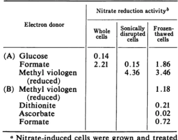
system present in the wild-type strains utilized in these studies corresponded tothosepreviously reported (2, 6, 8). In nitrate-induced cells of strains HfrH, formate was a more effective
electron donorfor nitrate reduction than glucose (Table 1). In frozen-thawed cells or in cell
ex-tracts, several reagents act as electron donors
for nitratereduction (Table 1). Reduced methyl viologen, which transfers electrons directly to
nitrate reductase (21), was the most effective electron donor, but formate was 30 to 40% as
active as reduced methyl viologen in
frozen-thawed cells. Nitrate reduction with formate as electron donor was completely inhibited by
0.01 mM n-heptyl hydroxyquinoline-N-oxide (HOQNO), whereas a 100-fold increase in the concentration of the inhibitor did not affect the reduction ofnitrate with reduced methyl viologen
or ascorbateasthe electron donor.
The formate dehydrogenase present in nitrate-induced cells of strain HfrH utilized several electronacceptors (Table 2). Methylene blue and
TABLE 1. Nitrate reduction with various
electron donorsa
Nitratereductionactivityb Electron donor
dwhole Sonically
Frozen-disrupted thawed
cells cells cells
(A) Glucose 0.14 Formate 2.21 0.15 1.86 Methyl viologen 4.36 3.46 (reduced) (B) Methyl viologen 1.18 (reduced) Dithionite 0.21 Ascorbate 0.02 Formate 0.72
aNitrate-induced cells were grownand treated
as described. Nitrate reduction was assayed as
described, with the following levels of electron donors in afinal volume of2.5 ml: 48 Amoles of glucose, 48 MAmoles of formate, 0.25 ,umoles of methyl viologen (reduced), 2.8,umoles of sodium dithionite, 10lAmoles of ascorbate, 6 ,umoles of
formate.
bExpressedasmicromoles of nitrateperminute
per milligram of protein.
TABLE 2. Activity offormate dehydrogenase with various electron acceptors
Electronacceptora Activity'
None 0.6
Methylene blue 65.2
Phenazine methosulfate + DCPIP 56.7
DCPIP 15.8
Ferricyanide 3.5
Benzylviologen 12.2
Triphenyltetrazolium chloride 9.0
Air 12.3
aThe concentration of electron acceptors used was 3.1
jsmoles/ml
(except forair).bNitrate-induced cells of HfrH were grown andbroken inaBransonSonifier, andthe extract wasprepared asindicted in Materials and Meth
-ods, but itwas frozen beforeuse.Formate dehy-drogenase was assayed by the release of radio-active CO2 from '4C-formate, and nanomoles of CO2 were calculated from the specific activity ofthe formate (8,230 counts per min per,mole)
utilized. Results shown are expressed as nano-molesofCO2 perminutepermilligramofprotein. phenazine methosulfate were the most effective acceptors, whereaspotassiumferricyanide, benzyl
viologen, and
triphenyltetrazolium
chloride were much less effective. Wehave routinelymeasuredformate dehydrogenase by following the
reduc-tion of dichlorophenol indophenol (DCPIP)
in the presenceofcatalyticamounts ofphenazine methosulfate (17). Although DCPIP is only slowly reduced by a mixture of crude extract and formate, in the presence of small amounts of
phenazine
methosulfate itis reduced at a rate whichcorresponds
to the rate observed with methylene blue. This assay does not involve anindirect flow of electrons through cytochrome b1, since reduced cytochrome
bi
is not oxidized by phenazine methosulfate in our preparations and formatedehydrogenase
can beassayed by
this
procedure
inmutantswhich lackcytochromebi
.Theavailability of anumber ofmutantswhich
lack formate dehydrogenase in nitrate-induced
cells (17)
permitted
us to test directly the possi-bility that distinct formatedehydrogenases
areinvolved in themetabolism ofE. coli.
Although
formate dehydrogenase
activity
which utilizesmethylene blue or phenazine methosulfate as
electronacceptors could not be detected or was
very low in such mutants, some of the mutants still formed gas whencultivatedunderconditions which lead to the formation of the formic
hy-drogenlyasesystem in the
wild-type
strain(Table
NITRATE REDUCIASE COMPLEX OF E. COLI acceptor, the evolution of CO2 from formate
or the reduction of the dye in the presence of formate was not linear with time and is, there-fore,expressed qualitatively.
These results support the hypothesis that two
biochemically distinct formate dehydrogenases
are involved in nitrate reduction and hydrogen
formation in E. coli. However, the formate de-hydrogenases may not be genetically distinct since, in some of the NR- mutants, both the
nitrate-specific formate dehydrogenase and the formate dehydrogenase involved in the formic
hydrogenlyase system are lost (Table 3) as the result of mutations which appear to be single, point mutations on the basis of reversion rates
(17).
When E. coli is grown anaerobically, the addition of nitratecauses anincrease inthe level
TABLE 3. Gasformation and formate
dehydro-genaseactivity inHfrHandselected NRtmutants Strain HfrH TW-15 TW-17 TW-22 TW-101 TW-112 TW-135 TW-140 TW-149 TW-153 Gas formation' + + + 0 0 Formate dehydrogenase activityb Methylene bluec 1.15 0.05 0.08 0.00 0.09 0.04 0.00 0.00 0.14 0.00 Phenazine metho-sulfate+ DCPIP 0.43 0.02 0.02 0.00 0.02 0.00 0.00 0.00 0.00 0.00 Benzyl vio-logen + + + 0 +
a Gas formation was assessed in cultures growing anaerobically incomplete medium
with-out nitrate in tubes containing inverted vials
to trap gas. Cultures were allowed to incubate
atleast5 daysandwereconsidered tobepositive
(+) if they formedmorethan 10% thegasobserved
inthe wildtype.
IThe assay of formate dehydrogenase with
benzyl viologen was qualitative. All assays were
performed with fresh cellextracts prepared from
cells grown anaerobically on complete medium
(benzyl viologen assay) or on complete medium
supplementedwithnitrate (radioassay and colori-metricassay). Resultsareexpressedasmicromoles
of C02 per minute per milligram of protein;
results for benzyl viologen areexpressed
qualita-tively.
cThe activity with methylene blue was
cal-culated as micromoles of CO2 released from formate (specific activity, 22,800 counts per min
per,mole).
ofcytochrome
bi
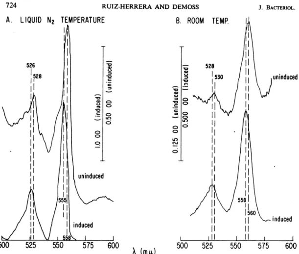
(17, 23). The following spectralanalysis demonstrates that the cytochrome bi
componentsformed in the presence and absence
ofnitrate arealsoqualitatively distinct. Absolute spectra of frozen-thawed cells reduced with
dithionite are shown in Fig. 1. The cells grown in the presence of nitrate exhibited only two major peaks between 500 and 600 nm. These were 526 nm
(3
band) and 555 nm (a band) atliquidnitrogentemperaturesand 528and 558nm at room temperature. On the other hand, the
cells grown in the absence of nitrate exhibited major peaks at 528 and 558 nm, a shoulder at 549 nm (cytochrome c) at liquid nitrogen tem-perature, and peaks at 530 and560 nm at room
temperature. The distinction between these b55a and b558 components (identified by their a band
peaks at liquid nitrogen
temperature)
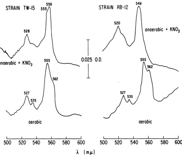
was clear insome NR- mutants which hadreduced levelsof cytochrome
b1.
When such mutants were grownunderanaerobic conditions in thepresenceof nitrate,bothcomponentswereapparentinthe reduced spectra (Fig. 2,mutant
TW-15).
Further-more, asecond type of mutant,whichformedno
detectable cytochrome b555 component when
grown anaerobically with nitrate (Fig. 2, strain RB-12), formed normal levels of the b558 com-ponents when grown
anaerobically
inthe absence of nitrate (not shown, cf.wild-type-uninduced
spectrum, Fig. 1).
When the wild-type
strains
were grownaero-bically, the bands of the major cytochrome b
components were at 555 and 562 nm (at
liquid
nitrogentemperature).Onthebasis of the follow-ing observations with NR- mutants, the b555 components found in anaerobic,
nitrate-induced
cells
would appear to be distinct from the b5a5 component in aerobiccells.NR- mutants, which have altered levels of the b555 component when grownanaerobically
with nitrate,formed
a normal distribution ofcytochrome components, including a b555 component, when they were grown under aerobic conditions (Fig. 2). Theabsolute spectra shown for the aerobic cells are
identical tothat found in aerobic wild-type cells
grown under these conditions, showing peaks
at555and 562
nm.
Thus the major cytochrome b component foundincellsgrownanaerobicallyinthepresence of nitrate appears to be physiologically and
genetically
distinct from the cytochrome b com-ponents found under other growth conditions.The
following
observations directly implicatethe
b555
component in the nitratereductionsystemandprovide evidencethat it is
composed
oftwo distinct cytochromes which possess identicalspectral
characteristics.723 VOL.99, 1969
A.
LIQUID
N2
TEMPERATURE
-W CD) C. lo 4) 0 -V U, 0B.
ROOM
TEMP.
,,
528 530C._1
,0
11
=°
> II U) I I n ci IH c'Juninduced
500
525
550
575
600
500
525
550
575
600
(mit)FIG. 1. Absolute dithionite-reducedspectraofstrainHfrHgrownwithandwithoutnitrate. Cellsweregrown
anaerobicallyincompletemedium with(inducedcells) andwithout (uninduced cells)potassium nitrateandtreated
asindicated.Frozen-thawed cellsuspensionswereusedatprotein concentrationof3.08mg/ml fortheuninduced cells and 1.74mg/mlfortheinducedcells.Spectraweredeterminedatliquid N2temperature (A)androom
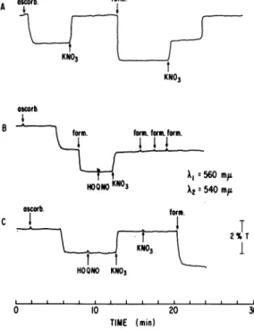
tem-perature(B).Thebars in thecenterofthefigureshow the scaleofopticaldensityunitsforeachofthecurves. When the kinetics of reduction and oxidation
ofcytochrome b1 were followed in an Aminco-Chance double-wavelength recording spectro-photometer, it wasfound that repeated additions
of1 mmreduced nicotinamide adenine
dinucleo-tide (NADH) did notcause the reduction of the cytochrome b1 component present in frozen-thawed preparations of nitrate-induced cells (Fig. 3). By increasing the NADH concentration
to 100 mM, it was possible toeffect a reduction of cytochrome
bi,
but we regarded this as anon-physiological level of NADH. Similar results were obtained by using crude extracts.
When 1 mm formate was added, an immediate
reduction ofcytochrome
b,
occurred. The subse-quent addition of nitrate caused the complete oxidation ofthe cytochrome, but with peculiar kinetics (Fig. 3). Only part of the cytochromewas immediately oxidized, the remainder being
oxidized after a definite time lag. With some
time lag, ascorbate also caused the reduction of cytochrome
bi
.However,inthiscasethereducedcytochrome was completely oxidized in a single
step upon addition of nitrate (Fig. 4A). Appar-entlyonlypartofthecytochrome b1 wasreduced,
since thesubsequent addition of formate caused the reduction ofmore cytochrome, and in this case the biphasic kinetics were observed for
oxidationby nitrate. This difference in theextent
ofreduction of the cytochromecomponentcaused by formate and ascorbate was verified by the
experiment showninFig. 4B, inwhich the addi-tion of formate immediately after ascorbate caused an additional increment of cytochrome reduction. These results suggestedthat two cyto-chrome
b,
components werereduced by formate, one of which is reduced by ascorbate, and thatnitrate oxidized both cytochrome components butone of them onlyaftera definite timelag.
The effects of HOQNO on the oxidation of
,uninduced
Iq
NITRATE
REDUCITASE
COMPLEX OF E. COLISTRAIN
TW-15
528 5'STRAIN
RB-12
0.025 O.D.
anaerobic
+KNO3
anaerobic
+KNO3
527aerobic
aerobic
I I500
520
540
560
580
600
I l500
520
540
560
580
600
X(mIt)
FIG. 2. Cytochrome spectra oftwo NR- mutantsgrownunderdifferent conditions. The spectra were
deter-minedatliquid N2temperatureonfrozen-thawedsuspensionsofcellsgrownandpreparedasdescribed. Protein
con-centrations were6.25 mg/ml for RB-12 (anaerobic), 4.32 mg/ml forRB-12 (aerobic), 6.82 mg/ml for TW-IS
(anaerobic), and 5.72 mg/mlfor TW-1S (aerobic).
cytochrome
bi
component provided additional evidence that two distinguishable cytochromeswerebeing reducedandoxidized inthese experi-ments. When 0.01 mm HOQNO was addedafter
the complete reduction of the cytochrome (Fig. 4B), only part ofthe cytochrome was reoxidized by nitrate and the oxidation took place imme-diately in a single step. Identical results were
obtained when the cytochrome was reduced by formate only, i.e., the second step of the
biphasic
kinetics was completely inhibited byHOQNO. Furthermore, the reduction again of the oxidized cytochrome by formate was
pre-vented by HOQNO (Fig. 4B). In contrast, that
portion of cytochrome b1 which was reduced by ascorbate was not prevented by HOQNO from being oxidized by nitrate (Fig. 4C). The
addition of more nitrate had no further effect and, in this case, HOQNO did not prevent
formate from reducing at least part of the
cyto-chrome.
To demonstrate that these results were due specifically to the behavior of cytochrome b555,
a similar set of
experiments
was carried out in whichspectra weredetermined at room tempera-ture with the split-beam attachment for the spectrophotometer (Fig. 5A). The formate-reduced cytochrome bexhibited
anca band,
at 558 nm at room temperature, whichcorresponds
to the b555 component (see
Fig.
IB).
Nitratecompletely reoxidized the cytochrome, but when
HOQNO was added to the
formate-reduced
preparation,
nitrate caused the oxidation of only part ofthe cytochrome,leaving
a reduced cytochrome with an identicalabsorption
maxi-mum. The results presented inFig. SB also con-firmed the results obtained with ascorbate as electron donor and demonstrated that a cyto-725 VOL. 99, 1969
AT 0 NADH
I
A2: Formote '7'T 0 2 FIG. 3. Reductio tion by nitrate. Chc werefollowed asthi \1 (560nm)andX2 leftshows thescaleCells of HfrH wer medium with 1% suspended as desc (1.74mg ofproteil phosphatebuffer, 1 Whereindicated, 3 ,umolesof sodiumi potassium nitrate
NADH Formate the remainder of the reduced cytochromewasnot oxidized, evenafter 8minof continuedbubbling
with air (curve c). When the concentration of formate was lower (5 X 10⁻⁴ M), HOQNO did not inhibit the oxidation of the
cytochrome
560m~~~~~~i
~(curves
d,
e,f).
560
mpz
lAgain,
these results may beinterpreted
by
540
mpt
assuming the existence of twocytochromes
inI______I______I______I_____
nitrate-induced cells which can be reducedby
2 3 4 5 6 formate and oxidized
by
oxygen(Fig. 7).
TheTIME (min) presence of HOQNO would inhibit the electron
flow between both cytochromes, allowing only one
cytochrome
to berapidly
oxidized in thepresenceof excess formate, since formate would continue to reduce the cytochrome before the block. The fact that HOQNO had no effect on oxidation of cytochromes when a low level of
formatewasused suggeststhat both cytochromes
areautooxidizable.
XXI1
560mul
DISCUSSIONKNO3
Theparticipation
of formatedehydrogenase,
cytochrome b1 and nitrate reductase in nitrate
4 6 8
TIME (min)
ascorb form.
)nof cytochromeb1and itsreoxida- A anges inthe stateofthecytochrome
edifferencein transmittancebetween
(540 nm) at35C.Thescaleatthe KNO3 e oftransmittance change observed.
re grown anaerobically in complete KNO3
potassium nitrate, washed, and re-*ribed. A frozen-thawed suspension
n) wasmixed with 0.05 Mpotassium ascorb.
pH
6.8,
inafinal
volumeof
2.8ml. B o f'.8Mnoles of NADH (0.01
ml),
3.8 B form. for.fomform.rormate (0.01
ml),
and 10 JAmoles of (0.01 ml) were added.-N3
Al560m,i
HO KNO3 A 540mu
ascorb. fo~~~~~~~~~~~rm. 1V w
chromewith anabsorption maximum of 558 nm at room temperature is involved. The kinetic
changes observed, therefore, reflect changes
occurring specifically in the cytochrome
b555
components.
These results can be explained assuming the
participation in nitrate reduction of two
cyto-chromes with identical spectra differing only in
their redox potentials (see Fig. 7).
Further evidence for such a hypothesis was
obtained by analyzingthe autooxidation of
cyto-chrome
b1
in nitrate-inducedcells
(Fig. 6). Formate at a concentration of 8.3 X 10³ Mrapidly reduced cytochrome
bi
(curvea).
Afterair wasbubbled through the
cell
suspension for 1 min, the cytochrome was completely oxidized(curve
b). When 8.3 X 10 M HOQNO wasadded, bubbling withair for 1 min caused rapid
oxidation of only half of the cytochrome, and
oscorb. form. C I I KNO3 HOONO KNO3 0 10 20 TIME (min)
FIG. 4. Reduction and oxidation of cytochrome b1 and effect of HOQNO. Cells were preparedandthe
conditions were as in Fig. 3 except that the
frozen-thawedcell suspension was treated with
deoxyribonu-clease (2.5 u.g/ml) to reduce the viscosity. Sodium
formate andsodium ascorbate (ascorbic acid adjusted topH 7.0 withsodium hydroxide) wereaddedas0.01
mlof 1.0 Msolutions to4.0 ml of cellsuspension (4.8
mg/mI).
2%T
I
NITRATE REDUCTASE COMPLEX OF E. COLI reduction by E. coli has been proposed by a
number ofinvestigators (6, 8, 17, 23). The results presented here, along withthe resultsfromstudies
A reducedwith
Ifo/
fite
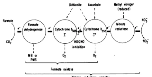
KNOX +HOONO ond KNO, reduced with ascorbate I +KNQ3 0.025OD0 +HOONO and KNO, Formate \ Farm deydoen_ C02 MBor PMSDithionite Ascorbate methyl viologen
/ \ (reduced) \ 0(\1 4 4 I IT( OONO NO2 inhibition 4 Formateoxidase
Nitrote reductaose comptex
FIG.7. Suggestedschemefor the nitrate reductase complex of Escherichia coli.
500520540 560580 600 520 54050 600
FIG.5. Differentialspectraofcytochrome b1 reduced by formate and ascorbate and oxidized by nitrate in thepresence of HOQNO. Cells of HfrHwere grown
anaerobically in complete medium supplemented with
1% potassium nitrate and used as a frozen-thawed
suspension. In this case, 10 gg of deoxyribonuclease
wasaddedperml ofsuspensiontoreducetheviscosity. Differential spectra were obtained with the split-beam
spectrophotometer at room temperature. A sample of cell suspension (3.0 ml containing 2.0 mg ofprotein
per ml) was added to each cuvette. To the reference
cuvette, 0.05mlof I Mpotassium nitratewasaddedto
oxidize the cytochrome b1. In the reduced cuvettes,
0.05 ml of 0.06Mformate orsolid ascorbic acidwas
added.
FIG.6. Auto-oxidation ofcytochrome b1 andeffect ofHOQNO. CellswerepreparedasinFig. 4and used
at a protein concentration of2.3 mg/ml. Difference
spectra weredeterminedat room temperaturewith the split-beam spectrophotometer. The reference cell
suspension wasoxidizedby bubbling with air through
apasteurpipette. (a) Cellsuspension plus8.3 pmoles of sodium formateperml; (b) suspension (a) bubbled
withairforI min; (c) suspension (a) plus8.3X 10- M HOQNO, bubbledwith airfor8min; (d) cell suspen-sionplus 0.5 Amoles ofsodium formate per ml; (e) suspension(d)bubbled with airfor I min: ()suspension (d) plus 8.3 X 10³ MHOQNO, bubbledwith airfor
I min.
on NR- mutants (17), indicate that distinct
forms of these components are involved in the
nitrate reduction system and that there is little interaction with other electrontransport chains,
i.e., nitrate reduction occurs mainly by the
sequential operation of formate dehydrogenase,
cytochrome
b555,
and nitrate reductase. Severla investigators have demonstrated that NADHmayserve as anelectrondonor for nitrate reduc-tion in cell-free extracts of E. coli (2, 5, 6, 13). However, NR- mutants which lack formate dehydrogenase but possess nitrate reductase
neither form nitrite nor remove nitrate from the
medium duringgrowth (unpublisheddata). When suchmutantsreach thestationary phase,however,
nitrite begins to accumulate, suggesting that NADH may serve as an electron donor for
nitrate reduction only in stationary-phase cells. In contrast, the wild-type parent accumulates
largeamounts ofnitriteduring growth underthe
sameconditions, and this accumulation continues
during the stationary phase. In none of these cases is nitrite further reduced, presumably
because we used 1 % nitrate, a condition which suppresses the formation ofthe nitritereductase system in E. coli (2). The specific role of the
NADH-linked nitrate reductase activity and its relationship to nitrate reductase activity
asso-ciated withformatedehydrogenase is, atpresent, unknown.
Theformate dehydrogenase involvedinnitrate
reductionappears tobe distinctfrom the formate
dehydrogenase involved in the formate hy-drogenlyase system. This distinction is based partly on the electron acceptor specificity of the formate dehydrogenase associated with these
two systems (5), but it is most clearly demon-strated by the existence ofa functional formate
hydrogenlyase system and a benzyl viologen-specific formate dehydrogenase in NR- mutants
which lack the phenazine methosulfate-specific
formate dehydrogenase associated with nitrate reduction. The fact that some NR- mutants lack both of these formate dehydrogenases, as
shown here and by others (14, 22), suggests that both activities depend on a common genetic element, perhaps the structural gene for formate dehydrogenase. If both are specified by the same gene, the genetic and physiological variations observed for the two formate dehydrogenases must result from an interaction and association of the gene product with distinct electron trans-port components. An alternative interpretation is that both activities are lost as a result of some pleiotropic alteration affecting the membrane components of the cell (1). In any case, a clear
understanding of the genetic variations observed
for the formate dehydrogenases will require a detailed genetic and biochemical analysis of this enzyme and its participation in distinct pathways. The scheme in Fig. 7 is proposed to explain theinteraction of the nitrate-specific cytochrome
b,
with the different electron donors, itsoxida-tion by nitrate, and the effects of the inhibitor HOQNO on these processes. Several-of the facts
presented here suggest that two distinct cyto-chrome b555 components are oxidized by nitrate. (i) Nitrate causes a biphasic oxidation of
formate-reduced cytochrome b555. (ii) Ascorbate, an
electron donor of moderately high redox
poten-tial, reduced only a part (about 50%) of the
nitrate-specific cytochrome b555 which dithionite or formate reduce. (iii) HOQNO inhibits the
oxidation of only part (about 50%) of the for-mate-reduced cytochrome b555 by nitrate but does not inhibit the oxidation of ascorbate-reduced
cytochrome b555 by nitrate. These observations,
along with the fact that HOQNO completely
inhibits the reduction of nitrate by formate, are most easily explained by the proposal in Fig. 7 that two cytochrome
b555
components withdifferent oxidation-reduction potentials function
sequentially in the transfer of electrons from
formate to nitrate. Whereas dithionite and
formate (via formate dehydrogenase) would
reduce both components, ascorbate would
reduce only the second cytochrome component. The selective effects of HOQNO would result
from its inhibition of the transfer of electron
between the two cytochrome components. The
biphasic kinetics of oxidation by nitrate would be due to the ability of formate, as long as it
is present, to keep cytochrome I reduced even
in the presence of oxidized cytochrome II.
Such a model might explain why many NR-mutants which lack either nitrate reductase or
formate dehydrogenase
also
exhibit decreasedbut intermediate levels of the cytochrome b555
(17);i. e., one or the other of the two cytochrome components might not be formed. Furthermore, the reports that cytochrome
bi
is associatedboth with formate dehydrogenase (7, 11) and
with nitrate reductase (9) after these enzymes had been resolved from one another can be explained by assuming that one cytochrome
bi
component remains associated with formate dehydrogenase and the other with nitrate re-ductase during the purification procedures.
It is clear that the nitrate-specific cytochrome
b555
components are distinct from the cyto-chrome b555 component formed under aerobic conditions, since NR- mutants which lack the nitrate-induced cytochrome b555 still form normal cytochrome b1 components under aerobic con-ditions. These observations do not establish whether that is the same cytochromebi
asso-ciated with different components or that E. colispecifies a series of distinct cytochrome
bi
com-ponents for various electron transport systems. In any case, the formation of cytochrome com-ponents must be specifically regulated by the other catalytic components with which theyarenormallyassociated.
Thatnitrate reduction is catalyzed by a
phys-ically associated complex of enzymes can be established only by the isolation of the catalytic unit. However, the membrane association of the components, the existence of pleiotropic
mutations affecting the activities, and the ap-parent lack of interaction of this system with other electron transport chains argue for the existence of such a complex.
ACKNOWLEDGMENTS
Weare indebted to William Cramerfor hishelpwith the cyto-chromemeasurementsandhis valuable advice and criticism. This workwassupported byaPublicHealth Service grant GM 13200-04 fromthe NationalInstitute of GeneralMedical Sciences.
LITERATURE CITED
1. Azoulay, E., J. Puig, and F. Pichinoty. 1967. Alteration of respiratory particles bymutation in Escherichia coli K12. Biochem. Biophys. Res. Commun. 27:270-274.
2. Cole, J. A., and J. Wimpenny. 1968.Metabolicpathwaysfor nitrate reduction in Escherichiacoli.Biochim.Biophys. Acta 162:39-48.
3. Deeb, S., and L. Hager. 1964.Crystallinecytochromebi from Escherichia coli. J. Biol. Chem. 239:1024-1031.
4. Gray,C. T., and H. Gest. 1965.Biologicalformation of molec-ularhydrogen.Science148:186-192.
5. Gray, C. T., J. Wimpenny, D.Hughes, and M. Mossman. 1966. Regulation ofmetabolism in facultative bacteria. I. Structural and functional changes in Escherichia coli as-sociated with shifts between the aerobic and anaerobic states.Biochim.Biophys.Acta117:22-31.
6. lida,K., and S.Taniguchi. 1959. Studiesonnitrate reductase system of Escherichia coli.L. Particulate electron transport system ofnitrate and itssolubilization. J. Biochem.(Tokyo) 46:1041-1055.
7. Itagaki, E., T.Fujita, and R. Sato. 1961. Solubilization and someproperties of formicdehydrogenasefrom Escherichia coli.Biochem.Biophys.Res.Commun.5:30-34. 8. Itagaki, E., T. Fujita, and R. Sato. 1962.Solubilization and
properties of formic dehydrogenase andcytochromebi from Escherichiacoil.J.Biochem.(Tokyo) 52:131-141.
NITRATE REDUCTASE COMPLEX OF E. COLI
9. Itagaki, E.,and S. Taniguchi. 1959. Studiesonnitrate
reduc-tase systemofEscherichia coli. Jl. Soluble nitrate reductase
systemofaerobicallygrowncellsinsynthetic medium. J. Biochem.(Tokyo) 46:1419-1436.
10. Keilen, D., and C. Harpley. 1941. Cytochrome system in Bacteriumcolicommune.Biochem. J.35:688-692.
11. Linnane, W., and C. Wrigley. 1963. Fragmentation of the
electrontransportchain of Escherichiacoil.Preparation ofa
soluble formate dehydrogenase-cytochrome bi complex. Biochim. Biophys. Acta77:408-418.
12. Lowry,0.H.,N. J.Rosebrough,A.L. Farr,and R. J.Randall.
1951.Proteinmeasurementwith theFolinphenolreagent.
J.Biol. Chem.193-.265-275.
13. Nicholas, D. J.D.,and A.Nason. 1955.Diphosphopyridine nucleotide-nitrate reductase from Escherichia coli. J. Bac-teriol.69:580-583.
14.O'Hara,J.,C. T. Gray, J.Puig, and F. Pichinoty. 1967. Defects in formate hydrogenlyase in nitrate-negative mutants of Escherichia coll. Biochrem. Biophys. Res. Commun. 28:951-957.
15.Peck, H. D. Jr.,andH.Gest. 1957. Formicdehydrogenaseand the hydrogenlyaseenzymecomplex in coli-aerogenes bac-teria.J.Bacteriol. 73:706-721.
16. Puig, J., and E.Azoulay. 1967. Etude gendtiqueetbiochimique
desmutants resistantauC103- (genes chlA,chlB,chl C). Compte Rend.(Paris) 264:1916.
17. Ruiz-Herrera, J., M. K. Showe, andJ. A. DeMoss. 1969. Nitrate reductasecomplexof Escherichiacoll K-12:isolation andcharacterization ofmutantsunabletoreduce nitrate.J. Bacteriol. 97:1291-1297.
18. Showe, M. K.,and J.A. DeMoss. 1968. Localization and regulation of synthesis ofnitrate reductase inEscherichia coll.J. Bacteriol. 95:1305-1313.
19. Smith, L. 1954. Bacterial cytochromes. Bacteriol. Rev. 18:106-130.
20. Sypherd, P. S., and N. Strauss. 1963. Chloramphenicol-pro-moted repression of,-galactosidase synthesis in Escherichia coil.Proc. Nat. Acad.Sci. U.S.A. 49:400-407.
21.Taniguchi, S., and E. Itagaki. 1960. Nitrate reductase of
ni-traterespirationtypefrom Escherichia coli. L.Solubilization
andpurification from theparticulatesystemwithmolecular characterization as a metalloprotein. Biochim. Biophys.
Acta44:263-279.
22.Venables, W. A., J. Wimpenny, andJ. A.Cole.1968.Enzymic properties ofa mutant ofEscherichia coli K12 lacking nitrate reductase. Arch. Mikrobiol. 63:117-121.
23. Wimpenny,J.,and J. A. Cole. 1967. The regulationof
me-tabolism in facultative bacteria. III.Theeffectof nitrate.
Biochim. Biophys. Acta 148:233-242.