Plasticity of the visual system after early brain damage
ANDREA GUZZETTA1 , 2
|
GIULIA D'ACUNTO3|
STEPHEN ROSE4|
FRANCESCA TINELLI1|
ROSLYN BOYD2|
GIOVANNI CIONI1 , 3
1Department of Developmental Neuroscience, Stella Maris Scientific Institute, Pisa, Italy.2Queensland Cerebral Palsy and Rehabilitation Research Centre, School of Medicine, University of Queensland, Brisbane, Australia.3Division of Child Neurology and Psychiatry, University of Pisa, Italy.4Centre for Magnetic Resonance, University of Queensland Centre for Clinical Research, Brisbane, Australia.
Correspondence to Dr Andrea Guzzetta at Department of Developmental Neuroscience, Stella Maris Scientific Institute, Via dei Giacinti, 2 I-56018 Calambrone, Pisa, Italy. E-mail: [email protected]
PUBLICATION DATA
Accepted for publication 29th March 2010. Published online 15th June 2010.
The aim of this review is to discuss the existing evidence supporting different processes of visual brain plasticity after early damage, as opposed to damage that occurs during adulthood. There is initial evidence that some of the neuroplastic mechanisms adopted by the brain after early damage to the visual system are unavailable at a later stage. These are, for example, the ability to differentiate functional tissue within a larger dysplastic cortex during its formation, or to develop new thalamo-cortical connections able to bypass the lesion and reach their cortical destination in the occipital cortex. The young brain also uses the same mechanisms available at later stages of development but in a more efficient way. For example, in people with visual field defects of central origin, the anatomical expansion of the extrastriatal visual network is greater after an early lesion than after a later one, which results in more efficient mechanisms of visual exploration of the blind field. A similar mechanism is likely to support some of the differences found in people with blind-sight, the phenomenon of unconscious visual perception in the blind field. In particular, compared with people with late lesions, those with early brain damage appear to have stronger subjective awareness of stimuli hitting the blind visual field, reported as a conscious feeling that something is present in the visual field. Expanding our knowledge of these mechanisms could help the development of early therapeutic interventions aimed at supporting and enhancing visual reorganization at a time of greatest potential brain plasticity.
Brain plasticity consists of the modifications to the central nervous system in response to environmental stimulation, which allow us to learn new skills, remember new information, and recover from brain injury.1Mechanisms of neuronal plas-ticity are more powerful during early development. For exam-ple, children are faster than adults in learning a new language or in achieving complex skills such as playing a musical instru-ment.2 Similarly, children lacking proper environmental inputs early in life are more susceptible to an abnormal devel-opment of the functions related to those inputs (the principle of sensitive periods3).
The presence of more powerful mechanisms of neuronal plasticity during early development should imply that recovery from brain damage is more effective after early lesions than after lesions occurring later in life. This principle was first sug-gested by Paul Broca in 18654and then more systematically explored by Margaret Kennard in the late 1930s.5Since then, most of the studies of different species have supported this general principle, although describing a more complex picture, which takes into consideration several other aspects beyond timing of the insult, including the location and extension of injury (e.g. focal versus diffuse), the clinical phenotype (e.g.
presence of seizures), or the genetic susceptibility of the indi-vidual.6Today, there is general agreement that the way the brain reacts to damage is influenced by the timing of the insult, as in the domain of language, and for the motor system, the somatosensory system and, to a lesser extent, the visual system.7
One of the first people to report an influence of the timing of brain damage on visual outcome was Hans-Lukas Teuber8in his 1975 study on 520 males with known brain injuries sustained in the Second World War. He observed how the longitudinal improvement of visual field impair-ment was more marked in the soldiers who sustained wounds in their late teens than in those who were older, and concluded that ‘the extent of recovery is correlated with age at the time of trauma, the youngest faring best’. Several other studies have contributed to the subject since then. In the present review we will summarize current knowledge on the plasticity of the visual system after early brain damage, focusing on post-chiasmatic lesions, as damage to pre-chiasmatic structures can be assimilated, to a great extent, to peripheral abnormalities, which are beyond the target of this review.
VISUAL DISORDERS IN CHILDREN WITH EARLY BRAIN LESIONS
More than 20 years ago, the expression ‘cortical visual impair-ment’ (with the term ‘cortical’ eventually replaced by ‘cere-bral’) was introduced to replace the term ‘cortical blindness’.9 This was to emphasize the differences between the profound vision loss observed in adults with damage to the central visual system, as opposed to the lesser visual impairment observed in children with congenital brain damage. Since then, interest in this field has grown enormously for several reasons. First, new tools have been developed to assess visual functions as early as the first days of life, based on simple behavioural tests measur-ing visual acuity, contrast sensitivity, visual fields, ocular motility, fixation shift, and attention at distance.10,11Second, improvement in advanced brain imaging techniques has allowed a detailed evaluation of the type and extent of brain damage immediately after birth.12This has made it possible to explore and follow longitudinally the correlation between brain damage and development of visual functions from the very early phases of development, significantly improving our understanding of the pathogenetic mechanisms of cerebral visual impairment.13
Pathogenesis of damage and vulnerability of the visual system
Many different conditions can expose the fetus or the newborn infant to brain damage. The type of lesion varies greatly according to the timing at which the insult occurred.14
During the first weeks of gestation, an insult generally results in malformation of cortical development.15Dysplastic tissue can involve visual structures, but the lesions are so heter-ogeneous in terms of extent and distribution that the corre-sponding visual impairment can be extremely variable. Also, functional visual tissue can develop within the dysplastic cor-tex, allowing significant levels of visual sparing.16These fac-tors make the prediction of visual outcome from early neuroimaging very difficult.
Early in the third trimester of gestation, the brain can be very vulnerable, especially when exposed to intrauterine infec-tion, or in association with intrauterine growth retardation or multiple pregnancies.17 Infants born preterm have an increased prevalence of visual impairment, which is related to the risk of ophthalmological abnormalities (e.g. retinopathy of prematurity),18and to the susceptibility of the white matter surrounding the ventricles, the site where, in normal condi-tions, geniculo-striate pathways are located (the optic radia-tions).19–21Using a combined clinical and imaging approach, it was shown that the presence and severity of visual abnormal-ities are related to the severity and the extent of the lesion (see Jacobson and Dutton21for a review; see also Kok et al.22). In infants with intraventricular haemorrhage, abnormalities of visual function are less common and generally less severe.23 Permanent effects may be present in cases complicated by ven-tricular dilation24or by parenchymal involvement (grade IV lesions), although in the latter case the incidence might be lower than expected owing to effective mechanisms of plastic reorganization (see below).
Around term age, and during the preterm period, ischaemic stroke can occur, often related to transient or genetic pro-thrombotic abnormalities of coagulation.25 In infants with neonatal stroke, the correlation between neurobehavioural visual tests and neonatal magnetic resonance imaging (MRI) is not always consistent.26 Although acuity and ocular move-ments are usually normal, other aspects of visual function, such as visual fields and visual attention, can be impaired. When the same infants are tested at school age, the proportion of children with visual abnormalities is lower than at the early assessment.27The low incidence of abnormal visual functions in children after cortical infarction, compared with adults with similar lesions, may be related to the existence of effective mechanisms of plasticity of the visual structures. This is evidenced by the presence of normal vision in children with damaged optic radiations and visual cortex.
Acute brain asphyxia typically occurs at term age.28 It usually results in a bilateral injury primarily involving the deep grey matter and the cortex, starting from the early myelinated perirolandic region.12The presence and severity of abnormali-ties of various aspects of visual function are correlated with the pattern of brain lesions on neonatal MRI rather than the severity of the hypoxic insult at birth.29,30 Not surprisingly, there is a strong association between visual impairment and involvement of basal ganglia and thalamus, where important subcortical visual structures are located, even when the involvement of subcortical nuclei is not associated with signifi-cant parenchymal or cortical damage.31
Many other types of brain damage can be observed, both during intrauterine life or after birth, which involve the central visual structures, including hydrocephalus, meningo-encepha-litis, neonatal hypoglycemia, or traumatic encephalopathy.32–34 Visual disorders are also among the most common clinical signs in infantile epileptic encephalopathies, such as in West syndrome.35
From the numerous studies exploring vision in individuals with early brain damage it can be concluded that visual impair-ment is common. Nevertheless, the correlation between the site of damage to the visual system and the corresponding functional picture is less definite than in individuals with lesions occurring at a later stage.36This is likely to be related to the different plastic potentials of the young brain, as we will highlight in the following sections.
IS RECOVERY OF NORMAL CONSCIOUS VISION POSSIBLE?
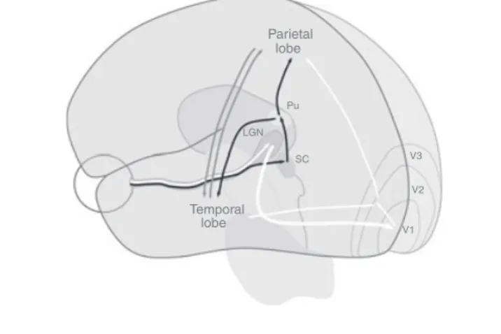
Full conscious vision relies on the integrity of the pathway between the retina and the primary visual cortex37(Fig. 1). To demonstrate the existence of a mechanism of cerebral plastic-ity that is able to restore normal conscious vision three criteria should be satisfied: (1) the documented damage to the
retino-What this paper adds
• It reviews the state of knowledge on the mechanisms of visual plasticity after early brain damage.
• It provides evidence of new mechanisms of reorganization.
• It provides new examples of the application of advanced brain imaging techniques in the study of structural reorganization of visual function.
geniculo-striate pathway; (2) the specific loss of conscious vision in part or whole of the visual field (the loss must be spe-cific in the sense that it does not depend on abnormalities of other functions, such as cognition or attention); (3) the sub-sequent partial or total recovery of the previously lost func-tion, possibly accompanied by the documented change of brain structure or activity.
There is little evidence that this type of plastic reorganiza-tion, i.e. changes in conscious perception in the blind field over time, can be found in adults with brain damage.38A lim-ited enlargement of the visual field can be commonly observed within the first few weeks after the insult. It results from the resolution of transient dysfunction of the perilesional tis-sue,39,40or from changes in the properties of neural circuits adjoining the lesion, such as excitability, receptive field size, and channel properties.41After this first spontaneous recovery, there is some evidence that intensive training can further increase visual field size by a few degrees, by recruiting poten-tially intact but under-performing visual circuits.42,43 How-ever, a recent systematic review concluded that there is no evidence that such a limited enlargement of visual field results in better ocular motor scanning strategies and leads to a better performance of daily-life activities.44 Indeed, most of the modifications in visual behaviour observed after acquired brain damage are not the result of a direct recovery of lost vision, but rather the effect of compensatory visuomotor strategies.45,46
Normal vision after early damage to primary visual cortex Is there evidence of restoration of normal conscious vision when the lesion occurs in the very early phases of develop-ment? In individuals with early brain damage it is extremely hard to document a specific and non-permanent loss of con-scious vision after damage. This is owing to the lack of active collaboration during early assessments and the necessity to
rely on behavioural responses, which are less reliable and more influenced by attention. In addition, functional responses to the same test can be related to different underlying mecha-nisms at different stages of development, making the compari-son between subacute and chronic phases of recovery potentially misleading. An example might clarify this concept. In 1996, Mercuri et al.26assessed visual field by kinetic peri-metry in a cohort of infants with perinatal arterial stroke, reporting a high percentage of field defects. The follow-up study of the same cohort at school age using the same visual field test showed a complete recovery in all of those who had previously shown a defect.27This could well be the effect of plastic reorganization of the geniculo-striate pathway, but a different interpretation is more likely. During the first months of life, visual fields are tested by first attracting the attention of the infant to the midline. The infant is then required to disen-gage attention from the central to a newly presented periph-eral stimulus, a task requiring the integrity of other cortical areas involved in shift of visual attention. Hence, the different behaviour observed at school age in infants with earlier appar-ent visual field restriction is likely to be the result of the matu-ration or recovery of the ability to shift attention, rather than the actual enlargement of the visual fields. This is also sup-ported by the finding in that study that all six children who had abnormal fields in the first year of life showed parietal lesions on MRI with sparing of the optic radiations and primary visual cortex, and that five of the six also had abnor-mal responses to a specific fixation shift test in infancy.27
To address whether individuals with early brain damage can show recovery of visual perception, another approach should be used. This is the demonstration of normal conscious vision in the presence of structural damage to the visual system that is so obvious to rule out the hypothesis that normal vision could be preserved without a process of plastic reorganization. In principle, the types of brain lesions more likely to be associ-ated with an effective reorganization of the visual cortex are the malformations of cortical development, as they develop during the very early phases of gestation when brain plasticity is thought to be highest. Cortical malformations of the occipi-tal lobes are not always associated with visual field defects.47–50 The underlying mechanisms are thought to be related to the presence of functioning tissue within the lesion (Fig. 2). In a recent study, three individuals with bilateral parasagittal occipital polymicrogyria and normal vision were analysed with retinotopic mapping.16Normal cortical responses and organi-zation of early visual areas were found, suggesting that dys-plastic tissue can be actively involved in the processing of visual information, presumably because of plastic reorganiza-tion within the polymicrogyric cortex. Similar conclusions were reached by other authors studying patients with cortical malformations51 or with perinatal incomplete lesions of the primary visual cortex.52In keeping with this is the finding of an increased risk of visual field defects after removal of dysplastic tissue in the occipital cortex.53,54 It is of note, however, that this happens with a frequency that is lower than expected, suggesting forms of reorganization outside the boundaries of the striate cortex.49
Parietal lobe Pu SC V3 V2 V1 Temporal lobe LGN
Figure 1: Schematic representation of major visual pathways. The retino-geniculo-striate and the post-striatal pathways are shown in white. The retino-tectal pathway and the connection to the extrastriatal visual areas are shown in black. The interconnections between parietal and temporal extrastriatal visual areas are shown in grey. LGN, lateral geniculate nucleus; Pu, pulvinar complex; SC, superior colliculus.
The possibility that early cortical damage to the primary visual cortex can result in its displacement to another region of the brain, which in normal conditions would not be in charge of conscious visual processing, is fascinating. The evidence that this could happen is, however, very weak. To the best of our knowledge, only one case reported in the literature sug-gests this type of plastic reorganization after a brain malforma-tion of the occipital cortex.55 This was a child with a right occipital cortical dysplasia, diagnosed when she was 12 years old after the onset of epileptic seizures. Despite normal visual fields (assessed by Goldmann perimetry), visual-evoked poten-tials showed a dislocation of P100 responses in the affected hemisphere towards the contiguous temporal and parietal regions, and functional MRI (fMRI) revealed cerebral activity in the same regions, suggesting a true dislocation of primary visual structures in areas outside the striate cortex. Another interesting case was described by Lambert et al. in 1990.56A child with a silent prenatal and perinatal history was found to be inattentive to one side at around the age of 6 weeks. The follow-up showed a normalization of the clinical picture with no detectable visual defect from the age of 9 months, and sub-sequent MRI documented a right occipital lobe hypoplasia. In this case, however, no visual mapping was performed, hinder-ing full understandhinder-ing of the underlyhinder-ing mechanisms of recov-ery. Based on the studies available in individuals with congenital brain damage, the possibility that primary visual function (i.e. full conscious vision), can be effectively processed by structures outside the boundaries of the classic primary visual cortex is still far from being demonstrated, and it is, in the best of the cases, the exception rather than the rule.
Normal vision after early damage to the geniculo-striate pathway
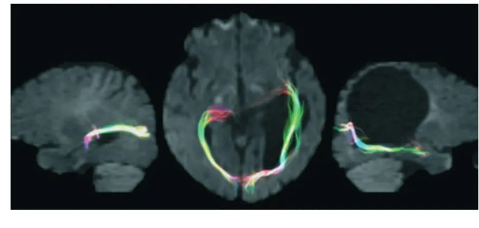
During early brain development, and in particular early in the third trimester of gestation, white matter can be particularly vulnerable to insult, exposing to injury the developing thalamo-cortical fibres, including the optic radiations. This is true in both bilateral periventricular ischaemic lesions and uni-lateral periventricular hemorrhagic infarction. At this stage of brain development, the plasticity of thalamo-cortical afferents is conspicuous. The afferents from the subplate zone are still migrating into the cortical plate, and there is a significant amount of growth-promoting molecules and axonal guidance cues, related to an increased expression of genes coding for such molecules.57This particular environment gives the brain additional potential strategies for plastic reorganization, especially when the periventricular damage is focal, such as for unilateral periventricular infarction. The somatosensory function, for example, was shown to be particularly resistant to brain damage in individuals with early brain lesions, both in terms of functional reorganization and topography of its cortical representations.58,59The underlying mechanisms were studied with diffusion tractography, a novel methodology that uses non-invasive brain MRI data to reconstruct the white matter pathway in the living brain. This is an indirect measure, as it implies the presence of fibre bundles along the path of least resistance to water diffusion. However, when correlated with electrophysiological or functional data, it provides impor-tant information on white matter anatomy. A recent study combining magneto-encephalography and diffusion tractogra-phy provided convincing evidence that, in patients with presumed preterm damage of the periventricular white matter, somatosensory projections might still develop after the lesion has occurred and bypass it to reach their cortical destination in the postcentral gyrus.60Using the same technique, we recently found some evidence that a similar process can be observed in the visual system of patients with unilateral periventricular brain damage early in the third trimester of gestation. One of these was a patient who showed normal visual fields despite a large lesion of the left periventricular white matter involving most of the tissue where optic radiations would normally sit (see Fig. 3). Diffusion tensor tractography showed how the trajectories of the optic radiations in the affected hemisphere deviated from their normal course, bypassing the cystic lesion and reaching their final target in the occipital lobe. These find-ings are consistent with animal studies,61–63but also with post-mortem findings in human fetuses showing that developing geniculo-striate axons after mid-gestation ‘wait’ in the subplate for weeks before entering the cortical plate.64
The exact characteristics and limits of this specific type of plasticity involving the thalamo-cortical pathway are far from being understood. It is hard to define the critical time window at which this type of reorganization is most effective. Some data suggest that at least up to term age, structural modifications of the geniculo-striate pathway can support functional reorganiza-tion of the visual system. Seghier et al.65,66 recently studied longitudinally an infant with perinatal left arterial stroke, spar-a
b
c
Figure 2: Possible mechanisms of functional reorganization underlying recovery of normal conscious vision in individuals with congenital brain damage. (a) Damage involves the primary visual cortex, but functional tis-sue is still present within the lesion;16,51,52(b) Primary visual function is
reorganized in areas of the occipital lobe that are outside the usual boundaries of the primary visual cortex;55(c) The geniculo-striate pathway curve around the lesion bypassing it, and reach the calcarine cortex.65,66
ing the primary visual cortex but involving the optic radiations, using a combination of fMRI and diffusion tensor tractography. When the infant was tested at 3 months of age with visual fMRI, cortical activation could only be observed in the unaffected side, and diffusion tensor imaging was unable to show the presence of the optic radiations in the affected hemisphere.65 At 20 months, the infant was tested again with the same protocol and, surprisingly, showed a clear fMRI activation, an indirect sign of functional reorganization, further supported by clear structural modifications on diffusion tractography.66 Unfortu-nately, the assessment of visual fields was not performed owing to the young age of the individual. However, regardless of the possible presence of a functional impairment, the imaging data seemed to support the existence of a process of reorganization at the level of the thalamo-cortical pathway, able to restore, at least partly, the functional connection between the lateral genic-ulate body and the occipital cortex.
To summarize, after early damage to the optic radiations or to the primary visual cortex, the young brain is capable of spe-cific strategies of plastic reorganization (Fig. 2), which to some extent seem to be more effective in restoring conscious vision, as opposed to those available at a later stage of brain develop-ment. These different modalities of cerebral plasticity rely on the presence of unique favourable conditions in the developing nervous system, which have definite time constraints. Full con-scious vision is possible, for example, in individuals with exten-sive occipital malformations of cortical development, or with clear damage of the optic radiations before birth. However, even when these efficient mechanisms of reorganization fail (i.e. if full conscious vision is not restored), the young brain seems to have an advantage over the more mature brain, as we highlight below.
BRAIN DAMAGE AND UNCONSCIOUS VISUAL PERCEPTION
Damage to primary visual cortex causes an essential inability to consciously perceive visual information in the contralateral
hemifield.67Vision, however, does not completely disappear, as first demonstrated almost 100 years ago.68Evidence from several individuals with damage to the primary visual cortex demonstrates the existence of basic residual visual perception in the blind field, including motion, form, and wavelength sen-sitivity (reviewed in Stoerig69). In its classic descriptions, the defect is generally characterized by no conscious experience and has been termed ‘blindsight’.67The key hallmark of blind-sight is the dissociation between below-chance performance for simple yes–no responses (e.g. do you see something?) and above-chance performance in forced-choice procedures (e.g. do you see something here or there?). The first can be consid-ered as a subjective measure of awareness (absent in blindsight) and the second as an objective measure of awareness (present in blindsight). By definition, blindsight implies subjective unawareness. In some patients, however, some awareness of the presence of the stimulus, either as an actual visual sensa-tion or as a more undefined feeling, can be present in specific conditions and can be increased by training.70This has led to a distinction between different types of blindsight, including a subgroup in which a sparing of some amount of awareness can be observed.71,72Most of the studies exploring blindsight have been performed in individuals with acquired focal lesions. It also has to be acknowledged that the existence of blindsight is not recognized by all scientists, and that different explanations of the phenomenon have been proposed (reviewed in Cowey73). A review of the literature in this field is beyond the scope of this paper. Here we address the question whether the timing of the insult (i.e. early vs late damage), differentially affects the quality of unconscious vision in the blind field, and should this be true, what the underlying mechanisms are.
Although this issue has never been studied systematically, there is some cumulative evidence, mostly from case reports, that reorganization of visual functions might be more effective after lesions that occurred during childhood. Two aspects in particular are influenced by the timing of the damage: the level Figure 3: Diffusion tensor tractography of the optic radiations of a patient overlaid on a co-registered DW image. Fibre tracking was obtained using two regions of interest for each hemisphere at the level of the lateral geniculate bodies and of the calcarine cortex. Left image: optic radiations of the left hemisphere; streamlines are displayed on the sagittal plane and show a trajectory circumscribing the dilated left ventricle, and reaching the final target at the level of the calcarine cortex. Central image: optic radiations of both hemisphere; streamlines are displayed on the axial plane. The optic radiations of the left hemisphere follow an abnormal trajectory going further anteriorly and laterally as opposed to the contralateral, unaffected side. Right image: optic radiations of the right hemisphere; streamlines are displayed on the sagittal plane and show a normal trajectory.
of perceptual awareness and the efficiency of compensating ocular motor strategies.
Level of subjective awareness of the blind visual field There is still controversy about where conscious perceptual experience arises in the brain (see Moutoussis74for a review). A major role is played by the primary visual cortex, either in rela-tion to its retinal input, its extrastriatal cortical output, or both.69The question here is whether some degree of visual awareness can be achieved without the contribution of primary visual areas, and if this is related to the timing of brain damage. The first to suggest that conscious vision is possible in patients blinded by damage to the primary visual cortex was George Rid-doch in 1917.68Several other reports have shown that different degrees of conscious vision, albeit highly degraded, are compat-ible with complete lesions of primary visual cortex (reviewed in Zeki and Ffytche;71see also Giaschi et al.75). What is generally reported by patients with acquired damage and residual aware-ness of the stimulus is a generic feeling of the presence of a stim-ulus and the impossibility of describing its visual elements.76,77 Things are different in the case of early damage.
In a study of 25 patients with large scotomata (areas of diminished vision surrounded by normal visual field) due to damage to the striate cortex, five were found to be capable of consistent responses to visual stimuli presented within their blind field.78Not only were they able to perform accurate eye movements in response to light flashes deep in the hemianopic field, but they also consistently reported the sensation of see-ing a dark shadow when stimulated by transient changes in illumination of either positive or negative contrast. In four of the five patients, the damage had presumably occurred before the age of 11 years, as opposed to only one of the remaining 20, suggesting a strong influence of timing of brain damage on residual visual awareness.
Reports on single cases have been informative. One of the patients with blindsight who has been most extensively studied in the literature, in some circumstances (specific object size, contrast, displacement, and velocity properties) is fully con-scious of the presence of a visual stimulus in the blind field and is able to report orally on its position, aspects of spectral con-tent, and direction and velocity of movement.71,79–81This indi-vidual had a lesion to the left primary visual cortex when he was 8 years old, and many authors have considered the timing of the insult as the main element supporting his remarkable visual performances (see, for example, Payne and Lomber,82Goebel et al.83). Another very informative case is a female born with a malformation of the left cerebral hemisphere and intractable epileptic seizures who underwent a complete left hemispherec-tomy at the age of 4 months.84Despite the total absence of one hemisphere, she was able immediately to redirect her gaze towards the visual target in each of the 72 different positions in the hemianopic field. In addition, she consistently reported that she had seen a light when questioned, showing some degree of subjective awareness. Others reported vision with awareness in individuals who underwent hemispherectomy later in life, but with overall performances way below those of this female patient (for a recent review see Ptito and Leh85). Recently,
visual awareness of moving stimuli was also found in an individ-ual with complete absence of both occipital lobes due to perina-tal damage, as confirmed by structural and functional neuroimaging, further supporting the possibility of maintain-ing visual awareness in case of early damage.75
In summary, early damage seems to be associated with an increased sensitivity towards the stimuli hitting the blind field, associated with an increased awareness of perception. The possible underlying mechanisms are discussed below. How-ever, it is important to notice that not only sensitivity to stim-uli, but also subjective awareness of the stimulus, have been shown to be enhanced by training in patients with blind-sight.81,86This suggests that the threshold of visual awareness is not unchangeable, even in case of permanent cortical dam-age, but is rather plastic and therefore trainable. It is not sur-prising then, that after early damage the enhanced neuronal plasticity of the young brain is able to induce higher levels of functional reorganization, including sensitivity to visual stimuli in the blind field and level of perceptual awareness. Efficiency of compensatory ocular motor strategies
Very high levels of accuracy can be reached in forced-choice tasks, so that, under specific conditions, patients with blind-sight can develop a sensitivity for detection that is superior in the blind field than in the intact one.87However, without a concomitant presence of subjective awareness, the level of dis-ability related to partial blindness is not alleviated, and blind-sight capabilities are not used in everyday life.88Consequently, the application of tests that can closely quantify functional mechanisms used in daily activities is crucial to have a more comprehensive picture of the adaptive reorganization of the system. One of the concepts most often explored to assess functional visual behaviour is visual search. It refers to the capacity of an individual to find a target among simultaneously presented distractors89and is based on visual abilities such as fast visual processing and accurate control of ballistic eye movements (saccades) that guide the fovea to the target loca-tion.90,91Studies in adults have shown that hemianopia due to unilateral damage to the visual pathway is often associated with visual search disorders.46,92,93 Patients cannot process images in the same way as typical controls and usually have difficulties with reading, detecting stimuli, or finding objects in the visual space corresponding to the impaired field. Their fixations typically dwell into the intact hemifield and their search pattern is characterized by frequent exploratory saccades into the blind part of the visual field93–95 with repeated saccades and fixations to the same object, resulting in overall longer visual search times.46,93This phenomenon has been defined as ‘slowness of vision’ in the contralateral hemi-field to the side of the lesion.93
Is visual search differentially affected in individuals with early damage to the visual system? Few studies have explored the effects of brain lesions acquired during childhood on visual search abilities, inconsistently reporting slower search responses in the contralesional visual field.96,97Other studies showed that patients who underwent hemispherectomy at 7 years of age or earlier showed greater sparing of visual
orient-ing abilities compared with patients who underwent surgery at 17 years of age.98In addition, single case studies documented residual visual exploration abilities in patients with bilateral damage to the visual cortex at birth,99patients who underwent hemi-decortication in the first year of life,100 and patients affected by congenital right hemi-hydranencephaly.101 We recently addressed the question of visual search and timing of brain damage by studying a group of age-matched children with hemianopia and brain damage occurred either pre- or perinatally or between 8 and 13 years of age.102As expected, children with hemianopia with acquired lesions showed signifi-cantly longer reaction times for stimuli presented in the blind hemifield. In contrast, children with congenital lesions showed normal reaction times irrespective of where the stimulus was presented. It is of interest that the ability to scan the blind field more efficiently is often combined with an anomalous head turn ipsilateral to the blind hemifield, as a further compensatory mechanism for the visual field deficit.103Altogether, these find-ings show that children with congenital brain damage are able to explore the environment efficiently, even when part of the visual field is blinded by damage to the geniculo-striate struc-tures. This further supports a more effective visual search strat-egy in children with congenital brain lesions, compared with acquired ones, in line with reports in the animal model. Underlying mechanisms
The differential effect of damage to the visual cortex between young and adult individuals has been thoroughly studied in the animal model. Striking differences in visual behaviour have been found when comparing monkeys with unilateral ablation of the striate cortex in adulthood with those undergoing the same procedure during early infancy.104 The animals with early lesion were able to detect and localize newly presented targets at most locations within the scotoma, whereas the ani-mals with later lesions appeared blind to targets at most sites. It is of great interest, however, that in the latter group, a sig-nificant improvement in visual behaviour was observed when the assessment procedure was modified by turning off the cen-tral target at the moment of presenting the peripheral one, thereby inducing the animal to saccade to a new location. In this new condition, all animals were able to detect and reorient their gaze to most targets, suggesting a type of perception sim-ilar to human blindsight, as previously proposed by other authors using different recognition paradigms.69,105,106How the timing of the insult affects visual behaviour has also been extensively studied in the cat (see Payne and Lomber82for a review). Despite having a similar impairment in visual perfor-mance based on acuity,107 cats with early lesions do signifi-cantly better in most visuotemporal tasks (e.g. simple pattern discrimination) and visuoparietal tasks (e.g. visual orientation, depth judgement), compared with cats with lesions sustained in adulthood.86,108–110Their visual behaviour is often indistin-guishable from that of healthy cats.
The most likely explanation for the striking residual visual capacities after damage to the primary visual cortex is the expansion of pathways that can bypass V1 and directly connect subcortical nuclei with extrastriate visual structures. The first
consequence of damage to the primary visual cortex is the retrograde degeneration of neurons in retinotopically corre-sponding areas of the dorsal lateral geniculate nucleus, and, transinaptically, of a large portion of retinal ganglion cells.111 However, several ganglion cells survive, and increase the den-sity of their projections to the dorsal lateral genicualte nucleus. From here, direct projections to extrastriate visual structures exist, for both the ventral stream112and the dorsal stream.113In addition, the network connecting the superior colliculus with the pulvinar and the extrastriate cortex is massively expanded, especially to dorsal stream areas such as V5 or middle tempo-ral⁄middle superior temporal (reviewed in Cowey and Stoerig114and Payne and Lomber82). It has also been suggested that in the case of focal lesions the contralesional hemisphere mediates residual functions through transcallosal or intertectal connections.115 A schematic representation of the potential mechanisms of reorganization after early damage, mainly based on evidence from animal studies, is shown in Figure 4.
The consequences at a cortical level of this network expan-sion, i.e. the increased activation⁄metabolism of the extrastriatal visual cortex, have been demonstrated with brain mapping techniques in humans.115–119Recently, experiments have also been able to show in vivo the key role played by the superior colliculus and its cortical connections in residual visual func-tions after early and late brain damage to the primary visual structures.120,121 Some initial evidence exists supporting a different mechanism of visual reorganization after early as opposed to late brain damage, paralleling the findings from the animal studies. For example, Goebel et al.83used fMRI to compare the activation of the dorsal stream in two patients with hemianopia due to brain damage that occurred at different times, at 8 and 42 years. Although both individuals showed ipsilesional activations of the dorsal pathway, the activity was much stronger in the individual with the earlier damage, and it was at least as strong as the activity elicited in the unaffected
Parietal lobe Pu SC LGN lobe Temporal
Figure 4: Possible mechanisms of functional reorganization underlying unconscious vision and visual orientation in individuals with congenital brain damage (mainly based on the animal model). In green are the expanded pathways; in red are the withdrawn pathways. See text for explanation. LGN, lateral geniculate nucleus; Pu, pulvinar complex; SC, superior colliculus.
hemisphere by stimulating the normal field, potentially sup-porting a more efficient connection between the retina and the extrastriatal visual areas. More recently we also presented behavioural and electrophysiological data from children with congenital brain damage that suggested the existence of com-pensatory mechanisms based on the expansion or sparing of ex-trastriatal visual structures.122We found that children with a damaged geniculo-striate pathway, as shown by abnormal visual evoked potentials and brain MRI, were able to reach bet-ter values of visual acuity when using techniques that could benefit more from the expansion of the extra-striatal pathways (i.e. behavioural techniques based on visual orientation). Taken together, human studies seem to suggest that mechanisms simi-lar to those observed in the animal models underlie the differ-ential visual outcomes of individuals with early and late brain damage. Future studies based on advanced brain imaging, including structural and functional connectivity or fibre track-ing, will further expand and possibly confirm these first reports. CONCLUSION
Despite the enormous advances since the seminal works of Margaret Kennard in the first half of the 20th century, our knowledge about the influence of timing on brain plasticity of the visual system is still very limited. There is increasing evi-dence supporting a better visual outcome in individuals with congenital brain damage, but our understanding of the possi-ble underlying mechanisms is still largely based on non-human models. Some of the strategies adopted by the immature brain are unavailable at a later stage, similarly to what is observed in the domain of language and sensorimotor function. There is, for example, the possibility of developing new cortico-thalamic connections capable of bypassing the lesion, or the ability to differentiate functional tissue within a larger dysplastic cortex, both mechanisms having very specific time constraints. In other circumstances, the lesion activates neuroplastic processes available at any stage of development, but more pronounced and efficient when the brain is still young. This probably applies to the pathways bypassing V1 and directly reaching the extrastriatal visual structures, most of which are normally present in the older brain, but less pre-disposed to the great expansion observed after early damage.
A crucial notion, strongly supported by animal studies, is that the extrastriatal visual networks are heavily reorganized
after early damage to primary visual structures. This is shown, for example, by studies using localized cooling for reversible cortical deactivation (reviewed in Payne and Lomber82). In normal adult animals, the deactivation of the visuoparietal cortex selectively abolishes visual orienting abilities, whereas the deactivation of the visuotemporal cortex selectively dis-ables object recognition processes. This is the result of the normal process of segregation and specialization of functions into distinct extrastriatal visual circuits. If primary visual cortex is removed early after birth, animals show a good development of extrastriatal visual functions. However, the effect of local deactivations only partly affects the corresponding functions, suggesting that they have been redistributed within the extra-striatal network (i.e. visual orienting is partly processed by the visuotemporal areas and object recognition is partly processed by the visuoparietal areas).82Although no study has directly explored the existence of such a mechanism in humans, it is of interest that the literature on dorsal and ventral stream func-tions in patients with brain damage shows very different pictures, depending on the timing of the insult. Although in adults with acquired brain injury clear cases of selective impairment of dorsal or ventral stream functions have been described,123,124it has been consistently shown that individuals with congenital brain damage generally show more subtle and mixed pictures, suggesting some degree of topographic redis-tribution of these functions (see, for example, Gunn et al.125).
To conclude, the question of why and to what extent the young visual brain reacts differently to damage is still open and will need extensive research to be answered. Only with this knowledge will we be able to modify the environment of infants with early brain damage to support and enhance the adaptive processes of visual reorganization at a time when brain plasticity potentials are highest. Evidence exists to suggest that not only this will have an impact on cerebral visual impairment, but more generally on neurodevelopment and cognition.126
ACKNOWLEDGEMENTS
This work was supported by a Mariani Foundation Grant 2006, PRIN 2007, from the Italian Ministry of University and Research (to GC), a National Health and Medical Research Council of Australia Project Grant (468300), a Career Development Grant (to RB), and a Queensland Smart State Fellowship (to RB).
REFERENCES
1.Pascual-Leone A, Amedi A, Fregni F, Merabet LB. The plastic human brain cortex.Annu Rev Neurosci2005;28:377–401.
2.Elbert T, Pantev C, Wienbruch C, Rockstroh B, Taub E. Increased cortical representation of the fingers of the left hand in string players.Science1995;270:305–7.
3.Lewis TL, Maurer D. Multiple sensitive periods in human visual development: evidence from visually deprived children.
Dev Psychobiol2005;46:163–83.
4.Berker EA, Berker AH, Smith A. Translation of Broca’s 1865 report. Localization of speech in the third left frontal convolution.Arch Neurol1986;43:1065–72.
5.Kennard M, Fulton JF. Age and reorganization of central nervous system.Mt Sinai J Med1942;9:594–606.
6.Anderson V, Spencer-Smith M, Leventer R, et al. Childhood brain insult: can age at insult help us predict outcome?Brain
2009;132:45–56.
7.Krageloh-Mann I, Horber V. The role of magnetic reso-nance imaging in elucidating the pathogenesis of cerebral palsy: a systematic review.Dev Med Child Neurol2007;49:
144–51.
8.Teuber HL. Recovery of function after brain injury in man. In: Porter R, Fitzsimmons DW, editors. Outcome of severe damage to the central nervous system. Ciba Found Symp, Amsterdam: Elsevier, 1975, 159–90.
9.Whiting S, Jan JE, Wong PK, Flodmark O, Farrell K, McCormick AQ. Permanent cortical visual impairment
in children. Dev Med Child Neurol 1985; 27: 730– 9.
10.Ricci D, Cesarini L, Groppo M, et al. Early assessment of visual function in full term newborns.Early Hum Dev2008;
84:107–13.
11.Atkinson J, van Hof-van Duin J. Visual assessment during the first years of life. In: Fielder A, Best A, Bax M, editors. The management of visual impairment in childhood. Clinics in Developmental Medicine No. 128. London: Mac Keith Press, 1993, 9–29.
12.Rutherford MA. MRI of the neonatal brain. London: WB Saunders, 2002.
13.Guzzetta A, Mercuri E, Cioni G. Visual disorders in children with brain lesions. 2. Visual impairment associated with cere-bral palsy.Eur J Paediatr Neurol2001;5:115–9.
14.Krageloh-Mann I, Horber V. The role of magnetic reso-nance imaging in furthering understanding of the pathogene-sis of cerebral palsy.Dev Med Child Neurol2007;49:948.
15.Barkovich AJ, Kuzniecky RI, Jackson GD, Guerrini R, Dobyns WB. A developmental and genetic classification for malformations of cortical development.Neurology2005;
65:1873–87.
16.Dumoulin SO, Jirsch JD, Bernasconi A. Functional organi-zation of human visual cortex in occipital polymicrogyria.
Hum Brain Mapp2007;28:1302–12.
17.Back SA, Riddle A, McClure MM. Maturation-dependent vulnerability of perinatal white matter in premature birth.
Stroke2007;38:724–30.
18.Larsson EK, Rydberg AC, Holmstrom GE. A population-based study on the visual outcome in 10-year-old preterm and full-term children.Arch Ophthalmol2005;123:825–32.
19.Banker BQ, Larroche JC. Periventricular leukomalacia of infancy. A form of neonatal anoxic encephalopathy.Arch
Neurol1962;7:386–410.
20.Cioni G, Fazzi B, Coluccini M, Bartalena L, Boldrini A, van Hof-van Duin J. Cerebral visual impairment in preterm infants with periventricular leukomalacia.Pediatr Neurol
1997;17:331–8.
21.Jacobson LK, Dutton GN. Periventricular leukomalacia: an important cause of visual and ocular motility dysfunction in children.Surv Ophthalmol2000;45:1–13.
22.Kok JH, Prick L, Merckel E, Everhard Y, Verkerk GJ, Scherjon SA. Visual function at 11 years of age in preterm-born children with and without fetal brain sparing.Pediatrics
2007;119:e1342–50.
23.Harvey EM, Dobson V, Luna B, Scher MS. Grating acuity and visual-field development in children with intraventricular hemorrhage.Dev Med Child Neurol1997;39:305–12.
24.Ricci D, Luciano R, Baranello G, et al. Visual development in infants with prenatal post-haemorrhagic ventricular dilata-tion.Arch Dis Child Fetal Neonatal Ed2007;92:F255–8.
25.Kirton A, deVeber G. Advances in perinatal ischemic stroke.
Pediatr Neurol2009;40:205–14.
26.Mercuri E, Atkinson J, Braddick O, et al. Visual function and perinatal focal cerebral infarction.Arch Dis Child Fetal
Neo-natal Ed1996;75:F76–81.
27.Mercuri E, Anker S, Guzzetta A, et al. Neonatal cerebral infarction and visual function at school age.Arch Dis Child
Fetal Neonatal Ed2003;88:F487–91.
28.Graham EM, Ruis KA, Hartman AL, Northington FJ, Fox HE. A systematic review of the role of intrapartum hypoxia– ischemia in the causation of neonatal encephalopathy.Am J
Obstet Gynecol2008;199:587–95.
29.Mercuri E, Atkinson J, Braddick O, et al. Visual function in full-term infants with hypoxic–ischaemic encephalopathy.
Neuropediatrics1997;28:155–61.
30.Mercuri E, Haataja L, Guzzetta A, et al. Visual function in term infants with hypoxic-ischaemic insults: correlation with neurodevelopment at 2 years of age.Arch Dis Child Fetal
Neonatal Ed1999;80:F99–104.
31.Mercuri E, Atkinson J, Braddick O, et al. Basal ganglia dam-age and impaired visual function in the newborn infant.Arch
Dis Child Fetal Neonatal Ed1997;77:F111–4.
32.Yalnizoglu D, Haliloglu G, Turanli G, Cila A, Topcu M. Neurologic outcome in patients with MRI pattern of damage typical for neonatal hypoglycemia.Brain Dev2007;29:285– 92.
33.Andersson S, Persson EK, Aring E, Lindquist B, Dutton GN, Hellstrom A. Vision in children with hydrocephalus.
Dev Med Child Neurol2006;48:836–41.
34.Woodward GA. Posttraumatic cortical blindness: are we missing the diagnosis in children?Pediatr Emerg Care1990;
6:289–92.
35.Guzzetta F, Frisone MF, Ricci D, Rando T, Guzzetta A. Development of visual attention in West syndrome.Epilepsia
2002;43:757–63.
36.Guzzetta A, Cioni G, Cowan F, Mercuri E. Visual disorders in children with brain lesions: 1. Maturation of visual func-tion in infants with neonatal brain lesions: correlafunc-tion with neuroimaging.Eur J Paediatr Neurol2001;5:107–14.
37.Felleman DJ, Van Essen DC. Distributed hierarchical pro-cessing in the primate cerebral cortex.Cereb Cortex1991;1:
1–47.
38.Huxlin KR. Perceptual plasticity in damaged adult visual sys-tems.Vision Res2008;48:2154–66.
39.Sabel BA, Kasten E, Kreutz MR. Recovery of vision after partial visual system injury as a model of postlesion neuro-plasticity.Adv Neurol1997;73:251–76.
40.Zhang X, Kedar S, Lynn MJ, Newman NJ, Biousse V. Natu-ral history of homonymous hemianopia.Neurology2006;66:
901–5.
41.Eysel UT. Perilesional cortical dysfunction and reorganiza-tion.Adv Neurol1997;73:195–206.
42.Kasten E, Poggel DA, Sabel BA. Computer-based training of stimulus detection improves color and simple pattern recog-nition in the defective field of hemianopic subjects.J Cogn
Neurosci2000;12:1001–12.
43.Sabel BA, Kasten E. Restoration of vision by training of residual functions.Curr Opin Ophthalmol2000;11:430–6.
44.Bouwmeester L, Heutink J, Lucas C. The effect of visual training for patients with visual field defects due to brain damage: a systematic review.J Neurol Neurosurg Psychiatry
2007;78:555–64.
45.Ishiai S, Furukawa T, Tsukagoshi H. Eye-fixation patterns in homonymous hemianopia and unilateral spatial neglect.
Neu-ropsychologia1987;25:675–9.
46.Pambakian AL, Wooding DS, Patel N, Morland AB, Ken-nard C, Mannan SK. Scanning the visual world: a study of patients with homonymous hemianopia.J Neurol Neurosurg
Psychiatry2000;69:751–9.
47.Burneo JG, Kuzniecky RI, Bebin M, Knowlton RC. Cortical reorganization in malformations of cortical development: a magnetoencephalographic study.Neurology2004;63:1818– 24.
48.Guerrini R, Dubeau F, Dulac O, et al. Bilateral parasagittal parietooccipital polymicrogyria and epilepsy.Ann Neurol
1997;41:65–73.
49.Kuzniecky R, Gilliam F, Morawetz R, Faught E, Palmer C, Black L. Occipital lobe developmental malformations and epilepsy: clinical spectrum, treatment, and outcome.Epilepsia
1997;38:175–81.
50.Zesiger P, Kiper D, Maeder P, Deonna T, Innocenti GM. Preserved visual function in a case of occipitoparietal microgyria.Ann Neurol2002;52:492–8.
51.Innocenti GM, Maeder P, Knyazeva MG, Fornari E, Deonna T. Functional activation of microgyric visual cortex in a human.Ann Neurol2001;50:672–6.
52.Knyazeva MG, Maeder P, Kiper DC, Deonna T, Innocenti GM. Vision after early-onset lesions of the occipital cortex: II. Physiological studies.Neural Plast2002;9:27–40.
53.Blume WT, Whiting SE, Girvin JP. Epilepsy surgery in the posterior cortex.Ann Neurol1991;29:638–45.
54.Williamson PD, Thadani VM, Darcey TM, Spencer DD, Spencer SS, Mattson RH. Occipital lobe epilepsy: clinical characteristics, seizure spread patterns, and results of surgery.
Ann Neurol1992;31:3–13.
55.Kong CK, Wong LY, Yuen MK. Visual field plasticity in a female with right occipital cortical dysplasia.Pediatr Neurol
2000;23:256–60.
56.Lambert SR, Kriss A, Taylor D. Detection of isolated occipi-tal lobe anomalies during early childhood.Dev Med Child
Neurol1990;32:451–5.
57.Kostovic I, Judas M. Prolonged coexistence of transient and permanent circuitry elements in the developing cerebral cor-tex of fetuses and preterm infants.Dev Med Child Neurol
2006;48:388–93.
58.Guzzetta A, Bonanni P, Biagi L, et al. Reorganisation of the somatosensory system after early brain damage.Clin
Neuro-physiol2007;118:1110–21.
59.Wilke M, Staudt M, Juenger H, Grodd W, Braun C, Krage-loh-Mann I. Somatosensory system in two types of motor reorganization in congenital hemiparesis: topography and function.Hum Brain Mapp2009;30:776–88.
60.Staudt M, Braun C, Gerloff C, Erb M, Grodd W, Krageloh-Mann I. Developing somatosensory projections bypass periventricular brain lesions.Neurology2006;67:522–5.
61.Kostovic I, Rakic P. Developmental history of the transient subplate zone in the visual and somatosensory cortex of the macaque monkey and human brain.J Comp Neurol1990;
297:441–70.
62.Rakic P. Prenatal genesis of connections subserving ocular dominance in the rhesus monkey.Nature1976;261:467– 71.
63.Rakic P. Prenatal development of the visual system in rhesus monkey.Philos Trans R Soc Lond B Biol Sci1977;278:245–60.
64.Hevner RF. Development of connections in the human visual system during fetal mid-gestation: a DiI-tracing study.
J Neuropathol Exp Neurol2000;59:385–92.
65.Seghier ML, Lazeyras F, Zimine S, et al. Combination of event-related fMRI and diffusion tensor imaging in an infant with perinatal stroke.Neuroimage2004;21:463–72.
66.Seghier ML, Lazeyras F, Zimine S, Saudan-Frei S, Safran AB, Huppi PS. Visual recovery after perinatal stroke evi-denced by functional and diffusion MRI: case report.BMC
Neurol2005;5:17.
67.Weiskrantz L, Warrington EK, Sanders MD, Marshall J. Visual capacity in the hemianopic field following a restricted occipital ablation.Brain1974;97:709–28.
68.Riddoch G. Dissociation of visual perceptions due to occipi-tal injuries, with especial reference to appreciation of move-ment.Brain1917;40:15–57.
69.Stoerig P. Blindsight, conscious vision, and the role of pri-mary visual cortex.Prog Brain Res2006;155:217–34.
70.Sahraie A, Trevethan CT, MacLeod MJ, Murray AD, Olson JA, Weiskrantz L. Increased sensitivity after repeated
stimu-lation of residual spatial channels in blindsight.Proc Natl
Acad Sci USA2006;103:14971–6.
71.Zeki S, Ffytche DH. The Riddoch syndrome: insights into the neurobiology of conscious vision.Brain1998;1:25–45.
72.Danckert J, Rossetti Y. Blindsight in action: what can the different sub-types of blindsight tell us about the control of visually guided actions?Neurosci Biobehav Rev2005;29:
1035–46.
73.Cowey A. The blindsight saga.Exp Brain Res2010;200:3–24.
74.Moutoussis K. Brain activation and the locus of visual aware-ness.Commun Integr Biol2009;2:265–7.
75.Giaschi D, Jan JE, Bjornson B, et al. Conscious visual abili-ties in a patient with early bilateral occipital damage.Dev
Med Child Neurol2003;45:772–81.
76.Sanders MD, Warrington EK, Marshall J, Wieskrantz L. ‘Blindsight’: vision in a field defect.Lancet1974;i:707–8.
77.Weiskrantz L. Varieties of residual experience.Q J Exp Psy-chol1980;32:365–86.
78.Blythe IM, Kennard C, Ruddock KH. Residual vision in patients with retrogeniculate lesions of the visual pathways.
Brain1987;4:887–905.
79.Barbur JL, Watson JD, Frackowiak RS, Zeki S. Conscious visual perception without V1.Brain1993;6:1293–302.
80.Brent PJ, Kennard C, Ruddock KH. Residual colour vision in a human hemianope: spectral responses and colour dis-crimination.Proc Biol Sci1994;256:219–25.
81.Weiskrantz L, Harlow A, Barbur JL. Factors affecting visual sensitivity in a hemianopic subject.Brain1991;5:2269–82.
82.Payne BR, Lomber SG. Plasticity of the visual cortex after injury: what’s different about the young brain?Neuroscientist
2002;8:174–85.
83.Goebel R, Muckli L, Zanella FE, Singer W, Stoerig P. Sus-tained extrastriate cortical activation without visual awareness revealed by fMRI studies of hemianopic patients.Vision Res
2001;41:1459–74.
84.Werth R. Visual functions without the occipital lobe or after cerebral hemispherectomy in infancy.Eur J Neurosci2006;
24:2932–44.
85.Ptito A, Leh SE. Neural substrates of blindsight after hemi-spherectomy.Neuroscientist2007;13:506–18.
86.Payne BR, Lomber SG, Gelston CD. Graded sparing of visually-guided orienting following primary visual cortex ablations within the first postnatal month.Behav Brain Res
2000;117:1–11.
87.Trevethan CT, Sahraie A, Weiskrantz L. Can blindsight be superior to ‘sighted-sight’?Cognition2007;103:491–501.
88.Schwiedrzik CM, Singer W, Melloni L. Sensitivity and per-ceptual awareness increase with practice in metacontrast masking.J Vis2009;9:1–18.
89.Treisman A. Perceptual grouping and attention in visual search for features and for objects.J Exp Psychol Hum Percept
Perform1982;8:194–214.
90.Findlay JM. Saccade target selection during visual search.
Vision Res1997;37:617–31.
91.Findlay JM. Visual search: eye movements and peripheral vision.Optom Vis Sci1995;72:461–6.
92.Meienberg O, Zangemeister WH, Rosenberg M, Hoyt WF, Stark L. Saccadic eye movement strategies in patients with homonymous hemianopia.Ann Neurol1981;9:537–44.
93.Zihl J. Visual scanning behavior in patients with homony-mous hemianopia.Neuropsychologia1995;33:287–303.
94.Tant ML, Cornelissen FW, Kooijman AC, Brouwer WH. Hemianopic visual field defects elicit hemianopic scanning.
Vision Res2002;42:1339–48.
95.Zangemeister WH, Meienberg O, Stark L, Hoyt WF. Eye-head coordination in homonymous hemianopia.J Neurol
1982;226:243–54.
96.Schatz J, Craft S, Koby M, DeBaun MR. Asymmetries in visual-spatial processing following childhood stroke.
Neuro-psychology2004;18:340–52.
97.Netelenbos JB, Van Rooij L. Visual search in school-aged children with unilateral brain lesions.Dev Med Child Neurol
2004;46:334–9.
98.Hecaen H, Perenin MM, Jeannerod M. The effects of corti-cal lesions in children: language and visual function. In: Almli CR, Finger S, editors. Early brain damage, vol. l:. Research orientations and clinical observations. New York Academic Press, 1984, 277–98.
99.Rizzo M, Hurtig R. The effect of bilateral visual cortex lesions on the development of eye movements and percep-tion.Neurology1989;39:406–13.
100.Braddick O, Atkinson J, Hood B, Harkness W, Jackson G, Vargha-Khadem F. Possible blindsight in infants lacking one cerebral hemisphere.Nature1992;360:461–3.
101.Porro G, Wittebol-Post D, de Graaf M, van Nieuwenhuizen O, Schenk-Rootlieb AJ, Treffers WF. Development of visual function in hemihydranencephaly.Dev Med Child Neurol
1998;40:563–7.
102.Tinelli F, Guzzetta A, Bancale A, et al. Assessment of visual search in children: normative data and effects of congenital and acquired brain lesions.Dev Med Child Neurol2008;50:35.
103.Paysse EA, Coats DK. Anomalous head posture with early-onset homonymous hemianopia.J AAPOS1997;1:209–13.
104.Gross CG, Moore T, Rodman HR. Visually guided behavior after V1 lesions in young and adult monkeys and its relation to blindsight in humans.Prog Brain Res2004;144:279–94.
105.Cowey A, Stoerig P. Blindsight in monkeys.Nature1995;
373:247–9.
106.Stoerig P, Cowey A. Blindsight in man and monkey.Brain
1997;3:535–59.
107.Mitchell DE. Behavioral analyses of the primary visual cortex contributions to vision. In: Payne BR, Peters A, editors. The cat primary visual cortex. San Diego: Academic Press, 2002, 655–94.
108.Cornwell P, Herbein S, Corso C, Eskew R, Warren JM, Payne B. Selective sparing after lesions of visual cortex in newborn kittens.Behav Neurosci1989;103:1176–90.
109.Cornwell P, Payne B. Visual discrimination by cats given lesions of visual cortex in one or two stages in infancy or in one stage in adulthood.Behav Neurosci1989;103:1191–9.
110.Shupert C, Cornwell P, Payne B. Differential sparing of depth perception, orienting, and optokinetic nystagmus after
neonatal versus adult lesions of cortical areas 17, 18, and 19 in the cat.Behav Neurosci1993;107:633–50.
111.Cowey A, Stoerig P, Perry VH. Transneuronal retrograde degeneration of retinal ganglion cells after damage to striate cortex in macaque monkeys: selective loss of P beta cells.
Neuroscience1989;29:65–80.
112.Cowey A, Stoerig P. Projection patterns of surviving neurons in the dorsal lateral geniculate nucleus following discrete lesions of striate cortex: implications for residual vision.Exp
Brain Res1989;75:631–8.
113.Sincich LC, Park KF, Wohlgemuth MJ, Horton JC. Bypass-ing V1: a direct geniculate input to area MT.Nat Neurosci
2004;7:1123–8.
114.Cowey A, Stoerig P. The neurobiology of blindsight.Trends
Neurosci1991;14:140–5.
115.Baseler HA, Morland AB, Wandell BA. Topographic organi-zation of human visual areas in the absence of input from pri-mary cortex.J Neurosci1999;19:2619–27.
116.Batista CE, Chugani HT, Juhasz C, Behen ME, Shankaran S. Transient hypermetabolism of the basal ganglia following perinatal hypoxia.Pediatr Neurol2007;36:330–3.
117.Brodtmann A, Puce A, Darby D, Donnan G. Serial func-tional imaging poststroke reveals visual cortex reorganization.
Neurorehabil Neural Repair2009;23:150–9.
118.Nelles G, de Greiff A, Pscherer A, et al. Cortical activation in hemianopia after stroke.Neurosci Lett2007;426:34–8.
119.Nelles G, Widman G, de Greiff A, et al. Brain representation of hemifield stimulation in poststroke visual field defects.
Stroke2002;33:1286–93.
120.Leh SE, Johansen-Berg H, Ptito A. Unconscious vision: new insights into the neuronal correlate of blindsight using diffusion tractography. Brain 2006; 129: 1822– 32.
121.Leh SE, Ptito A, Schonwiesner M, Chakravarty MM, Mullen KT. Blindsight mediated by an S-cone-independent collicu-lar pathway: an fMRI study in hemispherectomized subjects.
J Cogn Neurosci2010;22:670–82.
122.Tinelli F, Pei F, Guzzetta A, et al. The assessment of visual acuity in children with periventricular damage: a comparison of behavioural and electrophysiological techniques.Vision Res
2008;48:1233–41.
123.James TW, Culham J, Humphrey GK, Milner AD, Goodale MA. Ventral occipital lesions impair object recognition but not object-directed grasping: an fMRI study.Brain2003;
126:2463–75.
124.Karnath HO, Perenin MT. Cortical control of visually guided reaching: evidence from patients with optic ataxia.
Cereb Cortex2005;15:1561–9.
125.Gunn A, Cory E, Atkinson J, et al. Dorsal and ventral stream sensitivity in normal development and hemiplegia. Neurore-port2002;13:843–7.
126.Cioni G, Bertuccelli B, Boldrini A, et al. Correlation between visual function, neurodevelopmental outcome, and magnetic resonance imaging findings in infants with periventricular leucomalacia.Arch Dis Child Fetal Neonatal Ed2000;82: