Original Article
Serum
IRS-1
acts as a novel biomarker for diagnosis in
patients with nasopharyngeal carcinoma
Xiuxia Sun1, Yongbao Chen2, Jifang Tan3, Xuanchang Qi1
1Department of Otorhinolaryngology, Affiliated Hospital of Weifang Medical College, Weifang, Shandong,
China; 2Department of Otorhinolaryngology, Yishui Central Hospital of Linyi, Shandong, China; 3Department of
Otorhinolaryngology, Air force Jinan to Recruit Students in Flight Training, Jinan, Shandong, China
Received October 12, 2015; Accepted November 25, 2015; Epub July 1, 2018; Published July 15, 2018
Abstract: Background: Nasopharyngeal carcinoma (NPC) is a major head and neck cancer with high occurrence in Southeast Asia and southern China. Insulin receptor substrate 1 (IRS-1) plays an important role in the development, progression, invasion and metastasis of tumors. The purpose of this study was to evaluate whether IRS-1 could be used as biomarkers for the diagnosis of NPC through measuring their expression and assess their relationship with clinical pathological factors. Methods: Quantitative real-time reverse transcriptase-polymerase chain reaction (qRT-PCR) and Western blot were used to analyze the expression of IRS-1 in 133 NPC patients and 104 healthy controls. The relationship between IRS-1 expression and clinicopathological characteristics in NPC was estimated through chi-square test. We calculated diagnostic values of serum IRS-1 expression by receiver operating charac-teristic (ROC) curve. Results: This study reports that IRS-1 protein was weakly expressed in NPC specimens, but highly in healthy controls. Serum IRS-1 were up-regulation in NPC patients compared with healthy controls. Their
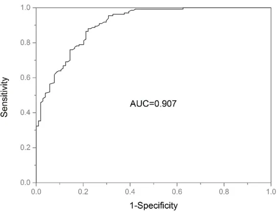
up-regulation was significantly correlated with lymph node status (P=0.029). Furthermore, the value of the area un-der the receiver-operating characteristic curve (AUC-ROC) was 0.907. The optimal cutoff value was 2.255, providing
a sensitivity of 88.0% and a specificity of 77.9% in differentiating NPC patients from healthy controls. Conclusion:
Our data indicates that serum IRS-1 might increase the sensitivity and accuracy in diagnosis of NPC, and may be a potential target for diagnosis and gene therapy.
Keywords: Insulin receptor substrate-1, diagnosis, nasopharyngeal carcinoma
Introduction
Nasopharyngeal carcinoma (NPC) is character-ized by peculiar epidemiologic and clinicopath-ologic features, affecting primarily middle-aged individuals [1]. NPC is a leading lethal malig-nancy that is most prevalent in Southeast Asia, especially in the Cantonese region of southern China [2, 3]. Despite growing incidence and awareness in the relevant populations, NPC is still characterized by diagnosed at a late stage, leading to a high mortality rate, usually through nasopharyngoscopy, a procedure that is sub-jective, skill-dependent, and expensive to main-tain. The standard treatment for NPC is radio-chemotherapy and 5-year survival rates have increased to approximately 60 to 70 % with the improvements in radiotherapy and chemother-apy regimens [4]. However, locoregional recur-rence and distant metastasis following
radio-therapy still have deleterious effects on the survival rate of patients with NPC [5]. The
clini-cal symptoms of NPC are usually nonspecific,
but its location is somewhat special and exami-nation of nasopharynx area requires expertise,
which render early detection of NPC very diffi
-cult. To date, it remains a challenge to find effi -cient biomarkers for early detection/diagnosis and prognosis of this type of malignant disease.
The insulin receptor substrate (IRS) proteins are cytoplasmic adaptor proteins that function as essential signaling intermediates downstream of activated cell surface receptors, many of which have been implicated in cancer. Until now, four IRS proteins (IRS-1 to IRS-4) have
initiation and progression. Overexpression of IRS-1 promotes cells growth, inhibits basal autophagy, reduces oxidative stress-induced autophagy, and diminishes oxidative stress-mediated autophagy-dependent cell death [7]. Recently, it is reported that IRS-1 exhibits increased expression in variety of tumors, including hepatocellular, pancreatic, prostatic, breast, ovarian and colorectal cancers [8-14]. However, so far whether the alteration of the expression of IRS-1 is associated with develop-ment and progression or clinicopathological/ diagnostic implication for NPC has not been reported. In the present study we performed the IRS-1 expression profiling in NPC serum
compared with healthy controls using qRT-PCR and constructed the receiver operating charac-teristic (ROC) curves in order to identify
wheth-er specific IRS-1 could discriminate between NPC patients and healthy controls.
Methods and materials
Patients and blood collection
All blood samples were collected from patients
at Affiliated Hospital of Weifang Medical College
(Fuzhou, China) between 2014 and 2015. A total of 133 patients with diagnosed NPC were recruited. Age, gender, diagnosis, clinical stage (stage I-stage IV), histology and lymph node status of these patients at diagnosis were recorded. The patients received a uniform pro-tocol of image-guided intensity modulated radiotherapy (IMRT). A total of 104 healthy indi-viduals served as controls. These were healthy volunteers without cancerous disease.
This study was approved by the institutional
ethical review boards of Affiliated Hospital of
Weifang Medical College, and written informed consent was obtained from all patients. All blood specimens were processed within 6 hr
after blood withdrawal. Briefly, whole blood was
drawn into EDTA-containing tubes and separat-ed into plasma by centrifugation at 1,500 g for 10 min.
RNA preparation, reverse transcription, and quantitative real-time PCR
Total RNAs were extracted from blood samples using Trizol (Invitrogen, Carlsbad, CA, USA) fol-lowing the manufacturer’s protocol. Extracted RNA samples were reverse transcription to
cDNA as soon as possible, using an All-in-One First-Strand cDNA Synthesis Kit (Genecopoeia).
Amplification of the appropriate product was confirmed by melting curve analysis following amplification. The IRS-1 expression profile was quantified by TaqMan miRNA assays (Applied
Biosystems), according to manufacturer’s instructions. Relative expression of IRS-1 was calculated using the comparative cycle thresh-old (CT) (2−ΔΔCT) method with
glyceraldehyde-3-phosphate dehydrogenase (GAPDH) as the endogenous control to normalize the data. Protein extraction and Western blot analysis
For protein extraction, miRVana PARIS kit (Ambion) was used according to the manufac-turer’s instructions. Protein concentration was determined by the Bradford method using Coomassie brilliant blue (Biofer, Italy). Samples were separated on 12.5% SDS-PAGE gels and electrotransferred to PVDF membranes (Millipore). Membranes were blocked in 5% non-fat milk and incubated with a rabbit anti-IRS-1 antibody (1:200, Boster, China) overnight at 4°C. The membranes were then incubated with a horseradish peroxidase (HRP)-con- jugated secondary antibody (1:4000, Boster, China) for 1 h at RT. Proteins of interest were detected and visualized by autoradiography after various exposure times. The GAPDH was used as internal control.
Statistical analysis
Graphical plotting was done using Origin Pro 9.0 software and statistical evaluations were performed using SPSS 18.0 (SPSS, Inc., Chicago, IL, USA). All the above tests were unpaired and two-tailed. Receiver operating characteristic (ROC) curves were constructed, and the area under the ROC curve (AUC) was
reported to assess the sensitivity and specific -ity. Chi-squared test was used to assess the correlation between the expression level of serum IRS-1 and clinical pathological factors of NPC patients. P value <0.05 was considered to
be statistically significant.
Results
Serum expression protein and mRNA level of IRS-1 in NPC and healthy controls
healthy controls. Analysis revealed that the area under the ROC curves (AUC) for serum IRS-1 was 0.907 (95% confidence interval
0.870-0.944). At an optimal cut-off value of
2.255, the sensitivity and specificity was 88.0%
and 77.9%, respectively (Figure 2).
Discussion
NPC is relatively rare on a global scale, but it is
endemic in a few well-defined populations [15].
It occurs commonly in men, and in the produc-tive age between 35-50 years. There are sev-eral host factors, including tobacco smoking,
consumption of salt-preserved fish, and history
of chronic respiratory tract diseases. In addi-tion, Epstein-Barr virus (EBV) infection is anoth-er well-established risk factor for NPC [3, 16]. The majority of NPC patients have a variety of EBV antigens, and anti-EBV antibody serologi-cal testing has become an important tool for NPC diagnosis [17, 18]. Traditional assays of anti-EBV antibodies have been very useful in expression in 133 cases of NPC serum and
104 healthy volunteer’s blood. Using RNA iso-lated from serum, we performed RT-qPCR to detect the expression levels of IRS-1 mRNA. Using GAPDH as normalization control, serum IRS-1 expression was significantly higher in
[image:3.612.93.374.70.284.2]NPC compared with healthy controls (P<0.05,
Figure 1).
Based on the above study, we further detected the expression of IRS-1 at protein level by Western blot assays in NPC and healthy con-trols. Results revealed that 89 cases (66.9%) exhibited the positive IRS-1 protein expression among all the 133 cases of NPC serum, where-as only 23 cwhere-ases (22.1%) displayed the positive IRS-1 protein expression in healthy volunteers blood (Table 1). Compared with healthy
con-trols, there was significantly increase in IRS-1
protein level in NPC (P<0.05), in line with the result of qRT-PCR. These results indicated that IRS-1 might act as oncogene in NPC.
Relationship between IRS-1 mRNA level and clinicopatho-logical characteristics of NPC
Our investigation revealed that IRS-1 protein expression was increased in NPC, which indicated that IRS-1 might be carcinogenesis. Therefore, we further investigated the asso-ciation of IRS-1 expression with the clinicopathological characteristics of the patients to explore the potential role of IRS-1 in NPC progression. The statistical analysis results, shown in Table 2, indicated that increased IRS-1 protein expression was associated with lymph node status (P=0.029). However, there were no relationships with other features, including age, gender, clinical stages and pathology (all P>0.05).
Diagnostic value of IRS-1 for NPC
[image:3.612.90.377.375.430.2]The ROC curve was plotted to identify a cut-off value that could distinguish NPC from
Table 1. Differential protein expression of IRS-1 between 133 NPC and 104 healthy controls
Group No. of cases IRS-1 expression Χ2 P values
Negative Positive
NPC 133 44 89 47.000 0.000 Healthy controls 104 81 23
fraction, but they suggested that higher IRS-1 levels enhance cancer growth and make earlier relapse possible [11]. IRS-1 is an estrogen-reg-ulated gene frequently expressed in ER positive breast cancer cells [24] where it has been involved in anchorage-independent growth, cell survival [23, 25], and estrogen independent growth [26].
clinical diagnosis of NPC [19]. However, EBV could proliferate in the lymphocytes and make the cells transform, and could long term subcul-ture. The virus-infected cells have the genome of EBV and can produce various EBV-related antigens, such as EBV nuclear antigen (EBNA), early antigen (EA), membrane antigen (MA), viral capsid antigen (VCA), and lymphocyte
membrane antigen (LYDMA). To date, the high false posi-tive rate of the screening bio-marker and low sensitivity of the diagnosis biomarker make accurate early
diagno-sis of NPC difficult [20]. EBV
DNA is considered to be a state-of-the-art quantitative blood biomarker in current NPC research. However, it has been reported that EBV DNA load is independent of serological parameters and
does not reflect the number
of intact tumor cells [21]. Therefore, investigation of the pathogenesis and
identi-fication of molecular markers
of NPC may facilitate early diagnosis, prediction, and development of effective therapeutic strategies for NPC patients.
IRS proteins are positioned to play a pivotal role in regulat-ing the response of tumor cells to many different micro-environmental stimuli and regulating cancer cell surviv-al, proliferation, and motility [22]. IRS-1, the first and most
[image:4.612.90.379.97.321.2]important (IRS) family mem-ber, is highly expressed in many cancers. For example, Surmacz et al. reported that IRS-1 overexpression has been associated with tumor development, hormone inde-pendence and antioestrogen resistance in breast cancer [23]. Rocha et al. found a lack of correlation between IRS-1 expression and mitotic activi-ty in cancer cells assessed by evaluation of the S-phase
Table 2. IRS-1 mRNA expression and clinicopathological features in NPC patients
Features No. of cases (n=133) IRS-1 expression P values Low (n=54) High (n=79)
Age (years)
<45 101 42 59 0.682
≥45 32 12 20
Gender
Male 94 41 53 0.272 Female 39 13 26
Clinical stages
I-II 46 21 25 0.388 III-IV 87 33 54
Pathology
Differentiated 102 45 57 0.134 Undifferentiated 31 9 22
Lymph node status
LNM 96 45 51 0.029
No LNM 37 9 26
LNM: lymph node metastasis.
[image:4.612.93.376.355.573.2]In the current study we compared the profile of
IRS-1 in serum from 133 NPC and 104 healthy controls and demonstrated that serum expres-sion of IRS-1 strongly differentiated the breast cancer patients from healthy controls. A highly
significant increase was found in serum IRS-1 of NPC compared with that of healthy individu-als. Moreover, receiver operating curve analy-sis indicated that the AUC of IRS-1 was 0.907
and the sensitivity and specificity at optimal
cutoff being 88.0% and 77.9%, indicating that it might be potential biomarkers in the diagnosis of NPC Luo et al. revealed that the expression
level of IRS-1 was significant higher in NPC than
that in the control nasopharyngeal epithelia [27]. Our results were consistent with previous studies.
We also analyzed the serum expression levels of IRS-1 in relation to the different clinical pathologic characteristics in NPC patients. The result showed that serum IRS-1 expression was relationship withlymph node status.However,
there were no significant relationships between
serum IRS-1 expression and other features, including age, gender, clinical stages and pathology.
In the present study, the results showed the expression levels of IRS-1 in NPC patients were higher than that of IRS-1 in healthy controls.
And the ROC results displayed significant diag -nostic accuracy of IRS-1. The findings indicated
serum IRS-1 appeared to be potentially useful biomarkers for NPC detection. Further study with larger sample involving in validation and optimizing improvement should be conducted
to confirm our results.
Disclosure of conflict of interest
None.
Address correspondence to: Xuanchang Qi, De-
partment of Otorhinolaryngology, Affiliated Hospital
of Weifang Medical College, Weifang 261000, Shandong, China. E-mail: xuchang14@126.com
References
[1] Li ZQ, Xia YF, Liu Q, Yi W, Liu XF, Han F, Luo W and Lu TX. Radiotherapy-related typing in 842 patients in canton with nasopharyngeal carci-noma. Int J Radiat Oncol Biol Phys 2006; 66: 1011-1016.
[2] Wei WI and Sham JS. Nasopharyngeal carci-noma. Lancet 2005; 365: 2041-2054. [3] Chang ET and Adami HO. The enigmatic
epide-miology of nasopharyngeal carcinoma. Cancer Epidemiol Biomarkers Prev 2006; 15: 1765-1777.
[4] Zhang L, Zhao C, Ghimire B, Hong MH, Liu Q, Zhang Y, Guo Y, Huang YJ and Guan ZZ. The role of concurrent chemoradiotherapy in the treatment of locoregionally advanced naso-pharyngeal carcinoma among endemic popu-lation: a meta-analysis of the phase III random-ized trials. BMC Cancer 2010; 10: 558. [5] Lee AW, Poon YF, Foo W, Law SC, Cheung FK,
Chan DK, Tung SY, Thaw M and Ho JH. Retrospective analysis of 5037 patients with nasopharyngeal carcinoma treated during 1976-1985: overall survival and patterns of failure. Int J Radiat Oncol Biol Phys 1992; 23: 261-270.
[6] Lee YH and White MF. Insulin receptor sub-strate proteins and diabetes. Arch Pharm Res 2004; 27: 361-370.
[7] Chan SH, Kikkawa U, Matsuzaki H, Chen JH and Chang WC. Insulin receptor substrate-1 prevents autophagy-dependent cell death caused by oxidative stress in mouse NIH/3T3 cells. J Biomed Sci 2012; 19: 64.
[8] Bergmann U, Funatomi H, Kornmann M, Beger HG and Korc M. Increased expression of insu-lin receptor substrate-1 in human pancreatic cancer. Biochem Biophys Res Commun 1996; 220: 886-890.
[9] Kornmann M, Maruyama H, Bergmann U, Tangvoranuntakul P, Beger HG, White MF and Korc M. Enhanced expression of the insulin re-ceptor substrate-2 docking protein in human pancreatic cancer. Cancer Res 1998; 58: 4250-4254.
[10] Hellawell GO, Turner GD, Davies DR, Poulsom R, Brewster SF and Macaulay VM. Expression of the type 1 insulin-like growth factor receptor is up-regulated in primary prostate cancer and commonly persists in metastatic disease. Cancer Res 2002; 62: 2942-2950.
[11] Rocha RL, Hilsenbeck SG, Jackson JG, VanDenBerg CL, Weng C, Lee AV and Yee D. Insulin-like growth factor binding protein-3 and insulin receptor substrate-1 in breast cancer: correlation with clinical parameters and dis-ease-free survival. Clin Cancer Res 1997; 3: 103-109.
[12] Koda M, Sulkowska M, Kanczuga-Koda L and Sulkowski S. Expression of insulin receptor substrate 1 in primary breast cancer and lymph node metastases. J Clin Pathol 2005; 58: 645-649.
sub-strate-1 is an important mediator of ovarian cancer cell growth suppression by all-trans retinoic acid. Cancer Res 2007; 67: 9266-9275.
[14] Esposito DL, Aru F, Lattanzio R, Morgano A, Abbondanza M, Malekzadeh R, Bishehsari F, Valanzano R, Russo A, Piantelli M, Moschetta A, Lotti LV and Mariani-Costantini R. The insu-lin receptor substrate 1 (IRS1) in intestinal epithelial differentiation and in colorectal can-cer. PLoS One 2012; 7: e36190.
[15] Parkin DM, Bray F, Ferlay J and Pisani P. Global cancer statistics, 2002. CA Cancer J Clin 2005; 55: 74-108.
[16] O’Neil JD, Owen TJ, Wood VH, Date KL, Valentine R, Chukwuma MB, Arrand JR, Dawson CW and Young LS. Epstein-Barr virus-encoded EBNA1 modulates the AP-1 transcrip-tion factor pathway in nasopharyngeal carci-noma cells and enhances angiogenesis in vi-tro. J Gen Virol 2008; 89: 2833-2842.
[17] Ai P, Wang T, Zhang H, Wang Y, Song C, Zhang L, Li Z and Hu H. Determination of antibodies directed at EBV proteins expressed in both la-tent and lytic cycles in nasopharyngeal carci-noma. Oral Oncol 2013; 49: 326-331. [18] Ng MH, Chan KH, Ng SP and Zong YS.
Epstein-Barr virus serology in early detection and screening of nasopharyngeal carcinoma. Ai Zheng 2006; 25: 250-256.
[19] Chang KP, Hsu CL, Chang YL, Tsang NM, Chen CK, Lee TJ, Tsao KC, Huang CG, Chang YS, Yu JS and Hao SP. Complementary serum test of antibodies to Epstein-Barr virus nuclear anti-gen-1 and early antigen: a possible alternative for primary screening of nasopharyngeal carci-noma. Oral Oncol 2008; 44: 784-792.
[20] Tsang RK, Vlantis AC, Ho RW, Tam JS, To KF
and van Hasselt CA. Sensitivity and specificity
of Epstein-Barr virus IGA titer in the diagnosis of nasopharyngeal carcinoma: a three-year in-stitutional review. Head Neck 2004; 26: 598-602.
[21] Lin SY, Tsang NM, Kao SC, Hsieh YL, Chen YP, Tsai CS, Kuo TT, Hao SP, Chen IH and Hong JH. Presence of Epstein-Barr virus latent mem-brane protein 1 gene in the nasopharyngeal swabs from patients with nasopharyngeal car-cinoma. Head Neck 2001; 23: 194-200. [22] Porter HA, Perry A, Kingsley C, Tran NL and
Keegan AD. IRS1 is highly expressed in local-ized breast tumors and regulates the sensitiv-ity of breast cancer cells to chemotherapy, while IRS2 is highly expressed in invasive breast tumors. Cancer Lett 2013; 338: 239-248.
[23] Surmacz E. Function of the IGF-I receptor in breast cancer. J Mammary Gland Biol Ne- oplasia 2000; 5: 95-105.
[24] Jackson JG, White MF and Yee D. Insulin recep-tor substrate-1 is the predominant signaling molecule activated by insulin-like growth fac-tor-I, insulin, and interleukin-4 in estrogen re-ceptor-positive human breast cancer cells. J Biol Chem 1998; 273: 9994-10003.
[25] Nolan MK, Jankowska L, Prisco M, Xu S, Guvakova MA and Surmacz E. Differential roles of IRS-1 and SHC signaling pathways in breast cancer cells. Int J Cancer 1997; 72: 828-834.
[26] Surmacz E and Burgaud JL. Overexpression of insulin receptor substrate 1 (IRS-1) in the hu-man breast cancer cell line MCF-7 induces loss of estrogen requirements for growth and transformation. Clin Cancer Res 1995; 1: 1429-1436.