Original Article
Age-related methylation of genomic DNA in human
adipose-derived stem cells
Ji-An Guo, Pei-Jun Yu, Lu-Ping Wang, Ying-Ying Shi, Yi Liu, Wei Chen
Department of Plastic Surgery, The Affiliated Shanghai Eighth People’s Hospital of Jiangsu University, Shanghai, China
Received January 15, 2017; Accepted February 22, 2017; Epub April 1, 2017; Published April 15, 2017
Abstract: Objective: Human adipose-derived stem cells (ADSCs) are multipotent stromal cells, andthe cellular func-tionsof ADSCs are regulated by genomic DNA methylation. The objective of this study was to research the relation-ship between age and ADSC genomic DNA methylation. Materials and methods: We examined the age-related gene expression and methylation of ADSCs from young (<25 years) and elderly (>55 years) patients. Real-time quantitative PCR was used to analyze OCT-4, NANOG and SOX-2 expression levels. Bisulfite sequencing was per-formed to determine the density of DNA methylation on target gene promoters. Results: After ADSCs from elderly patients (oldADSCs) were treated with the DNA-demethylating drug 5-aza-2’-deoxycytidine (5-Aza-dC), the OCT-4 and NANOG expression levels were significantly lower in oldADSCs than those in ADSCs from young patients (youngAD-SCs) (P=0.011 and P=0.030, respectively). Conversely, SOX-2 expression was significantly increased in oldADSCs (P=0.029). OCT-4 and NANOG promoter methylation was extremely dense in oldADSCs, but these promoters were hypomethylated in youngADSCs (P=0.031 and P=0.048, respectively). Moreover, significant associationswere found between methylation and the expression of OCT-4 and NANOG (R=-0.693, P=0.026 and R=-0.839, P=0.002, respec-tively). However, significant differencesin SOX-2 methylation were not observed between the two groups (P=0.179). Conclusion: ADSCs treated with 5-Aza-dC exhibited significant increases inOCT-4, NANOG, and SOX-2 expression. Our results suggest that DNA methylation plays an important role in ADSC aging and that DNA methylation density increases with patient age. More importantly, demethylation drugs may restore OCT-4 and NANOG expression inol-dADSCs and could have implications regarding the potential for autologous stem cell therapies in elderly patients.
Keywords: Aging, adipose-derived stem cells, methylation, genomic DNA
Introduction
Human adipose-derived stem cells (ADSCs) are mesenchymal stem cells (MSCs) that can be collected from adipose tissue [1-3]. ADSCs have the capacity for self-renewal and the potential to differentiate into multiple lineag- es, including adipocytes, osteocytes, myocytes and chondrocytes [4-8]. Due to their broad differentiation potential and convenient acces-sibility, ADSCs are an attractive source of adult MSCs for many clinical applications, including orthopedic and reconstructive surgery, as well as cell developmental plasticity research [9-11]. Unfortunately, the physiological function and differentiation potential of ADSCs dimin-ishes with advanced age, which limits the app- lication of ADSCs in elderly patients [12-14]. Epigenetic modifications (DNA methylation, genomic imprinting, maternal effects and gene
silencing) can regulate ADSC pluripotency and differentiation. Moreover, the epigenetic regu- lation of the genome is considered a crucial pathway affecting stem cell aging [15-18]. Recently, the effects of CpG methylation on proliferation, stemness, and differentiation have been carefully studied, particularly for important pluripotency genes, such as OCT-4, NANOG, and SOX-2 [19-23]. DNA methylation is a powerful mechanism regulating gene expression; hypermethylation of promoter regi- ons has been shown to silence gene expres-sion, whereas promoterhypomethylation can activate transcription [24].
wheth-er these age-related changes are revwheth-ersible after DNA demethylation.
Materials and methods
Cultivation and isolation of ADSCs
The study was approved by the Institutional Ethics Committee of the Affiliated Shanghai Eighth People’s Hospital of Jiangsu University. Human abdominal fat tissue was obtained from young (aged<25 years, n=5) and elderly (aged>55 years, n=5) donors who underwent abdominoplasty. Informed consent was recei- ved from all patients for the use of their tissues.
The subcutaneous fat was washed three tim- es with phosphate-buffered saline (HyClone, Logan, UT, USA) containing 100 U/ml penicillin and 100 μg/ml streptomycin (Gibco, Grand Island, NY, USA) and gently shaken to remove-blood. Then, the samples were cut into small pieces (1 mm3) using scissors. Adipose tissues were then treated with 0.075% collagenase type I (Sigma-Aldrich, St. Louis, MO, USA) and incubated for 40 minutes at 37°C in a vibra- ting, constant-temperature water bath. Then, Dulbecco’s Modified Eagle Medium/Nutrient Mixture F12 (DMEM/F12; Gibco) containing 10% fetal bovine serum (FBS; Gibco), 100 U/ml penicillin, and 100 μg/ml streptomycin was added to stop the digestion.
The digested fat was centrifuged at 300×g for 10 minutes. The supernatant was discarded, and the pellet was resuspended in complete medium (DMEM/F12 containing 10% FBS) and filtered through a 100 μm nylon mesh filter (Millipore, Billerica, MA, USA). The filtered cell fraction was incubated overnight, and the adherent cells were collected, maintained at 37°C in a humidified atmosphere containing
5% CO2, and cultured to passage 3 for experi- ments.
Immunophenotyping
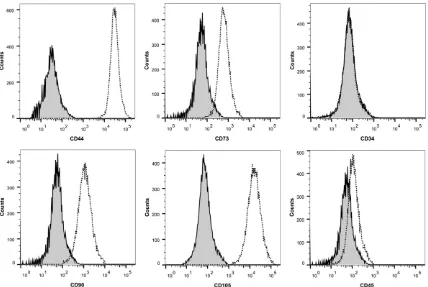
To characterize the ADSCs, we performed a flow cytometric analysis for six MSC markers, CD44, CD73, CD90 and CD105, and two nega-tive markers, CD34 and CD45. Fluorescence-conjugated antibodies were purchased from eBioscience (San Diego, CA, USA). The surface antigens were analyzed witha FACSVerse sys-tem (BD Bioscience, San Jose, CA, USA) using FlowJo software (Treestar, Inc., San Carlos, CA, USA).
Adipogenic differentiation of ADSCs
ADSCs from healthy donors (passage 3) were seeded at 2×10⁴ cells/cm2 in 6-well cell culture plates. Cells were allowed to grow to post- confluence in complete medium. Adipogenic differentiation of ADSCs was achieved using an adipogenic kit (Cyagen Biosciences, Guang- zhou, China) and was confirmed by Oil Red O (Cyagen) staining of lipid droplets after 16 days in culture.
Osteogenic differentiation of ADSCs
ADSCs (passage 3) were seeded at 2×10⁴ cells/cm2 in 6-well cell culture plates pre- coated with 0.1% gelatin solution (Cyagen). Cells were grown to 80-90% confluence in complete medium, which was then replaced with osteoinduction medium (Cyagen). Osteo- induction was stopped at day 18, and the cells were stained with alizarin red (Cyagen) for microscopic visualization.
5-Aza-2’-deoxycytidine (5-Aza-dC) treatment of ADSCs
For demethylation studies, ADSCs were seed- ed at an initial density of 1×10⁵ cells/cm2. After
[image:2.612.91.525.83.192.2]attachment, the cells were incubated with culture medium containing 5-Aza-dC (Sigma-Aldrich, Steinheim, USA) (final concentrations of 0 μM, 1 μM, or10 μM) for 72 hours. All the cells were cultured until harvested for RNA and DNA extraction.
RNA isolation, reverse transcription and real-time quantitative PCR (qRT-PCR)
TRIzol reagent (Invitrogen, Carlsbad, CA, USA) was used to isolate total RNA. Reverse tran-scription reactions (40 μL) contained 10 mM dNTPs, 10 μM random hexamers, 10 mM 5× buffer, 80 U RNasin, and 200 U MMLV reverse transcriptase (MBI Fermentas, Hanover, USA). Reverse transcription was performed using an iCycler Thermal Cycler (Eppendorf, Hamburg, Germany). The reaction mixtures were incuba- ted for 10 minutes at 25°C, 60 minutes at 42°C, and then stored at -20°C. qRT-PCR was performed on a 7500 Thermo cycler (Applied Biosystems, CA, USA). The primer sequences used to assess OCT-4, NANOG, and SOX-2 expression are listed in Table 1. The PCR reactions contained 20 ng of cDNA, 10 μM
SYBR Premix Ex Taq II (Takara, Japan), 0.4 μM 50× ROX (Takara, Japan) and 0.8 μM primers. The RT-PCR reaction conditions were 95°C for 30 s; 40 cycles of 95°C for 5 s, annealing temperature for 30 s (listed in Table 1), 72°C for 30 s, and 80°C for 30 s to collect fluores-cence, followed by 95°C for 15 s, 60°C for 60 s, 95°C for 15 s, and 60°C for 15 s. GAPDH was used as the endogenous control. All assays included positive and negative controls.
DNA isolation and chemical modification
A genomic DNA purification kit (Gentra, Min- neapolis, MN, USA) was used to isolate DNA. Genomic DNA was modified by the Cp Genome DNA Modification Kit (Chemicon, Ternecula, Canada) according to the manufacturer’s instructions.
Bisulfite sequencing (BSP)
[image:3.612.94.522.71.358.2]BSP was performed to analyze the DNA meth-ylation density on the three gene promoters. Bisulfite-treated DNA was amplified with the sequencing primers listed in Table 1. The reac-tion was performed on an iCycler Thermal
Cycler (Eppendorf, Hamburg, Germany) in 25-μL reaction mixtures containing 10×PCR buffer (0.25 mM KCl), 6.25 μM dNTP mixture, 0.5 μM primers, 0.75 U Hot Dtart DNA poly-merase (Takara, Tokyo, Japan), and 20 ng of modified DNA. PCR conditions were 98°C for 10 s; 40 cycles of 10 s at 98°C, 30 s at annealing temperature (listed in Table 1), and 30 s at 72°C; followed by a final 7-minute ex- tension step at 72°C. The PCR products were purified using the AxyPrep DNA Gel recovery kit (Axygen, Suzhou, China) and cloned into a pMD®19-T Vector (Takara, Dalian, China); five clones for each sample were sequenced at BGI (Shanghai, China).
Statistical analysis
Statistical analysis was performed using the Statistical Program for Social Science (SPSS) software 20.0 package (SPSS, Chicago, IL,
USA). Allresults are expressed as the mean ± SEM of at least 3 independent experiments and were compared with two-tailed Student’s t-tests. P values<0.05 were considered stati- stically significant.
Results
Phenotypic characterization of ADSCs
[image:4.612.91.530.72.267.2]Using our isolation and culture methods, ADSCs were easily expanded in vitro and exhibited a fibroblast-like morphology. Cell surface marker expressionwas analyzed using flow cytometry (Figure 1), which revealed the expression of the MSC markers CD44, CD73, CD90 and CD105 in the ADSCs. The hematopoietic lineage mark-ers CD34 and CD45 were not expressed in the derived ADSCs. Therefore, our results suggest that ADSCs isolated from fat tissue resemble MSCs rather than hematopoietic stem cells.
Figure 2. Characterization of isolated ADSCs by adipogenic and osteogenic differentiation. A: ADSCs were cultured in adipogenic media for 16 days and showed positive Oil Red O staining results. The lipid droplets appeared red under the fluorescence microscope (×100). B: ADSCs were cultured in osteogenic media for 18 days and showed positive alizarin red staining results. Red staining marked mineral deposition in the newly formed extracellular ma-trix (×100).
[image:4.612.91.527.346.441.2]Induction of multilineage differentiation
Adipogenesis: Adipogenic differentiation of AD- SCs was induced with an adipogenic kit. Pre- adipocytes with lipid droplets appeared after approximately one week of treatment. The lipid vacuoles became larger after 16 days of treat-ment and were clearly visible without staining under a phase microscope. After Oil Red O staining, the lipid nature of the red vacuoles was evident (Figure 2A). The ability of these cells to differentiate into adipocytes was observed in cells derived from both young and elderly donors.
Osteogenesis: After treatment with an osteo-genic kit for 18 days, the cell monolayer was covered with a layer of visible deposits indica-tive of cell differentiation. Posiindica-tive staining with alizarin red revealed that these deposits were calcified extracellular matrix (Figure 2B). The induction of osteogenesis in ADSCs deri- ved from both young and elderlydonorswas widespread, and these results indicated that ADSCs retained their capacity for osteogenic differentiation.
Gene expression levels in ADSCs from patients of different ages
The ADSCs were divided into two groups accor- ding to their age (<25 years and >55 years). The relative OCT-4, NANOG and SOX-2 expression levels were evaluated with qRT-PCR, and GA- PDH was used as an internal control. NANOG and OCT-4 expression levels were significantly lower in ADSCs from elderly patients than in those from young patients (P=0.011 and P=0.030, respectively) (Figure 3A, 3B). Con- versely, SOX-2 expression was significantly ele-vated in ADSCs from elderly patients (P=0.029) compared with ADSCs from young patients (Figure 3C).
Gene expression levels after 5-Aza-dC treat-ment
The cell lines with low OCT-4, NANOG and SOX-2 expression were selected for 5-Aza-dC treat-ment at different concentrations (0 μM, 1 μM or 10 μM).
Accordingly, OCT-4, NANOG and SOX-2 expres-sion was all significantly increased after 5-Aza-dC treatment (Figure 4).
DNA methylation status of OCT-4, NANOG and SOX-2
To investigate the methylation density of the OCT-4, NANOG and SOX-2 promoters, BSP was performed on cells from five young patients and five elderly patients. The methylation den-sity of OCT-4 and NANOG was extremely high in elderly patients, whereas OCT-4 and NAN- OG were hypomethylated in young patients (P=0.031 and P=0.048, respectively) (Figure 5A, 5B). However, no significant differences in SOX-2 methylation were observed between the two groups (P=0.179) (Figure 5C).
The cell lines were treated with different con-centrations of 5-Aza-dC, andthe BSP results showed that OCT-4, NANOG and SOX-2 promo- ter methylation was decreased and that OCT-4, NANOG and SOX-2 expression levels were sig-nificantly increased after 5-Aza-dC treatment (Figure 6).
The association between OCT-4, NANOG and SOX-2 methylation and expression
The expression levels of OCT-4, NANOG and SOX-2 in ten patients ranged from 0.109 to 1.000, 0.197 to 1.000 and 0.026 to 1.000, respectively. A significant correlation was observed between OCT-4 and NANOG
[image:5.612.90.519.71.175.2]sion and promoter methylation (R=-0.693, P=0.026 and R=-0.839, P=0.002). However, a significant correlation between SOX-2 expres-sion and promoter methylation was not obser- ved (R=-0.315, P=0.375).
Discussion
The study of ADSCs has become popular beca- use ADSCs have the potential to treat various diseases. Owing to their multilineage differen-tiation potential, ADSCs have been applied in regenerative medicine [21, 25, 26]. In addition,
[image:6.612.93.524.68.300.2]munication, mitochondrial dysfunction, proteos- tasis loss and deregulated nutrients [30-33]. The increasing age of MSCs and their hosts has been associated with decreased capacities for proliferation, differentiation and mobiliza-tion [34]. Campisi J posited that the presence of senescent cells, whether stem cells them-selves or surrounding cells in stem cell niches, could disrupt the stem cell microenvironment andthat this senescent microenvironment could then alter the functional capacity of the resident stem cells [35].
Figure 5. DNA methylation status of genes in the two groups. A: NANOG; B: OCT-4; and C: SOX-2. Upper: DNA meth-ylation density of genes in five young patients. Lower: DNA methmeth-ylation density of genes in five elderly patients.
Figure 6. Cell lines were treated with different concentrations of 5-Aza-dC (0 μM, 1 μM, or 10 μM). A: NANOG (0 μM, mean density: 82.5%; 1 μM, mean density: 55%, P=0.009; 10 μM: mean density: 42.5%, P=0.001); B: OCT-4 (0 μM, mean density: 69.33%; 1 μM, mean density: OCT-48%, P=0.013; 10 μM: mean density: 34.67%, P=0.001); and C: SOX-2 (0 μM, mean density: 71.58%; 1 μM, mean density: 55.79%, P=0.014; 10 μM: mean density: 43.16%, P<0.001).
[image:6.612.91.376.348.480.2]com-Due to their broad multipotent potential and convenient accessibility, ADSCs have become the focus of recent research. Several studies have focused on the mechanisms that affect ADSC functions, especiallythe methylation of certain genes [36-39]. CpG methylation of the CD31 and CD144 promoter regions in ADSCs was demonstrated to limit ADSC differentiation potential [40]. Berdasco M et al. reported that DNA methylation was necessary toregulate ADSC differentiation and proliferation [41]. Fur- thermore, the use of 5-Aza-dC may promote ADSC differentiation [20].
However, the status of gene expression and promoter methylation at different ages has not been previously described. This study exam-ined donor-age-related changes in the pattern of OCT-4, NANOG and SOX-2 expression and methylation. Furthermore, we assessed wheth-er these age-related changes could be revwheth-ersed after treatment with a demethylation agent. In this study, we unexpectedly discovered that OCT-4 and NANOG expression were significant-ly lower in oldADSCs than in youngADSCs. Additionally, the density of OCT-4 and NANOG methylation was extremely high in oldADSCs, but OCT-4 and NANOG were hypomethylated in youngADSCs. Furthermore, we also observed a significant association between the methyla-tion and expression of OCT-4 and NANOG, sug-gesting that the age-related methylation of OCT-4 and NANOG along their promoters regu-lates their expression. Moreover, in ADSCs treated with 5-Aza-dC, both OCT-4 and NANOG expression levels were significantly increa- sed, further confirming that both OCT-4 and NANOG expression can be restored by treat-ment with a demethylating drug, thereby pro-viding a method to study and treat aging. Zhou GS et al. reported that pretreating MSCs with 5-Aza-dC significantly accelerated their osteo-genic differentiation, resulting in DNA hypo- methylation and the increased expression of osteogenic genes [42]. Yan X et al. revealed that 5-Aza-dC improved the osteogenic diffe- rentiation potential of aged ADSCs by DNA demethylation [20]. Unfortunately, although SO- X-2 expression was significantly increased in oldADSCs, no significant differencesin SOX-2 methylation were observed between the two groups. Accordingly, more prospective studies are needed to confirm and elucidate the role of age-related DNA methylation in ADSCs.
In summary, our investigation discovered that DNA methylation plays an important role in ADSC aging and that DNA methylation density increases with donor age. Additionally, demeth-ylating agents can restore OCT-4 and NANOG expression in ADSCs from elderly patients. Acknowledgements
This study was supported by Shanghai Muni- cipal Commission of Health and Family Plan- ning (20154Y0198), Xuhui District Medical Research Project of Shanghai (SHXH201606), Clinical Medical Science and Technology Development Foundation of Jiangsu University (JLY20160077).
Disclosure of conflict of interest
None.
Addresscorrespondence to: Dr. Wei Chen, Depart- ment of Plastic Surgery, The Affiliated Shanghai Eighth People’s Hospital of Jiangsu University, Shanghai, China. Tel: +86-13701924355; E-mail: drchenwei@163.com
References
[1] Kaewsuwan S, Song SY, Kim JH and Sung JH. Mimicking the functional niche of adipose-de-rived stem cells for regenerative medicine. Ex-pert Opin Biol Ther 2012; 12: 1575-1588. [2] Halvorsen YD, Bond A, Sen A, Franklin DM,
Lea-Currie YR, Sujkowski D, Ellis PN, Wilkison WO and Gimble JM. Thiazolidinediones and glucocorticoids synergistically induce differen-tiation of human adipose tissue stromal cells: biochemical, cellular, and molecular analysis. Metabolism 2001; 50: 407-413.
[3] Erickson GR, Gimble JM, Franklin DM, Rice HE, Awad H and Guilak F. Chondrogenic potential of adipose tissue-derived stromal cells in vitro and in vivo. Biochem Biophys Res Commun 2002; 290: 763-769.
[4] Mendel TA, Clabough EB, Kao DS, Demidova-Rice TN, Durham JT, Zotter BC, Seaman SA, Cronk SM, Rakoczy EP, Katz AJ, Herman IM, Peirce SM and Yates PA. Pericytes derived from adipose-derived stem cells protect against reti-nal vasculopathy. PLoS One 2013; 8: e65691. [5] Zuk PA, Zhu M, Ashjian P, De Ugarte DA, Huang
JI, Mizuno H, Alfonso ZC, Fraser JK, Benhaim P and Hedrick MH. Human adipose tissue is a source of multipotent stem cells. Mol Biol Cell 2002; 13: 4279-4295.
tre JS, Laharrague P, Charriere G, Carriere A and Penicaud L. Plasticity of adipose tissue: a promising therapeutic avenue in the treatment of cardiovascular and blood diseases? Arch Mal Coeur Vaiss 2005; 98: 922-926.
[9] Zuk PA. The adipose-derived stem cell: looking back and looking ahead. Mol Biol Cell 2010; 21: 1783-1787.
[10] Tremolada C, Colombo V and Ventura C. Adi-pose tissue and mesenchymal stem cells: state of the art and lipogems(R) technology de-velopment. Curr Stem Cell Rep 2016; 2: 304-312.
[11] Caplan AI. Adult mesenchymal stem cells for tissue engineering versus regenerative medi-cine. J Cell Physiol 2007; 213: 341-347. [12] Maredziak M, Marycz K, Tomaszewski KA,
Kor-nicka K and Henry BM. The influence of aging on the regenerative potential of human adi-pose derived mesenchymal stem cells. Stem Cells Int 2016; 2016: 2152435.
[13] Mobarak H, Fathi E, Farahzadi R, Zarghami N and Javanmardi S. L-carnitine significantly de-creased aging of rat adipose tissue-derived mesenchymal stem cells. Vet Res Commun 2017; 41: 41-47.
[14] Ma N, Qiao C, Zhang W, Luo H, Zhang X, Liu D, Zang S, Zhang L and Bai J. Original Research: adipose-derived stem cells from younger do-nors, but not aging dodo-nors, inspire the host self-healing capability through its secreta. Exp Biol Med (Maywood) 2017; 242: 68-79. [15] Berdasco M and Esteller M. Hot topics in
epi-genetic mechanisms of aging: 2011. Aging Cell 2012; 11: 181-186.
[16] McGee-Lawrence ME and Westendorf JJ. His-tone deacetylases in skeletal development and bone mass maintenance. Gene 2011; 474: 1-11.
[17] Selvaraj V, Plane JM, Williams AJ and Deng W. Switching cell fate: the remarkable rise of in-duced pluripotent stem cells and lineage re-programming technologies. Trends Biotechnol 2010; 28: 214-223.
[18] Tahiliani M, Koh KP, Shen Y, Pastor WA, Ban-dukwala H, Brudno Y, Agarwal S, Iyer LM, Liu DR, Aravind L and Rao A. Conversion of 5- methylcytosine to 5-hydroxymethylcytosine in mammalian DNA by MLL partner TET1. Sci-ence 2009; 324: 930-935.
osteogenic differentiation potential of aged hu-man adipose-derived mesenchymal stem cells by DNA demethylation. PLoS One 2014; 9: e90846.
[21] Zhang RP, Shao JZ and Xiang LX. GADD45A protein plays an essential role in active DNA demethylation during terminal osteogenic dif-ferentiation of adipose-derived mesenchymal stem cells. J Biol Chem 2011; 286: 41083-41094.
[22] Lagarkova MA, Volchkov PY, Lyakisheva AV, Philonenko ES and Kiselev SL. Diverse epigen-etic profile of novel human embryonic stem cell lines. Cell Cycle 2006; 5: 416-420. [23] Koh KP, Yabuuchi A, Rao S, Huang Y, Cunniff K,
Nardone J, Laiho A, Tahiliani M, Sommer CA, Mostoslavsky G, Lahesmaa R, Orkin SH, Rodig SJ, Daley GQ and Rao A. Tet1 and Tet2 regulate 5-hydroxymethylcytosine production and cell lineage specification in mouse embryonic stem cells. Cell Stem Cell 2011; 8: 200-213. [24] Taby R and Issa JP. Cancer epigenetics. CA
Cancer J Clin 2010; 60: 376-392.
[25] Gimble JM, Bunnell BA, Chiu ES and Guilak F. Concise review: adipose-derived stromal vas-cular fraction cells and stem cells: let’s not get lost in translation. Stem Cells 2011; 29: 749-754.
[26] Mizuno H, Tobita M and Uysal AC. Concise re-view: adipose-derived stem cells as a novel tool for future regenerative medicine. Stem Cells 2012; 30: 804-810.
[27] Fang B, Song Y, Liao L, Zhang Y and Zhao RC. Favorable response to human adipose tissue-derived mesenchymal stem cells in steroid-re-fractory acute graft-versus-host disease. Tran- splant Proc 2007; 39: 3358-3362.
[28] Gonzalez-Rey E, Anderson P, Gonzalez MA, Rico L, Buscher D and Delgado M. Human adult stem cells derived from adipose tissue protect against experimental colitis and sep-sis. Gut 2009; 58: 929-939.
[30] Sethe S, Scutt A and Stolzing A. Aging of mes-enchymal stem cells. Ageing Res Rev 2006; 5: 91-116.
[31] Wheeler HE and Kim SK. Genetics and genom-ics of human ageing. Philos Trans R Soc Lond B Biol Sci 2011; 366: 43-50.
[32] Efimenko A, Dzhoyashvili N, Kalinina N, Koche-gura T, Akchurin R, Tkachuk V and Parfyonova Y. Adipose-derived mesenchymal stromal cells from aged patients with coronary artery dis-ease keep mesenchymal stromal cell proper-ties but exhibit characteristics of aging and have impaired angiogenic potential. Stem Cells Transl Med 2014; 3: 32-41.
[33] Fafian-Labora J, Fernandez-Pernas P, Fuentes I, De Toro J, Oreiro N, Sangiao-Alvarellos S, Ma-teos J and Arufe MC. Influence of age on rat bone-marrow mesenchymal stem cells poten-tial. Sci Rep 2015; 5: 16765.
[34] Rossi DJ, Bryder D, Seita J, Nussenzweig A, Hoeijmakers J and Weissman IL. Deficiencies in DNA damage repair limit the function of hae-matopoietic stem cells with age. Nature 2007; 447: 725-729.
[35] Campisi J. Senescent cells, tumor suppres-sion, and organismal aging: good citizens, bad neighbors. Cell 2005; 120: 513-522.
[36] Kim WS, Park BS, Sung JH, Yang JM, Park SB, Kwak SJ and Park JS. Wound healing effect of adipoderived stem cells: a critical role of se-cretory factors on human dermal fibroblasts. J Dermatol Sci 2007; 48: 15-24.
[37] Chen KH, Chen CH, Wallace CG, Yuen CM, Kao GS, Chen YL, Shao PL, Chen YL, Chai HT, Lin KC, Liu CF, Chang HW, Lee MS and Yip HK. In-travenous administration of xenogenic adi-pose-derived mesenchymal stem cells (ADM-SC) and ADMSC-derived exosomes markedly reduced brain infarct volume and preserved neurological function in rat after acute isch-emic stroke. Oncotarget 2016; 7: 74537-74556.
[38] Lee M, Ban JJ, Kim KY, Jeon GS, Im W, Sung JJ and Kim M. Adipose-derived stem cell exo-somes alleviate pathology of amyotrophic lat-eral sclerosis in vitro. Biochem Biophys Res Commun 2016; 479: 434-439.
[39] Berdasco M, Melguizo C, Prados J, Gomez A, Alaminos M, Pujana MA, Lopez M, Setien F, Or-tiz R, Zafra I, Aranega A and Esteller M. DNA methylation plasticity of human adipose-de-rived stem cells in lineage commitment. Am J Pathol 2012; 181: 2079-2093.
[40] Boquest AC, Noer A, Sorensen AL, Vekterud K and Collas P. CpG methylation profiles of endo-thelial cell-specific gene promoter regions in adipose tissue stem cells suggest limited dif-ferentiation potential toward the endothelial cell lineage. Stem Cells 2007; 25: 852-861. [41] Berdasco M and Esteller M. DNA methylation
in stem cell renewal and multipotency. Stem Cell Res Ther 2011; 2: 42.