Original Article
Association of serum vaspin levels with the presence
and severity of peripheral arterial disease in
type 2 diabetic patients
Qingxu Guo1, Youdong Chen2, Yunlong Liu1
1Department of Vascular Surgery, General Hospital of Beijing Military Region, Beijing, PR China; 2Department of
General Surgery, Beijing Huanxing Tumor Hospital, Chaoyang District, Beijing, PR China
Received November 28, 2015; Accepted January 26, 2016; Epub February 1, 2017; Published February 15, 2017
Abstract: Objective: Vaspin, a newly discovered adipokine, is implicated to play an anti-atherosclerosis role. This study aims to determine the association of serum levels of vaspin with the presence and severity of peripheral arte-rial disease (PAD) in patients with type 2 diabetes mellitus (T2DM). Methods: This study consisted of 340 T2DM patients (160 without PAD and 180 with PAD). Serum levels of vaspin were evaluated using enzyme-linked
immu-nosorbent assay method. Results: T2DM patients with PAD had significantly lower serum vaspin levels compared
with those without PAD. Multivariable logistic regression analysis indicated that serum vaspin levels were inversely associated with the presence of PAD in T2DM patients (OR 0.002, 95% CI 0.000 to 0.014; P<0.001). Simple linear regression analysis showed that the serum levels of vaspin were negatively correlated with body mass index (BMI), triglycerides (TG), homeostasis model assessment of insulin resistance (HOMA-IR), and positively correlated with ankle-brachial index (ABI) in T2DM patients. Furthermore, only HOMA-IR and ABI remained associated with serum vaspin after multiple stepwise regression analysis. Conclusion: Decreased serum vaspin levels are associated with the presence and severity of PAD in T2DM patients.
Keywords: Vaspin, peripheral arterial disease, type 2 diabetes
Introduction
Peripheral arterial disease (PAD) is a common macrovascular complication in patients with type 2 diabetes mellitus (T2DM). Clinical study showed that diabetic patients had a 2.6-fold increased risk of developing PAD compared with people without diabetes, and this fold increased with the increase of duration of dia-betes [1]. PAD is a common marker of athero-sclerosis and acts as a predictor of morbidity and mortality in the case of cardiovascular and cerebrovascular diseases [2]. Smoking, advan- ced age, hypertension, dyslipidemia, degree and duration of hyperglycemia are considered to be important traditional risk factors for PAD [3].
Visceral adipose tissue-derived serine prote-ase inhibitor (vaspin) has primarily been
identi-fied to be expressed in the visceral adipose of
Otsuka Long-Evans Tokushima Fatty (OLETF) rats [4]. It improves glucose tolerance and
enhances insulin sensitivity in mice [4]. Re- cently, vaspin was found to play an important protective role in the process of atherosclero-sis. Vaspin could prevent free fatty acid-induced endothelial apoptosis [5]. In addition, serum
vaspin levels were significantly reduced in
pa-tients with coronary artery disease than healthy controls [6]. Therefore, it is speculated that vaspin might be involved in the pathophysiology of PAD.
To our knowledge, the role of vaspin in the pathogenesis of PAD among T2DM patients has not been examined previously. The present study was designed to determine the associa-tion of serum levels of vaspin with the presence and severity of PAD in T2DM patients.
Materials and methods
Patients
made according to the criteria using a fasting
glucose level ≥7.0 mmol/L or 2-hour
postpr-andial plasma glucose (P2hPG) level ≥11.1
mmol/L. Patients were excluded if they had malignancy, renal impairment, type 1 diabetes, pregnancy, previous diagnosis of osteoporosis, macrovascular event, a recent or current frac-ture or foot ulcer/osteomyelitis. The diagnoses
of PAD were confirmed by color Doppler ultraso -nography with an ankle-brachial index (ABI) <0.9 on either side of the lower extremities.
Patients with ABI ≥1.4 in at least one limb were diagnosed as medial arterial calcification (MAC)
and were excluded from the current analysis. The lower of the two ABI was used for further analysis.
This study was planned according to the ethics guidelines of the Helsinki Declaration and was approved by the Institutional Research Ethics Board of our hospital. All patients gave writt- en informed consent regarding participation in this study.
model to calculate the Odds ratio values (OR)
and 95% confidence intervals (CI) for the pres -ence of PAD in T2DM patients. The correlation between serum vaspin and other parameters were analyzed using simple linear regression analysis. Then a multiple stepwise linear regres-sion analysis was used to determine the contri-bution of various factors to serum vaspin. P values less than 0.05 were considered to be
statistically significant.
Results
Baseline clinical characteristics
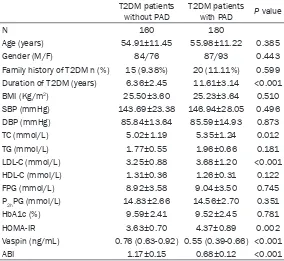
The clinical and laboratory characteristics of T2DM patients with and without PAD are pre-sented in Table 1. T2DM patients with PAD showed higher levels of duration of T2DM, total cholesterol (TC), low-density lipoprotein choles-terol (LDL-C), homeostasis model assessment of insulin resistance (HOMA-IR), and decreased ABI compared with those without PAD. There
[image:2.612.91.375.98.359.2]were no significant differences in other charac -teristics between the two groups.
Table 1. Clinical and biochemical characteristics of T2DM patients with and without PAD
T2DM patients
without PAD T2DM patients with PAD P value
N 160 180
Age (years) 54.91±11.45 55.98±11.22 0.385 Gender (M/F) 84/76 87/93 0.443 Family history of T2DM n (%) 15 (9.38%) 20 (11.11%) 0.599 Duration of T2DM (years) 6.36±2.45 11.61±3.14 <0.001 BMI (Kg/m2) 25.50±3.60 25.23±3.64 0.510
SBP (mmHg) 143.69±23.38 146.94±28.05 0.496 DBP (mmHg) 85.84±13.64 85.59±14.93 0.873 TC (mmol/L) 5.02±1.19 5.35±1.24 0.012 TG (mmol/L) 1.77±0.55 1.96±0.66 0.181 LDL-C (mmol/L) 3.25±0.88 3.68±1.20 <0.001 HDL-C (mmol/L) 1.31±0.36 1.26±0.31 0.122 FPG (mmol/L) 8.92±3.58 9.04±3.50 0.745 P2hPG (mmol/L) 14.83±2.66 14.56±2.70 0.351
HbA1c (%) 9.59±2.41 9.52±2.45 0.781 HOMA-IR 3.63±0.70 4.37±0.89 0.002 Vaspin (ng/mL) 0.76 (0.63-0.92) 0.55 (0.39-0.66) <0.001 ABI 1.17±0.15 0.68±0.12 <0.001
T2DM, type 2 diabetes mellitus; PAD, peripheral arterial disease; BMI, body mass index; SBP, systolic blood pressure; DBP, diastolic blood pressure; TG, triglycerides; TC, total cholesterol; HDL-C, high-density lipoprotein cholesterol; LDL-C, low-density lipoprotein cholesterol; FPG, fasting plasma glucose; P2hPG, 2-hour postprandial plasma glucose; HOMA-IR, homeostasis model assessment of insulin resistance; ABI, ankle-brachial index.
Measurements
Anthropometric (height, wei- ght and blood pressures), clin-ical and laboratory analysis were performed. Venous bl- ood was collected after a min-imum of 10 hours of absolute diet. Serum vaspin was deter-mined using an enzyme-linked immunosorbent assay (ALPCO Diagnostic, Salem NH, USA). Statistical analysis
Association of serum vaspin levels with the presence of PAD in T2DM patients
The results indicated serum vaspin levels were
significantly decreased in T2DM patients with
PAD compared with those without PAD (Table 1). Simple logistic regression analysis showed that duration of T2DM (OR 2.113, 95% CI 1.798 to 2.482; P<0.001), TC (OR 1.258, 95% CI 1.048 to 1.510; P=0.014), LDL-C (OR 1.487, 95% CI 1.192 to 1.856; P<0.001), HOMA-IR (OR 1.245, 95% CI 1.080 to 1.434; P=0.002), and serum vaspin (OR 0.003, 95% CI 0.001 to 0.012; P<0.001) showed relative significant
correlations with the presence of PAD in T2DM patients (Table 2). All these variables were then entered into a backward stepwise multivariate logistic regression model. Multivariate logistic regression of revealed that serum vaspin levels
remained as a significant and independent pre -dictor of T2DM (OR 0.002, 95% CI 0.000 to 0.014; P<0.001) (Table 2).
Association of serum vaspin levels with other clinical characteristics
[image:3.612.95.521.83.322.2]Simple regression analyses showed that serum vaspin levels in T2DM patients were negatively Table 2. Logistic regression Analysis for the presence of PAD in T2DM patients
Simple regression Multiple regression OR (95% CI) P OR (95% CI) P
Age (years) 1.008 (0.990-1.028) 0.384 Gender (M/F) 1.181 (0.771-1.810) 0.443 Family history of T2DM 1.208 (0.596-2.448) 0.599
Duration of T2DM (years) 2.113 (1.798-2.482) <0.001 2.320 (1.880-2.863) <0.001 BMI (Kg/m2) 0.980 (0.924-1.039) 0.495
SBP (mmHg) 1.005 (0.997-1.013) 0.249 DBP (mmHg) 0.999 (0.984-1.014) 0.872
TC (mmol/L) 1.258 (1.048-1.510) 0.014 1.157 (0.752-1.780) 0.508 TG (mmol/L) 1.121 (0.947-1.328) 0.184
LDL-C (mmol/L) 1.487 (1.192-1.856) <0.001 1.429 (0.857-2.380) 0.171 HDL-C (mmol/L) 0.602 (0.316-1.148) 0.123
FPG (mmol/L) 1.010 (0.951-1.073) 0.744 P2PG (mmol/L) 0.963 (0.889-1.043) 0.350 HbA1c (%) 0.988 (0.905-1.078) 0.780
HOMA-IR 1.245 (1.080-1.434) 0.002 1.399 (1.095-1.789) 0.007 Vaspin (ng/mL) 0.003 (0.001-0.012) <0.001 0.002 (0.000-0.014) <0.001
T2DM, type 2 diabetes mellitus; PAD, peripheral arterial disease; BMI, body mass index; SBP, systolic blood pressure; DBP, diastolic blood pressure; TG, triglycerides; TC, total cholesterol; HDL-C, high-density lipoprotein cholesterol; LDL-C, low-density lipoprotein cholesterol; FPG, fasting plasma glucose; P2hPG, 2-hour postprandial plasma glucose; HOMA-IR, homeostasis model assessment of insulin resistance; ABI, ankle-brachial index.
Table 3. Linear regression analyses between serum vaspin and other clinical parameters
Parameters R P value Age (years) 0.023 0.669 Gender (M/F) 0.077 0.155 Family history of T2DM 0.030 0.576 Duration of T2DM (years) -0.067 0.216 BMI (Kg/m2) -0.128 0.019
SBP (mmHg) -0.082 0.131 DBP (mmHg) -0.070 0.197 TC (mmol/L) -0.047 0.389 TG (mmol/L) -0.153 0.005 LDL-C (mmol/L) -0.088 0.106 HDL-C (mmol/L) 0.063 0.245 FPG (mmol/L) -0.034 0.537 P2PG (mmol/L) -0.061 0.261 HbA1c (%) -0.043 0.432 HOMA-IR -0.235 <0.001
ABI 0.186 0.001
[image:3.612.91.290.410.639.2]correlated with BMI (r=-0.128, P=0.019), tri-glycerides (TG) (r=-0.153, P=0.005), HOMA-IR (r=-0.235, P<0.001), and positively correlated with ABI (r=0.186, P=0.001) (Table 3). Then variables including BMI, TG, HOMA-IR, and ABI were incorporated into the stepwise linear regression model. Multiple stepwise regression
analysis shows that only HOMA-IR (β=-0.216,
P<0.001) and ABI (β=0.168, P=0.005) remain- ed associated with serum vaspin.
Discussion
In the present study, we found that T2DM
patients with PAD had significantly lower serum
vaspin levels compared with those without PAD. In addition, serum vaspin levels were neg-atively correlated with BMI, TG, HOMA-IR, and positively correlated with ABI. To the best of
our knowledge, this is the first cross-sectional
study that demonstrates the association of serum vaspin levels with the presence and severity of PAD in T2DM patients.
Nowadays, biomarkers are used in early dis-ease diagnosis, prognosis of disdis-ease progress, and evaluating therapeutic effects of many dis-eases. Adipose tissue is an active metabolic and endocrine organ. Adipocytes produce sev-eral substances, so called as adipokines, which
influence glucose and lipid metabolism. These adipokines includes tumor necrosis factor-α (TNF-α), interleukin-6 (IL-6), leptin, resistin, vis -fatin, and chemerin and so on [7]. These adipo-kines play important roles in many disease such as obesity, metabolic syndrome, diabetes, and cardiovascular disease [8]. The present study revealed that T2DM patients with PAD
had significantly lower serum vaspin levels
compared with those without PAD, which sug-gesting the potential role of vaspin in the patho-physiology of PAD in diabetes. Similar results were found in another study, which reported
that serum vaspin levels were significantly
lower in T2DM patients with carotid plaque than in those without [9]. Furthermore, the cur-rent results showed that serum levels of vas- pin were positively correlated with ABI. Serum vaspin levels are correlated with the severity of PAD. Therefore, serum vaspin levels are sug-gested to be an independent predicting bio-marker of the presence and severity of PAD in T2DM patients.
Vaspin plays an important role in the process of atherosclerosis. Vaspin increased endothelial
nitric oxide synthase activity by reducing asym-metric dimethylarginine level [10]. Vaspin inhib-ited high glucose-induced vascular smooth muscle cells (VSMC) proliferation and chemoki-nesis by preventing reactive oxygen species (ROS) activation and MAPK, PI3K/Akt, and
NF-κB signaling [11]. In addition, vaspin was
found to inhibit platelet-derived growth factor-BB-induced VSMC migration via preventing the ROS generation [12]. All these results indicate that vaspin play as an anti-atherogenic actor. Serum levels of vaspin were also found to be
significantly lower in patients with coronary
artery disease [13] and carotid stenosis [14], which point to the anti-atherogenic role of vaspin.
The limitation of the present study should be considered. First, the modest sample size in this study could impede our discovery of less-strong associations; larger sample size is re-
quired to determine whether the significant
results were false positive. Second, this study was cross-sectional. A prospective cohort study
may provide more definitive evidence about the
association of serum vaspin with the presence and severity of PAD in T2DM patients.
In conclusion, this study showed that T2DM
patients with PAD had significantly lower serum
vaspin levels compared with those without PAD.
Serum levels of vaspin were significantly corre -lated with ABI. Decreased serum vaspin levels are suggested to be an independent predicting marker of the presence and severity of PAD in T2DM patients.
Disclosure of conflict of interest
None.
Address correspondence to: Dr. Yunlong Liu, De- partment of Vascular Surgery, General Hospital of Beijing Military Region, 5 South Gate Warehouse, Dongcheng District, Beijing 100010, PR China. Tel: 86-10-66721629; Fax: 86-10-66721629; E-mail: liuyunlongyl@126.com
References
[1] Dormandy JA, Rutherford RB. Management of peripheral arterial disease (PAD). TASC Wor- king Group. TransAtlantic Inter-Society Con- sensus (TASC). J Vasc Surg 2000; 31: S1-S296.
ease in relation to glycaemic level in an elderly Caucasian population: the Hoorn study. Dia- betologia 1995; 38: 86-96.
[3] American Diabetes Association. Peripheral ar-terial disease in people with diabetes. Diabetes Care 2003; 26: 3333-3341.
[4] Hida K, Wada J, Eguchi J, Zhang H, Baba M, Seida A, Hashimoto I, Okada T, Yasuhara A, Nakatsuka A, Shikata K, Hourai S, Futami J, Watanabe E, Matsuki Y, Hiramatsu R, Akagi S, Makino H, Kanwar YS. Visceral adipose tissue-derived serine protease inhibitor: a unique in-sulin-sensitizing adipocytokine in obesity. Proc Natl Acad Sci U S A 2005; 102: 10610-10615. [5] Jung CH, Lee WJ, Hwang JY, Seol SM, Kim YM, Lee YL, Park JY. Vaspin protects vascular endo-thelial cells against free fatty acid-induced apoptosis through a phosphatidylinositol 3-ki-nase/Akt pathway. Biochem Biophys Res Co- mmun 2011; 413: 264-269.
[6] Kobat MA, Celik A, Balin M, Altas Y, Baydas A, Bulut M, Aydin S, Dagli N, Yavuzkir MF, Ilhan S. The investigation of serum vaspin level in ath-erosclerotic coronary artery disease. J Clin Med Res 2012; 4: 110-113.
[7] Trayhurn P, Wood IS. Adipokines: inflammation
and the pleiotropic role of white adipose tis-sue. Br J Nutr 2004; 92: 347-355.
[8] Blüher M. Clinical relevance of adipokines. Diabetes Metab J 2012; 36: 317-327.
[9] Li Z, Ma C, Li L, Pan X, Chen L. Vaspin serum concentration in patients with type 2 diabetes and carotid plaque. J Int Med Res 2012; 40: 1670-1676.
[10] Jung CH, Lee WJ, Hwang JY, Lee MJ, Seol SM, Kim YM, Lee YL, Kim HS, Kim MS, Park JY. Vaspin increases nitric oxide bioavailability through the reduction of asymmetric dimethyl-arginine in vascular endothelial cells. PLoS One 2012; 7: e52346.
[11] Li H, Peng W, Zhuang J, Lu Y, Jian W, Wei Y, Li W, Xu Y. Vaspin attenuates high glucose-in-duced vascular smooth muscle cells prolifera-tion and chemokinesis by inhibiting the MAPK,
PI3K/Akt, and NF-κB signaling pathways.
Ath-erosclerosis 2013; 228: 61-68.
[12] Phalitakul S, Okada M, Hara Y, Yamawaki H. A novel adipocytokine, vaspin inhibits platelet-derived growth factor-BB-induced migration of vascular smooth muscle cells. Biochem Bio- phys Res Commun 2012; 423: 844-849. [13] Kadoglou NP, Gkontopoulos A, Kapelouzou A,
Fotiadis G, Theofilogiannakos EK, Kottas G,
Lampropoulos S. Serum levels of vaspin and visfatin in patients with coronary artery dis-ease-Kozani study. Clin Chim Acta 2011; 412: 48-52.